management of the incidental renal mass on ct: a white … · the acr incidental findings committee...
TRANSCRIPT

ORIGINAL ARTICLE
Management of the Incidental RenalMass on CT: A White Paper of theACR Incidental Findings Committee
Brian R. Herts, MDa, Stuart G. Silverman, MDb, Nicole M. Hindman, MDc, Robert G. Uzzo, MDd,Robert P. Hartman, MDe, Gary M. Israel, MD f, Deborah A. Baumgarten, MD, MPHg,Lincoln L. Berland, MDh, Pari V. Pandharipande, MD, MPHiAbstract
The ACR Incidental Findings Committee (IFC) presents recommendations for renal masses that are incidentally detected on CT. Theserecommendations represent an update from the renal component of the JACR 2010 white paper on managing incidental findings in theadrenal glands, kidneys, liver, and pancreas. The Renal Subcommittee, consisting of six abdominal radiologists and one urologist, developedthis algorithm. The recommendations draw from published evidence and expert opinion and were finalized by informal iterative consensus.Each flowchart within the algorithm describes imaging features that identify when there is a need for additional imaging, surveillance, orreferral for management. Our goal is to improve quality of care by providing guidance for managing incidentally detected renal masses.
Key Words: Kidney, renal, small renal mass, cyst, Bosniak classification, incidental finding
J Am Coll Radiol 2017;-:---. Copyright � 2017 American College of Radiology
OVERVIEW OF THE ACR INCIDENTALFINDINGS PROJECTThe core objectives of the Incidental Findings Project areto (1) develop consensus on patient characteristics andimaging features that are required to characterize anincidental finding, (2) provide guidance to manage suchfindings in ways that balance the risks and benefits topatients, (3) recommend reporting terms that reflect thelevel of confidence regarding a finding, and (4) focusfuture research by proposing a generalizable managementframework across practice settings. The Incidental
aImaging Institute, Cleveland Clinic, Cleveland, Ohio.bDepartment of Radiology, Brigham and Women’s Hospital, Boston,Massachusetts.cDepartment of Radiology, NYU Medical Center, New York, New York.dDepartment of Urology, Fox Chase Cancer Center, Philadelphia,Pennsylvania.eDepartment of Radiology, Mayo Clinic, Rochester, Minnesota.fDepartment of Radiology, Yale University Medical Center, New Haven,Connecticut.gDepartment of Radiology, Emory University Hospital, Atlanta, Georgia.hDepartment of Radiology, University of Alabama Birmingham MedicalCenter, Birmingham, Alabama.iDepartment of Radiology, Massachusetts General Hospital, Boston,Massachusetts.
ª 2017 American College of Radiology1546-1440/17/$36.00 n http://dx.doi.org/10.1016/j.jacr.2017.04.028
Findings Committee (IFC) generated its first white paperin 2010, addressing four algorithms for managing inci-dental pancreatic, adrenal, kidney, and liver findings [1].
THE CONSENSUS PROCESS: THE INCIDENTALRENAL MASS ALGORITHMThe current publication represents the first revision of theIFC’s recommendations on incidental renal masses. Thealgorithm was created by a committee comprised ofthe renal subcommittee chair, five appointed abdominalradiologists, and one urologist. The subcommittee gained
Corresponding author and reprints: Brian R. Herts, MD, ImagingInstitute—Desk L10, Cleveland Clinic, 9500 Euclid Avenue, Cleveland,OH 44195; e-mail: [email protected].
Dr. Herts reports a research Grant from Siemens Healthineers.Dr. Silverman has nothing to disclose. Dr. Hindman has nothing todisclose. Dr. Uzzo reports personal fees as an advisory board member fromPfizer and Myriad, and receives speaker fees from Jannsen, all outside thesubmitted work. Dr. Hartman has nothing to disclose. Dr. Israel hasnothing to disclose. Dr. Baumgarten has nothing to disclose. Dr. Berlandreports personal fees from Nuance Communications, Inc., outside thesubmitted work. Dr. Pandharipande reports a research grant from theMedical Imaging and Technology Alliance, outside the submitted work.
1

consensus on a preliminary version using publishedevidence as their primary source. The team’s collectiveexpertise was invoked where evidence was not available.The preliminary algorithm underwent review by addi-tional IFC members, including the body commissionchair, the IFC chair, and additional IFC subcommitteechairs. The revised algorithm and correspondingwhite paper draft were submitted to additional ACRstakeholders to gain input and feedback. Consensus wasobtained iteratively by successive review and revision,after which the algorithm and white paper were finalized.The IFC’s consensus processes meet policy standards ofthe ACR. However, they do not meet any specific, formalnational standards. This algorithm and set of recom-mendations does not represent policy of the ACR practiceguidelines or the ACR appropriateness criteria. Ourconsensus may be termed “guidance” and “recommen-dations” rather than “guidelines,” which has a moreformal definition.
ELEMENTS OF THE FLOWCHARTS: COLOR-CODINGThis algorithm consists of five flowcharts (Figs. 1-5).Withineach flowchart, yellow boxes indicate using or acquiringclinical data (eg, imaging features, interval stability), greenboxes describe recommendations for action (eg, additionalimaging or referral for treatment), and red boxes indicatethat workup or surveillance may be terminated (eg, a
Fig 1. Flowchart for managing an incidental renal mass on noncointerest < �10 HU), refer to Figure 5. 2Too small to characterize. 3Ware visually much lower or much higher than the unenhanced renpreferred for characterizing smaller masses (<1.5 cm) and for deUltrasound may be able to characterize a homogeneous hyperattenold images are available, any renal mass that has been without chamm per year for at least 5 years is likely of no clinical significancTSTC ¼ too small to characterize; WO&W ¼ without and with; W
2
benign or indolent mass). To minimize complexity,each algorithm addresses most—but not all—imagingappearances and clinical scenarios. Radiologists should feelcomfortable deviating from the algorithm in circumstancesthat are not represented in the algorithm, based on thespecific imaging appearance of the finding in question andpatient characteristics—the algorithm content must beviewed as recommendations, and should not a priori beconsidered as “standard of care.”
NATURE AND SCOPE OF THE PROBLEMThe majority of renal masses are benign cysts. Theprevalence of cysts increases with age [2,3], and cysts canbe seen in as many as 40% of patients on CT [4].Incidental renal masses are concerning because mostrenal cell carcinomas (RCCs) are incidentally detected[5], and patient prognosis is better when RCC isdetected incidentally [6-8]. However, there is no higherprevalence of urologic symptoms in patients withsimple cysts [9]; thus, most cysts are also incidentalfindings. Moreover, cancers of the kidney and renalpelvis are relatively uncommon, estimated to be only3.7% of new cancer cases in 2016, less than breast,lung, prostate, lymphoma, colon, and bladder cancer[10]. Furthermore, many patients with small incidentalrenal cancers may not benefit from treatment [11-13].Therefore, when evaluating any incidental renal mass,the potential benefit of early detection of RCC needs to
ntrast CT. 1If the mass contains fat attenuation (a region ofell-circumscribed and homogeneous TSTC renal masses thatal parenchyma are probably benign cystic lesions. 4MRI istecting enhancement in suspected hypovascular masses.uating renal mass as a hemorrhagic or proteinaceous cyst. 5Ifnge in imaging features and has had an average growth of � 3e and does not need further workup. HU ¼ Hounsfield unit;/U ¼ work-up.
Journal of the American College of RadiologyVolume - n Number - n Month 2017

Fig 2. Flowchart for managing an incidental renal mass on contrast-enhanced CT. 1If the mass contains fat attenuation (a regionof interest < �10 HU), refer to Figure 5. 2Too small to characterize. 3Well-circumscribed and homogeneous TSTC renal massesthat are visually much lower than the enhanced renal parenchyma are probably benign cystic lesions. 4MRI is preferred forcharacterizing smaller masses (<1.5 cm) and for detecting enhancement in suspected hypovascular masses. Ultrasound may beable to characterize a homogeneous renal mass as a hemorrhagic or proteinaceous cyst. 5If old images are available, any renalmass that has been without change in imaging features and has had an average growth of �3 mm per year for at least 5 years islikely of no clinical significance and does not need further workup. HU ¼ Hounsfield unit; TSTC ¼ too small to characterize;WO&W ¼ without and with; W/U ¼ work-up.
be weighed against the potential harms of investigating ortreating a benign or small malignant renal mass that mayhave no clinical significance.
Definition of the Incidental Renal MassThe incidental renal mass is one that is initially detectedon an imaging study performed for an indication otherthan the assessment of urinary tract disease. For any
Fig 3. Flowchart for managing a cystic renal mass on CT or MRIcontains fat attenuation (a region of interest < �10 HU), refer tosepta, thickening of the wall or septa, or development of a solid nIV). Growth of a cystic mass without morphologic change is notwithout change in imaging features for at least 5 years is considereunit; IV ¼ intravenous; WO&W ¼ without and with; W/U ¼ work
Journal of the American College of RadiologyHerts et al n Incidental Renal Mass on CT
such mass, one of three conclusions can be drawn fromits imaging features: (1) it is completely characterized,with imaging features diagnostic of a simple orcomplicated cystic mass, or of a solid neoplasm (with orwithout fat), such that management can be recom-mended; (2) it is incompletely characterized requiringfurther evaluation before recommending management;and (3) it is incompletely characterized, but based on
performed both without and with IV contrast. 1If the massFigure 5. 2Morphologic change includes increasing number ofodular component (including reclassification as Bosniak III orindicative of malignancy. 3A Bosniak IIF cystic renal massd stable and likely of no clinical significance. HU ¼ Hounsfield-up.
3
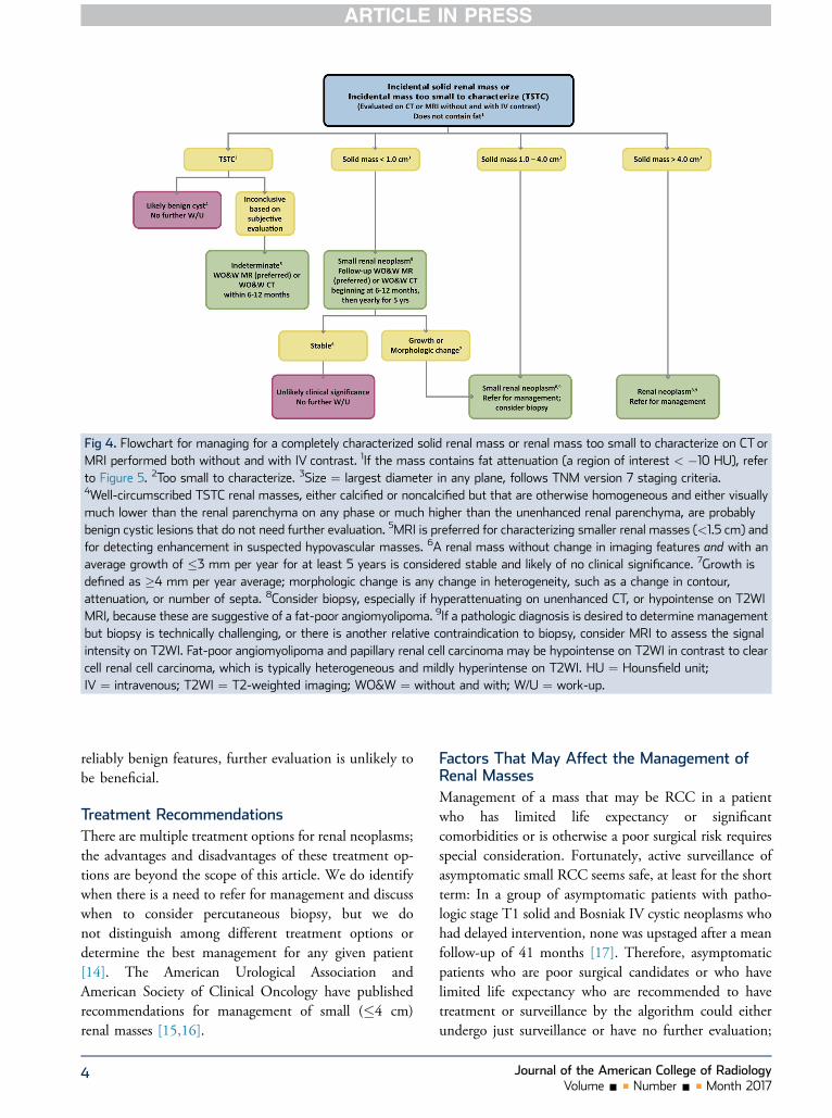
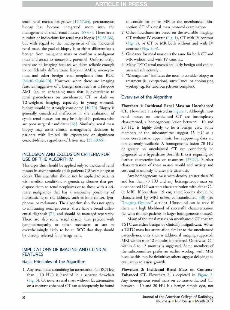
Fig 4. Flowchart for managing for a completely characterized solid renal mass or renal mass too small to characterize on CT orMRI performed both without and with IV contrast. 1If the mass contains fat attenuation (a region of interest < �10 HU), referto Figure 5. 2Too small to characterize. 3Size ¼ largest diameter in any plane, follows TNM version 7 staging criteria.4Well-circumscribed TSTC renal masses, either calcified or noncalcified but that are otherwise homogeneous and either visuallymuch lower than the renal parenchyma on any phase or much higher than the unenhanced renal parenchyma, are probablybenign cystic lesions that do not need further evaluation. 5MRI is preferred for characterizing smaller renal masses (<1.5 cm) andfor detecting enhancement in suspected hypovascular masses. 6A renal mass without change in imaging features and with anaverage growth of �3 mm per year for at least 5 years is considered stable and likely of no clinical significance. 7Growth isdefined as �4 mm per year average; morphologic change is any change in heterogeneity, such as a change in contour,attenuation, or number of septa. 8Consider biopsy, especially if hyperattenuating on unenhanced CT, or hypointense on T2WIMRI, because these are suggestive of a fat-poor angiomyolipoma. 9If a pathologic diagnosis is desired to determine managementbut biopsy is technically challenging, or there is another relative contraindication to biopsy, consider MRI to assess the signalintensity on T2WI. Fat-poor angiomyolipoma and papillary renal cell carcinoma may be hypointense on T2WI in contrast to clearcell renal cell carcinoma, which is typically heterogeneous and mildly hyperintense on T2WI. HU ¼ Hounsfield unit;IV ¼ intravenous; T2WI ¼ T2-weighted imaging; WO&W ¼ without and with; W/U ¼ work-up.
reliably benign features, further evaluation is unlikely tobe beneficial.
Treatment RecommendationsThere are multiple treatment options for renal neoplasms;the advantages and disadvantages of these treatment op-tions are beyond the scope of this article. We do identifywhen there is a need to refer for management and discusswhen to consider percutaneous biopsy, but we donot distinguish among different treatment options ordetermine the best management for any given patient[14]. The American Urological Association andAmerican Society of Clinical Oncology have publishedrecommendations for management of small (�4 cm)renal masses [15,16].
4
Factors That May Affect the Management ofRenal MassesManagement of a mass that may be RCC in a patientwho has limited life expectancy or significantcomorbidities or is otherwise a poor surgical risk requiresspecial consideration. Fortunately, active surveillance ofasymptomatic small RCC seems safe, at least for the shortterm: In a group of asymptomatic patients with patho-logic stage T1 solid and Bosniak IV cystic neoplasms whohad delayed intervention, none was upstaged after a meanfollow-up of 41 months [17]. Therefore, asymptomaticpatients who are poor surgical candidates or who havelimited life expectancy who are recommended to havetreatment or surveillance by the algorithm could eitherundergo just surveillance or have no further evaluation;
Journal of the American College of RadiologyVolume - n Number - n Month 2017
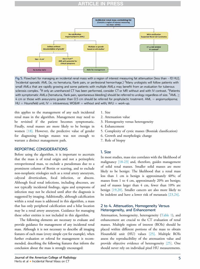
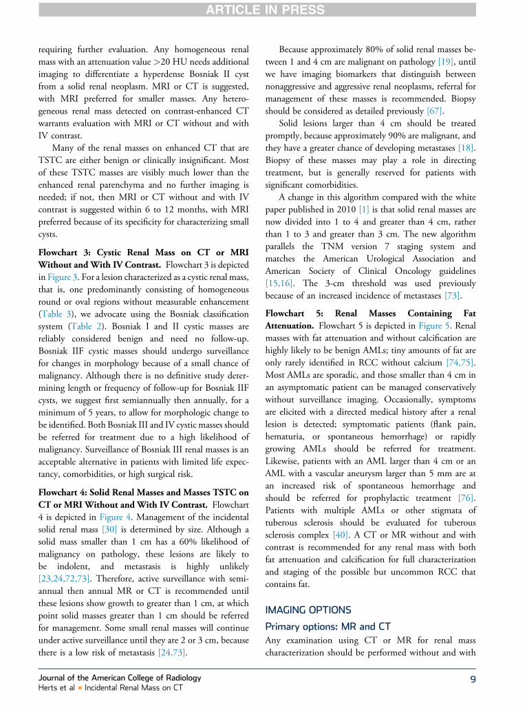
Fig 5. Flowchart for managing an incidental renal mass with a region of interest measuring fat attenuation (less than �10 HU).1Incidental sporadic AML (ie, no hematuria, flank pain, or perilesional hemorrhage.) 2Many urologists will follow patients withsmall AMLs that are rapidly growing and some patients with multiple AMLs may benefit from an evaluation for tuberoussclerosis complex. 3If only an unenhanced CT has been performed, consider CT or MR without and with IV contrast. 4Patientswith symptomatic AMLs (hematuria, flank pain, spontaneous bleeding) should be referred to urology regardless of size. 5AML �4 cm or those with aneurysms greater than 0.5 cm should be referred for prophylactic treatment. AML ¼ angiomyolipoma;HU ¼ Hounsfield unit; IV ¼ intravenous; WO&W ¼ without and with; W/U ¼ work-up.
this applies to the management of any such incidentalrenal mass in the algorithm. Management may need tobe revisited if the patient becomes symptomatic.Finally, renal masses are more likely to be benign inwomen [18]. However, the predictive value of genderfor diagnosing benign masses was not enough towarrant a distinct management path.
REPORTING CONSIDERATIONSBefore using the algorithm, it is important to ascertainthat the mass is of renal origin and not a perinephricretroperitoneal mass, to exclude a pseudomass due to aprominent column of Bertin or scarring, and to excludenon-neoplastic etiologies such as a renal artery aneurysm,calyceal diverticulum, focal infection, or abscess.Although focal renal infections, including abscesses, arenot typically incidental findings, signs and symptoms ofinfection may not be elicited until after the diagnosis issuggested by imaging. Additionally, although calcificationwithin a renal mass is addressed in this algorithm, a massthat has only peripheral calcification and a hilar locationmay be a renal artery aneurysm. Guidance for managingthese other entities is not included in this algorithm.
The following elements are necessary to evaluate andprovide guidance for management of any incidental renalmass. Although it is not necessary to describe all imagingfeatures of each mass (every simple cyst for example), whenfurther evaluation or referral for management is recom-mended, describing the following features that inform theconclusion about the mass is strongly encouraged:
Journal of the American College of RadiologyHerts et al n Incidental Renal Mass on CT
1. Size2. Attenuation value3. Homogeneity versus heterogeneity4. Enhancement5. Complexity of cystic masses (Bosniak classification)6. Growth and morphologic change7. Role of biopsy
1. SizeIn most studies, mass size correlates with the likelihood ofmalignancy [18-23] and, therefore, guides managementof solid renal masses. Smaller solid masses are morelikely to be benign: The likelihood that a renal massless than 1 cm is benign is approximately 40%; ofmasses from 1 to 4 cm, approximately 20% are benign;and of masses larger than 4 cm, fewer than 10% arebenign [19,20]. Smaller cancers are also more likely tobe indolent and have a lower risk of metastasis [23,24].
2 to 4. Attenuation, Homogeneity VersusHeterogeneity, and EnhancementAttenuation, homogeneity, heterogeneity (Table 1), andenhancement are crucial to the CT evaluation of renalmasses. Multiple regions of interest (ROIs) should beplaced within different portions of the mass to obtainHounsfield unit (HU) values [25]. Multiple ROIsassess the reproducibility of the attenuation value andprovide objective evidence of heterogeneity [25]. Oneshould never rely on individual pixel HU measurements.
5

Table 1. Features that indicate heterogeneity in a renal mass
Feature
Wall thickeningOne or more septaMural nodule(s)Measurable or visible attenuation differencesCalcification
When without and with contrast-enhanced CTscans are available, homogeneous masses between �10and þ20 HU without enhancement are simple renalcysts. Although an RCC may have an average attenuationvalue of less than 20 HU before contrast, it is almostalways heterogeneous on the unenhanced scan [26]. It israre for a homogeneous mass less than 20 HU on anunenhanced CT to be RCC [27,28]. Therefore, anyhomogeneous mass between �10 and þ20 HU on CTeither without or with contrast is also considered asimple cyst (Bosniak I, Table 2) and does not requirefurther evaluation.
On unenhanced CT, a homogeneous mass of 70HU or greater is almost always a hyperdense Bosniak IIcyst [27,29] and needs no further evaluation. However,on contrast-enhanced CT scans, both RCCs and hyper-dense cysts may have contrast-enhanced attenuation of70 HU or greater.
CT or MRI before and after intravenous (IV) contrastis necessary to characterize any homogeneous mass greaterthan 20 and less than 70 HU on unenhanced CT, orgreater than 20 HU on contrast-enhanced CT only. Onthese examinations, enhancement (Table 3) of the renalmass or any nodular component of a cystic renal massis concerning for neoplasm [30-32]. Although some CT
Table 2. Bosniak renal cyst classification system
BosniakClassification
I Benign simple cyst with a hairline thin wall wnear-water attenuation density (�10 to 20
II Benign minimally complicated cyst that may cnot measurable enhancement. Fine calcificapresent in the wall or septa. Also, a well-mdensity above simple fluid attenuation (hyp
IIF Usually benign complicated renal cyst with mwall or septa. Wall or septa may contain thimeasurable enhancement. Also, a well-marabove simple fluid.
III Indeterminate complicated cystic renal mass wenhancement.
IV Malignant cystic renal mass with enhancing s
6
and MRI enhancement patterns are associated withbenign tumors or specific RCC subtypes, no patternwas considered sensitive or specific enough to beincluded in the algorithm. However, percutaneousbiopsy should be considered when features suggest abenign or indolent etiology [33-38].
An ROI value of less than �10 HU within the massindicates the presence of fat [39]. The overwhelmingmajority of renal masses with fat are angiomyolipomas(AMLs). Most AMLs have ROIs with attenuation muchcloser to fatty tissue (�100 HU) [40]. Unfortunately, asmall percentage of AMLs have no fat detectable onunenhanced CT (termed fat-poor AMLs) and may bemisdiagnosed as RCCs [39-42]. Because some cysts in aphantom study assessing multidetector CT scannershad attenuation values lower than �10 HU [43], MR orultrasound should be considered to evaluate ahomogeneous mass with an attenuation valuebetween �20 and �10 HU to exclude a cyst. Calcificationis extremely rare in AML; therefore, a mass withinterspersed fat and calcification should be considered RCC.
Any heterogeneous renal mass, as defined by thepresence of wall thickening, septa, mural nodules,attenuation differences, or calcification (Table 1) [27],warrants complete characterization.
Some incidental renal masses will be too small tomeasure the attenuation accurately—that is, too small tocharacterize (TSTC). This occurs when the lesion size isless than twice reconstructed slice thickness [25].Creation and review of thin sections (w1 mm) mayminimize the number of these lesions. Fortunately,these are usually clinically insignificant and many canbe assessed subjectively.
Description
ithout septa, calcification, or solid component. HomogeneousHU) without enhancement.ontain a few hairline thin septa that may have “perceived” buttion or a segment of slightly thickened calcification may bearginated nonenhancing homogeneous mass < 3 cm witherdense cyst).ultiple hairline thin septa or minimal smooth thickening of theck and nodular calcification and may have “perceived” but notginated intrarenal nonenhancing mass > 3 cm with density
ith thickened irregular walls or septa that have measurable
oft tissue components (cystic renal cell carcinoma).
Journal of the American College of RadiologyVolume - n Number - n Month 2017

Table 3. CT and MRI criteria for defining enhancement in a renal mass
CT Criteria: Increase in Attenuation After Contrast
�20 HU Definite for enhancement>10 to < 20 HU Equivocal for enhancement; consider factors related to beam
hardening, intra-renal location*�10 HU No enhancement
MRI criteria for enhancement
�15% increase in signal intensity after contrast Enhancing lesionAlternative method Visible signal intensity on subtraction images
HU ¼ Hounsfield units.*Stricter criteria (15 HU) should be used as a cutoff for enhancement of exophytic or larger lesions not prone to these factors.
5. Complexity of Cystic Masses (BosniakClassification)As opposed to solid masses, where the likelihood ofmalignancy increases with size, the likelihood of malig-nancy of cystic renal masses is based on the degree of cystcomplexity. Cystic RCCs are also smaller, have lowerstage and grade, and have a more indolent biology [44].Patients with cystic RCC have better survival rates andless frequent metastatic disease [45]; thus, cystic andsolid masses are managed differently in the algorithm(Figs. 3, 4).
We recommend the use of the Bosniak classification(Table 2) for evaluating any cystic renal mass [46-48].Surveillance studies of Bosniak IIF and III cystic massesoften show no progression or development of locallyadvanced or metastatic disease [49-51]. Therefore,guidance for managing Bosniak IIF and III complicatedcystic lesions is based on the less aggressive nature ofcystic RCC and reported malignancy rates of 11% and54%, respectively [50,51], noting higher rates occur inpatients with a history of RCC or co-existing BosniakIV or solid mass [50]. Although management of BosniakIII masses varies among institutions, resection is generallyfavored [52]. Calcification, once a component of theBosniak system, is no longer considered as significant[47]. Size is not a factor in the Bosniak classificationsystem. Although necrosis in RCC may measure fluidattenuation, it is usually heterogeneous or poorlymarginated and should not be mistaken for cysticmorphology.
6. Growth and Morphologic ChangeMany small solid renal masses exhibit either slow or nogrowth, and RCC rarely metastasizes in the absence ofgrowth [53-58]. In a meta-analysis of over 200 small (lessthan 4.1 cm) renal masses with a mean follow-up of 34months and mean growth rate of 0.28 cm per year, only1% developed metastases [59]. Therefore, although lack
Journal of the American College of RadiologyHerts et al n Incidental Renal Mass on CT
of or slow growth does not assure benignity, it indicatesindolent disease. Ample and growing data supportsurveillance of small solid renal masses [25,60,61], andwe recommend surveillance of solid and indeterminaterenal masses smaller than 1.0 cm (Fig. 4) [5,48].Conversely, rapid growth of a renal mass correlates withhigher potential for metastatic disease duringsurveillance. In a study of patients with solid andBosniak IV masses 4 cm or larger, 13.8% (5 of 36)progressed to metastatic disease; their average growthrate was 2.8 cm per year [62]. For indeterminatecomplicated cystic masses (Bosniak IIF), growthwithout morphologic change is not suspicious formalignancy [46].
Any change in morphology (ie, increasing hetero-geneity) of a renal mass is concerning for RCC andwarrants referral for management. Neoplasm developingin an otherwise benign-appearing cyst is uncommonand does not occur without detectable morphologicchange [3].
Based on surveillance studies, for this algorithm weconsider lack of change in morphology and an averagegrowth rate of �0.3 cm per year over at least 5 years to bea stable lesion with an insignificant risk of metastasis[55,63]. This can be used retrospectively as well, and it isimportant to review old examinations, includingnonabdominal studies like spine MR, to assess thestability of renal lesions. Unfortunately, the follow-upperiod required to confidently diagnose a mass as indo-lent or benign no longer requiring surveillance, remainsundetermined [6]. Age, symptoms, imaging features, andmaximum diameter of clinical stage T1a masses atpresentation are not predictors of growth and, thus,cannot be used in lieu of surveillance [55,64].
7. Role of BiopsyRenal mass biopsy is both safe and effective [65,66]. Asour understanding of the nature and natural history of
7
small renal masses has grown [17,57,64], percutaneousbiopsy has become integrated more into themanagement of small renal masses [65-67]. There are anumber of indications for renal mass biopsy [30,65,66],but with regard to the management of the incidentalrenal mass, the goal of biopsy is to either differentiate abenign from malignant mass or confirm a malignantmass and assess its metastatic potential. Unfortunately,there are no imaging features we deem reliable enoughto confidently differentiate fat-poor AMLs, oncocyto-mas, and other benign renal neoplasms from RCC[34,40-42,68-70]. However, when there are imagingfeatures suggestive of a benign mass such as a fat-poorAML (eg, an enhancing mass that is hyperdense torenal parenchyma on unenhanced CT or dark onT2-weighted imaging, especially in young women),biopsy should be strongly considered [40,70]. Biopsy isgenerally considered ineffective in the evaluation ofcystic renal masses but may be helpful in patients whoare poor surgical candidates [65]. Similarly, renal massbiopsy may assist clinical management decisions inpatients with limited life expectancy or significantcomorbidities, regardless of lesion size [25,30,65].
INCLUSION AND EXCLUSION CRITERIA FORUSE OF THE ALGORITHMThis algorithm should be applied only to incidental renalmasses in asymptomatic adult patients (18 years of age orolder). This algorithm should not be applied to patientswith medical conditions or genetic syndromes that pre-dispose them to renal neoplasms or to those with a pri-mary malignancy that has a reasonable possibility ofmetastasizing to the kidneys, such as lung cancer, lym-phoma, or melanoma. The algorithm also does not applyto infiltrating renal processes; these have a broad differ-ential diagnosis [71] and should be managed separately.There are also some renal masses that present withlymphadenopathy or other metastases or are sooverwhelmingly likely to be an RCC that they shouldbe directly referred for management.
IMPLICATIONS OF IMAGING AND CLINICALFEATURES
Basic Principles of the Algorithm
1. Any renal mass containing fat attenuation (an ROI lessthan �10 HU) is handled in a separate flowchart(Fig. 5). Of note, a renal mass without fat attenuationon a contrast-enhanced CT can subsequently be found
8
to contain fat on an MR or the unenhanced thin-section CT of a renal mass protocol examination.
2. Other flowcharts are based on the available imaging:CT without IV contrast (Fig. 1), CT with IV contrast(Fig. 2), or CT or MR both without and with IVcontrast (Figs. 3, 4).
3. Guidance for renal masses is the same for both CT andMR without and with IV contrast.
4. Many TSTC renal masses are likely benign and can beassessed subjectively.
5. “Management” indicates the need to consider biopsy ortreatment (ie, extirpation), surveillance, or nonimagingworkup (eg, for tuberous sclerosis complex).
Overview of the Algorithm
Flowchart 1: Incidental Renal Mass on UnenhancedCT. Flowchart 1 is depicted in Figure 1. Although mostrenal masses on unenhanced CT are incompletelycharacterized, a homogeneous lesion between �10 and20 HU is highly likely to be a benign cyst. Somemembers of the subcommittee suggest 15 HU as amore conservative upper limit, but supporting data arenot currently available. A homogeneous lesion 70 HUor greater on unenhanced CT can confidently bediagnosed as a hyperdense Bosniak II cyst requiring nofurther characterization or treatment [27,29]. Furthercharacterization of these masses would add anxiety andcost and is unlikely to alter the diagnosis.
Any homogeneous mass with density greater than 20and less than 70 HU and any heterogeneous mass onunenhanced CT warrants characterization with either CTor MRI. If less than 1.5 cm, these lesions should becharacterized by MRI unless contraindicated [48] (see“Imaging Options” section). Ultrasound can be used ifthere is a high likelihood of successful characterization(ie, with thinner patients or larger homogeneous masses).
Many of the renal masses on unenhanced CT that areTSTC are either benign or clinically insignificant. Whena TSTC mass has attenuation similar to the unenhancedparenchyma, only then is additional imaging suggested;MRI within 6 to 12 months is preferred. Otherwise, CTwithin 6 to 12 months is suggested. Some members ofthe subcommittee prefer an earlier workup with MRIbecause this may be definitive; others suggest delaying theevaluation to assess growth.
Flowchart 2: Incidental Renal Mass on Contrast-Enhanced CT. Flowchart 2 is depicted in Figure 2.Any homogenous renal mass on contrast-enhanced CTbetween �10 and 20 HU is a benign simple cyst, not
Journal of the American College of RadiologyVolume - n Number - n Month 2017
requiring further evaluation. Any homogeneous renalmass with an attenuation value >20 HU needs additionalimaging to differentiate a hyperdense Bosniak II cystfrom a solid renal neoplasm. MRI or CT is suggested,with MRI preferred for smaller masses. Any hetero-geneous renal mass detected on contrast-enhanced CTwarrants evaluation with MRI or CT without and withIV contrast.
Many of the renal masses on enhanced CT that areTSTC are either benign or clinically insignificant. Mostof these TSTC masses are visibly much lower than theenhanced renal parenchyma and no further imaging isneeded; if not, then MRI or CT without and with IVcontrast is suggested within 6 to 12 months, with MRIpreferred because of its specificity for characterizing smallcysts.
Flowchart 3: Cystic Renal Mass on CT or MRIWithout and With IV Contrast. Flowchart 3 is depictedin Figure 3. For a lesion characterized as a cystic renal mass,that is, one predominantly consisting of homogeneousround or oval regions without measurable enhancement(Table 3), we advocate using the Bosniak classificationsystem (Table 2). Bosniak I and II cystic masses arereliably considered benign and need no follow-up.Bosniak IIF cystic masses should undergo surveillancefor changes in morphology because of a small chance ofmalignancy. Although there is no definitive study deter-mining length or frequency of follow-up for Bosniak IIFcysts, we suggest first semiannually then annually, for aminimum of 5 years, to allow for morphologic change tobe identified. Both Bosniak III and IV cystic masses shouldbe referred for treatment due to a high likelihood ofmalignancy. Surveillance of Bosniak III renal masses is anacceptable alternative in patients with limited life expec-tancy, comorbidities, or high surgical risk.
Flowchart 4: Solid Renal Masses and Masses TSTC onCT or MRI Without and With IV Contrast. Flowchart4 is depicted in Figure 4. Management of the incidentalsolid renal mass [30] is determined by size. Although asolid mass smaller than 1 cm has a 60% likelihood ofmalignancy on pathology, these lesions are likely tobe indolent, and metastasis is highly unlikely[23,24,72,73]. Therefore, active surveillance with semi-annual then annual MR or CT is recommended untilthese lesions show growth to greater than 1 cm, at whichpoint solid masses greater than 1 cm should be referredfor management. Some small renal masses will continueunder active surveillance until they are 2 or 3 cm, becausethere is a low risk of metastasis [24,73].
Journal of the American College of RadiologyHerts et al n Incidental Renal Mass on CT
Because approximately 80% of solid renal masses be-tween 1 and 4 cm are malignant on pathology [19], untilwe have imaging biomarkers that distinguish betweennonaggressive and aggressive renal neoplasms, referral formanagement of these masses is recommended. Biopsyshould be considered as detailed previously [67].
Solid lesions larger than 4 cm should be treatedpromptly, because approximately 90% are malignant, andthey have a greater chance of developing metastases [18].Biopsy of these masses may play a role in directingtreatment, but is generally reserved for patients withsignificant comorbidities.
A change in this algorithm compared with the whitepaper published in 2010 [1] is that solid renal masses arenow divided into 1 to 4 and greater than 4 cm, ratherthan 1 to 3 and greater than 3 cm. The new algorithmparallels the TNM version 7 staging system andmatches the American Urological Association andAmerican Society of Clinical Oncology guidelines[15,16]. The 3-cm threshold was used previouslybecause of an increased incidence of metastases [73].
Flowchart 5: Renal Masses Containing FatAttenuation. Flowchart 5 is depicted in Figure 5. Renalmasses with fat attenuation and without calcification arehighly likely to be benign AMLs; tiny amounts of fat areonly rarely identified in RCC without calcium [74,75].Most AMLs are sporadic, and those smaller than 4 cm inan asymptomatic patient can be managed conservativelywithout surveillance imaging. Occasionally, symptomsare elicited with a directed medical history after a renallesion is detected; symptomatic patients (flank pain,hematuria, or spontaneous hemorrhage) or rapidlygrowing AMLs should be referred for treatment.Likewise, patients with an AML larger than 4 cm or anAML with a vascular aneurysm larger than 5 mm are atan increased risk of spontaneous hemorrhage andshould be referred for prophylactic treatment [76].Patients with multiple AMLs or other stigmata oftuberous sclerosis should be evaluated for tuberoussclerosis complex [40]. A CT or MR without and withcontrast is recommended for any renal mass with bothfat attenuation and calcification for full characterizationand staging of the possible but uncommon RCC thatcontains fat.
IMAGING OPTIONS
Primary options: MR and CTAny examination using CT or MR for renal masscharacterization should be performed without and with
9

IV contrast using a dedicated renal mass imaging pro-tocol [30]. Although CT and MRI are both excellent fordetecting and characterizing renal masses [77], thesuperior contrast resolution of MRI provides severaladvantages. MRI is more sensitive to contrastenhancement and is recommended for renal masseswith inconclusive enhancement or for depictingenhancing nodules [78]. MRI better detects andcharacterizes small renal cysts by their T2hyperintensity and better detects enhancement insmall renal lesions and is not subject to pseudo-enhancement as is CT [79,80]. Therefore, we preferMR to characterize smaller renal masses. MRI mayalso be more specific for the diagnosis of a fat-poorAML [81,82]. MRI depicts more septa or thickenedwalls in complex cystic masses, which may result in ahigher Bosniak classification [83].
Newer Technologies and Other ModalitiesDual-energy CT (DECT) and contrast-enhanced ultra-sound (CEUS) both show great potential for characterizingincidental or indeterminate renal masses [84,85]. WithDECT, iodine mapping or virtual unenhanced imagesmay allow complete characterization of a renal lesiondetected only on a contrast-enhanced DECT [86,87].With CEUS, enhancement patterns may differentiatebetween benign and malignant tumors and guidemanagement [85,88]. Because neither DECT nor CEUSis in widespread use in the United States [89], thesemodalities are not directly included in the algorithm.Those using DECT and CEUS should integratethe additional data into the algorithm and directmanagement as for any fully characterized renal mass(Figs. 3 and 4). PET-CT and PET-MRI are not recom-mended because their role evaluating the incidental renalmass is limited.
10
TAKE-HOME POINTS
- Incidental renal masses are a common problem in im-aging;weprovide an algorithm toguidemanagementofthe incidental renal mass based on imaging features.
- Key properties of our algorithm include (1)guidance based on the CT examination on whichthe mass was detected; (2) guidance for solid,cystic, and fat-containing masses; (3) acknowl-edgment that many renal masses that are TSTCare either benign or otherwise insignificant; (4)incorporation of renal mass biopsy as a diagnostictool; and (5) surveillance of subcentimeter solidrenal masses.
- We emphasize the importance of shared decisionmaking between patients and physicians, particu-larly in patients with limited life expectancy andcomorbidities.
ACKNOWLEDGMENTSThe ACR thanks the Society of Abdominal Radiologyand the Society of Computed Body Tomography andMagnetic Resonance for their contributions to andendorsement of the recommendations in this whitepaper. In addition, we are grateful to Dr Alec Megibow(IFC Pancreas Subcommittee chair), Dr WilliamMayo-Smith (IFC Adrenal Subcommittee chair), andDr Richard Gore (IFC Liver Subcommittee chair), whoprovided substantial input and feedback for this whitepaper as members of the ACR IFC’s ExecutiveCommittee.
ADDITIONAL RESOURCESReferences can be found online at: http://dx.doi.org/10.1016/j.jacr.2017.04.028.
Credits awarded for this enduring activity are designated “SA-CME” by the AmericanBoard of Radiology (ABR) and qualify toward fulfilling requirements for Maintenance ofCertification (MOC) Part II: Lifelong Learning and Self-assessment. Scan the QR codeto access the SA-CME activity or visit http://bit.ly/ACRSACME.
Journal of the American College of RadiologyVolume - n Number - n Month 2017