management of osteoporotic compression vertebral fractures · pathological vertebral fractures...
TRANSCRIPT

Acta Orthopædica Belgica, Vol. 82 - 3 - 2016
Pathological vertebral fractures including osteopo-rotic compression fractures are common problems with an incidence which increases as the age of the population increases. The aim of this study is to evalu-ate the clinical outcome of percutaneous vertebro-plasty in patients with refractory pathological frac-tures. It is a clinical prospective study conducted on 56 patients. The patients were assessed pre- and post-operatively with (VAS) with a 0 to 10 scaling. Local anesthesia was used in 51 patients and general anes-thesia was used in 5 patients. Biplanar fluoroscopy was used. Unipedicular approach was used. 87.5% of patients experienced partial or complete pain relief within the first 24 hours after the procedure. The mean preoperative VAS was 8.4 ± 1.6, which im-proved to 2.5 ± 0.3 at four weeks after surgery. This mini-invasive procedure can immediately and signifi-cantly reduce pain and improve the quality of life of these patients.
Keywords : bone cements ; osteoporotic fractures ; per-cutaneous vertebroplasty ; vertebral compression frac-ture.
INTRODUCTION
Pathological vertebral fractures including osteo-porotic compression fractures (OCFs) are common problems with an incidence which increases as the age of the population increases. These fractures are symptomatic in 23-33% of patients (9). In these pa-tients, analgesic drugs, modifications in activity of
daily living and braces are supposed to be effec-tive (6).
Some patients are refractory to conservative treatment and are not suitable to prolonged surger-ies due to the various comorbidities with these elderly patients. To those patients, percutaneous cement augmentation (first reported in 1987) is a suitable modality of treatment (3). The aim of this study is to evaluate the clinical outcome of percuta-neous vertebroplasty (PVP) in patients with refrac-tory pathological fractures.
MATERIALS AND METHODS
It is a clinical prospective study conducted on 56 pa-tients (from January 2009 to December 2013). Inclusion criteria included failure of conservative treatment (anal-gesics and bracing) in the form of persistent back pain or its progression of more than 2 weeks, age more than 55 years with osteoporotic compression fractures, multi-ple myeloma or spinal metastasis, conformity of back pain with the location of the involved vertebra, recent fracture confirmed by magnetic resonance imaging (Fig. 1) or Technetium bone scan (8-10-11). Exclusion criteria included fractures with neurologic compromise,
No benefits or funds were received in support of this study.The authors report no conflict of interests.
Acta Orthop. Belg., 2016, 82, 462-466
Management of osteoporotic compression vertebral fractures
Tameem ElkhatEEb, Mohammed Zayan
From the Faculty of Medicine, Orthopedic Surgery, Ain-Shams University, Egypt
ORIGINAL STUDY
n Tameem Elkhateeb, MD.n Mohammed Zayan, MD. Orthopedic Surgery, Faculty of Medicine, Ain-Shams Uni-
versity, Egypt.Correspondence : Tameem Elkhateeb, Faculty of Medicine,
Ain Shams University, Egypt.© 2016, Acta Orthopædica Belgica.
elkhateeb-.indd 462 5/10/16 14:03

463 t. ElkhatEEb, m. Zayan
Acta Orthopædica Belgica, Vol. 82 - 3 - 2016
significant burst components involve the posterior verte-bral body wall and fractures have a morphology that restricts vertebral body access (2). The patients were assessed pre- and postoperatively with the visual analogue scale (VAS) with a 0 to 10 scaling (1). Poly-methylmethacrylate (PMMA) cement was routinely used.
Surgical Technique
Local anesthesia was used in 51 patients and general anesthesia was used in 5 patients. The patients were posi-tioned prone on two pillows. Biplanar fluoroscopy was used. Unipedicular approach was used with application of beveled needle number 11 into the anterior third of vertebra directed toward the fractured end plate and in-jection of PMMA (poly methyl methacrylate) (Fig. 2). Between 4 and 8 ml of cement is injected into the verte-
bral body. The patient was not moved from the prone position until the cement has cured.
RESULTS
There were 71 injected levels in 56 patients di-vided between 44 patients with one level injection, 10 patients with two levels, one patient with 3 levels and one patient with 4 levels (Fig. 3 & 4). The youngest patient was 57 years while the oldest was 84 years. The mean age was 72.7 years.15 patients were males while 41 patients were females. 87.5% of patients experienced partial or complete pain re-lief within the first 24 hrs. after the procedure. The mean preoperative VAS was 8.4 ± 1.6, which im-proved to 2.5 ± 0.3 at four weeks after surgery.
Fig. 1. — Male patient 79 yrs. with pathological fracture D12, L1 and L5
elkhateeb-.indd 463 5/10/16 14:03

Acta Orthopædica Belgica, Vol. 82 - 3 - 2016
managEmEnt of ostEoporotic comprEssion vErtEbral fracturEs 464
The complications encountered with the tech-nique are shown in Table I.
Clinically, 2 cases complained of sciatic pain postoperatively (possibly extravasation into neural foramen) and needed local injection with good improvement (Fig. 5).
Fig. 2. — Fluoroscopy shows vertebroplasty needle during Cement filling
Fig. 3. — Intraoperative picture showing 3 level cement injec-tion.
Fig. 4. — Level of injection (71 injected level)
elkhateeb-.indd 464 5/10/16 14:03
465 t. ElkhatEEb, m. Zayan
Acta Orthopædica Belgica, Vol. 82 - 3 - 2016
tures were reported, with a conclusion of being a safe and successful procedure in the treatment of OCFs. In our study, the mean preoperative VAS was 8.4 which became 2.5 at one month. Adjacent vertebral fracture occurred in three patients (5.3%). Li et al in 2012 (5) in a study on 166 cases reported a 38% re-fracture rate. Most of them occurred
DISCUSSION
Grados et al in 2000 reported a study on 25 pa-tients with OCF (4). The mean preoperative VAS scale was 8.0 which became 3.7 one month after surgery and 3.4 at the last visit. No significant com-plications other than some adjacent segment frac-
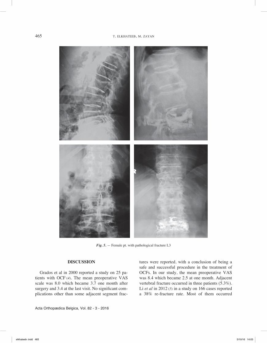
Fig. 5. — Female pt. with pathological fracture L3
elkhateeb-.indd 465 5/10/16 14:03

Acta Orthopædica Belgica, Vol. 82 - 3 - 2016
managEmEnt of ostEoporotic comprEssion vErtEbral fracturEs 466
REFERENCES
1. Cook F, Keefe F, Jensen MP, Roddey TS et al. Development and validation of the Visual Analogue Scale (VAS) Spine Score. Unfallchirurg 2001 ; 104 : 488-97.
2. Cotton A, Boutry N, Cortet B, Assaker R et al. Percutaneous vertebroplasty : state of the art.Radiographics. 1998 ; 18 : 311-20.
3. Galibert P, Deramond H, Rosat P, Le Gars D. Preliminary note on the treatment of vertebral angioma by percutaneous acrylic vertebroplasty. Neurochirurgie 1987 ; 33 : 166-8.
4. Grados F, Depriester C, Cayrolle G, Hardy et al. Long-term observations of vertebral osteoporotic fractures treated by percutaneous vertebroplasty. Rheumatology 2000 ; 39 : 1410-4.
5. Li A, Lin CL, Chang MC, Liu CL, Chen TH, Lai SC. Subsequent vertebral fracture after vertebroplasty : inci-dence and analysis of risk factors. Spine 2012 ; 37 : 179-83.
6. Lombardi I, Jr, Oliveira M, Mayer AF et al. Evaluation of pulmonary function and quality of life in women with osteoporosis. Osteoporos Int. 2005 ; 16 : 1247-53.
7. Lotfinia I, Sayyahmelli S. Complications of percutaneous vertebroplasty : a clinical study and literature review. Neurosurg Q. 2010 ; 20 : 241-6.
8. Lu Y, Genant K, Shepherd J et al. Classification of osteoporosis based on bone mineral densities. J Bone Miner Res. 2001 ; 16 : 901-10.
9. Phillips FM. Minimally invasive treatments of osteoporotic vertebral compression fractures. Spine 2003 ; 28 : 45-53.
10. Pizzoli AL, Brivio R, Caudana R, Vittorini E. CT-guided percutaneous vertebroplasty : personal experience in the treatment of osteoporotic fractures and dorsolumbar metastases. Radiol Med 2008 ; 113 : 114-33 .
11. Tanigawa N, Komemushi A, Kariya S, Kojima K, et al. Percutaneous vertebroplasty : relationship between vertebral body bone marrow edema pattern on MR images and initial clinical response. Radiology 2006 ; 239 : 195-200.
within the first three months and was positively correlated with the volume of injected cement. Our re-fracture rate was somewhat lower (5.3%), in comparison with Li et al study, and occurred adja-cent to the previously injected vertebra. Lotfinia and Sayyahmelli in 2010 reported that their main com-plication (7) is leakage of the bone cement the prev-alence was disc space leakage in 23.3%, epidural and foraminal leakage in 20%, and venous epidural leak in 6.7%. In comparison to our study, the preva-lence of disc space leakage was 32.3%, paraverte-bral leakage was 15.4% and spinal canal leakage was 1.4%.
CONCLUSION
This mini-invasive procedure should be carried out by experienced surgeons in a well equipped the-atre for the possibility of immediate neurologic de-compression. It can immediately and significantly reduce pain and improve the quality of life of these patients.
Table IType of complication IncidenceCement leakage into disc space 23 levelCement leakage paravertebral 11 levelCement leakage into spinal canal 1 levelAdjacent vertebral fracture 3 cases
elkhateeb-.indd 466 5/10/16 14:03