management of invasive cervical resorption: … of invasive cervical resorption: observations from...
TRANSCRIPT

Case Report/Clinical Techniques
Management of Invasive Cervical Resorption: Observationsfrom Three Private Practices and a Report of Three CasesRichard S. Schwartz, DDS, J. William Robbins, DDS, MA, and Eric Rindler, DDS
Abstract
Invasive cervical resorption (ICR) is a type of externalresorption that is not well understood or well knownin the dental community. It is often misdiagnosed,leading to improper treatment or unnecessary loss ofthe tooth. Treatment may involve the periodontium aswell as the tooth and pulp, and management can becomplex. Early diagnosis and appropriate treatmentare the keys to a successful outcome. This articlediscusses the decision-making process and manage-ment of ICR, with emphasis on the restorative aspectsof treatment. Three treatment cases are presentedthat include nonsurgical and surgical approaches, withrecalls of 4, 8, and 9.5 years. (J Endod 2010;36:1721–1730)Key WordsExternal resorption, extracanal invasive resorption, inva-sive cervical resorption, trichloroacetic acid
From Private Practice, San Antonio, TX.Address requests for reprints to Dr Richard Schwartz, 1130
E. Sonterra Blvd., Suite 140, San Antonio, TX 78258. E-mailaddress: [email protected]/$0 - see front matter
Copyright ª 2010 American Association of Endodontists.doi:10.1016/j.joen.2010.06.011
JOE — Volume 36, Number 10, October 2010
Invasive cervical resorption (ICR) is a common clinical entity. In 2009, the endodontistauthor and his partner diagnosed ICR 49 times in their private practice. Although
common, ICR is not well understood within the dental community and is often undiag-nosed or misdiagnosed. Even when diagnosed correctly, there is often disagreement orconfusion about the best course of treatment, even within the endodontic community.
ICR invades the tooth from the PDL, apical to the epithelial attachment (1–4), andis not evident clinically in the early stages. In many cases, it is first detectedradiographically (1–3) or when the tooth takes on a pink appearance because ofdeep red granulation tissue showing through the tooth structure (1–3).
On periapical radiographs, ICR may be a barely discernable radiolucency ordramatically evident. The lesions vary from well-delineated radiolucencies that are quiteobvious to poorly defined lesions with irregular borders and sometimes resemblecaries radiographically. When ICR is superimposed in the pulp space, pulp spaceanatomy is usually evident (4).
ICR is often seen in the cervical area of the tooth, but because it is initiated apical tothe epithelial attachment, it can present anywhere in the root (4). In the early stages, itmay be somewhat symmetrical, but the larger lesions tend to be asymmetrical (3). It canexpand apically or coronally (Fig. 1; see case 3).
Heithersay (1, 2, 5, 6) wrote a classic series of articles in which he described thefeatures, possible predisposing factors, and recommended treatment regimen for ICR.He described his treatment regimen (2, 6), which included mechanical and chemicaldebridement of the resorptive lesions, followed by restoration, and analyzed thetreatment results. For the small, localized lesions (class 1 or 2), he reported thatsuccessful treatment was close to 100%. For the moderate-size lesions (class 3), he re-ported a 77.8% success rate. For the extensive, class 4 lesions, his success rate was only12.5%.
Part of the confusion about ICR is that it is identified in the literature by at least ninedifferent names. Heithersay (1) coined the name invasive cervical resorption used inthis article. It is sometimes referred to as extracanal invasive resorption based on anarticle by Frank and Backland in 1987 (7) and was recently labeled as external cervicalresorption (ECR) in an excellent review article by Patel et al in 2009 (3).
Much of the literature pertaining to treatment of ICR is in the form of case reportswith a short-term follow-up. Hiremath et al (8) described the treatment of a tooth withICR with 1-year recall, and the restoration of ICR lesions is described in case reports byBaratto-Filho et al (9) with a 2-year recall, Park and Lee (10) with a 27-month recall,and Yilmaz et al (11) with a 1-year recall. The endodontic literature generally lackslong-term follow-up of teeth with the treatment of ICR.
Collectively, the authors, an endodontist, a periodontist, and a restorative dentist,have treated hundreds of patients with ICR, usually with an interdisciplinary approach.This article discusses their observations and provides clinical guidance for clinicians forwhat is often a difficult clinical challenge. Early diagnosis, elimination of the resorption,and restorative management are the keys to a successful outcome. Three clinical casesare presented using different treatment approaches with recalls of 4, 8, and 9.5 years.
Treatment PlanningWhen ICR is diagnosed, there are generally three choices for treatment: (1) no
treatment with eventual extraction when the tooth becomes symptomatic; (2) imme-diate extraction; or (3) access, debridement, and restoration of the resorptive lesion.
Management of Invasive Cervical Resorption 1721

Figure 1. An example of advanced ICR. The resorption has hollowed out thecoronal tooth structure but note that the outline of the pulp space is stillevident through the resorption. (Courtesy of Dr Tim Silbert, Perth, Australia.)
Case Report/Clinical Techniques
Dental implants have led to the increasing use of options 1 and 2. If notreatment is chosen, a tooth may go many years without symptoms(Fig. 2). The authors generally recommend options 1 or 2 to theirpatients for class 3 and class 4 lesions. Also, location and estheticconcerns may dictate option 1 or 2. If the decision is made to pursueoption 3, nonsurgical debridement may be possible, or a combinednonsurgical/surgical approach may be required (12). The cases thatfollow show examples of both.
Figure 2. The progression of ICR is unpredictable. These radiographs of a mandilittle change of the ICR as compared with the tooth shown in Figures 9A and B.
1722 Schwartz et al.
TreatmentThe authors use the Heithersay approach to debridement of the
resorptive lesion. The lesion is accessed, whether internally or exter-nally, and debrided with a carbide round bur in a slow-speed hand-piece. All the obvious resorptive tissue is removed until smooth,clean dentin is present, except a few small spots that are discoloredor bleeding, which represent communication of the resorption withthe PDL (Fig. 3). The dentin is then scrubbed for 1 minute with 90%aqueous trichloroacetic acid (TCA) (2, 6) on a cotton ball. TCA isvery caustic and cauterizes the residual resorptive tissue (2, 6),which makes it more obvious under magnification. Additional toothstructure is removed carefully with a slow-speed round bur, and theacid is applied again. This process is continued until all the penetrationpoints are eliminated (in the surgical approach) or perforation throughthe external root surface is imminent in the nonsurgical approach. Fornonsurgical treatment, it is impossible to eliminate all the penetrationpoints, and you must rely on the TCA to cauterize any resorptive tissuethat remains. If any of the invading resorptive tissue remains viable, theresorptive process is likely to continue.
Patient 1: A Moderate-size Lesion Treated Internallywithout Surgery
Patient 1 (Fig. 4) was a 47-year-old white man who presented in2002 with no symptoms. His restorative dentist noted the odd radio-graphic appearance of the pulp space in tooth #2 and referred himfor endodontic evaluation. There was no evidence of the lesion onthe external surface of the tooth. The referring doctor’s tentative diag-nosis was internal resorption. The patient reported none of the possiblepredisposing factors for ICR (2, 3, 5), such as orthodontics or trauma.
Endodontic testing found that the tooth was nontender to pressureand percussion and responded normally to cold compared with hisother teeth. There were no significant periodontal pockets. The
bular right central incisor were taken almost 10 years apart. There is relatively
JOE — Volume 36, Number 10, October 2010

Figure 3. A clinical photograph showing the ICR ‘‘penetration points’’ insidea tooth. (Courtesy of Dr Marga Ree, Amsterdam, Netherlands.)
Case Report/Clinical Techniques
endodontic diagnosis was normal pulp and normal periapex. Theradiolucent lesion was diagnosed as ICR.
The patient was presented with three options: (1) no treatmentwith eventual extraction of the tooth when it becomes symptomatic,(2) extraction now, and (3) endodontic treatment followed by internaldebridement and restoration. Because of the location and size of thelesion, excessive bone removal would have been necessary for a surgicalapproach, so this was not considered. The patient opted for option #3.
Access was made into the resorptive lesion through the occlusalsurface of the tooth. The lesion was extremely hemorrhagic. Grossdebridement was accomplished with a #6 round carbide bur in
Figure 4. Patient 1. The internal treatment of a maxillary right second molar is shotooth is asymptomatic, and there is no evidence of ICR.
JOE — Volume 36, Number 10, October 2010
a slow-speed handpiece. As the bulk of resorptive tissue was removed,the hemorrhage decreased. Periodic radiographs were taken as theexternal surface of the tooth was approached. The external surfacewas never penetrated with a bur. Debridement of the lesion was accom-plished by alternating the round bur with trichloroacetic acid as previ-ously described.
Numerous small bleeding points were evident in the cervical areaafter initial debridement. After the dentin was treated with TCA, thebleeding stopped, and the penetration points looked like small darkspots and could be seen clearly under the operating microscope.
The initial debridement caused the pulp to be exposed, andendodontic treatment was initiated. Four canals were located andprepared and dressed with calcium hydroxide paste (Ultracal; UltradentProducts, Provo, UT). At the second appointment, the canals were ob-turated with gutta-percha and Kerr sealer (Kerr Corp, Orange, CA.), thedentin surface was ‘‘refreshed’’ with a bur, and the lesion and accessopening were restored with a ‘‘fourth-generation’’ dentin bondingsystem (Optibond Dual Cure; Sybron Dental Specialties, Orange, CA)and a dual-cure buildup material (BuildIt; Sybron Dental Specialties,Orange, CA.). After a 3-month waiting period in which the tooth re-mained asymptomatic, it was restored with a metal-ceramic crown.The final radiograph in Figure 4 is an 8-year recall. The patient’s toothwas asymptomatic, and there was no evidence that the ICR had pro-gressed.
Patient 2: A Relatively Small Lesion with Easy SurgicalAccess
Patient 2 (Fig. 5) was a 39-year-old Hispanic woman who pre-sented in 2006 with no symptoms. Her restorative dentist noteda moderate-size oval radiolucency in the cervical area of #23 andreferred her for evaluation. He noted a pink discoloration on the labialsurface of the tooth and a ‘‘catch’’ in the subgingival area with an
wn. The last two radiographs were taken at an 8-year recall appointment. The
Management of Invasive Cervical Resorption 1723
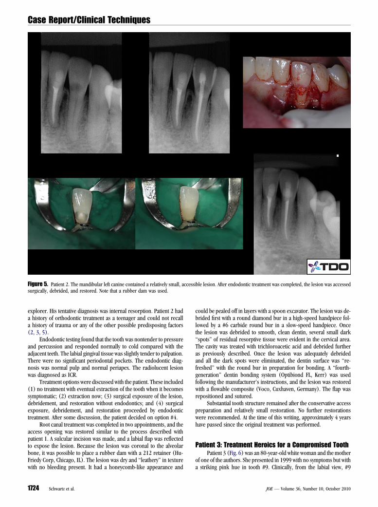
Figure 5. Patient 2. The mandibular left canine contained a relatively small, accessible lesion. After endodontic treatment was completed, the lesion was accessedsurgically, debrided, and restored. Note that a rubber dam was used.
Case Report/Clinical Techniques
explorer. His tentative diagnosis was internal resorption. Patient 2 hada history of orthodontic treatment as a teenager and could not recalla history of trauma or any of the other possible predisposing factors(2, 3, 5).
Endodontic testing found that the tooth was nontender to pressureand percussion and responded normally to cold compared with theadjacent teeth. The labial gingival tissue was slightly tender to palpation.There were no significant periodontal pockets. The endodontic diag-nosis was normal pulp and normal periapex. The radiolucent lesionwas diagnosed as ICR.
Treatment options were discussed with the patient. These included(1) no treatment with eventual extraction of the tooth when it becomessymptomatic; (2) extraction now; (3) surgical exposure of the lesion,debridement, and restoration without endodontics; and (4) surgicalexposure, debridement, and restoration proceeded by endodontictreatment. After some discussion, the patient decided on option #4.
Root canal treatment was completed in two appointments, and theaccess opening was restored similar to the process described withpatient 1. A sulcular incision was made, and a labial flap was reflectedto expose the lesion. Because the lesion was coronal to the alveolarbone, it was possible to place a rubber dam with a 212 retainer (Hu-Friedy Corp, Chicago, IL). The lesion was dry and ‘‘leathery’’ in texturewith no bleeding present. It had a honeycomb-like appearance and
1724 Schwartz et al.
could be pealed off in layers with a spoon excavator. The lesion was de-brided first with a round diamond bur in a high-speed handpiece fol-lowed by a #6 carbide round bur in a slow-speed handpiece. Oncethe lesion was debrided to smooth, clean dentin, several small dark‘‘spots’’ of residual resorptive tissue were evident in the cervical area.The cavity was treated with trichloroacetic acid and debrided furtheras previously described. Once the lesion was adequately debridedand all the dark spots were eliminated, the dentin surface was ‘‘re-freshed’’ with the round bur in preparation for bonding. A ‘‘fourth-generation’’ dentin bonding system (Optibiond FL, Kerr) was usedfollowing the manufacturer’s instructions, and the lesion was restoredwith a flowable composite (Voco, Cuxhaven, Germany). The flap wasrepositioned and sutured.
Substantial tooth structure remained after the conservative accesspreparation and relatively small restoration. No further restorationswere recommended. At the time of this writing, approximately 4 yearshave passed since the original treatment was performed.
Patient 3: Treatment Heroics for a Compromised ToothPatient 3 (Fig. 6) was an 80-year-old white woman and the mother
of one of the authors. She presented in 1999 with no symptoms but witha striking pink hue in tooth #9. Clinically, from the labial view, #9
JOE — Volume 36, Number 10, October 2010

Figure 6. Patient 3. This large lesion was debrided and restored. It lasted almost 10 years before the tooth fractured. A is preoperative, B is during the restorativephase, and C is after endodontic and restorative treatments were completed. D shows the postoperative clinical result. The fact that the labial enamel was intact madean acceptable esthetic result possible. The radiograph in E was taken at the 3-year recall. The tooth fractured shortly after the radiograph in F was taken, approx-imately 9.5 years after treatment was completed.
Case Report/Clinical Techniques
looked like a normal, intact tooth other than the color. From the palatalaspect, the external surface of the tooth had broken down in the cervicalarea. The radiographic appearance was somewhat shocking. The toothwas nontender, but the pulp was nonresponsive to cold. The endodonticdiagnosis was necrotic pulp with a normal periapex. The resorptivelesion was diagnosed as ICR.
After discussion among the authors, the following treatmentoptions were discussed: (1) extraction or (2) heroic efforts to treatthe tooth. The patient’s preference was to save the tooth if possible.Because her son was a dentist who would provide close follow-up,heroic efforts were undertaken.
Initial access was made through the palatal surface of the tooth,and initial debridement was completed without reflecting a flap. Theresorptive tissue was similar to patient 2 in that it was dry and leathery.Root canal treatment was completed, and the canal was temporarilysealed at the orifice. A palatal flap was reflected, and debridement ofthe resorptive lesion was completed as previously described. A smallamount of alveolar bone was removed in the cervical area on the palatalaspect of the tooth. A post space was created, and a zirconium post(Cosmopost; Ivoclar Vivadent, Amherst, NY) was cemented with Vi-tremer cement (3M, St Paul, MN). The tooth was restored with an‘‘open sandwich’’ technique. The dentin was conditioned with 10% pol-
JOE — Volume 36, Number 10, October 2010
yacrylic acid for 15 seconds. A resin-modified glass ionomer material(Fuji 2 LC; ESPE, St. Paul, MN) was used to restore the majority ofthe preparation. The incisal one quarter of the restoration was preparedwith a bevel of the remaining enamel and was restored with a bondedcomposite resin (Scotchbond Multipurpose, 3M) and Z100 composite(3M).
The patient had regular follow-ups by her son, and #9 functionednormally until it fractured in 2009. The final radiograph in Figure 6F isthe 9.5-year recall.
DiscussionPatient 3 is not an example of the typical treatment approach for
a patient with such extensive ICR. In most cases, her tooth would beextracted. The circumstances and patient desires strongly influencedthe treatment plan. This case shows that even with an extensivelesion, ICR can be arrested and treated successfully over the long-term. The eventual failure was structural and not endodontic orcaused by recurrence of the resorption. Not surprisingly, the crownfractured in the cervical area. The post remained cemented in thecanal. In retrospect, zirconium was probably not the best choicefor a post because it cannot be etched or effectively bonded to
Management of Invasive Cervical Resorption 1725

Figure 7. An example of internal resorption. Although ICR tends to be offcenter, diffuse, and asymmetrical, internal resorption tends to be symmetricaland centered on a canal. (Courtesy of Dr Charles Maupin, Lubbock, TX.)
Figure 8. External inflammatory resorption typically occurs as a result oftrauma. It is associated with a necrotic, infected pulp. The radiographicpresentation is a ‘‘moth eaten’’ appearance with multiple lesions at the lateralaspects of the root.
Case Report/Clinical Techniques
cements or restorative materials. Also, resin-modified glass ionomermaterials are generally not as strong as composite resins. It ispossible that had the tooth been restored with a different type ofpost in combination with a composite resin material, the restorativetreatment might have provided even greater longevity.
Purely internal treatment of ICR is preferable when possible. Thereis the obvious desire of most patients to avoid surgery, plus there is noneed to remove alveolar bone from the treated tooth and adjacent teeth.Although it is impossible to totally eliminate the resorption from insidethe tooth, careful mechanical and chemical debridement can stop theresorptive process and result in long-term success as shown withpatient 1. This approach is viable only if the external surface of the toothremains grossly intact.
When an external approach is necessary and the lesion is acces-sible, a rubber dam can sometimes be used for isolation as shown inpatient 2. The trichloroacetic acid is very caustic and will cause burnsif it comes in contact with the gingival tissues. The rubber dam alsoprovides better visualization and isolation for the restorative proce-dures. A rubber dam is always recommended for the endodontic treat-ment and the internal debridement approach.
Dentin that has been treated with TCA is severely demineralizedand is not suitable for bonding with either dentin-bonding agents orglass ionomer materials. It must be ‘‘refreshed’’ with a bur beforebonding procedures. Dentin bonding agents rely on a shallow demin-eralization of the dentin surface, which is infiltrated with a resin toform a hybrid layer with the exposed dentinal collagen matrix. Glass ion-omer materials rely primarily on ionic bonding to the calcium inhydroxyapatite. A TCA-treated surface is demineralized to too greata depth for the dentin-bonding agents to fully infiltrate, resulting in
1726 Schwartz et al.
a weak, leaky bond. TCA almost totally eliminates the hydroxyapatitefrom the dentin surface, so there is no calcium available for bondingby glass ionomer materials. Therefore, before any bonding proceduresare initiated, the surface should be refreshed with a bur to providea normal dentin surface.
Mineral trioxide aggregate (Dentsply International, York, PA) hasbeen recommended in several case reports (8–11) as the restorativematerial of choice because it is ‘‘biocompatible.’’ The authors useresin-modified glass ionomer materials or composite resins becausethey are stronger, they bond to tooth structure, and they can be exposedto the oral cavity. There are no known benefits to the use of MTA torestore ICR lesions.
The authors have observed three clinical presentations of theresorptive tissue. When entering an intact tooth, the resorptive tissueis usually hemorrhagic and soft. When the external tooth surface hasbroken down, ICR presents as a dry, leathery lesion with a honey-combed appearance under a microscope, which is sometimes mis-diagnosed as caries. Occasionally, ICR looks like bone that hasinvaded the tooth.
Patient 2 had endodontics performed before restoration of theresorptive lesion. Heithersay (6) reported high success rates withoutendodontic treatment in type 1 and type 2 cases of ICR. It has been
JOE — Volume 36, Number 10, October 2010
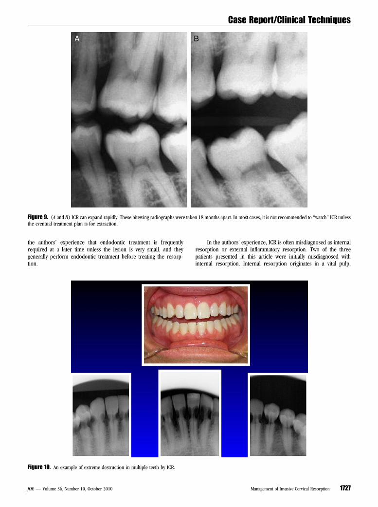
Figure 9. (A and B) ICR can expand rapidly. These bitewing radiographs were taken 18 months apart. In most cases, it is not recommended to ‘‘watch’’ ICR unlessthe eventual treatment plan is for extraction.
Case Report/Clinical Techniques
the authors’ experience that endodontic treatment is frequentlyrequired at a later time unless the lesion is very small, and theygenerally perform endodontic treatment before treating the resorp-tion.
Figure 10. An example of extreme destruction in multiple teeth by ICR.
JOE — Volume 36, Number 10, October 2010
In the authors’ experience, ICR is often misdiagnosed as internalresorption or external inflammatory resorption. Two of the threepatients presented in this article were initially misdiagnosed withinternal resorption. Internal resorption originates in a vital pulp,
Management of Invasive Cervical Resorption 1727
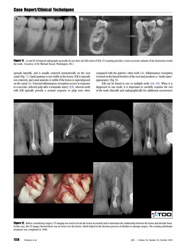
Figure 11. (A and B) Periapical radiographs generally do not show the full extent of ICR. CT scanning provides a more accurate estimate of the destruction insidethe tooth. (Courtesy of Dr Michael Pascal, Washington, DC.)
Case Report/Clinical Techniques
spreads laterally, and is usually centered symmetrically on the rootcanal (Fig. 7). Canal anatomy is not visible in the lesion. ICR is typicallynot centered, and canal anatomy is visible if the lesion is superimposedon the canal (4). External inflammatory resorption occurs in responseto a necrotic, infected pulp after a traumatic injury (13), whereas teethwith ICR typically provide a normal response to pulp tests when
Figure 12. Before considering surgery, CT imaging was used to locate the lesion accIn this case, the CT images showed there was no bone over the lesion, which helpedtreatment was completed in 1990.
1728 Schwartz et al.
compared with the patient’s other teeth (4). Inflammatory resorptionis found on the lateral borders of the root and produces a ‘‘moth-eaten’’appearance (Fig. 8).
ICR can be found in one or multiple teeth (14, 15). When it isdiagnosed in one tooth, it is important to carefully examine the restof the teeth clinically and radiographically for additional occurrences
urately and to determine the relationship between the lesion and alveolar bone.in the decision process of whether to attempt surgery. The existing endodontic
JOE — Volume 36, Number 10, October 2010

Figure 13. Clinical and radiographic views of an extracted tooth with ICR. Note how the resorptive tracts spread apically and out to the PDL. (Courtesy of Dr GaryCarr, San Diego, CA.)
Case Report/Clinical Techniques
of ICR. If a lesion is diagnosed when it is small, it can be treated withminimal morbidity, whereas large lesions may be untreatable. Whena small lesion is diagnosed in an accessible location, immediatetreatment is recommended because the lesions sometimes spreadrapidly (Fig. 9A and B) and can be extremely destructive (Fig. 10). Itis not a good idea to tell the patient, ‘‘We’ll watch it,’’ unless the planis for extraction at a later time.
Computed tomography (CT) scanning can be very useful intreating ICR. Periapical radiographs tend to underestimate the sizeof the resorptive lesion. CT scanning gives a more accurate estimateof lesion size (16, 17) and, in some cases, may discourage treatment(Fig. 11A and B). CT images also provide the precise location of the
Figure 14. The same tooth as shown in Figure 13 after clearing. (Courtesy of Dr
JOE — Volume 36, Number 10, October 2010
lesion in three dimensions and the relationship to the surroundingbone, which can be very helpful in decision making (Fig. 12). Acommon finding in the larger lesions is finger-like projectionsfrom the main body of resorption that extend apically and out tothe PDL (4), which can make it difficult to eliminate all the invadingresorptive tissue. An example is shown in an extracted tooth inFigures 13 and 14.
Orthodontic extrusion is sometimes helpful in patients with ICR.Extrusion provides better access to ICR lesions and allows the finalboney and gingival architecture to be more ideal when surgery is neces-sary (12). ICR is by far the most diagnosed type of resorption in theendodontist author’s private practice. In 2009, he and his partner
Carr.)
Management of Invasive Cervical Resorption 1729

Case Report/Clinical Techniques
diagnosed ICR 49 times, as previously stated. By comparison they diag-nosed internal resorption three times and inflammatory resorption 11times.These three cases show that ICR can be arrested using the ‘‘Hei-thersay approach’’ to treatment (ie, mechanical debridement, treatmentwith TCA, and restoration). Prudent case selection and proper execu-tion can lead to the successful treatment and long-term retention ofthe tooth. The keys are the location, size, and accessibility of the lesionand the structural integrity of the tooth and periodontium after treat-ment is completed.
It is important for endodontists to understand the periodontal andrestorative aspects of treating ICR. Teeth with ICR are often structurallycompromised and may eventually fail even though the endodontic treat-ment is successful. The endodontic treatment is irrelevant if the resorp-tion is not eliminated, and the restorative aspects are not managedproperly. Proper management requires knowledge and skills inendodontics, surgery, and restorative dentistry, and elimination of theresorption is performed most effectively under a microscope. Even ifthe endodontist does not perform all the treatment, he/she must beknowledgeable of all aspects of the treatment to direct the treatmentteam. Frequently, nobody else is quite sure what to do with ICR.
References1. Heithersay GS. Clinical, radiologic, and histopathologic features of invasive cervical
resorption. Quintessence Int 1999;30:27–37.2. Heithersay GS. Invasive cervical resorption. Endod Topics 2004;7:73–92.3. Patel S, Kanagasingam S, Pitt Ford T. External cervical resorption: a review. J Endod
2010;35:616–25.
1730 Schwartz et al.
4. Bergmans L, Van Cleynenbreugel J, Verbeken E, et al. Cervical external root resorp-tion in vital teeth. X-ray microfocustomographical and histopathological case study.J Clin Periodontol 2002;29:580–5.
5. Heithersay GS. Invasive cervical resorption: an analysis of potential predisposingfactors. Quintessence Int 1999;30:83–95.
6. Heithersay GS. Treatment of invasive cervical resorption: an analysis of results usingtopical application of trichloracetic acid, curettage, and restoration. QuintessenceInt 1999;30:96–110.
7. Frank Al, Backland LK. Non endodontic therapy for supra osseous extracanal inva-sive resorption. J Endod 1987;13:348–55.
8. Hiremath H, Yakub SS, Metgud S, et al. Invasive cervical resorption: a case report.J Endod 2007;33:999–1003.
9. Baratto-Filho F, Limongi O, Araujo Cde J, et al. Treatment of invasive cervical resorp-tion with MTA: case report. Aust Endod J 2005;31:76–80.
10. Park JB, Lee JH. Use of mineral trioxide aggregrate in the non-surgical repair ofperforating invasive cervical resorption. Med Oral Patol Oral Cir Bucal 2008;13:E678–80.
11. Yilmaz HG, Kalender A, Cengiz E. Use of mineral trioxide aggregate in the treatmentof invasive cervical resorption: a case report. J Endod 2010;36:160–3.
12. Smidt A, Nuni E, Keinan D. Invasive cervical root resorption: treatment rationale withan interdisciplinary approach. J Endod 2007;33:1383–7.
13. Andreasen JO. External root resorption: its implications in dental traumatology, pae-dodontics, periodontics, orthodontics and endodontics. Int J Endod 1985;8:109–18.
14. Coyle M, Toner M, Barry H. Multiple teeth showing invasive cervical resorption—anentity with little known histologic features. J Oral Pathol Med 2006;35:55–7.
15. Mattar R, Pereira SA, Rodor RC, et al. External multiple invasive cervical resorptionwith subsequent arrest of the resorption. Dent Traumatol 2008;24:556–9.
16. Cohenca N, Simon JH, Mathur A, et al. Clinical indications for digital imagingin dento-alveolar trauma. Part 2: root resorption. Dent Traumatol 2007;23:105–13.
17. Gulsahi A, Gulsahi K, Ungor M. Invasive cervical resorption: clinical and radiologicaldiagnosis and treatment of 3 cases. Oral Surg Oral Med Oral Pathol Oral RadiolEndod 2007;103:e65–72.
JOE — Volume 36, Number 10, October 2010