management of enterocutaneous fistula fistula.pdf · management of enterocutaneous fistula kelley...
TRANSCRIPT
Management of Enterocutaneous Fistula
Kelley A. Sookraj, MDKings County Hospital Center
August 25th 2011
www.downstatesurgery.org
• CC: drainage of fluid and air from umbilicus
• HPI: This is a 56 y/o female who underwent a subtotal colectomy with ileorectal anastomosis for recurrent diverticulitis with stricture and obstructive symptoms in December 2010 at another institution.
Postoperatively, she developed an intra-abdominal abscess which was drained by IR, and she was placed on TPN and broad-spectrum antibiotics for presumed leak.
Case Presentationwww.downstatesurgery.org

• HPI: cont’d
After discharge home on oral antibiotics, she presented to KCHC in March 2011 for a second opinion regarding drainage from her umbilicus which was being treated as a suture granuloma.
Case Presentationwww.downstatesurgery.org
• PMHx: asthma, diverticulitis, morbid obesity
• PSHx: subtotal colectomy 12/2010
• Allergies: NKDA
• Meds: zyvox, claritin, albuterol inhaler
• SHx: denies smoking, ETOH, IVDA
• FHx: non-contributory
www.downstatesurgery.org

• Radiologic StudiesBarium Enema: contrast filled tract arising from distal ileum adjacent to ileorectal anastomosis
www.downstatesurgery.org

CT scan of Abd/Pelvis: 2.3 x 1.6 x 9cm collection of contrast and gas which appears to be arising from the ileorectal anastomosis
www.downstatesurgery.org

•Flexible Sigmoidoscopy: no evidence of IBD/ lesions
•Vitals: Temp 97.8 F BP 132/64 HR 86 RR 16
• Physical Exam:General: AAOx3Chest: CTA bilaterallyCVS: S1S2, rrrAbdomen: midline scar, drainage at umbilicus, soft, NT, +BSRectal: good tone, no gross blood
www.downstatesurgery.org

• Labs:
CBC: 5.81 / 10.2 / 31.4 / 377
Chem: 139 / 5.0 / 106 / 24 / 15 / 0.58 / 105
LFTs: 6.8 / 3.4 / 32 / 18 / 72 / 0.1
Coags: 9.7 / 23 / 0.9
www.downstatesurgery.org

• intra-op: placement of bilateral ureteral stents, exploratory laparotomy, lysis of adhesions, revision of ileorectal anastomosis, intraoperative colonoscopy
• pathology: perforated ileorectal anastomosis, moderate chronic inflammation
www.downstatesurgery.org

• post-op:
POD #0-3: extubated, A-line and foley d/ced, abdominal binder in place, ambulating
POD #4: SOB, wheezing, tachypneic, tachycardic, pain over midline wound, RA ABG pO2 66, CTA neg PE
emesis x1, resp distress requiring intubation, transferred to SICU, febrile, hypotensive, sepsis protocol initiated, midline wound opened, fascial defect at level of umbilicus
POD #5: CT scan Abd/Pelvis: no free air or obvious leak
POD #6:malodorous/feculent drainage from midline wound, TPN, octreotide
www.downstatesurgery.org
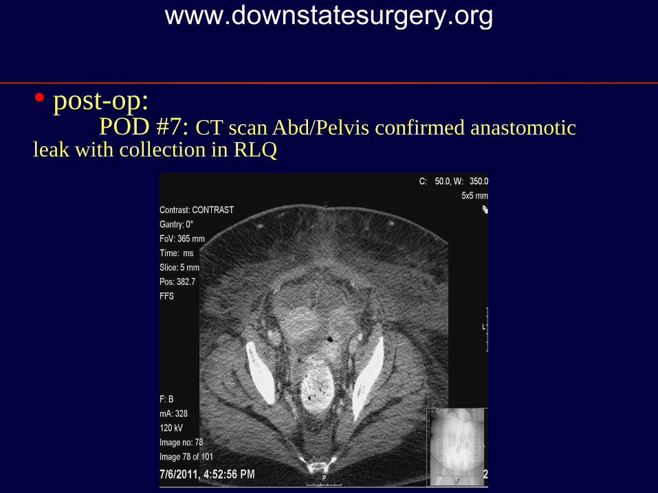
• post-op: POD #7: CT scan Abd/Pelvis confirmed anastomotic
leak with collection in RLQ
www.downstatesurgery.org

• post-op:
POD #8: Pt taken to OR for abdominal washout and possible creation of diverting ostomy
Intra-op: frozen abdomen, washout and drainage with placement of Malecot drain near anastomotic leak and 2 large sump drains ( LLQ anterior to anastomosis, RLQ posterior ), fascial defect closed with Surgisis and vac dressing applied
POD #15: CT scan abd/pelvis – evacuation of previous collection, drains in place with no collection
www.downstatesurgery.org

• post-op:
POD #27: tracheostomy performed
POD #51: BE showed no evidence of leak or filling of LLQ drains
Currently: hemodynamically stable, on TC, (+) flatus and BM, minimal drainage from Malecot and left sump drains, wound granulating with vac dressing in place
www.downstatesurgery.org

• Enterocutaneous Fistula – abnormal connection between small or large bowel and skin
• Complicated by abscess formation, sepsis, electrolyte derangements, dehydration and malnutrition
• Despite advances with antimicrobial and nutritional therapy, overall mortality rate ranges from 5-20%
Enterocutaneous Fistula www.downstatesurgery.org

• Epidemiology- 75% postoperative- 15-25% trauma, cancer, irradiation, IBD, ischemic
or infective diseases (TB)
• Etiology - majority of fistula formation occurs following
abdominal surgery and is related to tissue integrity and healing
Enterocutaneous Fistula www.downstatesurgery.org
• Presentation
- abdominal pain, distension- tachycardia- fever- drainage of enteric contents from wound- peritonitis
Enterocutaneous Fistula www.downstatesurgery.org

• Differential Diagnosis
Anastomotic leak Unrecognized enterotomy Fascial suture through bowel Ischemic bowel segment Erosion into bowel by suction drains or mesh Perforated viscous (e.g., peptic ulcer or colonic diverticula) Local inflammation (e.g., Crohn's disease)
Enterocutaneous Fistula www.downstatesurgery.org

• Classification
Low output < 200 cc/day
Moderate output 200-500 cc/day
High output >500 cc/day
Enterocutaneous Fistula www.downstatesurgery.org

• Factors Inhibiting Spontaneous Closure
High fistula output (>500 ml/24 hours) Short fistula tract Abscess Distal bowel obstruction Foreign body Malignancy Radiation Steroids Chronic epithelialization
Enterocutaneous Fistula www.downstatesurgery.org

• Diagnosis
- GI contrast study ( UGI series, BE )
- CT scan
- fistulogram
Enterocutaneous Fistula www.downstatesurgery.org

• Treatment
- goals of therapy are early recognition and stabilization to prevent life-threatening metabolic, septic and nutritional complications
- intervention should focus on resuscitation, correction of electrolyte abnormalities, control of sepsis, nutritional support and skin care
Enterocutaneous Fistula www.downstatesurgery.org

• Treatment: Resuscitation and Electrolyte Disturbances
- aggressive intravascular volume repletion with crystalloid
- strict Is and Os, monitor urine output- replace electrolytes ( K+, Mg, HCO3)- monitor fistula output and replace- octreotide ( ↓ GI secretions )
Enterocutaneous Fistula www.downstatesurgery.org
• Treatment: Control of Sepsis
- uncontrolled sepsis is major cause of mortality- broad-spectrum antibiotics - drainage of intra-abdominal abscess
- percutaneous- surgical (resection, drainage, ostomy)
- tincture of time and adequate control may result in spontaneous closure
Enterocutaneous Fistula www.downstatesurgery.org

• Treatment: Nutritional Support- severely malnourished, high catabolic state
secondary to sepsis
- protein and caloric requirements: 1.5-2 g/kg/day; 30 kcal/kg/day
- TPN: high output fistula, uncontrolled intra-abdominal sepsis
- enteral feeds: associated with increased mucosal integrity and immunologic host defenses; contraindicated with proximal fistulas, bowel obstruction, short gut syndrome
Enterocutaneous Fistula www.downstatesurgery.org

• Treatment: Skin Care
- prevent skin maceration and breakdown from corrosive effluent; wound infection
- utilize paste, zinc, stoma appliances, vac dressing and wound care nurse to control output, protect skin and promote healing
Enterocutaneous Fistula www.downstatesurgery.org

• Treatment: Definitive Care- spontaneous closure occurs approx. 10-40% with
conservative management
- if persistent 4-8 wks after resolution of sepsis and nutritional deficits, will require definitive, elective procedure
- typically requires delineation of anatomy ( preop imaging )
- Operation: mobilization of bowel, resection of fistula and involved intestinal segment, primary anastomosis
Enterocutaneous Fistula www.downstatesurgery.org

Management of enterocutaneous fistulae:A 10 years experienceTaggarshe D, Bakston D, Jacobs Michael, Mittal, V; World J Gastrointest Surg
2010 July;2(7):242-46.
• retrospective study; 83 patients; 1997-2007
• aim: compare outcomes of conservative vs surgical treatment of enterocutaneous fistulae
• 58 women; 25 men
• MC cause of fistula was post-operative
• 66 conservative management: NPO, TPN, somatostatin analogue, control of fistula output, skin care; 43 (65%) spontaneous closure
Enterocutaneous Fistula www.downstatesurgery.org

Management of enterocutaneous fistulae:A 10 years experienceTaggarshe D, Bakston D, Jacobs Michael, Mittal, V; World J Gastrointest Surg
2010 July;2(7):242-46.
• 17, plus 18 from conservative arm underwent surgery; 28 (80%) successfully treated
• no statistical difference: length of stay, recurrence rate, mortality
• concluded: mode of treatment is individually based however, conservative management is an important bridge to definitive surgical intervention as it allows for control of sepsis, correction of nutritional status and improvement in wound management
Enterocutaneous Fistula www.downstatesurgery.org

References
1. Cameron et al. Current Surgical Therapy 9th Ed. pgs. 143-1452. Townsend, Beauchamp et al. Sabiston Textbook of Surgery 17th Ed.3. Taggarshe D, Bakston D, Jacobs Michael, Mittal, V; World J Gastrointest Surg
2010 July;2(7):242-46.4. Pritts T, et al; Postoperative enterocutaneous fistula, Surgical Treatment:
Evidence-Based and Problem-Oriented, NIH5. Vikram, K et al; Enterocutaneous Fistula Treatment and Management
http://emedicine.medscape.com6. Kaushal, M et al; Management of Enterocutaneous Fistulas; Clinics in Colon and
Rectal Surgery, 2004 pgs 79-87 7. Schecter WP, Hirshberg A et al; Enteric Fistulas: Principles of Management,
JACS, 2009 article in press
Enterocutaneous Fistula www.downstatesurgery.org