Management of Crush Victims in Mass Disasters WEB PAGE 2011 - part1.pdf · Management of Crush Victims in Mass Disasters Mehmet Şükrü Sever Raymond Vanholder Istanbul School of
30
Management of Crush Victims Management of Crush Victims in Mass Disasters in Mass Disasters Mehmet Mehmet Ş Ş ü ü kr kr ü ü Sever Sever Raymond Vanholder Raymond Vanholder Istanbul School of Medicine, Turkey University Hospital Gent, Belgium
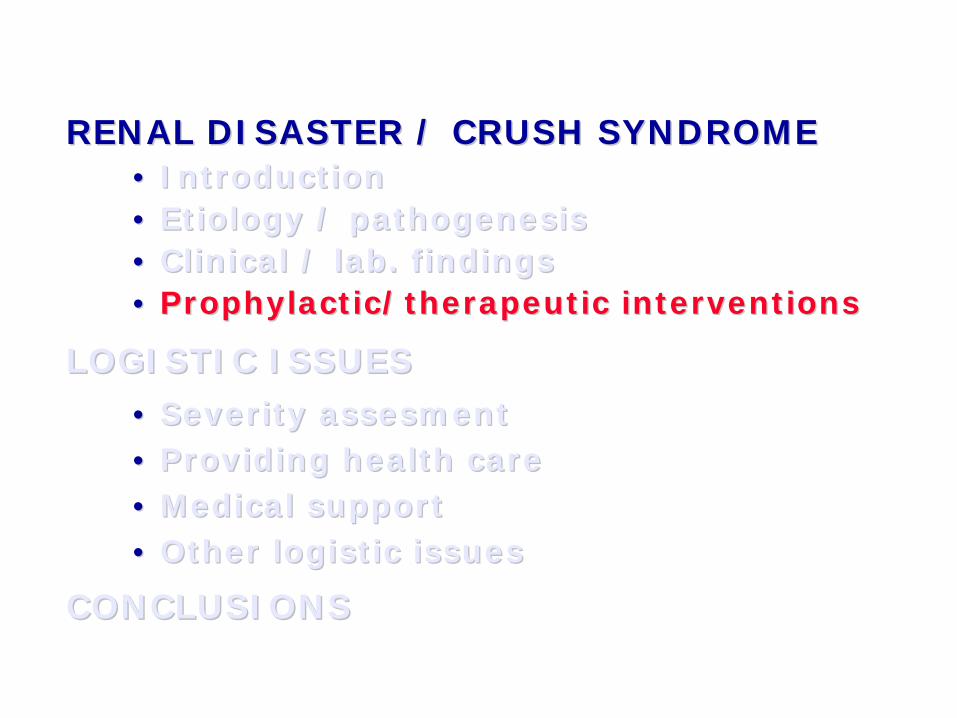
LOGISTIC ISSUESLOGISTIC ISSUES•• Severity assesment Severity assesment •• Providing health care Providing health care •• Medical support Medical support •• Other logistic issuesOther logistic issues
CONCLUSIONSCONCLUSIONS
GLOBAL SEISMIC HAZARD MAPGLOBAL SEISMIC HAZARD MAP
EARTHQUAKES: A WORLWIDE PROBLEMEARTHQUAKES: A WORLWIDE PROBLEM
Presenter
Presentation Notes
As seen on this map, many countries around the globe (indicated here in red) are located in very risky areas for earthquakes, while all countries carry the threat of man-made disasters. Thus, nephrologists, dialysis nurses and technicians should always be prepared for treating large numbers of crush syndrome casualties.
THE MARMARA EARTHQUAKE:THE MARMARA EARTHQUAKE:One of the most catastrophic DisastersOne of the most catastrophic Disasters
of the World in the 20th Centuryof the World in the 20th Century
•• 17 August, 199917 August, 1999
•• 7.4 (Richter scale)7.4 (Richter scale)
•• 45 sec45 sec
•• Deaths: 17Deaths: 17,,480480
•• Injured: 43Injured: 43,,953953
Presenter
Presentation Notes
During this presentation, the Marmara earthquake will be cited frequently because, from medical point of view, so far, this was one of the most catastrophic and best documented earthquakes. This disaster, was caused by the North Anatolian fault on the 17th of August 1999. The epicenter was close to Istanbul in the north-western part of Turkey and affected a large area. The intensity was 7.4 on the Richter’s scale; the seism lasted for 45 seconds causing more than 17,000 deaths and in total 43,000 injured.
Presenter
Presentation Notes
In the affected region, nearly 130,000 homes had partially or completely collapsed and approximately 600,000 people became homeless. Since many of the highways and railroads were heavily damaged, transport of victims was extremely difficult during the first two days of the disaster. A part of the city of Golcuk, the epicenter of the disaster, was partially submerged under the Marmara sea (right below).
The The HanshinHanshin--Awaji Awaji (Kobe) Earthquake(Kobe) Earthquake
Patients with AKI: 202Patients requiringDialysis:123
Oda et al. J Trauma 1997
The The Marmara Marmara EarthquakeEarthquake
Patients. with renal problems: 639Patients requiringdialysis: 477
Sever et al. Kidney Int 2001
The The largestlargest ““renal disasterrenal disaster”” documented so far !documented so far !
Presenter
Presentation Notes
One of the most dramatic consequences of the Marmara Earthquake was the presence of 639 crush syndrome patients, of whom 477 required dialysis support. In the until then best-documented severe earthquake, the Kobe disaster in Japan, 202 patients with acute kidney injury (AKI) of whom 123 needed dialysis support were reported. Therefore, the Marmara Earthquake was the largest “renal disaster” documented so far.
Br Med J Br Med J 1989; 298: 4431989; 298: 443--55
Presenter
Presentation Notes
Now, we are faced with an unusual terminology, “renal disaster”. What does “RENAL DISASTER” mean?? This terminology was first introduced after the Armenian earthquake, in 1988, because many victims rescued alive from under the rubble died soon afterwards due to renal failure. Why did this happen?
80% die instantly10% minor injuries10% major injuries
Ron et al. Arch Intern Med 1984
Crush syndrome
2nd most frequent cause of deaths (following direct effect of trauma)
Ukai. Ren Fail 1997
““R E N A L D I S A S T E RR E N A L D I S A S T E R””
Presenter
Presentation Notes
In a classic observation on the sudden collapse of an eight-story building, 80% of the entrapped victims instantly died by direct trauma, while 10% survived with minor injuries. The remaining 10% were badly injured; and of those, 7 out of 10 developed crush syndrome. Extrapolated to earthquakes, whereby thousands of buildings collapse, dramatic numbers of crush casualties can be expected. During earthquakes, injuries to vital organs can cause instant death. Late mortality is generally attributable to rhabdomyolysis resulting in the crush syndrome, which is the most frequent cause of death after earthquakes, apart from trauma. Therefore, next the original disaster, renal failure causes a second one, which is appropriately named “renal disaster”.
Mortality in crush s.:Mortality in crush s.:
•• Overall: 24.8% (50/202)Overall: 24.8% (50/202)
•• Dialyzed: 41% (50/123)Dialyzed: 41% (50/123)
Crush syndrome is a lifeCrush syndrome is a life--threatening disorder !threatening disorder !
Mortality rates in dialyzed crush victims:Mortality rates in dialyzed crush victims:Marmara: 17%, Taiwan: 17%; Pakistan: 19%; Iran: 13%Marmara: 17%, Taiwan: 17%; Pakistan: 19%; Iran: 13%Hwang et al. 2001; Sever et al 2004; Van der Tol et al. 2008; Hatamizadeh et al AJKD 2006
Presenter
Presentation Notes
Crush syndrome is a life-threatening disorder ! In an analysis of the 3651 victims of the Kobe earthquake, crush syndrome was found to be the second most frequent cause of death after asphyxia. In another report on this catastrophe, the mortality rate in the crush victims was high; overall mortality rate was 24.8% (50/202); and this figure turned to be 41% (50/123) in the dialyzed victims. Mortality figures are around 15 to 20% in more recent disasters, such as the Marmara, Taiwan, Iran and Pakistan earthquakes.
Crush: Crush: injury due to injury due to presspressureure between opposing between opposing elementselements
Literally, crush means “compression between opposing elements so as to break or injure”. Crush syndrome: is the whole of systemic manifestations caused by rhabdomyolysis as a result of crush. These manifestations can be summarized under 2 headings; among the medicalL features hypovolemic shock, AKI, hyperkalemia, heart failure, respiratory failure and infections may be cited, while the most important surgical features are the local consequences of trauma and most important of all, the compartment syndrome.
Disintegration of striated musclesDisintegration of striated muscles
RhabdomyRhabdomyoolysislysis
resulting in release of muscular cell contentsresulting in release of muscular cell contents
into the extracellular fluidinto the extracellular fluid
Muscles are the largest organ system in the body (40% of body weight) and their risk to be affected by trauma is very high. Rhabdomyolysis is: Disintegration of striated muscles that results in release of muscular cell contents into the extracellular fluid. Among these substances, lactic acid, thromboplastin, creatine kinase, nucleic acids, phosphate and creatine can be cited, but the most important ones are: myoglobin and potassium. Apart from fluid depletion, which will be discussed later, these substances play an important role in the pathogenesis of crush syndrome.
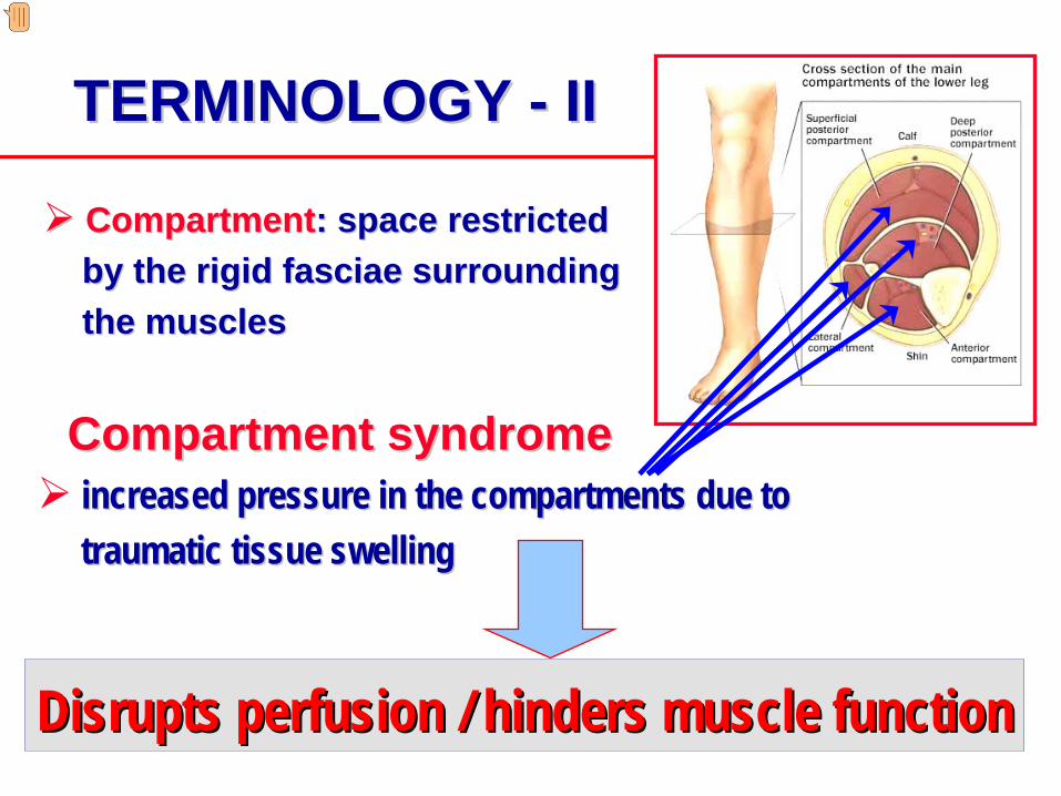
CompartmentCompartment: space : space restrictedrestrictedby the rigid fasciaby the rigid fasciaee surrounding surrounding the musclethe muscless
CCompartmompartmeenntt ssyndromeyndromeincreased pressure in the compartmentsincreased pressure in the compartments due to due to traumatic tissue swellingtraumatic tissue swelling
Striated muscles are located in spaces or ‘compartments’ formed by rigid, noncompliant fascias. Normally, the pressure in these spaces is very low (i.e. 0–20 mm Hg). An increased pressure in the compartment that disrupts the perfusion and hinders the function of the tissues is referred to as ‘compartment syndrome’ . In other words, compartment syndrome is some kind of muscle tamponade.
Presenter
Presentation Notes
This is a compartment syndrome observed in one of the casualties of the Marmara earthquake. Please note the increased circumference and edematous appearence of the left leg. If intracompartmental pressure is left to increase without any treatment, tissue necrosis can develop, which can result in deterioration in the clinical and laboratory findings of the patient.
surgical incision through surgical incision through the fasciathe fasciaee to reduce to reduce intracompartmental pressureintracompartmental pressure
The most effective way to decrease intracompartmental pressure is performing a surgical incision through the fascia, an intervention which is referred to as fasciotomy.
Presenter
Presentation Notes
This is a picture of a fasciotomy operation, again performed in one of the victims of the Marmara earthquake.
LOGISTIC ISSUESLOGISTIC ISSUES•• Severity assesment Severity assesment •• Providing health care Providing health care •• Medical support Medical support •• Other logistic issuesOther logistic issues
CONCLUSIONSCONCLUSIONS
ETIOLOGY of RHABDOMYOLYSISETIOLOGY of RHABDOMYOLYSIS
•• MetaboliMetabolic myopathiesc myopathies•• Drugs and toxinsDrugs and toxins•• InfectionsInfections•• Electrolyte abnormalitiesElectrolyte abnormalities•• Endocrine disordersEndocrine disorders•• Polymyositis, dermatomyositisPolymyositis, dermatomyositis
•• Traffic or working accidentsTraffic or working accidents•• Prolonged immobilizationProlonged immobilization•• Vessel clampingVessel clamping•• Strainful exercise of musclesStrainful exercise of muscles•• Electrical currentElectrical current•• HyperthermiaHyperthermia
•• DisastersDisasters
Vanholder et al. JASN 2000Brumback et al. Pediatr Clin N Am 1992
NonNon--traumatictraumatic TraumaticTraumatic
Presenter
Presentation Notes
Rhabdomyolysis may result from both non-traumatic and traumatic etiologies. Among the nontraumatic causes: metabolic myopathies, drugs and toxins, infections, electrolyte abnormalities, endocrine disorders and polymyositis, dermatomyositis can be cited. In conventional conditions nontraumatic etiology is more frequent. Considering traumatic etiology, traffic or working accidents, prolonged immobilization, vessel clamping, strainful exercise of muscles, electrical current, and hyperthermia can be mentioned. Disasters should be highlighted as an etiology since they cause hundreds or even thousands of casualties complicated by rhabdomyolysis within a very short time period.
Ca++
Ca++
Ca++
Ca++
Ca++
Ca++
Na+
H2 0Cl-
PO42+
CreatinMyoglobin
K+
RHABDOMYOLYSISRHABDOMYOLYSIS
H+
Uric acid
COMPARTMENTCOMPARTMENTSYNDROMESYNDROME
Ca++
PATHOGENESIS of TRAUMATIC RHABDOMYOLYSISPATHOGENESIS of TRAUMATIC RHABDOMYOLYSIS
Better et al. Miner Electrolyte Metab 1990; Better and Stein. NEJM 1990; Abassi et al. AJKD 1998
When the muscles are compressed, the permeability of sarcolemma (the cell membrane of the muscle cell) increases and substances abundant in the extracellular environment such as calcium, sodium, chloride and water move to the intracellular milieu. On the other hand, substances that are highly concentrated in the muscle cells (such as potassium, myoglobin, phosphate and creatine) are exteriorized into the extracellular environment. Among these different shifting elements, the most important one is calcium. Once a critical intracellular free Ca concentration is reached persistent muscle contraction occurs resulting in depletion of ATP stores; it also activates proteolytic enzymes causing myofibril and membrane phospholipid damage. The net result is myocyte lysis and release of toxic intracellular constituents into the extracellular microenvironment. Local accumulation of these products may cause microvasculature damage, producing capillary leak and further increasing intracompartmental pressure. Once the compartmental syndrome develops, increased pressure on the capillaries causes occlusion of the microcirculation and ischemia-related tissue injury. However, in this case most of the damage occurs after flow into the damaged tissue is restored (reperfusion injury). Leukocytes migrate into these tissues only after reperfusion has started and production of free radicals starts only after oxygen is available.
PATHOGENESIS of PATHOGENESIS of RHABDOMYOLYSISRHABDOMYOLYSIS--INDUCED AKIINDUCED AKI
Better and Stein. NEJM 1990 Zager. Kidney Int 1996 Vanholder et al. JASN 2000
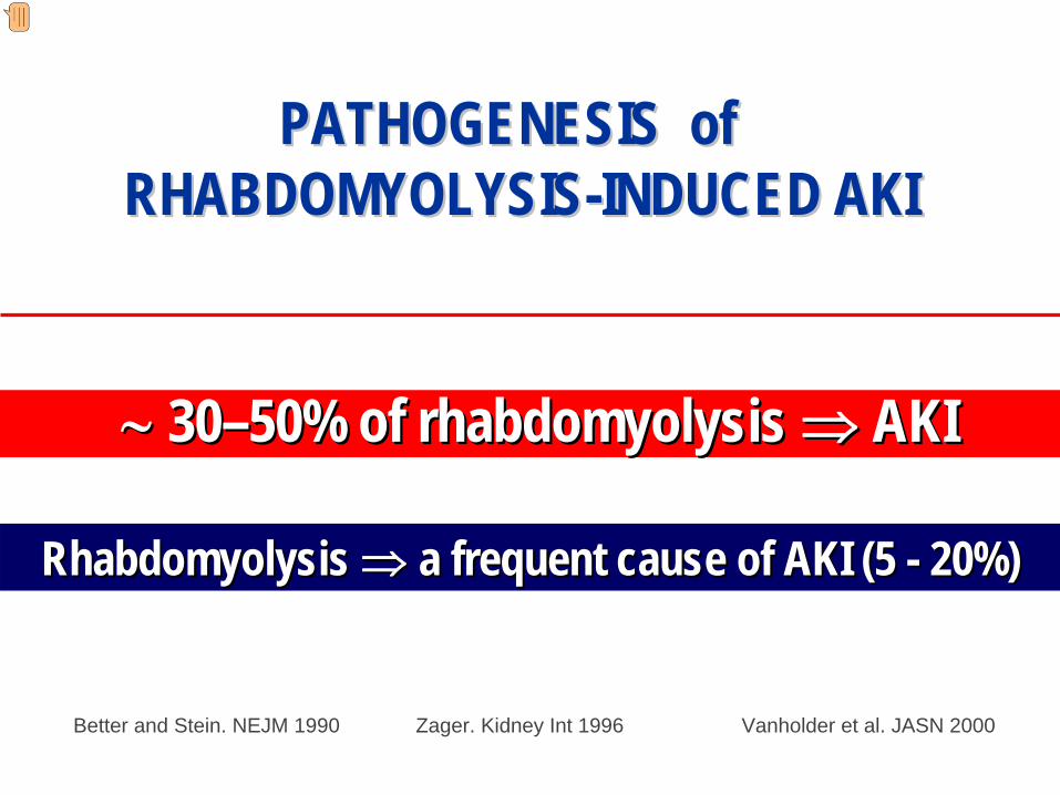
∼∼ 3030––50% of rhabdomyolysis 50% of rhabdomyolysis ⇒⇒ AKIAKI
RRhabdomyolysis habdomyolysis ⇒⇒ a frequent cause a frequent cause of AKI (5 of AKI (5 -- 20%)20%)
Presenter
Presentation Notes
In 30–50% of rhabdomyolysis cases, AKI due to crush syndrome can develop. Overall, 5-20% of AKI cases are due to rhabdomyolysis.
IVVIVVMyoglobinemia
Better and Stein, NEJM 1990,Zager, KI 1996,Vanholder et al. JASN 2000
Renal hypoperfusion/ischemia
Endotoxin/cytokines
Cast formation
Tubular damage
ATNATN
AKIAKI
Myoglobinuria
Luminal stasis
Uric ac. Lactic ac. NO scavenging
Potassium
Fe Fe loadingloading
Hyperphosphatemia
Hypocalcemia
CaPO4 salts
Thromboplastin
DICFree Free
radicalsradicals
Primary importance Secondary importance Tertiary importancePathogenesis of rhabdomyolysisPathogenesis of rhabdomyolysis--induced AKIinduced AKI
Presenter
Presentation Notes
Pathogenesis of rhabdomyolysis-induced AKI is complex. The "key organs” are the injured muscle itself and the kidney. Pathogenetic mechanisms can be of primary, secondary and tertiary importance. Beginning with the most important mechanism: -when the muscles are compressed by the rubble, compartment syndrome develops and dramatic fluid third spacing leads to intravascular volume depletion, which may cause renal hypoperfusion, ischemic damage and tubular necrosis. Among the secondary factors the most important one is myoglobin release from damaged muscles which results in subsequent myoglobinuria. Myoglobinuria results in many pathogenetic mechanisms, the most important one of which is intratubular cast formation that causes luminal stasis and tubular damage. In addition, nucleosides are released from disintegrating cell nuclei and metabolized in the liver to purines such as xanthine, hypoxanthine, and to uric acid, which may contribute to cast formation and tubular obstruction. Myoglobin has direct toxic effects on the tubular epithelial cells. There are tertiary factors as well, such as adverse effects of hyperphosphatemia, thromboplastin and hyperkalemia. We will not discuss these factors here; but would like to underline once more that intravascular volume depletion is the most critical event during this process.
LOGISTIC ISSUESLOGISTIC ISSUES•• Severity assesment Severity assesment •• Providing health care Providing health care •• Medical support Medical support •• Other logistic issuesOther logistic issues
CONCLUSIONSCONCLUSIONS
CLINICAL FINDINGS in CRUSH SYNDROMECLINICAL FINDINGS in CRUSH SYNDROME
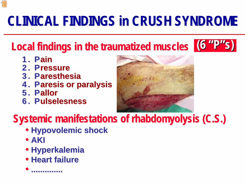
1.1. PPainain2.2. PPressureressure3.3. PParesthesiaaresthesia4.4. PParesis or paralysisaresis or paralysis5.5. PPallorallor6.6. PPulselesnessulselesness
(6 (6 ““PP””s)s)LLocalocal findings in the traumatized musclesfindings in the traumatized muscles
SSystemicystemic manifestations of manifestations of rhabdomyolysis (C.S.)rhabdomyolysis (C.S.)
Presenter
Presentation Notes
Clinical features of crush syndrome can be classified as: (1) local findings in the traumatized muscles, and (2) systemic findings of rhabdomyolysis. Local findings include Pain, Pressure, Paresthesia, Paresis or paralysis, Pallor and Pulselesness These findings have beenqualified as the “6 “P”s Systemic manifestations (or findings) of the crush syndrome are seen in 30 to 50% of rhabdomyolysis cases; they include hypovolemic shock, AKI hyperkalemia, heart failure and the involvement of many other systemic organs.
14,3
18,9
31,9
13,2
36,6
13,7
05
10152025303540
Mor
talit
y ra
te
(%)
Extremitytrauma
Thoracictrauma
Abdominaltrauma
Trauma (+) Trauma (-)
p<0.0001p<0.0001
P=0.19
Thoracic 69Abdominal 41
Skull 32Multiple 54
Others 51
TRAUMA PATTERN in the TRAUMA PATTERN in the MARMARA EARTHQUAKE CRUSH VICTIMS MARMARA EARTHQUAKE CRUSH VICTIMS
Victims with thoracic / abdominal traumas Victims with thoracic / abdominal traumas should be referred from the field as soon as possibleshould be referred from the field as soon as possible
Sever et al, NDT, 2002
No. of traum.Extremities
1 2742 2053 264 7
Global 790
Presenter
Presentation Notes
Considering the injury pattern of the crush victims; in the Marmara earthquake experience, the most frequently traumatized parts of the body were the extremities. Taken as a whole, 790 extremity traumas were noted. Among the other frequent injuries in crush cases, thoracic and abdominal trauma should be emphasized. In a univariate analysis, mortality rate of the patients who suffered from these traumas was significantly higher than that of those who did not have these traumas. In a multivariate analysis model, again, thoracic and abdominal trauma predicted mortality, with odds ratios of 2.8 and 3.8, respectively. Therefore, victims with thoracic / abdominal trauma should be evacuated from the field as soon as possible
LABORATORY FINDINGS in CRUSH SYNDROMELABORATORY FINDINGS in CRUSH SYNDROME
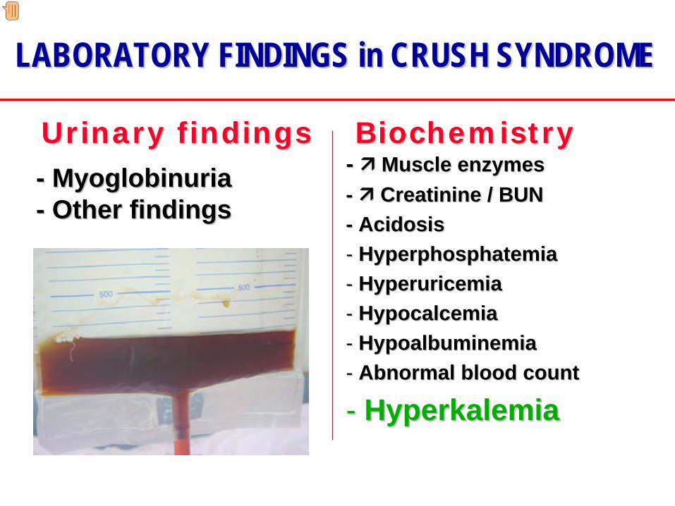
-- MyoglobinuriaMyoglobinuria-- Other findingsOther findings
Laboratory findings can be classified under 2 headings: Urinalysis, and biochemical findings. The most typical finding in urinalysis is a dirty-brownish discoloration of the urine as a result of myoglobinuria. Macroscopic hematuria and trace proteinuria due to penetrating or blunt traumas to the urinary system may also be observed. The pathologic findings in the biochemical analysis are related to increased serum levels of substances released from the injured muscles, such as: increased levels of muscle enzymes, urea and creatinine, phosphate and most importantly potassium. On the other hand, serum calcium and albumin often are decreased.
Many patients died at the field, during transportation or Many patients died at the field, during transportation or on admission to hospitals due to fatal hyperkalemia!on admission to hospitals due to fatal hyperkalemia!
SERUM POTASSIUMSERUM POTASSIUM(The Marmara Earthquake Experience)(The Marmara Earthquake Experience)
Presenter
Presentation Notes
In the Marmara earthquake experience, mean serum potassium on admission was 5.3 millimoles/L. As can be seen in the figure there is an overall tendency towards hyperkalemia. In 70 patients hyperkalemia above 7 millimoles/L was noted. All these figures indicate that probably many patients died due to fatal hyperkalemia at the disaster field before any major support could be offered.
Marmara E.: Marmara E.: 10% of the patients 10% of the patients were receiving were receiving KK++ containing solutions on admissioncontaining solutions on admission
Resulted in many patient deaths ?Resulted in many patient deaths ?
This was certainly This was certainly can be calledcan be callednothing less thannothing less than malpracticemalpractice
K+ containing solutions should NEVER be administered empiricallK+ containing solutions should NEVER be administered empirically ! y !
SERUM POTASSIUM SERUM POTASSIUM -- II II (The Marmara Earthquake Experience)(The Marmara Earthquake Experience)
Presenter
Presentation Notes
Interestingly, in the Marmara earthquake experience, 10% of the casualties were receiving potassium-containing solutions at admission, before any laboratory test had been performed. It is very well possible that this practice (or malpractice) caused some deaths by further increasing serum potassium level in many already hyperkalemic or hyperkalemia-prone casualties. Hence empiric administration of potassium containing solutions should be strictly discouraged.
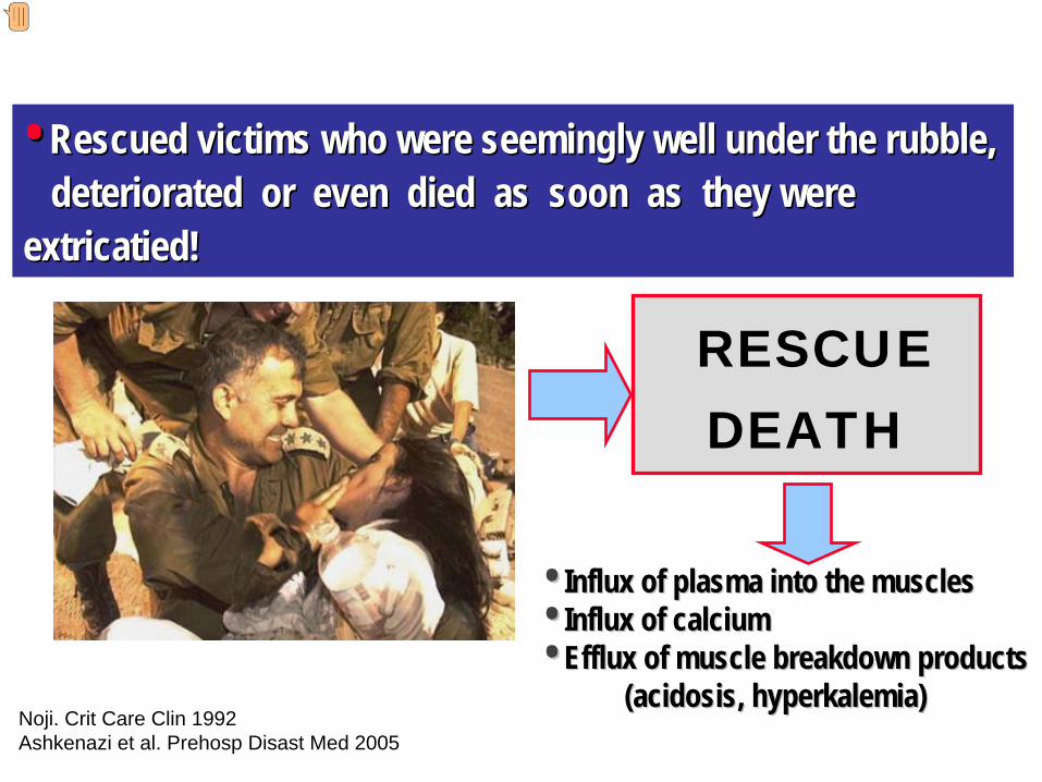
RESCUE DEATH
•• Rescued victims who were seemingly well under the rubble, Rescued victims who were seemingly well under the rubble, deteriorated or even died as soon as deteriorated or even died as soon as they were they were
extricatiextricatieded!!
•• IInflux of plasma into the musclesnflux of plasma into the muscles•• Influx of calciumInflux of calcium•• Efflux of muscle breakdown productsEfflux of muscle breakdown products
(acidosis, hyperkalemia)(acidosis, hyperkalemia)Noji. Crit Care Clin 1992Ashkenazi et al. Prehosp Disast Med 2005
Presenter
Presentation Notes
An unfortunate finding has been noted in some victims of several earthquakes, amongst which the Marmara disaster. Rescued victims who were seemingly well under the rubble got sicker or even died as soon as they were extricated, which is named “Rescue death”. The underlying pathology in this phenomenon is that the victims are spared from the hemodynamic and systemic effects of rhabdomyolysis when they are trapped under rubble. After they have been rescued and the extremities are decompressed however, an influx of plasma, sodium and importantly calcium occurs, while muscle breakdown products diffuse into the circulation, and severe metabolic acidosis as well as fatal hyperkalemia develop.
LOGISTIC ISSUESLOGISTIC ISSUES•• Severity assesment Severity assesment •• Providing health care Providing health care •• Medical support Medical support •• Other logistic issuesOther logistic issues
CONCLUSIONSCONCLUSIONS
IVVIVVMyoglobinemia
Better and Stein, NEJM 1990,Zager, KI 1996,Vanholder et al. JASN 2000
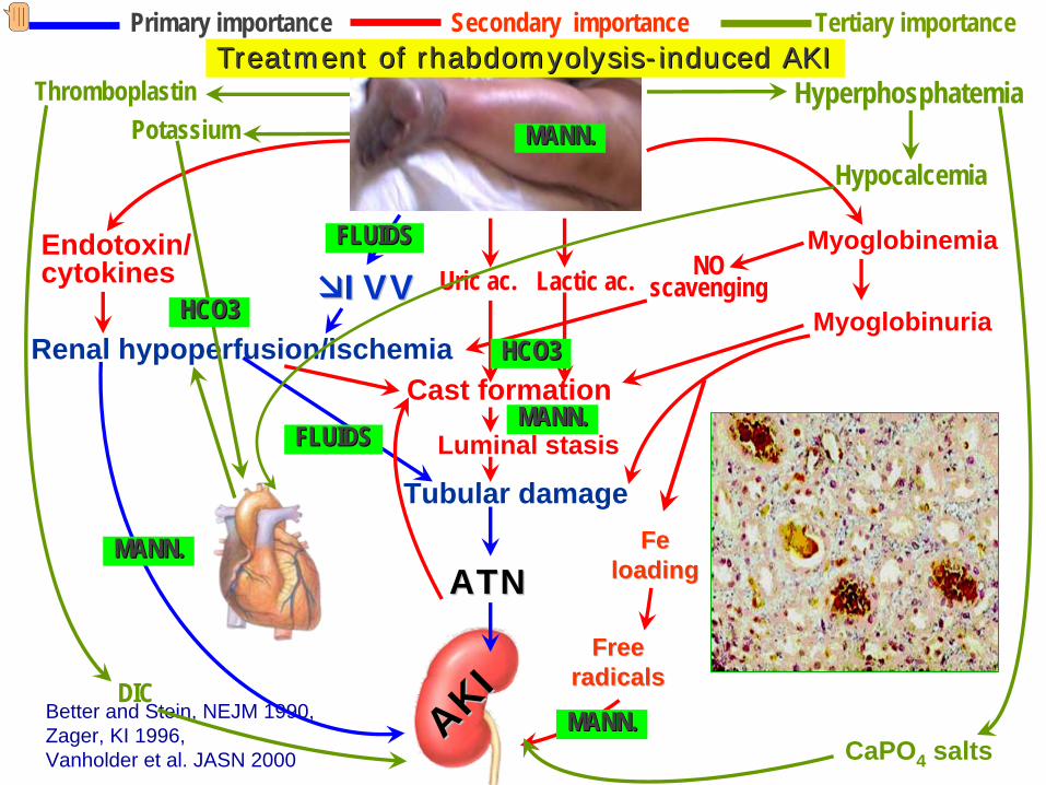
TreatmentTreatment of rhabdomyolysisof rhabdomyolysis--induced AKIinduced AKI
Presenter
Presentation Notes
What type of prophylactic and therapeutic interventions should we do for preventing this sad outcome? In prophylaxis of crush syndrome the most important therapeutic intervention in this busy scenario is giving iv fluids at the earliest occasion to correct hypovolemia and improve renal perfusion. Bicarbonate may be beneficial in alkalinizing urine and preventing cast formation. It can also decrease hyperkalemia. Lastly, it has been suggested that mannitol may be beneficial through many mechanisms, which include improvement of compartment syndrome and renal perfusion, interfering with cast formation and free radical production. It can decrease intracompartmental pressure as well. However, as will be explained later, mannitol should be used with extreme care, and is absolutely contraindicated in oligo-anuric patients.
Presenter
Presentation Notes
In the “Recommendatons for the Management of Crush Victims in Mass Disasters", a joint project for European Renal Best Practice (ERBP) and Renal Disaster Relief Task Force (RDRTF) of the ISN, the largest section was devoted to "Interventions at the disaster field". In this text, we underlined the importance for fluid administration (typically isotonic saline) as soon as possible (even when the victim is still under the rubble) at a rate of 1000 mL/hr. We suggest to continue isotonic saline during the extrication as well (if a vein could be punctured previously). The extrication period varies significantly (usually 45 – 90 min, but sometimes 4 - 8 hrs), because of differing disaster severity, efficacy of relief logistics, and local and patient conditions. Therefore, we suggest to administer fluids at a rate of 1 L/hr only during the first 2 hrs of extrication; fluid administration rate should be decreased by at least 50% (<500 mL/h) afterwards. Otherwise, volume overload may develop in oliguric victims. Of course, these are general comments and volume of fluids should be individualized considering demographic characteristics of the victim, trauma pattern, environmental conditions, time spent under the rubble and many other factors.