mammalian remains from a new site near the classical
TRANSCRIPT

109
Scientific Annals, School of Geology, Aristotle University of Thessaloniki
Proceedings of the XIX CBGA Congress, Thessaloniki, Greece Special volume 99 109-119
Thessaloniki
2010
MAMMALIAN REMAINS FROM A NEW SITE NEAR THE CLASSICAL
LOCALITY OF PIKERMI (ATTICA, GREECE)
Theodorou G.E.1, Roussiakis S.J.
2, Athanassiou A.
3, Filippidi A.
4
University of Athens, Department of Historical Geology–Palaeontology, 15784 Zographou, Greece, [email protected], [email protected], [email protected], [email protected]
Abstract: We present the first results on the fossil mammalian fauna recovered during the first excava-
tion season at the new site Pikermi Valley-1 (PV1). The fauna comprises two hipparionine species (C.
cf. mediterraneum, H. cf. brachypus), a giraffid (Bohlinia cf. attica), five bovids (Palaeoreas linder-
mayeri, Protragelaphus skouzesi, Tragoportax cf. amalthea, Gazella sp., Bovidae indet.) and two carni-
vores (? Adcrocuta eximia, Felidae indet.). The composition of the fauna suggests a Turolian age.
Keywords: Pikermi, Late Miocene, Turolian, Mammalia
1. Introduction
Pikermi constitutes one of the oldest known (dis-
covered in 1835 or 1836) and most famous locali-
ties of the Eurasian Late Miocene. It is perhaps the
most important reference locality for the European
continental Upper Miocene and for the so-called
Pikermian biome due to the richness of its fauna
and due to the fact that it is the type locality of
several Turolian genera and species. The Pikermi
fossil fauna is the product of several excavations
carried out mainly during the 19th and the early 20
th
century. Most of the available material derives
from the excavations of Albert Gaudry (1855-56
and 1860), A. Smith Woodward and Theodor Sku-
fos (1901) and Othenio Abel (1912) (Gaudry 1855,
1862-1867; Woodward 1901; Abel 1922), but nu-
merous other minor excavations yielded smaller
fossil bone collections. After Abel’s excavation
there was no other research activity in the area of
the classical site. In 1971 a new site has come to
light during quarrying activity east of the classical
site. This site, known as Kisdári or Chomaterí or
Chomaterés, was partly excavated from 1972 to
1980 (Bachmayer et al. 1982).
In 2008 the major of Pikermi Mr. A. Adamopoulos
started collaboration with the first author trying to
raise funds for founding a local Museum, protect
the fossiliferous sites and organize a presentation
of the fossils in situ, a project pending since at
least 1901, when the demand for a local museum
was expressed by the mining engineer Andreas
Kordellas in a newspaper article. Geological pros-
pecting carried out in the same year by a Universi-
ty of Athens team, in order to locate sites for the
purposes of the project, resulted in the discovery of
the new fossiliferous locality ―Pikermi Valley-1‖
(PV1). The new site is situated at the bottom of the
ravine of Megálo Réma stream (locally known as
Valanáris), 500 m east-southeast of the alleged lo-
cation of the classical Pikermi locality and about
1700 m southwest of Chomateri site (Fig. 1). The
findings come from a single fossiliferous lens on
the northern bank of the stream, slightly above the
present water level.
The first excavation season (June 15 – July 15,
Fig. 1. Geographical distribution of the Pikermi sites: 1,
classical locality; 2, Chomateri or Kisdari; 3, Pikermi
Valley-1. The plotted position of the classical site is tra-
ditionally accepted as the location excavated by Albert
Gaudry, but this is not confirmed by any historical or
field data. Pikermi is located 20 km east-northeast of
Athens. Satellite image © 2009 Digital Globe / Google.
110
2009), sponsored by the Municipality of Pikermi
(University of Athens Project Research Account
70/3/9494), revealed a promising fossil mammal
assemblage. The specimens were rather sparse,
particularly when compared to the tight accumula-
tion observed in the classical Pikermi locality (ob-
servation based on fossiliferous blocks stored in
several museums), or at Chomateri locality
(Bachmayer et al. 1982). Most long bones exhi-
bited a NE–SW orientation, which probably was
the direction of the palaeocurrent (Fig. 2).
2. Material and Methods
The excavated sample consists of more than 200
specimens; the identifiable ones are described be-
low. All material is stored in the Museum of Pa-
laeontology and Geology, University of Athens,
and was prepared in its laboratory using mechani-
cal methods (hand and pneumatic tools). The bio-
metric study of the equids followed the methodol-
ogy of Eisenmann et al. (1988), while the artiodac-
tyl specimens were measured according to the sug-
gestions of Heintz (1970). All measurements are in
mm. The upper and lower teeth positions are given
using upper and lower case letters respectively
(e.g. M1 and m1).
3. Systematics
Perissodactyla OWEN, 1848
Equidae GRAY, 1821
Hipparionini QUINN, 1955
The hipparionine material (Figs 3–5, Table 1) con-
sists of a few mainly isolated teeth and some post-
cranial elements. The best preserved teeth belong
to aged individuals. The upper teeth exhibit nu-
merous enamel plications at the fossettes, simple
pli caballin and rather shallow, V-shaped hypocon-
al groove. The very worn lower premolars have a
shallow ectoflexid that does not penetrate the isth-
mus and rounded metaconid and metastylid, sepa-
rated by a shallow linguaflexid. In a very worn
lower molar the linguaflexid and the ectoflexid are
in contact. The flexids show practically no plica-
tions. A fresh isolated lower molar has a hypso-
donty index of 47 (calculated following Eisenmann
et al. 1988).
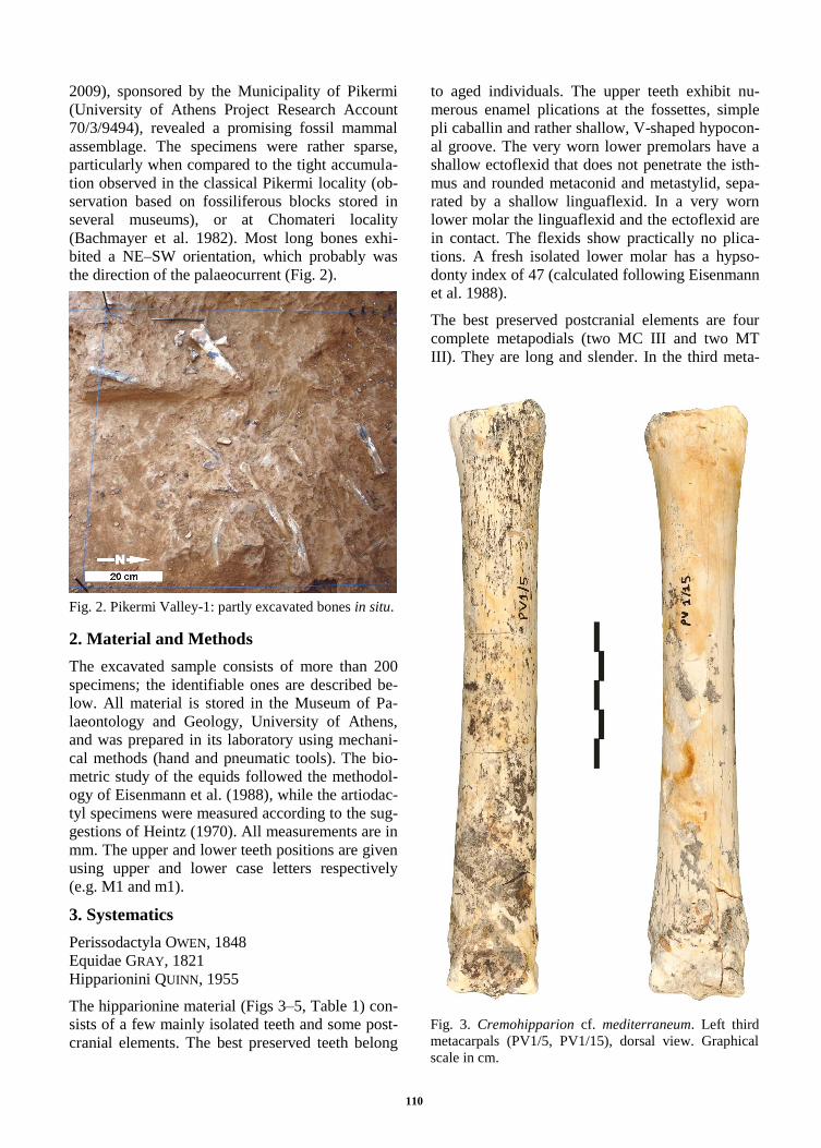
The best preserved postcranial elements are four
complete metapodials (two MC III and two MT
III). They are long and slender. In the third meta-
Fig. 2. Pikermi Valley-1: partly excavated bones in situ.
Fig. 3. Cremohipparion cf. mediterraneum. Left third
metacarpals (PV1/5, PV1/15), dorsal view. Graphical
scale in cm.

111
carpals, the slenderness index (3/1) is 12.3% and
12.1%; another similar index (11/1) is calculated to
be 15.1% and about 16.6% in the two specimens
respectively. The same indices on the two third
metatarsals are 10.1% and about 10.1% (for 3/1),
and 12.4% and 12.6% (for 11/1). The above spe-
cimens are plotted inside the cluster of the small
sized Pikermi hipparions provided by Dermitzakis
(1976, figs 6-7); their dimensions are also in ac-
cordance with the metrical data provided by Kou-
fos (1987a, tables 33, 41) for H. mediterraneum
from Pikermi. Based on their morphology, abso-
lute size and proportions, these metapodials are re-
ferred to the species Cremohipparion cf. mediter-
raneum (ROTH & WAGNER, 1854).
A fragmentary left calcaneus (PV1/53) presents
large distal maximal breadth (measurement ―7‖ of
Eisenmann et al. 1988), measuring 51.0 mm. It can
be referred to Hippotherium cf. brachypus (HEN-
SEL, 1862), since it is plotted to the cluster of the
large sized Pikermi hipparions provided by Theo-
dorou (1997) and corresponds in size to the larger
values provided by Koufos (1987a) for this spe-
cies.
There are two tibias, both missing their proximal
ends. One of these (PV1/4) is more slender and has
similar dimensions with the C. mediterraneum Pi-
kermi sample studied by Koufos (1987a, 1987b).
The other one (PV1/1) is larger and more robust;
compared to the data provided by Koufos (1987b,
fig. C-18) it is plotted inside the H. brachypus
sample.
Two complete proximal phalanges III (possibly
pedal) also present similar size differences, one of
them (PV1/57) being significantly more robust
than the other (PV1/12), and could belong to dif-
ferent hipparionine species. The dimensions of the
robust one plot inside the range of the H. brachy-
pus sample (Koufos 1987b, fig. C-18).
Despite the inadequate hipparionine material avail-
able to date from PV1, the presence of two species
—Cremohipparion cf. mediterraneum (ROTH &
WAGNER, 1854) and Hippotherium cf. brachypus
(HENSEL, 1862)— is documented in the site. The
rest of the hipparionine material, which is not de-
terminable to the species level, is referred to as
Hipparionini indet.
Fig. 4. Cremohipparion cf. mediterraneum. Left third
metatarsals; PV1/8, lateral view; PV1/40, dorsal view.
Graphical scale in cm.
Fig. 5. Hipparionini indet. Proximal phalanges III
(PV1/12, PV1/57), dorsal view. Graphical scale in cm.

112
Artiodactyla OWEN, 1848
Giraffidae GRAY, 1821
Bohlinia MATTHEW, 1929
Bohlinia cf. attica (GAUDRY & LARTET, 1856)
The giraffids are represented in the new material
by only one specimen (PV1/72), a right radius-ulna
missing its proximal part (Fig. 6). Its preserved
length measures about 68 cm, the greatest width of
the distal end 107 mm, and the medio-lateral di-
ameter of the diaphysis about 63 mm. The crest
that defines the lateral margin of the lunar facet is
more curved and oblique than in the extant giraffe.
The specimen is referable to B. attica based on its
morphological and metrical characters (Gaudry
1862-1867; Bohlin 1926; Geraads 1974; Geraads
1979).
Bovidae GRAY, 1821
The Bovidae material comprises dental and cranial
remains, and few postcranial elements. The dental
and cranial remains indicate the presence of five
taxa.
Palaeoreas GAUDRY, 1861
Palaeoreas lindermayeri (WAGNER, 1848)
The material referred to this species comprises a
frontlet (PV1/42) and a right mandible (PV1/25).
The frontlet (Fig. 7) is badly damaged and dis-
torted by compression. The horn-cores are in a
poor state of preservation, but present an anterior
and a posterior keel. The former is blunt and des-
cends anteriorly. The latter, preserved at the distal
part of the left horn-core, is acute. Only the antero-
posterior basal diameter of the left horn-core can
be provided, that measures about 48 mm. The
postcornual fossae are deep. Between the horn-
cores, the frontal region is strongly elevated above
the level of the supraorbital margins and strongly
bent, even if the latter is probably exaggerated by
Fig. 6. Bohlinia cf. attica. Right radius-ulna (PV1/72),
anterior view. Graphical scale: 10 cm.
Table 1: Long bone measurements of mediterraneum from PV-1. Measurement methodology accord-
ing to Eisenmann et al. 1988. The Hipparion measurements in parentheses are inaccurate.
Metacarpal III Metatarsal III Tibia Prox. Phalanx III
PV1/5 PV1/15 PV1/8 PV1/40 PV1/1 PV1/4 PV1/12 PV1/57
1 204.4 202.5 259.5 253.3 — — 61.8 63.7
2 196.8 196.3 251.7 245.1 — — 56.9 58.2
3 25.2 24.5 26.1 (25.6) 40.6 38.5 24.2 27.6
4 21.1 22.7 28.7 (27.1) 29.1 27.0 35.5 39.2
5 32.6 33.9 38.9 39.1 — — 27.2 31.0
6 24.2 23.9 28.0 — — — 29.1 32.7
7 28.5 29.2 36.7 (35.6) 66.7 59.2 29.0 32.2
8 — 8.8 10.3 — 44.5 41.2 18.3 22.1
9 — 3.4 8.1 — — — 19.1 20.3
10 31.0 34.5 35.9 36.9
11 30.8 (33.7) (32.2) 32.0
12 25.3 26.3 30.3 28.5
13 21.4 22.4 22.8 22.1
14 21.9 23.8 26.9 25.6
11/1 15.1 (16.6) (12.4) 12.6

113
distortion. The supraorbital foramens open into
deep frontal depressions and communicate with the
orbital fossae. The distance between the lateral
borders of the frontal depressions measures about
35 mm.
The mandible (Fig. 8) is completely preserved
(Lp=25.2, Lp3-p4=19.2, Lm=41.6, Lpm=66.2,
Lp3-m3=60.2). The mandibular ramus is only
slightly deeper below m3 than below p2, and the
premolar row is large in relation to the molar one
(Lp/Lm=60.6%, Lp3-p4/Lm1-m3=46.2%). The p3
and p4 are elongated, and the p3 is long in relation
to the p4 (Lp3/Lp4=90.4%). On p3 the metaconid
is small. On the labial wall, the groove in front of
the hypoconid is faint. The p4 is generally similar
to p3. The paraconid is well separated from the pa-
rastylid. The metaconid is columnar and the 2nd
and 3rd valleys of the tooth are open. The base of
the 2nd valley bears a small tubercle. The groove
in front of the hypoconid is well marked. The mo-
lars bear moderately developed goat fold. The ec-
tostylids are moderately developed on m1, m2, less
in m3. The specimen is metrically similar to P. lin-
dermayeri from Pikermi (Fig. 9).
Both specimens exhibit all characters of P. linder-
mayeri (Bouvrain 1980; Bouvrain 1992; Geraads
et al. 2003) and are referred to this species.
Fig. 7. Palaeoreas lindermayeri. Frontlet (PV1/42), an-
terior view. Graphical scale in cm.
Fig. 8. Palaeoreas lindermayeri. Right mandible
(PV1/25); a) occlusal view, b) lingual view. Graphical
scale in cm.
Fig. 9. Comparison of P. lindermayeri and P. skouzesi
from PV1 with specimens from Pikermi and Maragha.
Based on personal data.

114
Protragelaphus DAMES, 1883
Protragelaphus skouzesi DAMES, 1883
A frontlet with both horn-cores (PV1/69; Fig. 10)
is referred to P. skouzesi. This specimen could be-
long to a not fully grown individual since its horn-
cores’ surface is porous. The horn-cores are mod-
erately separated (about 27.5 mm) and diverge
from the sagittal plane by an angle of about 30°,
and they are slightly compressed mediolaterally at
their bases (DAP×DT=(44.0)×35.8 mm, left horn-
core). The horn-core axis is almost straight, the
torsion follows a close spiraling, and there is no
anterior keel. The posterior keel can be traced in
two revolutions. The left horn-core is almost com-
pletely preserved and measures about 210 mm in
length (in straight line). There are moderately deep
postcornual fossae. The intercornual region is on a
higher level than the supraorbital margins. The pa-
rietofrontal suture is open. The interfrontal suture
cannot be traced since the specimen is damaged
along it. The supraorbital foramens are small, not
sunken in frontal depressions, and open about 27
mm below the horn-cores’ bases. The frontlet
PV1/69 is quite similar to other specimens referred
to P. skouzesi, though, its horn-cores are slightly
less robust compared to already known Pikermi
specimens (Fig. 11; Roussiakis 2009).
A left mandible (Fig. 12; PV1/68; Lp3-p4=20.8,
Lm=48.9, Lp3-m3=68.0) can also be attributed to
P. skouzesi. The premolars are short in relation to
the molars (Lp3-p4/Lm=42.5%). The p3 is rather
triangular and clearly smaller than the p4
(Lp3/Lp4=84.1%). The metaconid is oblique and
extends distolingually, closing the distal part of the
lingual wall of the tooth. The 2nd valley remains
open, while there is no groove in front of the hypo-
conid on the labial wall of the tooth. The p4 is sim-
ilar to p3, but the metaconid forms a mesiodistally
directed wall that also constricts the 2nd valley of
the tooth. The groove in front of the hypoconid is
moderately marked. On the molars, the ectostylids
are rather strong on m1 and m2, less on m3. There
is a faint goat fold on m1 and m2. The lower denti-
tion of P. skouzesi is not definitely known. Pilgrim
and Hopwood (1928, pl. 9, fig. 3), followed by
Gentry (1971, pl. 6), referred some mandibular
rami from Pikermi to P. skouzesi, while Mecque-
nem (1925, pl. 6, fig. 1) referred to this species a
mandibular ramus from Maragha. In addition, to
the same species might belong two mandibular ra-
Fig. 11. Comparison of the basal horn-core dimensions
(DT vs DAP) of P. skouzesi from PV1 with Protragela-
phus, Ouzocerus and Helladorcas species from various
localities. Data according to Bouvrain (1978, 1997), Ge-
raads and Güleç (1999), and Roussiakis (2009). PNT:
Pentalophos-1, RZ1: Ravin des Zouaves-1.
Fig. 12. Protragelaphus skouzesi. Left mandible
(PV1/68); a) occlusal view, b) lingual view. Graphical
scale in cm.
Fig. 10. Protragelaphus skouzesi. Frontlet (PV1/69); a)
anterior view, b) anterolateral view. Graphical scale in
cm.

115
mi from Pikermi stored in AMPG: one of this,
PA1842/91, was referred to P. skouzesi by Rous-
siakis (1996), while the other (Π89/740) was re-
cently located in the AMPG Pikermi collections.
On these specimens the Lp3-p4/Lm ratio ranges
from 40.3% to 45.1% and the metaconid of p4
tends to be fused with the paraconid. This tendency
is also exhibited by PV1/68 that appears compara-
ble in size and proportions to the Pikermi speci-
mens referred to P. skouzesi but slightly smaller
compared to the Maragha specimen (Fig. 9).
Tragoportax PILGRIM, 1937
Tragoportax cf. amalthea (ROTH & WAGNER,
1854)
We refer to T. cf. amalthea a left mandibular ra-
mus (PV1/38; Fig. 13), with the complete dentition
preserved (Lp=45.7, Lp3-p4=32.3, Lm=60.7, Lp3-
m3=91.0, Lpm=104.3). The mental foramen opens
about 40 mm in front of p2. The premolar series is
elongated (Lp/Lm=75.3%, Lp3-p4/Lm=53.2%).
On p3, the 2nd valley is widely open while the 3rd
one is narrow. The paraconid is transverse to the
mesiodistal axis of the tooth and rather bulbous
towards its base. The metaconid is distolingually
directed. The groove in front of the hypoconid is
faint. On p4, the 2nd and 3rd valleys are narrow
due to the mesiodistally directed T-shaped meta-
conid. The paraconid is well separated from the
parastylid and directs distolingually. The groove in
front of the hypoconid is strong. A trace of goat
fold is observed on m1. The ectostylid is well de-
veloped on m1, but decreases towards the m3. The
m1 and m2 bear a lingual basal pillar between the
paraconid and metaconid; this is faint on m3.
Metrically PV1/38 is comparable to the larger Pi-
kermi specimens of Miotragocerus valenciennesi
and the smaller of T. amalthea (Fig. 14). However,
the rather wide p3 and the T-shaped metaconid of
p4 are typical characters of Tragoportax (Spassov
and Geraads 2004; Kostopoulos 2005), so we ten-
tatively refer this specimen to T. amalthea.
Gazella sp.
An isolated but damaged horn-core (PV1/70; Fig.
15) with preserved length of about 85 mm (in
straight line) can be referred to Gazella sp. The
surface of the horn-core is deeply grooved. The
lateral surface is flatter than the medial, and the
posterior surface appears gently curved in lateral
view. The base of the horn-core is badly broken
and no measurements can be provided. However, 2
cm above the base the cross section of the horncore
Fig. 14. Premolar versus molar length (lower dentition)
in Tragoportax and Miotragocerus from various locali-
ties. Based on personal data, Kostopoulos (2005, 2009,
pers. com.), Spassov and Geraads (2004) and Bouvrain
(2001).
Fig. 15. Gazella sp. Right horn-core (PV1/70), medial
view. Graphical scale in cm.
Fig. 13. Tragoportax cf. amalthea. Left mandible
(PV1/38); a) occlusal view, b) lingual view. Graphical
scale in cm.

116
is subelliptical (DAP×DT=24.0×21.4 mm) while
about 7 cm above the base is more circular
(L×W=17.1×16.6 mm). The general characters of
this specimen show similarities to Gazella capri-
cornis (WAGNER, 1848), but a specific determina-
tion cannot be established because of the scanti-
ness of the available material.
Bovidae indet.
A right maxilla fragment (PV1/36; Fig. 16) pre-
serving the complete dentition (LP=38.7,
LM=48.1, LP3-P4=25.4, LPM=80.4) is characte-
rized by the long premolar series in relation to the
molars (LP/LM=80.5%, LP3-P4/LM=52.8%). P2
is elongated (L×W=14.0×10.1). Its parastyle is
thin, while the paracone rib is thick, labially pro-
jected and rather mesially situated. The protocone
projects lingually slightly more than the hypocone.
It is separated from the latter by a deep lingual
groove located opposite the paracone rib. P3 is on-
ly slightly larger in length than in width
(L×W=13.9×12.8). The parastyle and paracone rib
are strong. The metastyle is stronger and projects
labially more than the parastyle and the paracone
rib. Lingually it is strongly bilobated, as P2, but
the hypocone projects lingually much more than
the protocone. P4 is larger transversely than mesi-
odistally (L×W=11.6×13.6). The parastyle, para-
cone rib and metastyle are moderately developed.
The hypocone bears a hypoconal spur that forms a
hypoconal islet. The upper molars are characte-
rized by the strong parastyle, paracone rib and me-
sostyle, while the metacone rib and the metastyle
(apart from M3) are weak. A hypoconal islet is
present on M2 and a free hypoconal spur on M3. A
central islet is not observed. A slightly developed
entostyle is present on M1. It is faint on M2 and
absent on M3.
A left mandibular ramus with p3-m3 (Fig. 17;
PV1/17; Lp3-m3=77.8, Lp3-p4=28.2, Lm=51.1)
agrees dimensionally, proportionally and morpho-
logically to the above described upper toothrow.
The p3 and p4 are elongated in relation to the mo-
lar series (Lp3-p4/Lm=55.2%). The toothrow
length is estimated to 85 mm and the premolar
length to 35 mm. The p3 is elongated relative to its
width (L×W=19.4×7.1). The paraconid is indepen-
dent from the parastylid and does not project lin-
gually. The metaconid is distolingually directed
but does not fuse with the entoconid. A faint
groove is present in front of the hypoconid. The p4
is similar to p3 but the metaconid appears stronger.
The m1 and m2 bear a weakly developed goat fold.
The parastylid and metastylid are weak on all mo-
lars. A moderated developed ectostylid is present
on m1; it is faint on m2 and absent on m3.
The maxilla PV1/36 exhibits similarities to a spe-
cimen from Maragha referred to Prostrepsiceros
houtumschindleri (RODLER & WEITHOFER, 1890)
by Mecquenem (1925, pl. 5, fig. 5) on its relative
long premolar series and elongated and bilobated
P2, but it is larger in size. The mandible PV1/17 is
very similar to NHML M13007 from Pikermi, re-
ferred by Gentry (1971), with reservation, to Pro-
strepsiceros rotundicornis (WEITHOFER, 1888),
but the latter is smaller (Lp3-m3=65.8), presents
stronger goat fold and a deep groove in front of the
hypoconid. PV1/17 also exhibits similarities to P.
rotundicornis from Akkaşdağı that ranges in p2-
m3 length from 68.8 to 75.1 mm (Kostopoulos
2005).
Carnivora BOWDICH, 1821
Hyaenidae GRAY, 1821
? Adcrocuta eximia (ROTH & WAGNER, 1854)
Fig. 16. Bovidae indet. Right upper toothrow (PV1/36);
a) labial view, b) occlusal view. Graphical scale in cm.
Fig. 17. Bovidae indet. Left mandible (PV1/17); a) oc-
clusal view, b) lingual view. Graphical scale in cm.

117
Hyaenids are represented by a right P2 and a right
upper canine (PV1/49, PV1/22). The canine
(L×W=17.3×13.5 mm) presents a moderately de-
veloped mesiolingual crest as well a distal one.
The P2 (L×W=16.2×11.1 mm) is rectangular in
outline. It presents two accessory cusps: a strong
distal one on the long axis of the tooth, and a
smaller one situated mesiolingually. Both speci-
mens can be provisionally referred to Adcrocuta
eximia.
Felidae FISCHER DE WALDHEIM, 1817
Felidae indet.
The proximal half of a proximal phalange
(PV1/71; preserved length=29.6 mm,
DTprox×DAPprox = 17.9×16.2 mm) has typical
felid morphology. Its dimensions indicate a large
felid, larger than Metailurus parvulus (HENSEL,
1862) (Roussiakis et al. 2006). It may belong to
Metailurus major ZDANSKY, 1924 or Machairodus
giganteus (WAGNER, 1848).
4. Discussion
The recently discovered PV1 locality, though very
promising, has not yet yielded a large number of
specimens and taxa; however, all findings come
from a single fossil accumulation, a fact that is im-
portant for the study of the complicated local bios-
tratigraphy. The Pikermi fauna is commonly con-
sidered as chronologically and taxonomically ho-
mogenous, though the early authors already no-
ticed that the fossils occur in at least two strati-
graphic levels. This was first observed by Gaudry
(1855; 1862-1867, p.14), who mentioned the pres-
ence of two fossiliferous horizons. Later, Wood-
ward (1901) referred to two or locally three hori-
zons, which is well documented in a photograph by
Th. Skufos, published by Abel (1922, fig. 132).
According to this photograph the two lower hori-
zons have a level difference οf about 2 m, while
the third (upper) lays less than 1 m above the
second. In spite of these observations, the strati-
graphic provenance of the excavated material at
the classical site is not specified in the relevant
museum collections and publications, raising con-
siderations on the exact faunal content of each lev-
el (e.g. Theodorou and Nicolaides 1988; Theodo-
rou 1997). Moreover, the various excavators dur-
ing the 19th and early 20th centuries collected ma-
terial from different quarries at undefined positions
in the broader area of Pikermi (Woodward 1901;
Abel 1922), which are impossible to locate today.
This means that what is known as ―Pikermi clas-
sical site‖ is in reality a complex of quarries and
stratigraphic levels along or close to the ravine,
and that many Pikermian museum collections may
comprise heterogeneous faunal samples, mixing
two or more quarries or stratigraphic levels. The
old collections may represent a fairly homogene-
ous palaeocommunity, only if the sedimentation
and the fossil accumulation have been very rapid,
lasting just a few thousand years, and providing
―snapshots‖ of the same fauna in slightly different
time slices.
The importance of the new site PV1 consists in its
potential to provide new stratigraphic data on the
relationship of the two/three fossiliferous horizons,
in case the recently discovered accumulation is
rich enough to produce an adequate statistical
sample. A direct stratigraphic correlation of PV1
with the alleged classical site or Chomateri is not
possible at the moment, as the ravine sections are
disturbed, mainly by agricultural activities, and be-
cause of the lack of any lithologically characteris-
tic horizon. The rapid urban development of the
Pikermi area also hinders the stratigraphic correla-
tion.
Despite the unknown stratigraphic provenance of
the Pikermi fauna, its age is collectively consi-
dered as middle Turolian (MN12) (Koufos 2006).
The newly excavated PV1 sample —though still
poor— does not differ taxonomically from the
classical fauna, as it is also the case with the Cho-
materi fauna. All the taxa recognized at PV1 are
already known in the Pikermi fauna.
Palaeoreas lindermayeri, a bovid geographically
confined to Greece, S. Bulgaria and W. Turkey, is
known from localities dated to middle–late Turo-
lian (MN12–MN13), while closely related forms
(P. zouavei Bouvrain, 1980) are known from early
Turolian (MN11) (Geraads and Güleç 1999;
Geraads et al. 2003; Koufos 2006). Protragelaphus
skouzesi is a rarer Late Miocene faunal element,
but it spans a wider palaeogeographic area. It is
known mainly from the middle Turolian of Greece
(Pikermi, Chomateri, Halmyropotamos), as well as
from Maragha (Iran), but it is also reported from
the early Turolian locality of Novoelisavetovka-2
in Ukraine (Gentry et al. 1999). Closely related or
identical forms also occur in Samos and W. Turkey
(Andree 1926; Geraads and Güleç 1999; Roussi-
akis 2009). The rest of the referred PV1 taxa
(Tragoportax cf. amalthea, Bohlinia cf. attica, Ad-
crocuta cf. eximia) point generally to Turolian or
even late Vallesian age.

118
Despite the faunal similarity with classical Pikermi
fauna, a biochronologic attribution of PV1 to mid-
dle Turolian cannot be supported for the moment,
since the faunal list is still rather short and insuffi-
cient for a detailed study. The biochronological da-
ta for the currently available material allow a pre-
liminary biostratigraphic attribution of PV1 to Tu-
rolian (MN11–MN13). Future excavations at PV1
will hopefully enrich the faunal and stratigraphic
data available for this site.
5. Conclusion
The material excavated during the first field season
documents the presence of the following taxa in
PV1: Cremohipparion cf. mediterraneum, Hippo-
therium cf. brachypus, Bohlinia cf. attica, Palaeo-
reas lindermayeri, Protragelaphus skouzesi, Tra-
goportax cf. amalthea, Gazella sp., Bovidae indet.,
? Adcrocuta eximia and Felidae indet.
In spite of the limited number of available speci-
mens, PV1 presents close taxonomic similarity
with the classical Pikermi fauna, as all taxa are
common between the two sites. This allows us to
date the new site in the Turolian. A more precise
dating requires more material, as well as a resolu-
tion of the stratigraphic problems of the classical
site.
Acknowledgements
We thank the Municipality of Pikermi, which sup-
ported financially the field work. Apart from the
authors, the following colleagues and students also
participated at the excavation: Theodoros Arghy-
riou, Grigorios Vassilopoulos, Alexandros Daniili-
dis, Dimitrios Michailidis, Elizabeth Stathopoulou,
Christos Solomos, George Konidaris, Vassiliki
Mitsopoulou, Zoi Modianaki, Dionysia Liakopou-
lou, Evi Tzortzaki, Maria Maglara, Tina Sklavou-
nou, Eleftheria Eleftheriou and Dionysia Liako-
poulou. Several other students also joined the team
occasionally.
References Abel O., 1922. Lebensbilder aus der Tierwelt der Vor-
zeit. Jena, 644p.
Andree J., 1926. Neue Cavicornier aus dem Pliocän von
Samos. Palaeontographica, 67, 135-175.
Bachmayer F., Symeonidis N. and Zapfe H., 1982. Die
Ausgrabungen in Pikermi-Chomateri bei Athen.
Eine Dokumentation. Annalen des Naturhistorischen
Museums in Wien, 84, 7-12.
Bohlin B., 1926. Die Familie Giraffidae. Palaeontologia
Sinica, 4, 1-178.
Bouvrain G., 1978. Protragelaphus theodori n. sp.
(Mammalia, Artiodactyla, Bovidae) du Miocène de
Macédoine (Grèce). Géologie Méditerranéenne, 5,
229-236.
Bouvrain G., 1980. Le genre Palaeoreas (Mammalia,
Artiodactyla, Bovidae), systématique et extension
géographique. Paläontologische Zeitschrift, 54, 55-
65.
Bouvrain G., 1992. Antilopes à chevilles spiralées du
Miocène supérieur de la province gréco-iranien :
Nouvelles diagnoses. Annales de Paléontologie
(Vertébrés-Invertébrés), 78, 49-65.
Bouvrain G., 1997. Les Bovidés du Miocène supérieur
de Pentalophos (Macédoine, Grèce). Münchner
Geowissenschaftliche Abhandlungen, 34, 5-22.
Bouvrain G., 2001. Les bovidés (Mammalia, Artiodac-
tyla) des gisements du Miocène supérieur de Vathy-
lakkos (Grèce du Nord). Neues Jahrbuch für Geolo-
gie und Paläontologie, Abhandlungen, 220, 225-244.
Dermitzakis M.D., 1976. Observations on the metapo-
dials of Hipparion from Pikermi (Attica, Greece).
Proceedings of the Koninklijke Akademie van We-
tenschappen, 79 (1), 18-28.
Eisenmann V., Alberdi M.T., De Giuli C. and Staesche
U., 1988. Methodology. In: Studying fossil horses,
Woodburne M. and Sondaar P. (eds), Brill, Leiden,
1-71.
Gaudry A., 1855. Sur les premiers résultats de la mis-
sion qui lui a été confiée par l’Académie pour
l’exploration du gîte fossilifère de Pikermi (Attique).
(Extrait d’une lettre de M. A. Gaudry à M. le Secré-
taire perpétuel). Comptes Rendus Hebdomadaires
des Séances de l’Académie des Sciences, 41, 894-
897.
Gaudry A., 1862-1867. Animaux fossiles et géologie de
l’Attique. F. Savy, Paris, 475p.
Gentry A.W., 1971. The earliest goats and other ante-
lopes from the Samos Hipparion fauna. Bulletin of
the British Museum of Natural History (Geology),
20, 231-296.
Gentry A.W., Rössner G.E. and Heizmann E.P.J., 1999.
Suborder Ruminatia. In: The Miocene land mam-
mals of Europe, Rössner G.E. and Heissig K. (eds),
Dr. Friedrich Pfeil, München, 225-258.
Geraads D., 1974. Les Giraffides du Miocène supérieur
de la region de Thessalonique (Grèce). Doctorat de
3ème cycle, Université de Paris VI, 103p.
Geraads D., 1979. Les Giraffinae (Artiodactyla, Mam-
malia) du Miocène supérieur de la région de Thessa-
lonique (Grèce). Bulletin du Muséum national
d’Histoire naturelle, 1, 377-389.
Geraads D. and Güleç E., 1999. On some spiral-horned
antelopes (Mammalia: Artiodactyla: Bovidae) from
the Late Miocene of Turkey, with remarks on their
distribution. Paläontologische Zeitschrift, 73, 403-
409.
Geraads D., Spassov N. and Kovachev D., 2003. Pa-
laeoreas lindermayeri (WAGNER, 1848) (Mammalia,

119
Bovidae) from the upper Miocene of Bulgaria, and a
revision of the species. Geodiversitas, 25, 405-415.
Heintz E., 1970. Les Cervidés villafranchiens de France
et d’Espagne. Mémoires du Muséum National
d’Histoire Naturelle, 22 (1, 2), 1-303, 1-206.
Kostopoulos D.S., 2005. The Bovidae (Mammalia, Ar-
tiodactyla) from the late Miocene of Akkaşdağı,
Turkey. Geodiversitas, 27, 747-791.
Kostopoulos D.S., 2009. The Late Miocene Mammal
Faunas of the Mytilinii Basin, Samos Island, Greece:
New Collection. 14. Bovidae. Beiträge zur Paläonto-
logie, 31, 345-389.
Koufos G.D., 1987a. Study of the Pikermi hipparions.
Part I: Generalities and taxonomy. Bulletin du Mu-
séum national d’Histoire naturelle, 9, 197-252.
Koufos G.D., 1987b. Study of the Pikermi hipparions.
Part II: Comparisons and odontograms. Bulletin du
Muséum national d’Histoire naturelle, 9, 327-363.
Koufos G.D., 2006. The Neogene mammal localities of
Greece: faunas, chronology and biostratigraphy.
Hellenic Journal of Geosciences, 41, 183-214.
Mecquenem R. de, 1925. Contribution à l’étude des fos-
siles de Maragha. Annales de Paléontologie, 14, 1-
34.
Pilgrim G.E. and Hopwood A.T., 1928. Catalogue of
the Pontian Bovidae of Europe in the Department of
Geology. British Museum (Natural History), Lon-
don, 106p.
Roussiakis S.J., 1996. Contribution to the study of the
mammals of the classical locality of Pikermi. PhD
Thesis, University of Athens, 259p. (in Greek).
Roussiakis S.J., 2009. Prostrepsiceros and Protragela-
phus (Artiodactyla, Mammalia) from the Late Mi-
ocene locality of Chomateri (Attica, Greece). An-
nales de Paléontologie, 95, 181-195.
Roussiakis S.J., Theodorou G.E. and Iliopoulos G.,
2006. An almost complete skeleton of Metailurus
parvulus (Carnivora, Felidae) from the late Miocene
of Kerassia (Northern Euboea, Greece). Geobios, 39,
563-584.
Spassov N. and Geraads D., 2004. Tragoportax PIL-
GRIM, 1937 and Miotragocerus STROMER, 1928
(Mammalia, Bovidae) from the Turolian of Had-
jidimovo, Bulgaria, and a revision of the late Mio-
cene Mediterranean Boselaphini. Geodiversitas, 26,
339-370.
Theodorou G.E., 1997. Observations on the carpus and
tarsus of Hipparion from Pikermi (Attica, Greece).
Annales Géologiques des Pays Helléniques, 37, 921-
980.
Theodorou G.E. and Nicolaides S.N., 1988. Stratigraph-
ic horizons at the classic mammal locality of Piker-
mi, Attica, Greece. Modern Geology, 13, 177-181.
Woodward A.S., 1901. On the bone beds of Pikermi,
Attica and on similar deposits in Northern Euboea.
Geological Magazine, 8, 481-486.