malleolar fractures anna ekman, lena brauer
TRANSCRIPT

Malleolar fractures
Anna Ekman, Lena Brauer
Ankle factures are one of the most common of bone and joint injuries.
Pain, inability to walk or concern that the bone might be broken is what usually causes people to seek care for an ankle injury.
1

Acknowledgments
• Editing:
• Chris Colton
• Susanne Baeuerle
• Isabel Van Rie Richards
• Review:
• Khalida Alsmadi
• All anatomical pictures are used from the 3D human anatomysoftware Primal Pictures
2

Learning outcomes
At the end of this lecture you will be able to:
• Describe principles of malleolar fracture classification
• Outline indications for nonoperative and surgical treatment
• Discuss principles of malleolar fracture fixation
At the end of this lecture you will be able to• describe principles of malleolar fracture classification• outline indications for nonoperative and surgical treatment• discuss principles of malleolar fracture fixation
3

• Includes injuries of bones (malleoli) and/or ligaments:
1. Medial malleolus• With deltoid complex
2. Lateral malleolus• With lateral ligaments
11
22
Malleolar fractures
Common symptoms of a malleolar fracture are:
• Deformity around the ankle
• Swelling
• Heamatoma
• Bony tenderness
• Instability and pain on attempting to walk
In the presence of such symptoms, clinical examination follows the principles of “look – feel –move”.
The biomechanics of the subtalar joint are such that violent inversion (supination) of the hindfoot produces external rotation of the talus, causing a fracture of the distal fibula or a rupture of the lateral ligament.
If talar rotation continues, the medial malleolus is avulsed and the deltoid ligament may rupture.
The anterior tubercle gives origin to the anterior syndesmotic ligament and at the posterior tubercle the very strong posterior syndesmotic ligaments are attached.
4

The important facts about malleolar fractures are:
• They are intraarticular injuries.
• Soft-tissue injury is common as the bones are subcutaneous.
• Uni malleolar fractures are the most common (68%), followed by bi malleolar fractures (25%) and tri malleolar fractures (7%).
4

• Includes injuries of bones (malleoli) and/or ligaments:
3. Anterior tubercle• With anterior syndesmotic ligaments
4. Posterior tubercle• With posterior syndesmotic
ligaments
33
44
Malleolar fractures
Common symptoms of a malleolar fracture are:
• Deformity around the ankle
• Swelling
• Heamatoma
• Bony tenderness
• Instability and pain on attempting to walk
In the presence of such symptoms, clinical examination follows the principles of “look – feel –move”.
The biomechanics of the subtalar joint are such that violent inversion (supination) of the hindfoot produces external rotation of the talus, causing a fracture of the distal fibula or a rupture of the lateral ligament.
If talar rotation continues, the medial malleolus is avulsed and the deltoid ligament may rupture.
The anterior tubercle gives origin to the anterior syndesmotic ligament and at the posterior tubercle the very strong posterior syndesmotic ligaments are attached.
5

The important facts about malleolar fractures are:
• They are intraarticular injuries.
• Soft‐tissue injury is common as the bones are subcutaneous.
• Uni malleolar fractures are the most common (68%), followed by bi malleolar fractures (25%) and tri malleolar fractures (7%).
5

Classification of malleolar fractures
AO/OTA Fracture and Dislocation Classification:
• Developed by Danis and Weber
• Based on the level of fracture of the fibula
The most common malleolar classification is the Müller AO classification• originally developed by Danis and later Weber• based on the level of fracture of the fibula
6
C
A
B
A
Supra-syndesmotic orabove the syndesmosis
Trans-syndesmotic orat level of the syndesmosis
Infra-syndesmotic orbelow the syndesmosis
B
C
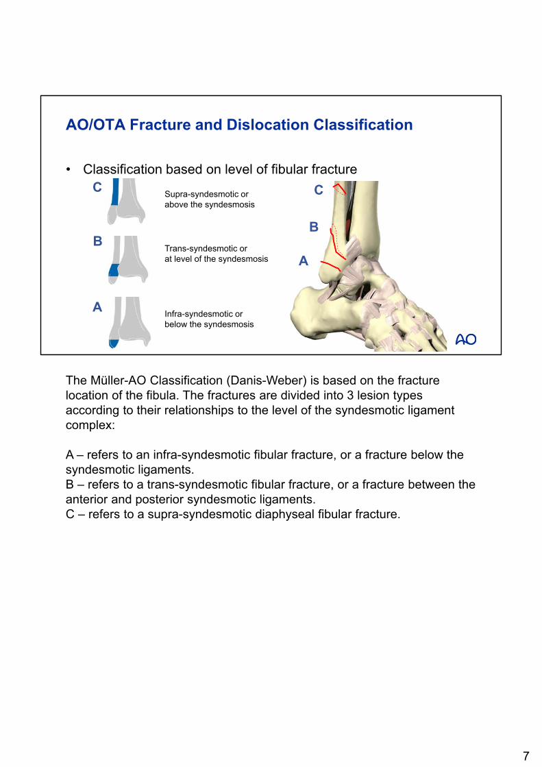
AO/OTA Fracture and Dislocation Classification
• Classification based on level of fibular fracture
The Müller-AO Classification (Danis-Weber) is based on the fracture location of the fibula. The fractures are divided into 3 lesion types according to their relationships to the level of the syndesmotic ligament complex:
A – refers to an infra-syndesmotic fibular fracture, or a fracture below the syndesmotic ligaments.B – refers to a trans-syndesmotic fibular fracture, or a fracture between the anterior and posterior syndesmotic ligaments.C – refers to a supra-syndesmotic diaphyseal fibular fracture.
7

Interosseous membrane
Posterior tibio-fibular ligament
Anterior tibio-fibular ligament
What is the syndesmosis?
There are several ligaments/membranes referring to the syndesmosis or structures which hold the tibia and fibula in position. These are:• the interosseous membrane • the syndesmotic ligaments
• posterior tibio-fibular ligament• anterior tibio-fibular ligament
8

Goal of reduction
Restoration of articular congruity• Fibula must be reduced both in length and
rotation
An x-ray of the uninjured ankle will serve as a guide to reduction
What do we want to achieve?
• In order to achieve a congruent ankle joint, the fibula should be restored to its original length and rotational alignment. An x‐ray of the uninjured ankle will serve as a guide to reduction.
9

Goal of reduction
• Restoration of articular congruity• Fibula must be reduced both in length and rotation
• Beak on fibula forms smooth curve with tibial articular surface
What do we want to achieve?
• In order to achieve a congruent ankle joint, the fibula should be restored to its original length and rotational alignment. An x‐ray of the uninjured ankle will serve as a guide to reduction.
• A key to correct length and rotation is a small beak on the fibula that must form a smooth curve with the tibial articular surface.
10

• Undisplaced fractures
• Stable fractures
• Uninjured medial ligaments
• Presence of contraindication to surgery• Patient comorbidities
• Soft-tissue status
Eg, isolated undisplaced44B-fracture of lateral malleolus
Indications for nonoperative treatment
Indications for nonoperative treatment are:• Undisplaced and stable fractures (for example isolated B fracture of fibula).• Clinically no injury of medial ligament.• Patients with diabetes and impaired circulation. They have delayed healing
entailing prolonged immobilization time in cast.• Unfit patients or limbs (e.g. swollen).
X-ray control after 10 days is undertaken to check stability. If fracture displacement has occurred, then surgery must be considered.
11

Eg, displaced, unstable 44B2 injury
Indications for operative treatment
• Articular fractures
• Displaced fractures
• Unstable fractures
• Condition• Soft tissue suitable for surgery
Indications for operative treatment of ankle fractures is dictated by the stability of the ankle joint.
Indications for surgery are: Articular fractures Displaced fractures Unstable fractures
An important condition is that the soft tissue is in a good condition allowing surgery.
Surgery involves open reduction and internal fixation (ORIF).
12

• Timing of surgery• Patient factors
• Status of soft tissue
• Positioning
• Surgical approach and reduction
• Implant choice
• Pitfalls and hazards
Principles of fracture treatment―preop. preparation
Preoperative preparation is very important. Before the surgery starts, many items must be prepared, checked and discussed.
• On clinical examination, it is important to assess condition of nerves and vessels.
• It is important to palpate the entire fibula because there may be an associated fracture proximally.
• Radiographic examination includes AP, lateral and mortise views (The mortise view is an anteroposterior x-ray taken with the ankle internally rotated some 10º-20º, so that the joint space between the lateral malleolus and the lateral facet of the talus is clearly seen).
• Timing of surgery depends on
• Patient details – e.g., co-morbidities, such as diabetes, allergies etc.
• Status of soft tissue – good condition of soft tissues is the key to a successful outcome.
• Positioning of the patient• Supine is standard with supports under hip and ankle to position ankle
joint laterally. • Perform a preoperative check:
• correct patient
13

• correct side
• correct site• Approach and reduction
• Surgical approach(es) depend on fracture type(s). Several may be necessary.
• Reduction - are all the instruments required for reduction available?• Implant choice - choice depends on availability in the hospital (1/3 tubular plate
for types A and B; LC DCP or LCP for type C, 4.0 partially threaded cancellous bone screws for medial malleolus and posterior malleolus).
• Pitfalls and hazards
13

Timing of surgery
• Immediate surgery whenfracture is displaced
In all cases a displaced ankle fracture most be reduced and splinted immediately.Because of the delicate soft-tissue cover over the two malleoli, the timing of surgery is crucial to avoid compromised healing.
14

Timing of surgery
• Immediate surgery when displaced fracture
• Postpone definitive surgery when • Ankle is swollen
• Local skin has blisters
• Skin looks like orange peel
If the ankle is very swollen and the skin has blisters, definitive surgery is postponed, ankle and foot splinted and the limb elevated, until the swelling has reduced.
15

Timing of surgery
• Immediate surgery when displaced fracture
• Postpone definitive surgery
• Apply external fixator as temporary stabilization
Temporary stabilization with large spanning external fixator in a triangular configuration is recommended as splintage until soft tissues have recovered sufficiently to allow definitive surgery. This can take up to 15 days.
16

Lateral and posterolateralaccess
Medial and posteromedial access
Positioning—supine
• Place the patient supine on a radiolucent operating table.• Put a sandbag under the ipsilateral buttock to internally rotate the limb for
lateral and posterolateral access• The figure of 4 position affords good medial and posteromedial access.
17

Positioning
• Apply a tourniquet on the thigh of the affected side, but inflate it only if required.
• Position the image intensifier and screen on the opposite side from the injured leg.
18

Procedure of surgical treatment
The surgical procedure follows the following steps:
1. Lateral malleolus
2. Medial malleolus
3. Posterior malleolus
4. Syndesmosis
Procedure of surgical treatment:
The surgical procedure follows generally the following steps:
1. The operation is usually started laterally with the fibula. Generally, in B fractures, reduction of the fibula also reduces any posterior malleolar fragment (Volkmann triangle).
2. If reduction is difficult, the medial side should be exposed to clear any soft tissue trapped in the medial side of the ankle joint.
3. Fixation of a posterior malleolus fracture is only required if the fragment bears more than one quarter of the tibial articular surface, as seen on the lateral x-ray of the ankle.
4. If the syndesmotic ligament is ruptured, a positioning screw, between the fibula and tibia, may be needed, provided the fibular fracture has been reduced, thereby restoring length and rotational alignment.
19

1. Lateral malleolus—approach
A 10–15 cm straight lateral incision over the distal fibula is made.
20

1. Lateral malleolus—reduction
• Anatomical reduction:• Length
• Rotation
The fracture is reduced anatomically. Reduction of both length and rotation are important.
21

R Teasdale
1 32
1. Lateral malleolus—fixation: options
1. Interfragmentary lag screw and neutralization plate
2. Tension band wiring
3. Intramedullary fixation
1. A 3,5 mm cortex screw is inserted as a lag screw. A neutralization (protection) plate is added.
2. Other options for fixation are tension band wiring and…3. …intra medullary fixation of the fibula with a large screw.
The distal screws in the fibula do not penetrate the joint. The position must be checked with the image intensifier.
22

1 2
2. Medial malleolus—approach
1. Incision• The incision starts 2 cm distal to the anterior tip of the medial malleolus and
curves towards the anterior edge of the medial malleolus and in the direction of the middle of the distal tibia.
• The saphenous vein and nerve are retracted with a vessel loop.• The fracture is exposed and any interposed soft tissue that may
preventing reduction is removed.
Note:Be careful not to damage the saphenous vein and nerve, especially distally.
1. Inspection of joint• The anterior part of the fracture site is exposed, the periosteum is removed
from the edges of the medial malleolus to the distal tibial joint surface and the joint inspected.
• If necessary, a vertical incision at the anteromedial edge of the joint capsule is made. The capsule is dissected as far as necessary to visualize the fracture and the joint surfaces.
23

2. Medial malleolus—reduction
Reduction• The fracture is reduced anatomically.
24

1 32
2. Medial malleolus―fixation (options)
1. Partially threaded cancellous bone screws
2. Tension band wiring
3. Medial buttress plate
1. The medial malleolus is fixed with two partially threaded cancellous bone screws 4,0 mm.
2. If the quality of the bone is not so good, or the fragment is small, a tension band wiring can be used.
3. If the fragment is large and the fracture plane is vertical, as in some type A fractures, the fracture is fixed with a medial buttress plate.
25

3. Posterior malleolus—approach and reduction
This approach is indicated in cases of posterior comminution and/or posterior extension of a medial malleolar fracture. • The incision starts 1 cm distal and 1 cm anterior to the middle of the tip of the medial
malleolus. The incision curves proximally and posteriorly over the tip of the medial malleolus and then follows the direction of the posterior crest of the distal tibia.
Note:Be careful not to damage the saphenous vein and nerve, especially distally.
The fracture is reduced anatomically.
26

21
3. Posterior malleolus—fixation
• After fixation of medial and lateral malleoli
• When there is posterior subluxation of the talus (1)
• When fragment bears more than 20–25% of articular surface (2)
Posterior malleolar fractures of significant size are usually associated with B fracture patterns.Fracture fixation of the posterior malleolus should be undertaken:
• After the medial and lateral malleoli have been reduced and fixed anatomically.
• If there is persistent posterior subluxation of the talus.
• If the posterior fragment bears more than 20–25% of articular surface.
27

21
3. Posterior malleolus—fixation
1. Anteroposterior screw fixation
2. Posteroanterior screw fixation and buttress plate
• Fixation is usually with one or two partially threaded cancellous bone screws, after reduction and temporary K‐wire stabilization.
• In large, long fragments, fixed via a posterolateral approach, a small buttress plate (1/3 tubular plate 3.5) can be added.
28

3. Posterior malleolus—fixation
1. Antero-posterior screw fixation
2. Postero-anterior screw fixation & buttress plate
3. Cannulated screws are helpful, if available
• Cannulated screws can be used if they are available in the OR.
29

4. Syndesmosis―stability testing
• Bone hook test
After plating of the fibular shaft in type C fractures, if there is no anterior tibial tubercular fracture, then stability of the syndesmosis is tested by attempting to distract the fixed fibula from the tibia, using a bone hook.
30

4. Syndesmosis―fixation
• Position screw(s): one or two cortex screws 3.5 mm
Fixation of the syndesmosis only if unstable on hook test:
• The fully threaded positioning screw(s) must grip in all 3 cortices.
• In certain fracture types 2 screws must be used (Maisonneuve injury).
• The position screw should be above, not through, the inferior tibio-fibular syndesmosis.
• The position screw may be inserted through the fibular plate.
31

Removal of position screw
• In young and active patients
• After 8–12 weeks
• If not, screw(s) might break
Removal of the position screw (syndesmosis screw):• Remove the position screws in young and active patients after 8-12 weeks.• In very unstable fractures (Maisonneuve injuries), in smokers, or in patients
with diabetes, wait a least 12 weeks.
• If not removed, the screw may eventually break.
32

After care
• Back-slab for a few days until swelling reduced
• Start early active joint motion exercises out of plaster slab
• Cooperative patients are allowed immediate touch weight bearing
• Remove sutures and do x-ray control after 2–3 weeks
• Remove position screws after 8–12 weeks if bone healing is satisfactory
The after care:• Apply a plaster of Paris back-slab to the lower leg with the foot in neutral
position.• Start physiotherapy on postoperative day 1, with active range of motion
exercises out of the splint, reapplying splint after exercise.
• Allow immediate partial weight bearing (10-15kg) to cooperative patients.
• Apply a short leg cast once a good range of motion is obtained.
• Remove sutures and make X-ray after 2-3 weeks.
• Weight bearing once full bone and ligamentous healing is assured (6-10 weeks), depending on fracture pattern.
• Eventually remove the syndesmosis screws after 8-12 weeks if bone healing is satisfactory.
33

Questions
OptionalInsert questions to check learning
34

What is the syndesmosis?
1. The anterior talofibular ligament of the ankle joint
2. A ligamentous complex holding fibula and tibia in correct anatomical position
3. A bony structure to which the Müller AO Classification refers
OptionalInsert questions to check learning
35

What is the syndesmosis?
1. The anterior talofibular ligament of the ankle joint
2. A ligamentous complex holding fibula and tibia in correct anatomical position
3. A bony structure to which the Müller AO Classification refers
OptionalInsert questions to check learning
36

What are the indications for operative treatment?
1. Articular, displaced, and unstable fractures
2. Articular, displaced, and stable fractures
3. Articular, undisplaced, and unstable fractures
OptionalInsert questions to check learning
37

What are the indications for operative treatment?
1. Articular, displaced, and unstable fractures
2. Articular, displaced, and stable fractures
3. Articular, undisplaced, and unstable fractures
OptionalInsert questions to check learning
38
Which implant is most commonly used for fixation of a lateral malleolar fracture?
1. One third tubular plate and lag screw
2. LC-DCP and lag screw
3. LCP and lag screw
OptionalInsert questions to check learning
39

Which implant is most commonly used for fixation of a lateral malleolar fracture?
1. One third tubular plate and lag screw
2. LC-DCP and lag screw
3. LCP and lag screw
OptionalInsert questions to check learning
40

Learning outcomes
You should now be able to:
• Describe principles of malleolar fracture classification
• Outline indications for nonoperative and surgical treatment
• Discuss principles of malleolar fracture fixation
You should now be able to• describe principles of malleolar fracture classification• outline indications for nonoperative and surgical treatment• discuss principles of malleolar fracture fixation
41