major drug classes abbreviations
TRANSCRIPT

21-1
Treatment of Seizure DisordersMichael A. Rogawski
21
THERAPEUTIC OVERVIEWEpilepsy is a chronic episodic disorder of brain function characterized by the unpredictable occurrence of seizures. Epileptic seizures are transitory alterations in behavior, sensation, or consciousness caused by abnormal, excessive, or synchronous neuronal activity in the brain that can be detected with the electroencephalogram (EEG). Approximately 0.8% of the population suffers from epilepsy. Epilepsy can occur at any age, but onset is more frequent in children younger than about age 10 and in adults over age 50. Recurrent seizures, if frequent, interfere with a patient’s ability to carry out day-to-day activities. However, daily oral use of antiseizure medications allows approximately 70% of patients to remain seizure free.
Seizures are classified into two major types: focal onset (formerly partial onset) seizures and generalized onset seizures. Focal seizures arise in a localized region in one cerebral hemisphere and are accom-panied by EEG abnormalities that are restricted to the epileptic focus. In contrast, generalized seizures are associated with EEG features indicating simultaneous hemispheric activation.
Focal seizures are further classified as aware, impaired awareness, or focal to bilateral tonic-clonic. The seizures are termed aware (formerly simple) if consciousness is preserved and impaired awareness (formerly complex) if consciousness is impaired or lost. In impaired awareness seizures, motor activity often appears as a complicated and seemingly purposeful movement referred to as an automatism. If a focal seizure spreads to encompass both hemispheres, the focal seizure can transition to a bilateral tonic-clonic seizure (formerly secondarily generalized) resulting in tonic-clonic manifestations, which involve rigid extension of the trunk and limbs (tonic phase) followed by rhythmic contractions of the arms and legs (clonic phase).
In generalized seizures, both hemispheres are involved at the onset. There are various types of generalized seizures, including generalized
tonic-clonic seizures, which are similar to focal to bilateral tonic-clonic seizures except that they do not begin focally; absence seizures, character-ized by impaired consciousness and minimal motor manifestations; and other types of seizures, including myoclonic, clonic, tonic, or atonic (astatic), depending on the specific clinical manifestations. The classifica-tion of seizures and their characteristics are presented in the Therapeutic Overview Box.
Status epilepticus, clinically defined as abnormally prolonged or repetitive seizures, presents in several forms, including (1) tonic-clonic (convulsive) status epilepticus, (2) nonconvulsive status epilepticus, (3) focal status epilepticus, and (4) absence status epilepticus. Con-vulsive status epilepticus is a life-threatening medical emergency that requires immediate treatment. Traditionally, convulsive status epilepticus was defined as more than 30 minutes of either (1) continuous seizure activity or (2) two or more sequential seizures without full recovery of consciousness between seizures. Because persistent seizure activity is believed to cause permanent neuronal injury and the majority of seizures terminates in 2 to 3 minutes, it is now generally accepted that treatment should begin when the seizure duration reaches 5 minutes for generalized tonic-clonic seizures and 10 minutes for focal seizures with or without impairment of awareness.
Convulsive status epilepticus can lead to systemic hypoxia, acidemia, hyperpyrexia, cardiovascular collapse, and renal shutdown. Nonconvulsive status epilepticus, a persistent change in behavior or mental processes with continuous epileptiform EEG but without major motor signs, also requires urgent treatment.
All people are capable of experiencing seizures. Brain insults such as fever, hypoglycemia, hypocalcemia, hyponatremia, and extreme lactic acidosis, or exposure to certain drugs or toxins, can trigger a seizure, but if the condition is corrected, seizures do not recur, and the condition is not considered epilepsy. Epilepsy is a disease (also variously described as a disorder) characterized by an enduring predisposition to epileptic seizures and by the neurobiological, cognitive, psychological, and social consequences of this condition. The diverse causes of seizures and epilepsy are listed in Box 21.1.
The goal of antiseizure drug therapy is to prevent seizures while minimizing adverse effects. If seizures continue after drug therapy is initiated, the dose may be increased until unacceptable adverse effects
Voltage-gated ion channel modulatorsVoltage-gated sodium channel blockersT-type voltage-gated calcium channel blockersGabapentinoids (α2δ ligands)Kv7 voltage-gated potassium channel openers
GABA enhancersGABAA receptor modulatorsGABA transporter inhibitorsGABA transaminase inhibitors
AMPA receptor antagonistsSV2A ligandsMixed-acting compounds
MAJOR DRUG CLASSESACTH AdrenocorticotropinARS Acute repetitive seizuresEEG ElectroencephalogramGABA γ-Aminobutyric acidGAT-1 GABA transporter
ABBREVIATIONS
ISBN: 978-0-323-47652-2; PII: B978-0-323-47652-2.00021-1; Author: Wecker; 00021
c00021
b0010
p0015
u0015
u0020
u0025
u0030
u0035
u0040
u0045
u0050
u0055
u0060
u0065
b0015
p0080
t0010
s0010
p0085
p0090
p0095
p0100
p0105
p0110
p0115
p0210
Chapter 21.indd 1 3/29/2018 4:01:27 PM
To protect the rights of the author(s) and publisher we inform you that this PDF is an uncorrected proof for internal business use only by the author(s), editor(s), reviewer(s), Elsevier and typesetter Toppan Best-set. It is not allowed to publish this proof online or in print. This proof copy is the copyright property of the publisher and is confidential until formal publication. These proofs may contain color(colour) figures. Those figures may print black and white in the final printed book if a color(colour) print product has not been planned. The color(colour) figures will appear in color(colour) in all electronic versions of this book.
CHAPTER 21 Treatment of Seizure Disorders 21-1.e1
Key Wordsacute repetitive seizuresantiepileptic drugantiseizure drugepilepsyseizurestatus epilepticus
AbstractAntiseizure drugs are used chronically to treat epilepsy and on an as needed basis to terminate status epilepticus and acute repetitive seizures.
ISBN: 978-0-323-47652-2; PII: B978-0-323-47652-2.00021-1; Author: Wecker; 00021
abs0010
Chapter 21.indd 1 3/29/2018 4:01:29 PM
To protect the rights of the author(s) and publisher we inform you that this PDF is an uncorrected proof for internal business use only by the author(s), editor(s), reviewer(s), Elsevier and typesetter Toppan Best-set. It is not allowed to publish this proof online or in print. This proof copy is the copyright property of the publisher and is confidential until formal publication. These proofs may contain color(colour) figures. Those figures may print black and white in the final printed book if a color(colour) print product has not been planned. The color(colour) figures will appear in color(colour) in all electronic versions of this book.

21-2 SECTION 3 Drug Treatment for Disorders Affecting the Central Nervous System
It is noteworthy that many of the genes in the monogenic epi-lepsies encode subunits of ion channels, which are the fundamental mediators of neuronal excitability. These types of epilepsies can be considered channelopathies. However, some monogenic epilepsies are caused by mutations in non–ion channel genes, including neural adhesion molecules, such as PCDH19 (protocadherin 19), and proteins involved in synapse development, such as LGI1 (leucine-rich glioma inactivated 1).
The cellular and molecular events leading to the development of focal epilepsies in cases of cortical injury are poorly understood. There is better understanding of the physiology of the seizures. Focal seizures are thought to occur as a consequence of the loss of surround inhibition, a process that normally prevents the activation of neurons adjacent to a focus (Fig. 21.1). This loss of surround inhibition may result from impaired γ-aminobutyric acid (GABA) transmission, loss of GABA interneurons, changes in GABA type A (GABAA) receptors, or alterations in intracellular chloride or bicarbonate ion concentrations. Excessive glutamate-mediated excitation may also lead to focal seizures. Impaired GABA-mediated inhibition or excessive glutamate-mediated excitation predisposes to abnormal hypersynchronous activity manifest as epi-leptiform discharges, which, if they encompass a large enough area of cortex, are associated with the motor, sensory, psychic, or autonomic symptoms of a focal seizure.
Generalized seizures involve both hemispheres and thalamic syn-chronizing mechanisms. In tonic-clonic convulsions, the tonic phase of muscle contraction is thought to reflect prolonged neuronal depolariza-tion as a consequence of the loss of GABA-mediated inhibition and the dominance of excitatory glutamate neurotransmission. As the seizure evolves, neurons repolarize and afterhyperpolarizations are apparent, which reflect the reappearance of GABA-mediated inhibition and diminished glutamate excitation, producing the clonic phase. Drugs that increase surround inhibition and prevent the spread of synchronous activity are effective in the treatment of focal seizures.
Our understanding of the onset of generalized tonic-clonic seizures is limited. However, there are some clues concerning the cellular mechanisms underlying absence seizures, which are characterized by the sudden appearance of spike-wave discharges synchronized throughout the brain. The EEGs recorded during an absence seizure compared with a generalized tonic-clonic seizure are shown in Fig. 21.2. Studies support a major role of thalamocortical circuits in the pathogenesis of absence seizures with abnormal oscillations generated by excitatory glutamatergic cortical pyramidal and thalamic relay neurons and inhibitory GABAergic thalamic reticular neurons (Fig. 21.3). Thalamic relay neurons project to the cortex, and cortical pyramidal neurons project back to the thalamus
prevent further dosage increases, at which point another drug can be substituted or a second drug added. Children who are seizure free for periods longer than 2–4 years while on antiseizure medications will remain so when medications are withdrawn in 70% of cases so that a trial of discontinuation may be warranted. Resolution of seizures is common for certain syndromes, such as childhood absence epilepsy and benign epilepsy of childhood with centrotemporal spikes (BECTS), but infrequent for others, such as juvenile myoclonic epilepsy. Resolution is unlikely in adults with an abnormal neurologic examination or an abnormal EEG so that drug treatment will likely be required for the life of the patient.
PathophysiologyMany cases of epilepsy are the result of damage to the brain, as occurs in traumatic brain injury, stroke, or infections, whereas in other cases, the epilepsy is caused by a brain tumor or developmental lesion such as a cortical or vascular malformation; these epilepsies are referred to as symptomatic. Mesial temporal lobe epilepsy associated with hip-pocampal sclerosis is a symptomatic epilepsy that is a common cause of medication refractory seizures.
In 40% of all epilepsies, genetic factors are believed to be the root cause. In some cases, the epilepsy is a component of a genetic syndrome, such as tuberous sclerosis, that has other associated structural or meta-bolic brain abnormalities. In other cases, the genetic epilepsy has seizures as its only clinical manifestation, and there is no apparent structural or metabolic disorder of the brain. Such idiopathic epilepsies include benign epilepsy of childhood with centrotemporal spikes (BECTS), benign familial neonatal convulsions, childhood absence epilepsy, and juvenile myoclonic epilepsy. In most genetic epilepsies, the inheritance is complex (polygenic); rarely, a single gene defect can be identified. Some monogenic epilepsies and associated gene mutations are listed in Box 21.2. In some cases, these genetic epilepsies are benign, and in other cases, they are severe and termed epileptic encephalopathies.
Causes of SeizuresIn newborns:
• Neonatal hypoxia; intracranial hemorrhage; maternal drug useIn infants and children:
• Fever; infections (meningitis or encephalitis)In adults and the elderly:
• Traumatic brain injury; stroke; metabolic disturbances (hypoglycemia, hypocalcemia, hypomagnesemia, hyponatremia, lactic acidosis, uremia); drugs, alcohol, and toxins including withdrawal from barbiturates and other central nervous system depressants
Causes of Epilepsy• Traumatic brain injury• Status epilepticus• Genetic syndromes with seizures in conjunction with intellectual disability,
brain structural or metabolic abnormalities, or congenital malformations• Genetic syndromes with isolated seizures (idiopathic)• Congenital malformations• Birth and perinatal injuries• Stroke• Brain tumor• Infections such as neurocysticercosis• Alzheimer’s disease and other degenerative neurological conditions
BOX 21.1 Causes of Seizures and Epilepsy
Disorder Mutation(s)
Autosomal dominant nocturnal frontal lobe epilepsy (ADNFLE)
Nicotinic acetylcholine receptor subunit genes CHRNA4, CHRNB2, or CHRNA2
Autosomal dominant juvenile myoclonic epilepsy
GABAA receptor subunit gene GABRA1
Benign familial neonatal seizures Voltage-gated potassium channel genes KCNQ2 or KCNQ3
Familial febrile seizures and Dravet syndrome
Voltage-gated sodium channel genes SCN1A and rarely SCN8A
BOX 21.2 Examples of Monogenic Epilepsies
GABAA, γ-Aminobutyric acid type A.
ISBN: 978-0-323-47652-2; PII: B978-0-323-47652-2.00021-1; Author: Wecker; 00021
b0020
s0015
p0120
u0075
u0080
u0085
u0090
u0095
s0020
p0155
u0105
u0110
u0115
u0120
u0125
u0130
u0135
u0140
u0145
b0025
p0225
t0015
p0230
s0025
p0215
p0220
p0235
p0240
p0245
p0250
Chapter 21.indd 2 3/29/2018 4:01:27 PM
To protect the rights of the author(s) and publisher we inform you that this PDF is an uncorrected proof for internal business use only by the author(s), editor(s), reviewer(s), Elsevier and typesetter Toppan Best-set. It is not allowed to publish this proof online or in print. This proof copy is the copyright property of the publisher and is confidential until formal publication. These proofs may contain color(colour) figures. Those figures may print black and white in the final printed book if a color(colour) print product has not been planned. The color(colour) figures will appear in color(colour) in all electronic versions of this book.

CHAPTER 21 Treatment of Seizure Disorders 21-3
MECHANISMS OF ACTIONSelection of the correct antiseizure drug depends on accurate diagnosis of the patient’s seizure type and epilepsy syndrome. Focal onset seizures must be distinguished from generalized onset seizures because some drugs effective for focal seizures do not prevent and may exacerbate some generalized seizure types. Certain epilepsy syndromes, such as infantile spasms, require treatment with special agents.
Epileptic activity may occur as a consequence of either decreased inhibition or increased excitation of neurons. Agents used for the treatment of epilepsy depress aberrant neuronal firing primarily by
in a recurrent excitatory loop. Thalamic relay neurons exhibit spike-wave discharges that generate normal cortical rhythms and participate in the generation of sleep spindles. The normal bursting pattern of these neurons results from the activation of low voltage-gated T-type calcium channels during depolarization, followed by GABA release from thalamic reticular neurons and hyperpolarization. The circuit transitions to abnormal rhythmicity at the onset of an absence seizure. T-type calcium channels in relay neurons and thalamic reticular neurons play a critical role in the pathological behavior of absence seizures, as blockade of these channels, most notably by ethosuximide, is effective for the treat-ment of such seizures.
Epilepticfocus
Inhibitorysurround
Inhibitorysurround
InhibitorysurroundEpileptic focus
Pyramidalneuron
GABAergicinterneuron
+ +
+ +-- -- -- --
FIG. 21.1 Seizure Generation and Spread in a Focal Seizure. Epileptiform activity begins in a localized area (epileptic focus) and spreads to adjacent and contralateral cortical regions. Epileptic activity reflects the synchronized activity of excitatory (+) glutamate cortical pyramidal neurons. Spread is restrained by inhibitory (–) γ-aminobutyric acid (GABA) interneurons. Seizure spread is believed to reflect the loss of surround inhibition. Antiseizure drugs that enhance GABA inhibition restrain seizure spread. Spread can also be prevented by sodium-channel blocking drugs (Fig. 21.4) and AMPA receptor antagonists.
GENERALIZED TONIC-CLONIC SEIZURE GENERALIZED ABSENCE SEIZURE
Tonic phase
SurfaceEEG
Intra-cellular
SurfaceEEG
Intra-cellularSustained
depolarization
Clonic phaseSpike Wave
FIG. 21.2 Comparison of the Electrical Behavior of the Surface Electroencephalogram (EEG) and Single-Neuron Recording During Generalized Tonic-Clonic and Absence Seizures on a Time Scale of Hundreds of Milliseconds. Surface EEG signals are field responses detected with flat metal electrodes on the scalp. Intracellular recordings represent the membrane potential changes of individual cortical neurons. A generalized tonic-clonic seizure begins with a tonic phase consisting of rhythmic high-frequency discharges recorded in the surface EEG; cortical neurons undergo sustained depolarization, which generates high-frequency action potential firing. Subsequently, the seizure converts to a clonic phase, character-ized by groups of spikes on the EEG; cortical neurons exhibit periodic depolarizations with clusters of action potentials on the crests. In an absence seizure, 3-Hz spike-and-wave discharges are recorded in the surface EEG. During the spike phase, cortical neurons generate short-duration depolarizations, which trigger a brief burst of action potentials. During the wave phase, cortical neurons are hyperpolarized. Recognizing the dif-ferences between the single neuron electrical events in tonic-clonic and absence seizures helps in understanding why antiseizure drugs that inhibit sustained repetitive firing of action potentials (see Fig. 21.4) are effective in the treatment of tonic-clonic, but not absence, seizures.
ISBN: 978-0-323-47652-2; PII: B978-0-323-47652-2.00021-1; Author: Wecker; 00021
f0010
f0015
s0040
p0335
p0340
Chapter 21.indd 3 3/29/2018 4:01:27 PM
To protect the rights of the author(s) and publisher we inform you that this PDF is an uncorrected proof for internal business use only by the author(s), editor(s), reviewer(s), Elsevier and typesetter Toppan Best-set. It is not allowed to publish this proof online or in print. This proof copy is the copyright property of the publisher and is confidential until formal publication. These proofs may contain color(colour) figures. Those figures may print black and white in the final printed book if a color(colour) print product has not been planned. The color(colour) figures will appear in color(colour) in all electronic versions of this book.

21-4 SECTION 3 Drug Treatment for Disorders Affecting the Central Nervous System
Cerebral corticallayers 4 and 5
Cortex
Thalamicreticular n.
Thalamus
T-type calciumchannel
T-type calciumchannel
Relayneuron
Thalamus
+
+
++ --
Thalamocorticalloop
+
+
++ --
Cerebral cortex
Thalamus
Corpus callosum
FIG. 21.3 Thalamocortical Circuitry Generating Absence Seizures According to the “Corticoreticular” Theory. The thalamus and cortex are both essential for the spike-wave discharges of absence seizures; bilateral synchrony depends on the corpus callosum connecting the two hemispheres. Spike-wave bursts are likely initiated in the cortex (the perioral region of the somatosensory cortex has been implicated) by the discharge of a network of massively interconnected excitatory neurons in the presence of insufficient γ-aminobutyric acid (GABA) inhibition. This initial event is followed by entrainment of the thalamus leading to synchronized oscillations in which the thalamus and cortex drive each other. Excitatory (+) glutamate thalamic relay neurons project to the cortex, and excitatory glutamate cortical neurons project back to the thalamus, forming a recurrent loop. The thalamic reticular nucleus, a shell-like structure covering the thalamus, is composed of GABA interneurons that provide massive inhibitory (–) input to thalamic relay neurons and may contribute to the pathological oscillations. T-type voltage-gated calcium channels are necessary for burst firing in thalamic relay neurons and thalamic reticular neurons. Shown below the diagram is a coronal fluo-rodeoxyglucose positron emission tomography image of a human brain superimposed on T1 magnetic resonance image, illustrating the location of the relevant structures. (From Johnson KA, Becker JA. Whole Brain Atlas, with permission. http://www.med.harvard.edu/aanlib/cprt.html)
Focal Onset (Partial Onset) SeizuresFocal aware seizure (formerly simple partial seizure)
Sensory, motor, autonomic, or psychic symptoms, without altered awareness
Focal impaired awareness seizure (formerly complex partial seizure)Dreamy disaffective state with or without automatisms, with altered
awarenessFocal to bilateral tonic-clonic seizure (formerly secondarily generalized tonic-clonic
seizure or grand mal seizure)Evolution of focal aware or focal impaired awareness seizure to convulsion
with rigid extension of trunk and limbs (tonic phase) and rhythmic contractions of arms and legs (clonic phase)
Generalized Onset SeizuresGeneralized tonic-clonic seizure (formerly primary generalized tonic-clonic seizure
or grand mal seizure)Similar to focal to bilateral tonic-clonic seizure except that onset is in both
hemispheres; occurs in patients with genetic (idiopathic) generalized epilepsies
Generalized absence seizuresAbrupt loss of consciousness with staring and cessation of ongoing activity
with or without eye blinks; occurs in patients with genetic (idiopathic) generalized epilepsies, including childhood absence epilepsy
Other types of generalized onset seizuresMyoclonic seizure: rapid shock-like (jerking) muscle contractionAtonic seizure (drop seizure or astatic seizure): loss of muscle toneEpileptic spasm: sudden flexion, extension or flexion-extension of neck, trunk,
arms, and legs
THERAPEUTIC OVERVIEW
ISBN: 978-0-323-47652-2; PII: B978-0-323-47652-2.00021-1; Author: Wecker; 00021
f0020
b0030
s0030
p0255
u0155
u0160
u0165
u0170
u0175
s0035
p0290
u0185
u0190
u0195
u0200
u0205
u0210
u0215
Chapter 21.indd 4 3/29/2018 4:01:28 PM
To protect the rights of the author(s) and publisher we inform you that this PDF is an uncorrected proof for internal business use only by the author(s), editor(s), reviewer(s), Elsevier and typesetter Toppan Best-set. It is not allowed to publish this proof online or in print. This proof copy is the copyright property of the publisher and is confidential until formal publication. These proofs may contain color(colour) figures. Those figures may print black and white in the final printed book if a color(colour) print product has not been planned. The color(colour) figures will appear in color(colour) in all electronic versions of this book.
CHAPTER 21 Treatment of Seizure Disorders 21-5
the repetitive firing of neurons by producing a use-dependent and voltage-dependent blockade of sodium channels (Fig. 21.4). By prolonging the inactivated state of the sodium channel and thus the relative refractory period, these drugs do not alter the first action potential in a train but rather reduce the likelihood of repetitive action potentials. Neurons retain their ability to generate action potentials at the lower frequencies common during normal brain function. The discrimination between normal firing from usual membrane potential levels and high-frequency firing under the abnormally depolarized conditions of the epileptic discharge allows these drugs to inhibit seizures without affecting normal brain function at therapeutic concentrations.
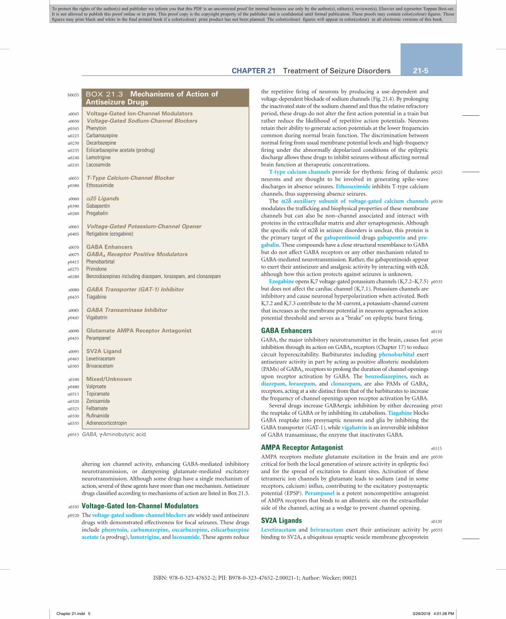
T-type calcium channels provide for rhythmic firing of thalamic neurons and are thought to be involved in generating spike-wave discharges in absence seizures. Ethosuximide inhibits T-type calcium channels, thus suppressing absence seizures.
The α2δ auxiliary subunit of voltage-gated calcium channels modulates the trafficking and biophysical properties of these membrane channels but can also be non–channel associated and interact with proteins in the extracellular matrix and alter synaptogenesis. Although the specific role of α2δ in seizure disorders is unclear, this protein is the primary target of the gabapentinoid drugs gabapentin and pre-gabalin. These compounds have a close structural resemblance to GABA but do not affect GABA receptors or any other mechanism related to GABA-mediated neurotransmission. Rather, the gabapentinoids appear to exert their antiseizure and analgesic activity by interacting with α2δ, although how this action protects against seizures is unknown.
Ezogabine opens Kv7 voltage-gated potassium channels (Kv7.2–Kv7.5) but does not affect the cardiac channel (Kv7.1). Potassium channels are inhibitory and cause neuronal hyperpolarization when activated. Both Kv7.2 and Kv7.3 contribute to the M-current, a potassium-channel current that increases as the membrane potential in neurons approaches action potential threshold and serves as a “brake” on epileptic burst firing.
GABA EnhancersGABA, the major inhibitory neurotransmitter in the brain, causes fast inhibition through its action on GABAA receptors (Chapter 17) to reduce circuit hyperexcitability. Barbiturates including phenobarbital exert antiseizure activity in part by acting as positive allosteric modulators (PAMs) of GABAA receptors to prolong the duration of channel openings upon receptor activation by GABA. The benzodiazepines, such as diazepam, lorazepam, and clonazepam, are also PAMs of GABAA receptors, acting at a site distinct from that of the barbiturates to increase the frequency of channel openings upon receptor activation by GABA.
Several drugs increase GABAergic inhibition by either decreasing the reuptake of GABA or by inhibiting its catabolism. Tiagabine blocks GABA reuptake into presynaptic neurons and glia by inhibiting the GABA transporter (GAT-1), while vigabatrin is an irreversible inhibitor of GABA transaminase, the enzyme that inactivates GABA.
AMPA Receptor AntagonistAMPA receptors mediate glutamate excitation in the brain and are critical for both the local generation of seizure activity in epileptic foci and for the spread of excitation to distant sites. Activation of these tetrameric ion channels by glutamate leads to sodium (and in some receptors, calcium) influx, contributing to the excitatory postsynaptic potential (EPSP). Perampanel is a potent noncompetitive antagonist of AMPA receptors that binds to an allosteric site on the extracellular side of the channel, acting as a wedge to prevent channel opening.
SV2A LigandsLevetiracetam and brivaracetam exert their antiseizure activity by binding to SV2A, a ubiquitous synaptic vesicle membrane glycoprotein
altering ion channel activity, enhancing GABA-mediated inhibitory neurotransmission, or dampening glutamate-mediated excitatory neurotransmission. Although some drugs have a single mechanism of action, several of these agents have more than one mechanism. Antiseizure drugs classified according to mechanisms of action are listed in Box 21.3.
Voltage-Gated Ion-Channel ModulatorsThe voltage-gated sodium-channel blockers are widely used antiseizure drugs with demonstrated effectiveness for focal seizures. These drugs include phenytoin, carbamazepine, oxcarbazepine, eslicarbazepine acetate (a prodrug), lamotrigine, and lacosamide. These agents reduce
Voltage-Gated Ion-Channel ModulatorsVoltage-Gated Sodium-Channel BlockersPhenytoinCarbamazepineOxcarbazepineEslicarbazepine acetate (prodrug)LamotrigineLacosamide
T-Type Calcium-Channel BlockerEthosuximide
α2δ LigandsGabapentinPregabalin
Voltage-Gated Potassium-Channel OpenerRetigabine (ezogabine)
GABA EnhancersGABAA Receptor Positive ModulatorsPhenobarbitalPrimidoneBenzodiazepines including diazepam, lorazepam, and clonazepam
GABA Transporter (GAT-1) InhibitorTiagabine
GABA Transaminase InhibitorVigabatrin
Glutamate AMPA Receptor AntagonistPerampanel
SV2A LigandLevetiracetamBrivaracetam
Mixed/UnknownValproateTopiramateZonisamideFelbamateRufinamideAdrenocorticotropin
BOX 21.3 Mechanisms of Action of Antiseizure Drugs
GABA, γ-Aminobutyric acid.
ISBN: 978-0-323-47652-2; PII: B978-0-323-47652-2.00021-1; Author: Wecker; 00021
b0035
s0045
s0050
p0345
u0225
u0230
u0235
u0240
u0245
s0055
p0380
s0060
p0390
u0260
s0065
p0405
s0070
s0075
p0415
u0275
u0280
s0080
p0435
s0085
p0445
s0090
p0455
s0095
p0465
u0305
s0100
p0480
u0315
u0320
u0325
u0330
u0335
p0515
s0105
p0520
p0525
p0530
p0535
s0110
p0540
p0545
s0115
p0550
s0120
p0555
Chapter 21.indd 5 3/29/2018 4:01:28 PM
To protect the rights of the author(s) and publisher we inform you that this PDF is an uncorrected proof for internal business use only by the author(s), editor(s), reviewer(s), Elsevier and typesetter Toppan Best-set. It is not allowed to publish this proof online or in print. This proof copy is the copyright property of the publisher and is confidential until formal publication. These proofs may contain color(colour) figures. Those figures may print black and white in the final printed book if a color(colour) print product has not been planned. The color(colour) figures will appear in color(colour) in all electronic versions of this book.

21-6 SECTION 3 Drug Treatment for Disorders Affecting the Central Nervous System
well understood. Zonisamide blocks voltage-gated sodium and T-type calcium channels, while rufinamide also modulates voltage-gated sodium-channel activity but may also have other actions.
ACTH, which is used for the treatment of infantile spasms, stimulates glucocorticoid (cortisol) synthesis and release from the adrenal cortex. The cortisol could influence infantile spasms through an antiinflam-matory action or in some other fashion. Synthetic glucocorticoids such as prednisone and prednisolone also have therapeutic activity in the treatment of infantile spasms.
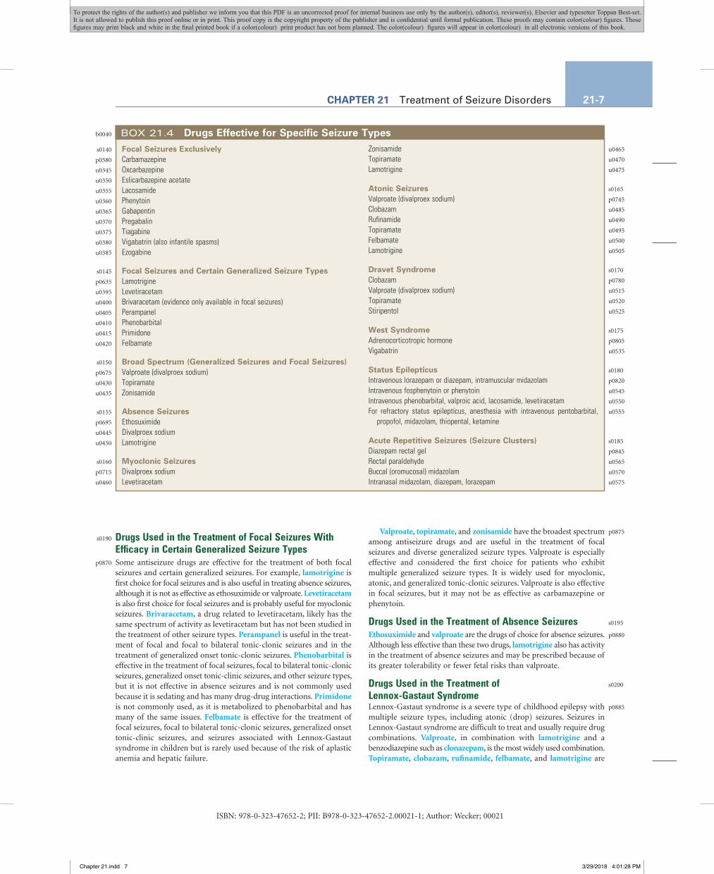
RELATIONSHIP OF MECHANISMS OF ACTION TO CLINICAL RESPONSEDrugs Used in the Treatment of Focal SeizuresThe sodium-channel blockers (except lamotrigine), gabapenti-noids, and tiagabine are used exclusively for the treatment of focal seizures and focal to bilateral tonic-clonic seizures (Box 21.4). Some of these drugs have also shown efficacy in generalized onset tonic-clonic seizures, including oxcarbazepine and phenytoin. These drugs may exacerbate certain types of generalized onset seizures, including absence and myoclonic seizures, and seizures in Dravet syndrome.
involved in exocytosis. The precise role of SV2A in the exocytotic process is not well understood, but evidence suggests that it interacts with synaptotagmin, a trigger for calcium-mediated exocytosis. It is possible that the binding of levetiracetam and brivaracetam to SV2A reduces the release of glutamate during trains of high-frequency activity as occurs during epileptic activity.
Mixed-Acting CompoundsThe mechanisms of action of many antiseizure drugs involve mixed effects or are poorly understood. These compounds include valproate, felbamate, topiramate, zonisamide, rufinamide, and adrenocortico-tropin (ACTH).
The antiseizure effects of valproate have been attributed to increases in the turnover of GABA in a regionally selective manner, which might be associated with enhanced synaptic or extrasynaptic inhibition, but this mechanism is not well established and there is no consensus on the drug’s antiseizure mechanism. Felbamate acts as a PAM at GABAA receptors and blocks glutamate NMDA receptors, although the relation-ship between this latter action and the antiseizure activity of felbamate is questionable. Topiramate may modulate voltage-gated sodium channels and glutamate receptors and may potentiate GABA activity, but again, the relationship between these actions and antiseizure activity is not
Na+ Extracellularside
Membrane
Intracellularside
--
+
CLOSED
AG
IG
AG
IG
Na+
Na+
+
--
OPEN
AG
IG
Sodium channel blockingantiseizure drug
Na+
+
--
INACTIVATED
10 mV
EPSP
0 mV
10 ms
Actionpotentialupstroke(sodium channelopening)
Sodiumchannelinactivation
-- 60 mV
Control Sodium channelantiseizure drug
Wash
200 ms10 mV
A
B C
FIG. 21.4 Action of Sodium-Channel Blocking Antiseizure Drugs on Voltage-Gated Sodium Chan-nels. (A) At hyperpolarized membrane potentials, the sodium-channel activation gate (AG) is closed, blocking sodium influx. Depolarization of the neuron causes AG to open, allowing sodium flux. Within less than 1 ms, the inactivation gate (IG) closes, terminating sodium flux. When the membrane potential is repolarized, the AG closes (not shown), and after 1–2 ms, the IG opens, and the channel reverts to its closed (resting) state, where it can be opened again by depolarization. Sodium-channel blocking drugs selectively bind to and stabilize the inactivated state of the channel. (B) Membrane potential changes in a neuron activated by an excitatory postsynaptic potential (EPSP) generated by stimulation of excitatory (glutamate) afferents. When the depolarization of the EPSP reaches threshold, an action potential is generated. The upstroke of the action potential results from sodium influx through voltage-gated channels opened by the EPSP depolarization. The downstroke is largely due to sodium-channel inactivation. (C) Depolarization of a neuron in vitro causes a high-frequency train of action potentials. In the presence of a sodium-channel blocking drug, the train terminates because sodium channels are progressively inhibited in a use-dependent fashion as the sodium channels cycle into the inactivated state where drug binding occurs. Wash out of the drug restores the ability of the neuron to discharge long trains.
ISBN: 978-0-323-47652-2; PII: B978-0-323-47652-2.00021-1; Author: Wecker; 00021
f0025
s0125
p0560
p0565
p0570
s0130
s0135
p0575
Chapter 21.indd 6 3/29/2018 4:01:28 PM
To protect the rights of the author(s) and publisher we inform you that this PDF is an uncorrected proof for internal business use only by the author(s), editor(s), reviewer(s), Elsevier and typesetter Toppan Best-set. It is not allowed to publish this proof online or in print. This proof copy is the copyright property of the publisher and is confidential until formal publication. These proofs may contain color(colour) figures. Those figures may print black and white in the final printed book if a color(colour) print product has not been planned. The color(colour) figures will appear in color(colour) in all electronic versions of this book.
CHAPTER 21 Treatment of Seizure Disorders 21-7
Valproate, topiramate, and zonisamide have the broadest spectrum among antiseizure drugs and are useful in the treatment of focal seizures and diverse generalized seizure types. Valproate is especially effective and considered the first choice for patients who exhibit multiple generalized seizure types. It is widely used for myoclonic, atonic, and generalized tonic-clonic seizures. Valproate is also effective in focal seizures, but it may not be as effective as carbamazepine or phenytoin.
Drugs Used in the Treatment of Absence SeizuresEthosuximide and valproate are the drugs of choice for absence seizures. Although less effective than these two drugs, lamotrigine also has activity in the treatment of absence seizures and may be prescribed because of its greater tolerability or fewer fetal risks than valproate.
Drugs Used in the Treatment of Lennox-Gastaut SyndromeLennox-Gastaut syndrome is a severe type of childhood epilepsy with multiple seizure types, including atonic (drop) seizures. Seizures in Lennox-Gastaut syndrome are difficult to treat and usually require drug combinations. Valproate, in combination with lamotrigine and a benzodiazepine such as clonazepam, is the most widely used combination. Topiramate, clobazam, rufinamide, felbamate, and lamotrigine are
Drugs Used in the Treatment of Focal Seizures With Efficacy in Certain Generalized Seizure TypesSome antiseizure drugs are effective for the treatment of both focal seizures and certain generalized seizures. For example, lamotrigine is first choice for focal seizures and is also useful in treating absence seizures, although it is not as effective as ethosuximide or valproate. Levetiracetam is also first choice for focal seizures and is probably useful for myoclonic seizures. Brivaracetam, a drug related to levetiracetam, likely has the same spectrum of activity as levetiracetam but has not been studied in the treatment of other seizure types. Perampanel is useful in the treat-ment of focal and focal to bilateral tonic-clonic seizures and in the treatment of generalized onset tonic-clonic seizures. Phenobarbital is effective in the treatment of focal seizures, focal to bilateral tonic-clonic seizures, generalized onset tonic-clinic seizures, and other seizure types, but it is not effective in absence seizures and is not commonly used because it is sedating and has many drug-drug interactions. Primidone is not commonly used, as it is metabolized to phenobarbital and has many of the same issues. Felbamate is effective for the treatment of focal seizures, focal to bilateral tonic-clonic seizures, generalized onset tonic-clinic seizures, and seizures associated with Lennox-Gastaut syndrome in children but is rarely used because of the risk of aplastic anemia and hepatic failure.
Focal Seizures ExclusivelyCarbamazepineOxcarbazepineEslicarbazepine acetateLacosamidePhenytoinGabapentinPregabalinTiagabineVigabatrin (also infantile spasms)Ezogabine
Focal Seizures and Certain Generalized Seizure TypesLamotrigineLevetiracetamBrivaracetam (evidence only available in focal seizures)PerampanelPhenobarbitalPrimidoneFelbamate
Broad Spectrum (Generalized Seizures and Focal Seizures)Valproate (divalproex sodium)TopiramateZonisamide
Absence SeizuresEthosuximideDivalproex sodiumLamotrigine
Myoclonic SeizuresDivalproex sodiumLevetiracetam
ZonisamideTopiramateLamotrigine
Atonic SeizuresValproate (divalproex sodium)ClobazamRufinamideTopiramateFelbamateLamotrigine
Dravet SyndromeClobazamValproate (divalproex sodium)TopiramateStiripentol
West SyndromeAdrenocorticotropic hormoneVigabatrin
Status EpilepticusIntravenous lorazepam or diazepam, intramuscular midazolamIntravenous fosphenytoin or phenytoinIntravenous phenobarbital, valproic acid, lacosamide, levetiracetamFor refractory status epilepticus, anesthesia with intravenous pentobarbital,
propofol, midazolam, thiopental, ketamine
Acute Repetitive Seizures (Seizure Clusters)Diazepam rectal gelRectal paraldehydeBuccal (oromucosal) midazolamIntranasal midazolam, diazepam, lorazepam
BOX 21.4 Drugs Effective for Specific Seizure Types
ISBN: 978-0-323-47652-2; PII: B978-0-323-47652-2.00021-1; Author: Wecker; 00021
b0040
s0140
p0580
u0345
u0350
u0355
u0360
u0365
u0370
u0375
u0380
u0385
s0145
p0635
u0395
u0400
u0405
u0410
u0415
u0420
s0150
p0675
u0430
u0435
s0155
p0695
u0445
u0450
s0160
p0715
u0460
u0465
u0470
u0475
s0165
p0745
u0485
u0490
u0495
u0500
u0505
s0170
p0780
u0515
u0520
u0525
s0175
p0805
u0535
s0180
p0820
u0545
u0550
u0555
s0185
p0845
u0565
u0570
u0575
s0190
p0870
p0875
s0195
p0880
s0200
p0885
Chapter 21.indd 7 3/29/2018 4:01:28 PM
To protect the rights of the author(s) and publisher we inform you that this PDF is an uncorrected proof for internal business use only by the author(s), editor(s), reviewer(s), Elsevier and typesetter Toppan Best-set. It is not allowed to publish this proof online or in print. This proof copy is the copyright property of the publisher and is confidential until formal publication. These proofs may contain color(colour) figures. Those figures may print black and white in the final printed book if a color(colour) print product has not been planned. The color(colour) figures will appear in color(colour) in all electronic versions of this book.
21-8 SECTION 3 Drug Treatment for Disorders Affecting the Central Nervous System
oxcarbazepine, which is activated to S-licarbazepine, and fosphenytoin, which is converted to phenytoin. Some antiseizure drugs, including gabapentin, pregabalin, vigabatrin, and levetiracetam, are not metabolized and are excreted unchanged in the urine. These drugs have a low propensity for drug-drug interactions.
Some antiseizure drugs, including phenytoin, tiagabine, valproate, diazepam, and perampanel, are highly (>90%) bound to plasma proteins and can be displaced by other protein-bound drugs, resulting in a transitory rise in the active free fraction that may be associated with adverse effects until metabolism or renal excretion reduces the free levels. The usual clinical laboratory determination of blood concentra-tions represents the total drug exposure (bound plus free) in plasma and may fail to reveal the cause of such toxicity. In states of hypoalbu-minemia, free levels may be increased, leading either to toxicity, more rapid hepatic metabolism, or both. The half-life (t1/2) of antiseizure drugs varies with the age of the patient and exposure to other drugs. The pharmacokinetic parameters of antiseizure agents are summarized in Table 21.1. The pharmacokinetics of the benzodiazepines are presented in Chapter 17.
Carbamazepine is nearly completely metabolized by CYP3A4 (although CYP2C8 and CYP3A5 may contribute) in the liver to produce carbamazepine-10,11-epoxide, which is relatively stable, accumulates in the blood, and has antiseizure activity. Carbamazepine also induces its own metabolism, with the rate of metabolism increasing during the first 4 to 6 weeks. After this time, larger doses become necessary to maintain constant plasma concentrations.
Oxcarbazepine is a prodrug and completely absorbed and extensively metabolized by hepatic cytosolic enzymes to its active 10-hydroxy metabolite licarbazepine, which is responsible for its clinical effects; both enantiomeric forms of licarbazepine [R(+) and S(−)] have antiseizure activity. Oxcarbazepine is administered twice daily. Eslicarbazepine acetate is a prodrug for S(–)-licarbazepine and is available as a marketed antiseizure drug recommended for once-daily administration.
Ethosuximide has a long half-life, which allows for once-a-day dosing. However, it has significant gastrointestinal side effects that are frequently intolerable with once-a-day dosing and may be mitigated with divided dosing, which reduces the peak plasma concentration and thereby reduces the incidence of side effects.
Lamotrigine is well absorbed and has negligible first-pass metabolism so that its bioavailability is >95%. However, it has a variable half-life, dependent on concomitant medications. Lamotrigine with valproate is considered to be a particularly effective combination, but valproate inhibits the metabolism of lamotrigine, decreasing its clearance by 60% so that plasma lamotrigine levels are increased. The interaction is a consequence of the effect of valproate on the UGT1A4 glucuronidation of lamotrigine. The addition of lamotrigine to a patient already taking valproate must be done especially slowly to avoid a skin rash (potentially Stevens-Johnson syndrome or toxic epidermal necrolysis) caused by lamotrigine levels rising too rapidly. In contrast, the addition of valproate to a patient already on a stable lamotrigine regimen does not increase the risk, as the patient is desensitized to the immunotoxic effect that causes Stevens-Johnson syndrome.
Phenytoin metabolism is characterized by saturation (zero order) kinetics (Chapter 3). At low doses, there is a linear relationship between the dose and the plasma concentration of the drug. At higher doses, however, there is a much greater rise in plasma concentration for a given increase in dose (nonlinear) because when plasma concentrations rise above a certain value, the liver enzymes that catalyze phenytoin metabolism become saturated. The dose at which this transition occurs varies from patient to patient but is usually between 400 and 600 mg/day (Fig. 21.5). Because of the unusual pharmacokinetic properties of phenytoin, dosing must be individualized.
also used. Sodium-channel blocking antiseizure drugs other than lamotrigine are not used, as they may worsen atonic seizures.
Drugs Used in the Treatment of Status Epilepticus and Acute SeizuresThe initial treatment of choice for status epilepticus is a benzodiazepine administered intravenously; lorazepam or diazepam are most commonly used. Recent evidence indicates that intramuscular midazolam delivered with an autoinjector system is equally effective and may be easier and more rapid to administer in an out-of-hospital setting. If seizures continue, or if there is a concern that seizures may recur, a second therapy is administered. Intravenous fosphenytoin or phenytoin is most common in the United States, although there is no evidence that these choices are superior to intravenous valproate or levetiracetam. Phenobarbital is also an acceptable second therapy, but it causes persistent sedation and may have serious cardiorespiratory adverse effects, including respiratory depression and hypotension. Lacosamide is available in an intravenous formulation, but there is little published experience to assess its efficacy. If the second therapy fails to stop the seizures, an additional second therapy agent is often tried.
Refractory status epilepticus occurs when seizures continue or recur at least 30 minutes following treatment with first and second therapy agents. Refractory status epilepticus is treated with anesthetic doses of pentobarbital, propofol, midazolam, thiopental, or ketamine, usually in combination. If status epilepticus continues or recurs 24 hours or more after the onset of anesthesia, the condition is considered super-refractory. Often superrefractory status epilepticus is recognized when anesthetics are withdrawn and seizures recur. There are no established therapies for superrefractory status epilepticus other than to reinstitute general anesthesia.
Acute repetitive seizures (ARS), or seizure clusters, are groups of seizures that occur more frequently than usual, typically three or more seizures within 24 hours. There is complete recovery between seizures so that patients do not meet the definition of status epilepticus. However, ARS can progress to status epilepticus and may be associated with other medical complications, including injury. Optimal management of ARS begins at home, before the need for emergency room care arises. In the United States, diazepam rectal gel is the only approved treatment for ARS. Although it has been demonstrated to be effective, administering the rectal gel can be a cumbersome, time-consuming, and embarrassing experience for the patient and caregivers. Rectal gel is most commonly used in children and rarely in adults because of stigma and difficulty positioning the patient. Buccal (oromucosal) midazolam, in which the treatment solution is administered to the buccal mucosa using an oral syringe, is commonly used in Europe and elsewhere in the world. Intranasal midazolam, diazepam, and lorazepam have also been shown to be efficacious; these drugs are not approved for this route of administration in the United States, but some clinicians use intranasal midazolam or oral benzodiazepines on an off-label basis.
PHARMACOKINETICSAntiseizure drugs used in the chronic therapy of epilepsy must be orally bioavailable. Even fosphenytoin and benzodiazepines, including mid-azolam, which are primarily administered parenterally, have excellent oral bioavailability and can be administered orally if necessary.
Seizures are a disorder of brain circuits; consequently, antiseizure agents must cross the blood-brain barrier to be active. Many antiseizure drugs are metabolized by the hepatic cytochrome P450 (CYP) system, and several have active metabolites, including primidone and diazepam. Some of these agents are prodrugs that require activation, including
ISBN: 978-0-323-47652-2; PII: B978-0-323-47652-2.00021-1; Author: Wecker; 00021
s0205
p0890
p0895
p0900
s0210
p0905
p0910
p0915
p0920
p0925
p0930
p0935
p0940
Chapter 21.indd 8 3/29/2018 4:01:28 PM
To protect the rights of the author(s) and publisher we inform you that this PDF is an uncorrected proof for internal business use only by the author(s), editor(s), reviewer(s), Elsevier and typesetter Toppan Best-set. It is not allowed to publish this proof online or in print. This proof copy is the copyright property of the publisher and is confidential until formal publication. These proofs may contain color(colour) figures. Those figures may print black and white in the final printed book if a color(colour) print product has not been planned. The color(colour) figures will appear in color(colour) in all electronic versions of this book.

CHAPTER 21 Treatment of Seizure Disorders 21-9
TABLE 21.1 Pharmacokinetic Parameters
Drug t1/2 (Hours)a
Bound to Plasma Proteins (%) Disposition
Therapeutic Concentration Range (µg/mL)
Brivaracetam 7–8 17 M, R Not availableCarbamazepine 3–55 75 M 4–12Clobazam 10–30; 36–46 (N-desmethylclobazam) 70–90 M 0.03–0.30; 0.3–3.0
(N-desmethylclobazam)Eslicarbazepine acetate <2 h conversion to eslicarbazepine 30 M 3–35 (based on licarbazepine
value for oxcarbazepine)Ethosuximide 30–60 <10 M, R 40–100Felbamate 16–22 25 M, R 30–60Gabapentin 5–9 <3 R 2–20Lacosamide 13 <15 M, R 10–20Lamotrigine 7–70 55 M, R 3–15Leviracetam 6–8 0 M, R 12–46Oxcarbazepine 7–15 (licarbazepine) 60 (parent), 40 (licarbazepine) M 3–35 (licarbazepine)Perampanel 51–129; with inducing comedications, 25 95 M 0.1–1Phenobarbital 53–118 55 M, R 10–40Phenytoin 12–36 90 M, R 10–20Pregabalin 5–7 0 R 0.9–14.2Primidone 6–8 10 M, R 5–10 (primidone), 10–40
(phenobarbital)Rufinamide 6–10 35 M 30–40Tiagabine 7–9 96 M 20–200Topiramate 10–30 15 R 5–20Valproate 8–17 90 M 50–100Vigabatrin 5–8 0 R 0.8–36Zonisamide 60–65 40 M, R 10–40
aAge dependent.M, Metabolized by liver; R, renal elimination (>3%); t½, half-life.
250 300 350 400 450
Dose (mg/day)
Therapeuticrange
500
10
20
30
40
Pla
sma
conc
entr
atio
n (µ
g/m
l)
Patient APatient B
FIG. 21.5 Relationship between the dose and steady-state plasma concentration of phenytoin is illustrated for two patients. In both patients, there is a linear relationship between the dose and plasma concentration at low doses. As the dose increases, there is saturation of metabolism and a shift from first-order to zero-order kinetics, in which a small increase in dose results in a large increase in concentration. This transition occurs at different doses in the two patients so that Patient A would not tolerate an increase in dose from 300 mg/day to 400 mg/day, whereas Patient B would require the higher dose to obtain a therapeutic plasma concentration.
Valproate has a relatively short half-life and is metabolized by both the hepatic microsomal cytochrome P450 system and mitochondria to approximately the same extent. In excess of 25 metabolites have been identified, but valproic acid glucuronide and 3-oxo-valproic acid are the most abundant.
Gabapentin is absorbed in the proximal small intestine by the L-amino acid transport system. Bioavailability is dose limited because of transporter saturation (<60%). Gabapentin blood levels increase linearly with dose, up to about 1.8 g per day. Plasma levels continue to increase at higher doses, but less than expected. Once absorbed, gabapentin is not bound to plasma proteins and is not metabolized. It has a relatively short half-life in the circulation and is excreted unchanged by the kidneys. Pregabalin is absorbed throughout the small intestine and by the ascending colon; it is not subject to saturation. Pregabalin is absorbed more rapidly and has higher bioavailability than gabapentin (>90%). Gabapentin can be administered as a gastroretentive formulation, which swells in gastric fluid and remains in the upper gastrointestinal tract, gradually releasing gabapentin over approximately 10 hours. The prodrug gabapentin enacarbil is absorbed through the small intestine by the proton-linked monocarboxylate transporter MCT-1, increasing the bioavailability somewhat (about 75%), and eliminating saturation kinetics inasmuch as MCT-1 is expressed at high levels in the intestine.
Levetiracetam is nearly completely absorbed and is not bound to plasma proteins. It is partially metabolized by hydrolysis of the acetamide group to the acid metabolite ucb L057 (24% of the dose), and
ISBN: 978-0-323-47652-2; PII: B978-0-323-47652-2.00021-1; Author: Wecker; 00021
f0030
t0020
p0945
p0950
p0955
Chapter 21.indd 9 3/29/2018 4:01:28 PM
To protect the rights of the author(s) and publisher we inform you that this PDF is an uncorrected proof for internal business use only by the author(s), editor(s), reviewer(s), Elsevier and typesetter Toppan Best-set. It is not allowed to publish this proof online or in print. This proof copy is the copyright property of the publisher and is confidential until formal publication. These proofs may contain color(colour) figures. Those figures may print black and white in the final printed book if a color(colour) print product has not been planned. The color(colour) figures will appear in color(colour) in all electronic versions of this book.
21-10 SECTION 3 Drug Treatment for Disorders Affecting the Central Nervous System
are serious (Stevens-Johnson syndrome and toxic epidermal necrolysis); these reactions are more common in patients of Chinese ancestry, and there is a strong association with the inherited HLA-B*1502 variant. Testing for this variant is recommended in patients of Chinese ancestry. HLA-B*3101 has also been associated with increased risk of the serious skin reactions and is present in a broader ethnic population. It may be worthwhile to test for this allele prior to initiating carbamazepine therapy in all ethnic groups. Carbamazepine causes leukopenia in 12% of children and 7% of adults, which may be transient or persistent and does not usually require discontinuation of treatment. The most problematic hematological effect is aplastic anemia (pancytopenia), which is a rare (less than 1 in 50,000), idiosyncratic (non–dose related) complication that usually occurs early in treatment. Oxcarbazepine is associated with similar adverse effects as carbamazepine. Both drugs may cause hyponatremia, which is usually asymptomatic, but the risk is greater with oxcarbazepine. Multiorgan hypersensitivity reactions have been reported with oxcarbazepine, and cross-reactivity with carbamazepine is not uncommon.
Ethosuximide causes a variety of dose-related side effects including nausea, vomiting, sleep disturbance, drowsiness, and hyperactivity. Psychotic behaviors can be precipitated, and blood dyscrasias and bone marrow suppression have been reported, but rarely.
Lamotrigine produces dose-related side effects that include dizziness, headache, diplopia, nausea, and sleepiness. A rash can occur as either a dose-related or idiosyncratic reaction. The rash may progress to Stevens-Johnson syndrome, toxic epidermal necrolysis, or angioedema, which can be life-threatening. Slow dose titration is essential to reduce the risk of developing a rash.
Phenytoin has many dose-related adverse effects, including ataxia and nystagmus, commonly detected when total plasma concentrations exceed 20 µg/mL. Other adverse effects of long-term therapy are hirsut-ism, coarsening of facial features, gingival hyperplasia, and osteomalacia. Less common reactions are hepatitis, a lupus-like connective tissue disease, lymphadenopathy, and pseudolymphoma. Because of the propensity for adverse effects and the availability of safer agents with fewer drug-drug interactions, phenytoin is rarely prescribed except for patients who initiated therapy prior to the availability of newer agents.
Valproate may cause nausea, vomiting, and lethargy, particularly early in therapy. The availability of an enteric-coated formulation containing valproate in the form of divalproex sodium has led to a decrease in the incidence of gastrointestinal side effects. Today, the divalproex form is almost always used for oral dosing. Common adverse effects of valproate are weight gain, alopecia, and tremor. Elevation of liver enzymes and blood ammonia levels is common. Fatal hepatitis may occur, but overall the risk is small (approximately 1 in 40,000). The risk is increased considerably in patients younger than 2 years of age treated with multiple antiseizure drugs. Two uncommon dose-related adverse effects of valproate are thrombocytopenia and changes in coagulation parameters resulting from depletion of fibrinogen.
Felbamate is used only in patients with seizures uncontrolled by other medications because of the risk of potentially fatal aplastic anemia, which occurs in approximately 1 in 5000 patients and is more common in individuals with blood dyscrasias and autoimmune disease. Felbamate use has also been associated with hepatic failure, but the risk may not be greater than that of valproate. Felbamate treatment is also associated with typical antiseizure drug adverse effects of anorexia, headache, nausea, dizziness, and gait disturbance. The drug is not sedative and often causes insomnia.
The gabapentinoids gabapentin and pregabalin are relatively safe drugs that are well tolerated and devoid of pharmacokinetic interactions with other agents. They can produce transient fatigue, dizziness, somnolence, and ataxia, which are dose related and usually transitory,
approximately two-thirds of an administered dose is excreted unchanged by the kidneys.
Perampanel is completely absorbed following oral administration and exhibits linear dose-proportional kinetics. Plasma protein binding is 95%–96%. Perampanel is extensively metabolized, primarily by CYP3A4 followed by glucuronidation. Clearance is increased with inducing drugs such as carbamazepine, oxcarbazepine, and phenytoin, and the con-comitant use of these drugs reduces perampanel exposure, necessitating higher doses of perampanel. Because of the long half-life and the propensity for adverse effects, the dose should be up titrated slowly, generally no more rapidly than 2 mg in a 2-week interval.
Phenobarbital is a weak acid that is absorbed with a bioavailability of >90% and rapidly distributed to all tissues. The drug is metabolized by hepatic CYP (CYP2C9 with minor contributions from CYP2C19 and CYP2E1) and by N-glucosidation. Phenobarbital is a major inducer of CYP, accelerating its own metabolism and that of other drugs taken concurrently. Phenobarbital dosing may need upward adjustment as a result of autoinduction. Approximately 20%–40% of an administered dose is excreted unchanged, while the metabolites are excreted as glucuronide conjugates in the urine. Phenobarbital has a long half-life and is usually administered on a once-daily schedule.
Primidone is an analog of phenobarbital with antiseizure activity but is metabolized slowly to phenobarbital, which gradually accumulates to plasma concentrations comparable to those in patients receiving therapeutic doses of phenobarbital itself. Another active metabolite is phenylethylmalonamide (PEMA). Because of its metabolism to phe-nobarbital, primidone leads to cytochrome P450 induction. Approxi-mately 65% of the administered dose of primidone is excreted unchanged in the urine.
Tiagabine is well absorbed (bioavailability >90%), but its rate of absorption is decreased by the presence of food. Tiagabine is oxidized primarily by CYP3A4 to inactive metabolites excreted in both the urine and feces. Drug-drug interactions with tiagabine are minimal. However, CYP3A4 induction by the concurrent administration of drugs such as phenobarbital, carbamazepine, or phenytoin increases the clearance of tiagabine by approximately 60%, resulting in approximately a 50% decreased half-life.
Topiramate is well absorbed (bioavailability >80%), and it is not extensively metabolized; typically 40%–50% is excreted unchanged by the kidneys. Elimination is accelerated in the presence of enzyme-inducing antiseizure drugs.
Zonisamide is well absorbed (bioavailability >90%) and undergoes moderate metabolism in the liver, primarily by acetylation (20%) and reduction by CYP3A4 (50%). Zonisamide binds extensively to erythrocytes, resulting in an approximate eightfold higher concentration in erythrocytes than the plasma.
PHARMACOVIGILANCE: ADVERSE EFFECTS AND DRUG INTERACTIONSAntiseizure drugs have dose-limiting adverse effects that can be avoided by reducing the dose. In addition, there are diverse serious idiosyncratic reactions, including allergic reactions, that are rare but can be life-threatening. These usually occur within several weeks or months of starting a new drug and tend to be dose-independent. Most antiseizure drugs should be introduced slowly to minimize adverse effects.
The adverse effects associated with the use of the benzodiazepines are presented in Chapter 17.
Carbamazepine often causes nausea and visual disturbances during initiation of therapy, but these effects can be minimized by slow titration. With high initial doses or rapid dose escalation, carbamazepine has been associated with rash. In some cases, the dermatological reactions
ISBN: 978-0-323-47652-2; PII: B978-0-323-47652-2.00021-1; Author: Wecker; 00021
p0960
p0965
p0970
p0975
p0980
p0985
s0215
p0990
p0995
p1000
p1005
p1010
p1015
p1020
p1025
p1030
Chapter 21.indd 10 3/29/2018 4:01:29 PM
To protect the rights of the author(s) and publisher we inform you that this PDF is an uncorrected proof for internal business use only by the author(s), editor(s), reviewer(s), Elsevier and typesetter Toppan Best-set. It is not allowed to publish this proof online or in print. This proof copy is the copyright property of the publisher and is confidential until formal publication. These proofs may contain color(colour) figures. Those figures may print black and white in the final printed book if a color(colour) print product has not been planned. The color(colour) figures will appear in color(colour) in all electronic versions of this book.

CHAPTER 21 Treatment of Seizure Disorders 21-11
offspring, and there may be an increased risk of autism spectrum disorders. The risk with valproate increases with dose. Phenobarbital use during pregnancy is associated with a risk of major congenital malformations, including cardiac defects. Topiramate increases the risk of oral clefts. Other antiseizure drugs may also present a risk of congenital malformations, but the risk may be lower than that of valproate, phenobarbital, and topiramate. Lamotrigine is often considered for use in pregnancy, as pregnancy registries have failed to find evidence of a substantial increase in the risk of major birth defects. The prevalence of malformations following levetiracetam exposure is not significantly different from lamotrigine and the rate in controls. Based on current evidence, lamotrigine and levetiracetam present the lowest level of risk to the fetus, whereas the risk with valproate is clear. Despite the risks, most pregnant patients exposed to antiseizure drugs deliver normal infants. Children of mothers who have epilepsy are at increased risk for malformations even if antiseizure drugs are not used during pregnancy. Whenever possible, women with epilepsy should be counseled before they become pregnant. It is recommended that the lowest possible doses of antiseizure drug be used during pregnancy.
Newborn infants of mothers who have received enzyme-inducing antiseizure drugs during pregnancy may develop a deficiency of vitamin K–dependent clotting factors, which can result in serious hemorrhage during the first 24 hours of life. This situation can be prevented by administering vitamin K to the newborn by intramuscular injection shortly after birth.
NEW DEVELOPMENTSSeveral potential new drug treatments for seizures and epilepsy are in clinical development. Many of the treatments are being studied for rare childhood epilepsy syndromes. For example, cannabidiol, a nonpsychoactive component of the cannabis plant, is being studied for the treatment of Dravet syndrome and Lennox-Gastaut syndrome. Fenfluramine is also being studied for these two syndromes. Stiripentol, which is available in Europe, Canada, and Japan as a treatment for Dravet syndrome, is being evaluated for marketing in the United States. The neurosteroid allopregnanolone and related compounds, which act as positive modulators of synaptic and extrasynaptic GABAA receptors, are being studied for various clinical indications. Allopregnanolone is being evaluated for refractory status epilepticus and its 3β-methyl analog ganaxolone for status epilepticus and rare epilepsy syndromes. Various treatments for ARS are under investigation, including thermal aerosol (inhaled) alprazolam and intranasal midazolam and diazepam. Finally, the carbamate cenoba mate (YKP3089) is being studied for focal seizures.
CLINICAL RELEVANCE FOR HEALTHCARE PROFESSIONALSIndividuals with seizure disorders often require long-term medication. Because many of these antiseizure drugs induce CYPs, it is incumbent on all healthcare professionals to ensure that their patients present a complete drug history. It is also incumbent on healthcare professionals to be aware of the primary adverse reactions associated with the antiseizure drugs so that they can recognize issues readily when they arise.
as well as edema and weight gain. Gabapentinoids can exacerbate myoclonic seizures.
Levetiracetam is generally well tolerated, but the drug can cause sedation and behavioral adverse effects, including irritability. In some patients, agitation and aggression have been a problem, particularly for those who are intellectually disabled and have a history of behavioral disturbances. Psychotic-like reactions can occur, especially in individuals with a previous psychiatric illness.
Perampanel is generally well tolerated but can cause dizziness, somnolence, headaches, and falls. Because of the tendency to produce sedation, administration at bedtime is advised. Some patients receiving perampanel experience troubling adverse behavioral effects, including irritability, aggression, hostility, anger, and homicidal ideation and threats. The incidence of these symptoms increases with dose, and younger patients are at greater risk.
Phenobarbital is highly sedative, although the sedation may resolve with chronic therapy. Cognitive disturbances are not uncommon, particularly in children. Additional adverse effects in children include hyperactivity, irritability, decreased attention, and mental slowing.
Tiagabine produces abdominal pain and nausea and should be taken with food to minimize these effects. Additional major side effects include dizziness, lack of energy, somnolence, nervousness, tremor, and difficulty concentrating. Tiagabine can also impair cognition and produce confu-sion and in some circumstances may have proconvulsant actions causing nonconvulsive status epilepticus.
Topiramate often leads to cognitive disturbances characterized by impairment in working memory, cognitive processing speed, motor speed, and verbal fluency and naming. It may also produce nervousness, weight loss, and diplopia. Renal stones have been reported, likely as a consequence of the ability of topiramate to cause a metabolic acidosis resulting from carbonic anhydrase inhibition.
Vigabatrin is only used in exceptional cases where other treatments have failed, or in catastrophic infantile spasms, as it can cause permanent bilateral concentric visual field constriction that is often asymptomatic but can be disabling. In addition, vigabatrin can damage the central retina. Other adverse effects are somnolence, headache, dizziness, and weight gain. Vigabatrin can worsen myoclonic seizures and cause nonconvulsive status epilepticus.
Zonisamide adverse effects include lethargy, dizziness, ataxia, anorexia, and weight loss. Zonisamide is a carbonic anhydrase inhibitor and, like topiramate, is rarely associated with renal stones. In children, oligohy-drosis may lead to hyperthermia and heat stroke.
Common adverse effects of the antiseizure drugs are listed in the Clinical Problems Box.
Antiepileptic Drugs During PregnancySeizures during pregnancy present risks to the mother and fetus. Therefore most women with epilepsy who become pregnant require antiseizure drug therapy. If at all possible, valproate, phenobarbital, and topiramate should be avoided, most importantly at the time of conception and early in the pregnancy. Valproate exposure during pregnancy is associated with neural tube defects and other malformations including cardiac, orofacial/craniofacial, and skeletal and limb malforma-tions. In addition, there is evidence of reduced cognitive ability in the
ISBN: 978-0-323-47652-2; PII: B978-0-323-47652-2.00021-1; Author: Wecker; 00021
p1035
p1040
p1045
p1050
p1055
p1060
p1065
p1070
s0220
p1075
p1080
s0225
p1085
s0230
p1090
Chapter 21.indd 11 3/29/2018 4:01:29 PM
To protect the rights of the author(s) and publisher we inform you that this PDF is an uncorrected proof for internal business use only by the author(s), editor(s), reviewer(s), Elsevier and typesetter Toppan Best-set. It is not allowed to publish this proof online or in print. This proof copy is the copyright property of the publisher and is confidential until formal publication. These proofs may contain color(colour) figures. Those figures may print black and white in the final printed book if a color(colour) print product has not been planned. The color(colour) figures will appear in color(colour) in all electronic versions of this book.
21-12 SECTION 3 Drug Treatment for Disorders Affecting the Central Nervous System
CarbamazepineInduction of its own metabolism; nausea, dizziness, blurred vision, ataxia
(dose-related); rash and rarely Stevens-Johnson syndrome; hyponatremia; leukopenia; aplastic anemia; hepatic failure
Divalproex (Valproate)Nausea, vomiting, and other gastrointestinal complaints; fine tremor; hair loss;
weight gain; thrombocytopenia; teratogenicity; hepatic failure, pancreatitis, hyperammonemia, aplastic anemia; many drug interactions
EthosuximideAbdominal pain and vomiting; valproate increases ethosuximide levels; abrupt
discontinuation may precipitate absence status epilepticus
FelbamateAnorexia; aplastic anemia; hepatic failure
Gabapentin and PregabalinAt initiation of therapy: sedation, fatigue, dizziness, ataxia
LacosamideDizziness, headache, nausea, vomiting, diplopia; prolonged PR interval
LamotrigineDizziness, blurred vision, headache, insomnia; rash, Stevens-Johnson syndrome,
toxic epidermal necrolysis; hepatic failure
LevetiracetamIrritability, aggression
OxcarbazepineNausea and vomiting, dizziness, blurred vision, ataxia (dose-related); rash and
rarely Stevens-Johnson syndrome; hyponatremia; leukopenia; aplastic anemia
PerampanelBehavioral adverse effects: irritability, aggression, hostility, anger; dizziness,
somnolence, headache, falls; increases clearance of carbamazepine, oxcarbazepine, phenytoin
PhenobarbitalFatigue, dizziness, ataxia, confusion; in children: hyperactivity; hepatic failure,
rash, Stevens-Johnson syndrome; many drug interactions; rebound seizures on abrupt discontinuation
PhenytoinNystagmus (benign sign); diplopia and ataxia (dose-related); cognitive impairment;
hirsutism, coarsening of facial features, gingival hyperplasia; saturation metabolism kinetics
TiagabineFatigue, dizziness, somnolence, irritability; spike-wave status epilepticus
TopiramateImpaired expressive language function, impaired verbal memory, slowing of
cognition; paresthesias at initiation of therapy; anorexia and weight loss; kidney stones; heat stroke (children); metabolic acidosis; acute close-angle glaucoma; teratogenicity (oral clefts)
VigabatrinFatigue, somnolence; irreversible visual loss
ZonisamideAnorexia and weight loss; kidney stones; heat stroke (children)
CLINICAL PROBLEMSMany antiseizure drugs are available in generic form, but some are proprietary. Trade names for some branded products available in the United States are shown in this table.Acetazolamide (Diamox)Carbamazepine (Tegretol, Carbatrol,a Equetro,a Carnexivb)Clobazam (Onfi)Clonazepam (Klonopin)Diazepam (Valium, Diastat Acudialc)Divalproex (Depakote)Eslicarbazepine acetate (Aptiom)Ethosuximide (Zarontin)Ezogabine (Potiga)Felbamate (Felbatol)Fosphenytoin (Cerebyx)Gabapentin (Neurontin, Gralisea,e)Gabapentin enacarbil (Horizanta)Lacosamide (Vimpat)Lamotrigine (Lamictal)Levetiracetam (Keppra, Keppra XR,a Spritam)Lorazepam (Ativan)Methsuximide (Celontin)Oxcarbazepine (Trileptal, Oxtellar XRa)Perampanel (Fycompa)Phenobarbital (Luminal)Phenytoin (Dilantin)Pregabalin (Lyrica)Primidone (Mysoline)Rufinamide (Banzel)Stiripentol (Diacomit)d
Tiagabine (Gabitril)Topiramate (Topamax, Trokendi XR,a Qudexy XRa)Valproic acid (Depakene)Valproate sodium injection (Depaconb)Zonisamide (Zonegran)
TRADE NAMES
aExtended release.bIntravenous.cRectal.dNot yet available in United States.eGastroretentive.
ISBN: 978-0-323-47652-2; PII: B978-0-323-47652-2.00021-1; Author: Wecker; 00021
b0045
s0235
p1095
s0240
p1105
s0245
p1115
s0250
p1125
s0255
p1135
s0260
p1145
s0265
p1155
s0270
p1165
s0275
p1175
s0280
p1185
s0285
p1195
s0290
p1205
s0295
p1215
s0300
p1225
s0305
p1235
s0310
p1245
b0050
p1255
u0660
u0665
u0670
u0675
u0680
u0685
u0690
u0695
u0700
u0705
u0710
u0715
u0720
u0725
u0730
u0735
u0740
u0745
u0750
u0755
u0760
u0765
u0770
u0775
u0780
u0785
u0790
u0795
u0800
u0805
u0810
Chapter 21.indd 12 3/29/2018 4:01:29 PM
To protect the rights of the author(s) and publisher we inform you that this PDF is an uncorrected proof for internal business use only by the author(s), editor(s), reviewer(s), Elsevier and typesetter Toppan Best-set. It is not allowed to publish this proof online or in print. This proof copy is the copyright property of the publisher and is confidential until formal publication. These proofs may contain color(colour) figures. Those figures may print black and white in the final printed book if a color(colour) print product has not been planned. The color(colour) figures will appear in color(colour) in all electronic versions of this book.

CHAPTER 21 Treatment of Seizure Disorders 21-13
FURTHER READINGBrodie MJ. Pharmacological treatment of drug-resistant epilepsy in adults: a
practical guide. Curr Neurol Neurosci Rep. 2016;16:82.Burakgazi E, French JA. Treatment of epilepsy in adults. Epileptic Disord.
2016;18:228–239.Patsalos PN. Antiepileptic Drug Interactions: A Clinical Guide. 2nd ed. Springer
Verlag, Switzerland; 2013.Pellock JM, Nordli DR Jr, Sankar R, Wheless JW. Pediatric Epilepsy: Diagnosis
and Therapy. 4th ed. Demos Medical; 2016.Rao VR, Lowenstein DH. Epilepsy. Curr Biol. 2015;25:R742–R746.Rogawski MA, Löscher W, Rho JM. Mechanisms of action of antiseizure
drugs and the ketogenic diet. Cold Spring Harb Perspect Med. 2016;6:pii: a022780.
Wyllie E, Gidal BE, Goodkin HP, et al. Wyllie’s Treatment of Epilepsy: Principles and Practice. Wolters Kluwer; 2015.
WEBSITES
http://www.epilepsy.com/learn/treating-seizures-and-epilepsyThis site is maintained by the Epilepsy Foundation and is an excellent
resource for both healthcare professionals and patients, as it has links to many resources.
https://www.aesnet.org/clinical_resources/guidelinesThe American Epilepsy Society website presents current evidence-based
guidelines for the treatment of seizure disorders.https://www.aan.com/Guidelines/Home/ByTopic?topicId=23The American Academy of Neurology also maintains evidence-based
guidelines for the treatment of seizure disorders.
1. A 6-year-old girl and her mother come to see you because the girl’s teacher observed episodes of staring and inability to communicate. These episodes last 3–5 seconds and occur 10–20 times during the school day. An EEG shows synchronized three-per-second spike-wave discharges generalized over the entire cortex. Which antiepileptic medication would you try first in this young girl? A. Phenytoin B. Clonazepam C. Primidone D. Carbamazepine E. Ethosuximide
2. A young patient’s seizures have been well controlled with phenytoin for many years, but he recently had two seizures. You determine that the phenytoin concentration in his blood is low because of his recent growth, and increase the phenytoin dose, calculating it based on his weight gain (same mg/kg as before). Several weeks later, the patient calls and tells you that he has not had any seizures, but he is having trouble walking and is dizzy. Which of the following statements best describes what has happened? A. The patient did not follow your instructions and has been taking
too many pills. B. After the dose increase, phenytoin was eliminated by zero-order
kinetics, and plasma concentrations were in the toxic range. C. His metabolism of phenytoin has increased as a result of induction
of liver microsomal enzymes. D. His phenytoin concentrations are too low. E. An inner ear infection has developed.
3. What is the best initial treatment for a 3-year-old girl experiencing generalized tonic-clonic seizures daily? A. Brain surgery to remove the focus of her seizures B. Monotherapy with primidone C. Treatment with carbamazepine D. Treatment with phenytoin E. No drug therapy at this time
4. Generalized tonic-clonic seizures are characterized by a sustained depolarization of cortical neurons with high-frequency repetitive action potential firing. An antiseizure drug that acts by which of the following mechanisms is best suited to treat such seizures? A. A voltage-gated sodium-channel blocker B. A T-type calcium-channel blocker C. A GABAA receptor positive modulator D. A GABA transporter inhibitor E. A GABA transaminase inhibitor
5. A 45-year-old woman with newly diagnosed epilepsy is started on an antiseizure drug. She initially does well, but she has two seizures approximately 4 weeks after the start of treatment. She has taken the same number of pills each day, but during therapeutic drug monitoring, it is noticed that the plasma level of her drug has decreased. Which antiseizure drug is she taking? A. Ethosuximide B. Primidone C. Phenytoin D. Carbamazepine E. Valproic acid
S E L F - A S S E S S M E N T Q U E S T I O N S
ISBN: 978-0-323-47652-2; PII: B978-0-323-47652-2.00021-1; Author: Wecker; 00021
s0315
p1415
u0820
u0825
u0830
u0835
u0840
em0010
p1450
o0850
o0855
o0860
o0865
o0870
o0875
o0880
o0885
o0890
o0895
o0900
o0905
o0910
o0915
o0920
o0925
o0930
o0935
o0940
o0945
o0950
o0955
o0960
o0965
o0970
o0975
o0980
o0985
o0990
Chapter 21.indd 13 3/29/2018 4:01:29 PM
To protect the rights of the author(s) and publisher we inform you that this PDF is an uncorrected proof for internal business use only by the author(s), editor(s), reviewer(s), Elsevier and typesetter Toppan Best-set. It is not allowed to publish this proof online or in print. This proof copy is the copyright property of the publisher and is confidential until formal publication. These proofs may contain color(colour) figures. Those figures may print black and white in the final printed book if a color(colour) print product has not been planned. The color(colour) figures will appear in color(colour) in all electronic versions of this book.