maintenance of ribosomal protein s19 in plasma by complex formation with prothrombin
TRANSCRIPT

Maintenance of ribosomal protein S19 in plasma by complexformation with prothrombinHiroshi Nishiura1, Sumio Tanase2, Kenichi Tsujita3, Seigo Sugiyama3, Hisao Ogawa3, TomohiroNakagaki4, Umeko Semba1, Tetsuro Yamamoto1
1Department of Molecular Pathology, Faculty of Life Science, Kumamoto University Graduate School, Kumamoto; 2Department of Biomedical
Laboratory Sciences, Faculty of Life Science, Kumamoto University Graduate School, Kumamoto; 3Department of Cardiovascular Medicine,
Faculty of Life Science, Kumamoto University Graduate School, Kumamoto; 4Third Production Department, The Chemo-Sero Therapeutic
Research Institute, Kumamoto, Japan
We have reported the presence of ribosomal protein S19
(RP S19) in plasma (1), although the source of plasma
RP S19 is still unclear. During blood coagulation, a
plasma transglutaminase (FXIIIa) that has been acti-
vated by thrombin intermolecularly cross-links RP S19
between Lys122 and Gln137, causing it to oligomerise.
The cross-linking reaction occurs on surface-exposed
phosphatidylserine molecules of platelets activated by
thrombin. For this reaction, interaction between the
phosphatidylserines and the glycosaminoglycan binding
moiety (Lys23–Lys29) of RP S19 is crucial.
The cross-linkage endows RP S19 with the capacity to
bind the C5a receptor and recruit monocytes ⁄macrophag-
es (2–4). To validate a role of RP S19 in plasma, we
recently developed a coagulum resorption model in the
guinea pig peritoneal cavity. When coagulum preformed
in vitro was inserted in the abdominal cavity, mono-
cytes ⁄macrophages were quickly recruited to the coagulum
resulted in a rapid clearance of it (5). The clearance was
prevented by the immunological neutralisation of the RP
S19 oligomer with anti-RP S19 antibodies or by blocking
the functional oligomer formation with the Gln137Asn-
RP S19 mutant. These data indicated a role of the plasma
RP S19 in the coagulum and thrombus resorption (6–8).
RP S19 is usually synthesised as a component of the
small ribosomal subunit. It is intriguing that such a small
protein (16 kDa) stays in plasma and escapes from filtra-
tion into urine in the renal glomeruli. Recently, it was
reported that macrophage migration inhibitory factor
has the ability to form a complex with RP S19 (9).
Our working hypothesis was that RP S19 complexes
with another plasma protein under non-inflammatory
Abstract
We have demonstrated that the cross-linking of ribosomal protein S19 (RP S19) on platelets by activated
factor XIII provides chemotactic potency to monocytes ⁄ macrophages for a resolution of coagulum. Factor
XIII is activated by an active form of prothrombin, thrombin. We here report that RP S19 is present as a
complex with prothrombin in the blood stream. Formation of this complex was blocked by a mutation of
the glycosaminoglycan-binding basic cluster (Lys23–Lys29) in RP S19. Prothrombin–RP S19 interaction was
enhanced by an absence of Ca2+ and the plasma RP S19 concentration was significantly low in the patient
treated with warfarin, indicating participation of the c-carboxyl glutamic acid domain of prothrombin making
a salt bridge with the basic cluster. The complex formation likely explains why a protein as small as RP
S19 can prevent from a filtering system of renal glomeruli at a steady state. The translocation of RP S19
from prothrombin to platelets during blood coagulation seems to be also advantageous for RP S19 from
the perspective of oligomerisation by activated factor XIII, which should have been activated by thrombin.
Key words Gla domain; plasma proteins; prothrombin; ribosomal protein S19; warfarin treatment
Correspondence Hiroshi Nishiura, Department of Molecular Pathology, Faculty of Life Science, Kumamoto University Graduate
School, Honjyo 1-1-1, Kumamoto 860-8556, Japan. Tel: +81(96)373 5306; Fax: +81(96)373 5308; e-mail: [email protected]
Accepted for publication 2 February 2011 doi:10.1111/j.1600-0609.2011.01585.x
ORIGINAL ARTICLE
European Journal of Haematology 86 (436–441)
436 ª 2011 John Wiley & Sons A/S

conditions. In this report, we describe the identification
of this binding partner as prothrombin. For the reasons
described above, the RP S19–prothrombin complex must
facilitate the intermolecular cross-linking of RP S19
during blood coagulation.
Materials and methods
Patient’s plasma
Plasmas of patients treated with or without warfarin at
Department of Cardiovascular Medicine in the Kuma-
moto University Hospital were collected with 3.2% citrate
under an informed consent according to the guideline of
Faculty of Life Science, Kumamoto University.
Regents
Anti-human RP S19 rabbit IgG was coupled to BrCN-
activated Sepharose 4B beads (Pharmacia, Uppsala,
Sweden) (anti-RP S19 IgG beads: 5 mg ⁄mL) (10).
Recombinant wild-type RP S19 and Lys23, 24, 27, 29Ala
mutant RP S19 (mutant RP S19) or Trx ⁄His ⁄S-Taqwild-type RP S19 were prepared using an Escherichia coli
expression system using a pET-11a or a pET32a vector,
respectively (11, 12). Wild-type RP S19 was coupled to
NHS-activated HP beads (RP S19 column: 5 mg ⁄mL) or
to BrCN-activated Sepharose 4B beads (RP S19 beads:
5 mg ⁄mL). Trx ⁄His ⁄S-Taq wild-type RP S19 was also
coupled to BrCN-activated Sepharose 4B beads
(Trx ⁄His ⁄S RP S19 beads: 50 lg ⁄mL).
Partial purification of plasma RP S19-binding protein
To partially purify a plasma RP S19 binding protein(s),
individual plasma samples from four healthy volunteers
containing 1 mm diisopropylfluorophosphate, 4 mm eth-
ylene diamine-N,N,N’,N’,-tetra acetic acid trisodium salt,
0.05 mm pepstatin-A and 0.5 mm N-ethylmaleimide
(Peptide Institute, Osaka, Japan) were subjected to col-
umn chromatography using a blue column and a heparin
column (Pharmacia), in that order. The RP S19 binding
protein in a breakthrough fraction of the blue column
was recovered from an 800 mm NaCl fraction in the
heparin column (data not shown).
SDS-PAGE and western blotting analyses
Plasma proteins eluted from a 5C4 AR-300 column
(Nakarai, Kyoto, Japan) were dried in a VC-960 speed-
vac concentrator (TAITEC, Aichi, Japan). Proteins
eluted from a Superdex 200HR 10 ⁄30 column (Pharma-
cia), from RP S19 beads or from anti-RP S19 IgG beads
were precipitated in cold acetone. Samples were
resuspended the initial volume of loading buffer (10 mm
Tris–HCl containing 9 m urea, 2% sodium deoxycholate
and 2% 2-mercaptoethanol, pH 6.8) and subjected to
SDS-PAGE (13). Proteins in 12.5% polyacrylamide gels
were then transferred to an Immobilon-PSQ membrane
(Millipore, Bedford, MA, USA) using an AE-6677 semi-
dry blotting machine (Atto, Tokyo, Japan). After treat-
ing with 1% Block AceTM (Dainippon Pharmaceutical,
Suita, Osaka, Japan) for 1 h at 22�C, proteins were incu-
bated with a-RP S19 rabbit IgG (12) or with anti-human
prothrombin rabbit IgG (Dako, Tokyo, Japan)
(200 ng ⁄mL) for 1 h at 22�C. After treatment with HRP-
conjugated anti-rabbit IgG goat IgG (50 ng ⁄mL) for
30 min at 22�C, the bound HRP was detected using the
ECL Plus western blotting detection system (Amersham
Biosciences KK, Tokyo, Japan). The concentration of
RP S19 in plasma was sometimes determined using NIH
Image 1.63 image analysis software. Statistical signifi-
cance was calculated by the non-parametric or para-
metric tests offered in the two-way analysis of variance
window, respectively. Values are expressed as
mean ± SD. A P-value <0.05 was considered statisti-
cally significant and shown as **P < 0.01.
NH2-terminal amino acid sequence analysis
Limited NH2-terminal amino acid sequencing was carried
out in duplicate for the initial 20 cycles with a protein
sequencer (477A; Applied Biosystems, Tokyo, Japan)
equipped with a PTH amino acid analyser (120A; Applied
Biosystems, Tokyo, Japan) according to the manufac-
turer’s manual. A homology search for the amino acid
sequence was carried out using BLAST network service at
Swiss Institute of Bioinformatics.
Immunological pull-down of RP S19–prothrombincomplex in plasma
Normal plasma (100 lL) of 3.2% citrate treated or
20 lm hirudine treated (Sigma, St.Louis. MO, USA) was
incubated with anti-RP S19 IgG beads (25 lL) for 3 h at
22�C. Unbound proteins were recovered by centrifuga-
tion. After washing, proteins bound to the beads were
eluted with 100 lL of 50 mm glycine, pH 3.0.
Competitive inhibition of prothrombin–RP S19 beadsinteraction with soluble RP S19 or RP S19 mutant
Prothrombin (0.3 mg ⁄mL, 50 lL) was incubated with
10 lL of RP S19 beads for 1 h at 37�C in the presence
of either wild-type RP S19 or mutant RP S19
(0.3 mg ⁄mL, 50 lL). After centrifugation, proteins
bound to the RP S19 beads were eluted with 100 lL of
50 mm glycine, pH 3.0.
Nishiura et al. RP S19–prothrombin complex in plasma
ª 2011 John Wiley & Sons A/S 437

Results and discussion
Identification of a plasma protein binding to anRP S19 column
A partially purified plasma sample (see Method) was
applied to an RP S19 column, and the bound proteins
were eluted with a NaCl gradient. As shown in Fig. 1A,
the bound protein(s) eluted with a spike at 400 mm NaCl
and a front shoulder at 280 mm NaCl. The protein(s) in
the spike fraction were subjected to reverse-phase column
chromatography with a 5C4 AR-300 column (Fig. 1B).
The protein(s) eluted as a single peak at an acetonitrile
concentration of 41%. The NH2-terminal amino acid
sequence for twenty residues in the peak fraction consti-
tuted a single set that completely overlapped with the
sequence of prothrombin (Fig. 1C). Consistently, the
peak fraction migrated as a single band with an apparent
molecular mass of 70 kDa in SDS-PAGE (Fig. 1D).
Ca2+-regulated complex formation between RP S19and prothrombin
To examine complex formation in the physiological
liquid phase and the effect of Ca2+ on it, prothrombin
(T) was incubated with an excess amount of RP S19 (S)
in 10 mm Tris–HCl containing 100 mm NaCl (pH. 7.5)
with or without 2.5 mm Ca2+ for 10 min at 22�C, andeach mixture containing or not containing Ca2+ was
subjected to gel filtration column chromatography with
the Superdex 200HR 10 ⁄ 30 column in the presence or
absence of Ca2+, respectively. A typical elution pattern
of the proteins in the absence of Ca2+ is shown in
Fig. 2A and 2C. BSA (67 kDa), ovalbumin (OVA;
0.32A B
C D
0.16
015
Time (min)
Binding protein
94 kDa70 kDa67 kDa
43 kDa30 kDa
20 kDa
14 kDa
Prothrombin (44)
ANTFLEEVRK
ANTFLEEVRK
GNLERECVEE
GNLERECVEE
Con
cent
rati
on o
f N
aCl
A23
5
300
0.5
1.0 0.32
0.16
0.000 15
Time (min)
Con
cent
rati
on o
f ac
eton
itri
l
A21
0
30
100
50
0
Figure 1 Identification of ribosomal protein S19
(RP S19)-binding protein(s) in plasma. (A)
Plasma proteins bound to an RP S19 column
equilibrated with 10 mM Tris–HCl buffer contain-
ing 140 mM NaCl (pH 7.5) were eluted with a
NaCl gradient. (n = 4) (B) RP S19-binding pro-
tein(s) bound to a 5C4AR-300 column equili-
brated with 0.1% trifluoroacetic acid containing
5% acetonitrile were eluted with an acetonitrile
gradient. (C) The NH2-terminal amino acid
sequence of the binding protein was deter-
mined with a protein sequencer. (D) The appar-
ent molecular weight of the RP S19 binding
protein was analysed by SDS-PAGE stained
with Coomassie blue.
0.16 0.08 100
50
0
T
0.04
0.000 15 30
Time (min)
RPS19 +Prothrombin +
CaCl2 –
RPS19 +Prothrombin +
CaCl2 –
RPS19
S T T T
ProthrombinCaCl2
++
+++
–++
0.08
0.000 30
Time (min)
A23
5
A21
0
Con
cent
rati
on o
f ac
eton
itri
l
T S
BSA
94 kDa67 kDa43 kDa30 kDa20 kDa
14 kDa
Cyt-COVA
60
B CA
Figure 2 Identification of the ribosomal protein S19 (RP S19) binding moiety in prothrombin. (A) RP S19 (S) mixed with the same molar of pro-
thrombin (T) in 10 mM Tris–HCl buffer containing 100 mM NaCl (pH. 7.5) without Ca2+ for 10 min at 22�C was applied to a Superdex 200HR
10 ⁄ 30 column. BSA (67 kDa), OVA (43 kDa) and Cys-C (12 kDa) denote eluate positions. (n = 3) The molar ratio of RP S19 to prothrombin in the
70-kDa fraction was analysed (B) by SDS-PAGE stained with Coomassie blue or (C) by reverse-phase column chromatography with a 5C4 AR-300
column.
RP S19–prothrombin complex in plasma Nishiura et al.
438 ª 2011 John Wiley & Sons A/S

43 kDa) and cytochrome-C (Cyt-C; 12 kDa) (Sigma)
were used as the molecular weight markers.
The average molar ratio between RP S19 (S) and pro-
thrombin (T) was analysed by SDS-PAGE and by 5C4
AR-300 column chromatography (Fig. 2B–C). In the
latter, RP S19 and prothrombin eluted separately at
38% and 41% acetonitrile, respectively. The RP S19 ⁄prothrombin ratio was 3 ⁄ 1 or 1 ⁄3 in the absence or
presence of Ca2+, respectively (data not shown).
Co-precipitation of prothrombin with RP S19 inplasma
To obtain direct evidence for the RP S19–prothrombin
complex in plasma, we immunoprecipitated plasma RP
S19 with anti-RP S19 IgG beads and examined whether
prothrombin co-precipitated. In this experiment, we used
citrated plasma and hirudine-treated plasma, because the
RP S19–prothrombin interaction was affected by Ca2+
concentration. As shown in Fig. 3, not only RP S19 but
also prothrombin was observed by western blotting in
the fraction precipitated with the antibody beads in
either plasma. In the supernatant, RP S19 was undetect-
able whereas a large amount of prothrombin remained.
These results indicated that almost all of the RP S19
molecules were present as a complex with prothrombin
in blood stream even under the normal plasma concen-
tration of Ca2+.
Decreased plasma concentration of RP S19 in patientstreated with warfarin
The Ca2+ sensitivity of the RP S19–prothrombin com-
plex suggested the involvement of the c-carboxylglutamic
acid (Gla) domain of prothrombin in the interaction with
RP S19. It is known that the mechanism of anti-coagu-
lant therapy with warfarin is to prevent the c-carboxyla-tion of glutamate residues in the Gla domains; therefore,
a batch of prothrombin and its family proteins in the
warfarin-treated patient plasma should be Gla-less or
proteins-produced in vitamin K absence form. In this
case, plasma RP S19 could not interact with prothrom-
bin and others and should be filtrated at renal glomeruli.
As a consequence, the plasma RP S19 concentration of
the warfarin-treated patient would be low. We examined
it by the western blotting analysis of plasma RP S19
comparing cardiovascular disease patients suffering from
ischaemic heart diseases between warfarin-treated and
non-treated groups (n = 6 in each group). Ages of the
patients were matched between the group; 68.5 ± 14.3
and 69.0 ± 8.5, respectively. The sex ratio (male ⁄ female)
slightly differed between the groups; 1 ⁄ 1 and 2 ⁄ 1, respec-tively. Activated partial thromboplastin time did not dif-
fer between the groups; 80.8 ± 32.0 and 83.0 ± 18.7 in
percentage, respectively. However, partial thromboplastin
time significantly differ between the groups; 54.5 ± 33.1
and 100.3 ± 2.3 in percentage, respectively, indicating
significant effect of the warfarin treatment. As shown in
Fig. 4, the band with an apparent molecular weight of
16 kDa in a membrane stained with the anti-RP S19
antibodies is very faint in the warfarin-treated group. We
quantified the density of band by the NIH Image J soft-
ware. The RP S19 densities in the warfarin-treated group
(22.5 ± 5.2) were significantly lower than those in the
non-treated group (115.9 ± 14.5). These results indicate
that RP S19 is stabilised by interacting with prothrombin
in plasma and that the submolecular region of prothrom-
bin interacting with RP S19 is a Gla domain.
αα-prothrombin Ab
Prothrombin
RPS19
PrecipitateCont rabbitIgG-beads
Citrate-treated Hirudine-treated
Human plasma
Precipitateα-RPS19
IgG-beads
Precipitateα-RPS19
IgG-beads
Supernatantα-RPS19
IgG-beads
α-RPS19 Ab
Figure 3 Validation of the ribosomal protein S19 (RP S19)–prothrom-
bin complex in plasma. Plasma proteins pretreated with citrate or hiru-
dine were bound to anti-RP S19 IgG beads or control rabbit IgG beads
for 3 h at 22�C and were eluted in 50 mM glycine buffer, pH 3.0, and
then western blotting was performed with a-RP S19 antibodies or a-
prothrombin antibodies, respectively (n = 3).
αα-RPS19 Ab**
150
100
50
0Warfarin-treated
Patient’s plasmasNon-treatedWarfarin-treated
Patient’s plasmas
16 kDa
Den
sity
of
RP
S19
Non-treated
Figure 4 Depletion of ribosomal protein S19 (RP S19) from patient’s
plasma by warfarin treatment. Patient’s plasma treated with or with-
out warfarin was applied to SDS-PAGE and examined a concentration
of RP S19 by western blotting (n = 6 in each group). A density of an
apparent molecular weight of 16-kDa band reacted with anti-RP S19
antibodies was measured by the NIH IMAGE J software (n = 4). Value
is expressed as mean ± SD. A P-value <0.05 is considered statistically
significant and shown as **P < 0.01.
Nishiura et al. RP S19–prothrombin complex in plasma
ª 2011 John Wiley & Sons A/S 439

Binding of factor X to RP S19-immobilised beads
As a rational consequence, it is expected that other Gla
domain family members such as factor X would also
interact with RP S19. To examine it, factor X purified
from normal human plasma demonstrated two bands in
the silver staining with apparent molecular weights of 57
and 68 kDa. We assume these are a less glycosylated
form and a fully glycosylated form of factor X mole-
cules, respectively (14). Trx ⁄His ⁄S-Taq RP S19 possessed
an apparent molecular weight of 32 kDa; 200 lL of fac-
tor X (75 lg ⁄mL) was mixed with 25 lL of the
Trx ⁄His ⁄S RP S19 beads or the control beads for 30 min
at 22�C (Fig. 5). The 57-kDa and 68-kDa bands were
clearly visible in the Trx ⁄His ⁄S RP S19 bead fraction but
not in the control-bead fraction, indicating that factor X
bound to RP S19. This result supports again the involve-
ment of Gla domains in the interaction with RP S19.
However, considering the difference of plasma concentra-
tions between prothrombin (100 lg ⁄mL) and the other
Gla domain family proteins including factor X
(7.5 lg ⁄mL), we assumes that the latter proteins would
not have chance to form complex with RP S19 under the
presence of large excess amount of prothrombin in
plasma at the normal condition.
Identification of a submolecular region of RP S19involved in complex formation with prothrombin
We finally examined the submolecular region of RP S19
as the interaction partner of Gla domain of prothrom-
bin. Because the interaction was enhanced in the absence
of Ca2+, we hypothesised that a salt bridge between the
highly acidic Ca2+-free Gla residue of prothrombin and
a basic residue(s) in a basic cluster region of RP S19 con-
tributed to complex formation. To test this, Lys23, 24,
27, 29Ala-mutant RP S19 as well as wild-type RP S19
was used to competitively inhibit the prothrombin–RP
S19 bead interaction. If soluble mutant RP S19 is capa-
ble of binding to prothrombin, when it is used as a com-
petitor, prothrombin should be recovered as a soluble
complex in the liquid fraction, as is the case when solu-
ble wild-type RP S19 is used as a competitor. As shown
in Fig. 6, unlike the case with wild-type RP S19, pro-
thrombin did not stay in the liquid phase but bound to
the RP S19 beads when the mutant RP S19 was used as
the competitor. This result indicates the involvement of
the basic glycosaminoglycan-binding cluster of RP S19
composed of Lys23, 24, 27, 29 residues in the interaction
with prothrombin.
The present study showed that RP S19 is stably pres-
ent in blood stream because of complex formation with
prothrombin. We have estimated the concentration of
RP S19 oligomers in serum to be about 10)8m from its
chemoattractant capacity to monocytes (15). A concen-
tration range such as 10)8m is not high enough to mea-
sure accurately in plasma by conventional means. We
have estimated RP S19 monomer in plasma to be
500 ng ⁄mL (2.5 · 10)8m) based on the strength of bands
immunoreactive to anti-RP S19 antibodies in western
blotting. On the other hand, the plasma prothrombin
concentration is as high as 140 lg ⁄mL (2 · 10)6m) (16),
indicating that prothrombin is present in plasma at a
100-fold higher molar concentration than is RP S19.
Under physiological salt and Ca2+ concentrations, the
Silver stain
68 kDa57 kDa
32 kDa
Sup beadsControl-
beads
Sup beadsRP S19-
beads
Figure 5 Binding of factor X to the Trx ⁄ His ⁄ S-Taq ribosomal protein
S19 (RP S19) beads. Purified factor X was incubated with control
beads or with the Trx ⁄ His ⁄ S-Taq RP S19 beads. After separation of
the supernatant fractions and the beads fractions, aliquots of these
fractions were analysed in 12% SDS-PAGE stained with silver-staining
solution (n = 3). A less glycosylated and a fully glycosylated factor X
molecules or Trx ⁄ His ⁄ S-Taq RP S19 demonstrated apparent molecular
weights of 57 and 68 kDa or 32 kDa, respectively.
αα-prothrombin Ab
Prothrombin
RPS19-beadsProthrombinMutant RPS19
RPS19-beadsProthrombinWild type RPS19
RPS19
BeadsSup
α-RPS19 Ab
Figure 6 Identification of the prothrombin-binding moiety in ribosomal
protein S19 (RP S19). Prothrombin was incubated with the RP S19
beads in the presence of wild-type RP S19 or mutant RP S19 (n = 4).
The supernatant (Sup) and the bead eluate (beads) were analysed by
western blotting with anti-RP S19 antibodies or anti-prothrombin anti-
bodies.
RP S19–prothrombin complex in plasma Nishiura et al.
440 ª 2011 John Wiley & Sons A/S
complex formed at an RP S19 to prothrombin molar
ratio of one to three (data not shown). Therefore, all RP
S19 molecules in plasma are presumably in the complex.
The fact that one prothrombin molecule of 100 in
plasma forms a complex with RP S19 indicates the irrele-
vance of the complex to prothrombin biology. However,
it must be important from the side of RP S19, which
would be filtered out at renal glomeruli if present in the
free form because of its small molecular size. The com-
plex must also be advantageous for RP S19 from the
perspective of oligomerisation by factor XIIIa, which
should have been activated by thrombin. We assumes
that local Ca2+ concentration would be somehow
increased beside activated platelets at blood coagulation
site thereby the RP S19–prothrombin complex would be
solved.
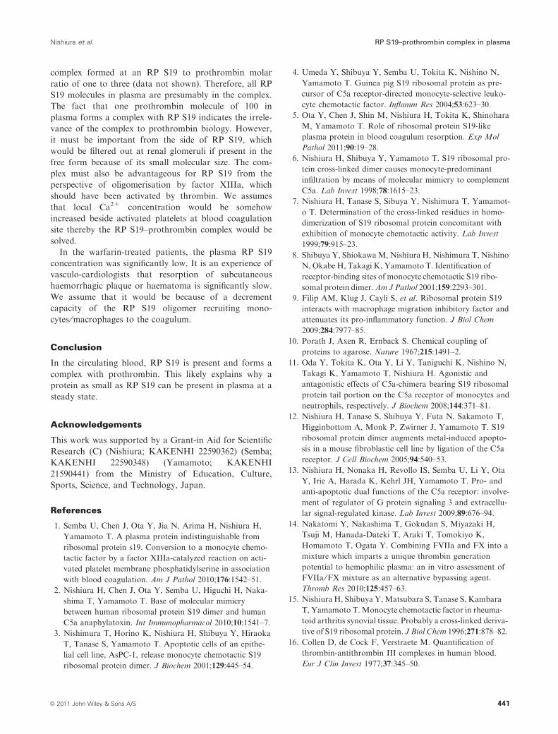
In the warfarin-treated patients, the plasma RP S19
concentration was significantly low. It is an experience of
vasculo-cardiologists that resorption of subcutaneous
haemorrhagic plaque or haematoma is significantly slow.
We assume that it would be because of a decrement
capacity of the RP S19 oligomer recruiting mono-
cytes ⁄macrophages to the coagulum.
Conclusion
In the circulating blood, RP S19 is present and forms a
complex with prothrombin. This likely explains why a
protein as small as RP S19 can be present in plasma at a
steady state.
Acknowledgements
This work was supported by a Grant-in Aid for Scientific
Research (C) (Nishiura; KAKENHI 22590362) (Semba;
KAKENHI 22590348) (Yamamoto; KAKENHI
21590441) from the Ministry of Education, Culture,
Sports, Science, and Technology, Japan.
References
1. Semba U, Chen J, Ota Y, Jia N, Arima H, Nishiura H,
Yamamoto T. A plasma protein indistinguishable from
ribosomal protein s19. Conversion to a monocyte chemo-
tactic factor by a factor XIIIa-catalyzed reaction on acti-
vated platelet membrane phosphatidylserine in association
with blood coagulation. Am J Pathol 2010;176:1542–51.
2. Nishiura H, Chen J, Ota Y, Semba U, Higuchi H, Naka-
shima T, Yamamoto T. Base of molecular mimicry
between human ribosomal protein S19 dimer and human
C5a anaphylatoxin. Int Immunopharmacol 2010;10:1541–7.
3. Nishimura T, Horino K, Nishiura H, Shibuya Y, Hiraoka
T, Tanase S, Yamamoto T. Apoptotic cells of an epithe-
lial cell line, AsPC-1, release monocyte chemotactic S19
ribosomal protein dimer. J Biochem 2001;129:445–54.
4. Umeda Y, Shibuya Y, Semba U, Tokita K, Nishino N,
Yamamoto T. Guinea pig S19 ribosomal protein as pre-
cursor of C5a receptor-directed monocyte-selective leuko-
cyte chemotactic factor. Inflamm Res 2004;53:623–30.
5. Ota Y, Chen J, Shin M, Nishiura H, Tokita K, Shinohara
M, Yamamoto T. Role of ribosomal protein S19-like
plasma protein in blood coagulum resorption. Exp Mol
Pathol 2011;90:19–28.
6. Nishiura H, Shibuya Y, Yamamoto T. S19 ribosomal pro-
tein cross-linked dimer causes monocyte-predominant
infiltration by means of molecular mimicry to complement
C5a. Lab Invest 1998;78:1615–23.
7. Nishiura H, Tanase S, Sibuya Y, Nishimura T, Yamamot-
o T. Determination of the cross-linked residues in homo-
dimerization of S19 ribosomal protein concomitant with
exhibition of monocyte chemotactic activity. Lab Invest
1999;79:915–23.
8. Shibuya Y, ShiokawaM, Nishiura H, Nishimura T, Nishino
N, Okabe H, Takagi K, Yamamoto T. Identification of
receptor-binding sites of monocyte chemotactic S19 ribo-
somal protein dimer.Am J Pathol 2001;159:2293–301.
9. Filip AM, Klug J, Cayli S, et al. Ribosomal protein S19
interacts with macrophage migration inhibitory factor and
attenuates its pro-inflammatory function. J Biol Chem
2009;284:7977–85.
10. Porath J, Axen R, Ernback S. Chemical coupling of
proteins to agarose. Nature 1967;215:1491–2.
11. Oda Y, Tokita K, Ota Y, Li Y, Taniguchi K, Nishino N,
Takagi K, Yamamoto T, Nishiura H. Agonistic and
antagonistic effects of C5a-chimera bearing S19 ribosomal
protein tail portion on the C5a receptor of monocytes and
neutrophils, respectively. J Biochem 2008;144:371–81.
12. Nishiura H, Tanase S, Shibuya Y, Futa N, Sakamoto T,
Higginbottom A, Monk P, Zwirner J, Yamamoto T. S19
ribosomal protein dimer augments metal-induced apopto-
sis in a mouse fibroblastic cell line by ligation of the C5a
receptor. J Cell Biochem 2005;94:540–53.
13. Nishiura H, Nonaka H, Revollo IS, Semba U, Li Y, Ota
Y, Irie A, Harada K, Kehrl JH, Yamamoto T. Pro- and
anti-apoptotic dual functions of the C5a receptor: involve-
ment of regulator of G protein signaling 3 and extracellu-
lar signal-regulated kinase. Lab Invest 2009;89:676–94.
14. Nakatomi Y, Nakashima T, Gokudan S, Miyazaki H,
Tsuji M, Hanada-Dateki T, Araki T, Tomokiyo K,
Homamoto T, Ogata Y. Combining FVIIa and FX into a
mixture which imparts a unique thrombin generation
potential to hemophilic plasma: an in vitro assessment of
FVIIa ⁄FX mixture as an alternative bypassing agent.
Thromb Res 2010;125:457–63.
15. Nishiura H, Shibuya Y,Matsubara S, Tanase S, Kambara
T, Yamamoto T.Monocyte chemotactic factor in rheuma-
toid arthritis synovial tissue. Probably a cross-linked deriva-
tive of S19 ribosomal protein. J Biol Chem 1996;271:878–82.
16. Collen D, de Cock F, Verstraete M. Quantification of
thrombin-antithrombin III complexes in human blood.
Eur J Clin Invest 1977;37:345–50.
Nishiura et al. RP S19–prothrombin complex in plasma
ª 2011 John Wiley & Sons A/S 441