macrophage angiotensin ii type 2 receptor triggers ...did not influence sni-induced mechanical...
TRANSCRIPT

Macrophage angiotensin II type 2 receptor triggersneuropathic painAndrew J. Shepherda,b,1, Aaron D. Micklea,b,2, Judith P. Goldena,2, Madison R. Mackc, CarmenM. Halabid, Annette D. de Kloete,Vijay K. Saminenia, Brian S. Kimc, Eric G. Krausef, Robert W. Gereau IVa,g, and Durga P. Mohapatraa,b,h,1
aWashington University Pain Center, Department of Anesthesiology, Washington University School of Medicine, St. Louis, MO 63110; bDepartment ofPharmacology, The University of Iowa Carver College of Medicine, Iowa City, IA 52242; cCenter for the Study of Itch, Department of Dermatology,Washington University School of Medicine, St. Louis, MO 63110; dDivision of Nephrology, Department of Pediatrics, Washington University School ofMedicine, St. Louis, MO 63110; eDepartment of Physiology and Functional Genomics, College of Medicine, University of Florida, Gainesville, FL 32610;fDepartment of Pharmacodynamics, College of Pharmacy, University of Florida, Gainesville, FL 32610; gDepartment of Neuroscience, Washington UniversitySchool of Medicine, St. Louis, MO 63110; and hCenter for the Investigation of Membrane Excitability Diseases, Washington University School of Medicine,St. Louis, MO 63110
Edited by Ardem Patapoutian, The Scripps Research Institute, La Jolla, CA, and approved July 17, 2018 (received for review December 14, 2017)
Peripheral nerve damage initiates a complex series of structural andcellular processes that culminate in chronic neuropathic pain. Therecent success of a type 2 angiotensin II (Ang II) receptor (AT2R)antagonist in a phase II clinical trial for the treatment of postherpeticneuralgia suggests angiotensin signaling is involved in neuropathicpain. However, transcriptome analysis indicates a lack of AT2R gene(Agtr2) expression in human and rodent sensory ganglia, raisingquestions regarding the tissue/cell target underlying the analgesiceffect of AT2R antagonism. We show that selective antagonism ofAT2R attenuates neuropathic but not inflammatory mechanical andcold pain hypersensitivity behaviors in mice. Agtr2-expressing mac-rophages (MΦs) constitute the predominant immune cells that in-filtrate the site of nerve injury. Interestingly, neuropathic mechanicaland cold pain hypersensitivity can be attenuated by chemogeneticdepletion of peripheral MΦs and AT2R-null hematopoietic cell trans-plantation. Our study identifies AT2R on peripheral MΦs as a criticaltrigger for pain sensitization at the site of nerve injury, and there-fore proposes a translatable peripheral mechanism underlyingchronic neuropathic pain.
neuropathic pain | angiotensin | AT2R | macrophage | chemogenetics
Neuropathic pain is caused by diseases or lesions affecting thesomatosensory nervous system. Often intractable in nature,
neuropathic pain is estimated to affect ∼3–17% of the chronicpain population (1). The etiology of neuropathic pain is complex,therefore presenting a formidable challenge to its effectivemanagement. It is associated with drugs used in cancer chemo-therapy, diabetic neuropathy, postherpetic neuralgia (PHN),traumatic injury, and trigeminal neuralgia, among others (2, 3).The lack of a precise mechanistic understanding of neuropathicpain, which is poorly managed by existing drugs, has undoubtedlyhampered the development of effective analgesics (3–6). How-ever, angiotensin II (Ang II) type 2 receptor (AT2R) antagonistshave recently proven efficacious in preclinical models of neuro-pathic, inflammatory, and cancer pain (7), and the AT2R an-tagonist EMA401 has shown effective pain relief in PHNpatients in a phase II clinical trial (8). However, the site of actionfor this antagonist remains controversial: prior reports suggestedthat Ang II acts directly on dorsal root ganglia (DRG) neuronsto induce peripheral pain sensitization (9–12), whereas activationof sensory neuron AT2R by a bacterial mycolactone toxin hasbeen reported to be analgesic in mice (13). Recent transcriptomeanalysis in human and rodent sensory ganglia and neurons showa lack of AT2R gene (Agtr2) expression therein, which raisesimportant questions about the tissue/cell target underlying theanalgesic action of AT2R antagonism (14–18).The major effector of the renin-angiotensin system (RAS),
Ang II is generated from angiotensinogen (Agt) and angiotensinI by renin and angiotensin-converting enzyme (ACE), respectively(19). The regulation of blood pressure by Ang II has been well-
established, with most of these physiological actions ascribed totype 1 receptor (AT1R) signaling; however, the role of AT2R hasremained more enigmatic (19). Expression of AT2R in the brainhas been reported contributing to a number of functions, such asregulation of drinking behavior and motor activity (19–21). Recentfindings have also suggested that AT2R in peripheral sensoryneurons is involved in pain modulation (7). Signaling originatingfrom Gαs-coupled AT2R in sensory neurons was shown to elicitperipheral pain sensitization (9, 10). In contrast, activation of Gαi/o-coupled AT2R in sensory neurons by a mycolactone toxin wasshown to induce analgesia in mice (13). Interestingly, a recentfollow-up study demonstrated that the AT2R antagonistEMA401 was unable to prevent the mycolactone toxin’s effect onsensory neurons in vitro (22). This raises the possibility that theanalgesic actions of EMA401 could result from targeting non-neuronal AT2R, or could be entirely independent of AT2R an-tagonism. This further emphasizes that establishing the tissue/celltarget of angiotensin signaling in chronic neuropathic pain states isessential for further therapeutic developments.Using the spared nerve injury (SNI) model of experimental
neuropathy (23) and the complete Freund’s adjuvant (CFA)
Significance
Neuropathic pain is a widespread problem that is undermanagedby currently available analgesic drugs. An antagonist of the typeII angiotensin II receptor (AT2R) reduces pain behaviors related toneuropathy, suggesting that angiotensin receptor signaling isinvolved in this pain. We find that AT2R expression is detectednot in sensory neurons themselves, but in macrophages that in-filtrate the site of nerve injury. Inducible depletion of peripheralmacrophages attenuates mechanical and cold pain hypersensi-tivity related to neuropathy, as does transplantation of AT2R-nullbone marrow into an otherwise WT recipient. Our observationsprovide powerful evidence that neuropathic pain is dependentupon angiotensin signaling, macrophages, and the AT2R-mediateddownstream signaling therein.
Author contributions: A.J.S. and D.P.M. designed research; A.J.S., A.D.M., J.P.G., M.R.M.,C.M.H., A.D.d.K., V.K.S., B.S.K., E.G.K., and D.P.M. performed research; A.J.S., C.M.H.,A.D.d.K., E.G.K., R.W.G., and D.P.M. contributed new reagents/analytic tools; A.J.S.,A.D.M., and D.P.M. analyzed data; and A.J.S. and D.P.M. wrote the paper.
The authors declare no conflict of interest.
This article is a PNAS Direct Submission.
Published under the PNAS license.1To whom correspondence may be addressed. Email: [email protected] or [email protected].
2A.D.M. and J.P.G. contributed equally to this work.
This article contains supporting information online at www.pnas.org/lookup/suppl/doi:10.1073/pnas.1721815115/-/DCSupplemental.
Published online August 6, 2018.
www.pnas.org/cgi/doi/10.1073/pnas.1721815115 PNAS | vol. 115 | no. 34 | E8057–E8066
NEU
ROSC
IENCE
Dow
nloa
ded
by g
uest
on
May
17,
202
0

model (24) of inflammation in mice, we corroborate the effec-tiveness of AT2R antagonism selectively in neuropathic pain hy-persensitivity. Elevated levels of Ang II are detected specifically ininjured sciatic nerves. Our observations in Agtr2GFP reporter micesuggest the absence of its expression on any cell type in the DRG.Instead, we report that Agtr2 expression is detected on macro-phages (MΦs) that infiltrate the site of peripheral nerve injury.Accordingly, chemogenetic depletion of peripheral MΦs, andtransplantation of AT2R-null bone marrow into WT recipientswere both able to attenuate neuropathic mechanical and cold painhypersensitivity. These findings describe the critical role for MΦsexpressing AT2R at the site of nerve injury in the development ofneuropathy-induced chronic pain hypersensitivity.
ResultsAT2R Activation Is Critical for Nerve Injury-Induced Mechanical andCold Pain Hypersensitivity. We began by evaluating the ability ofAT1R and AT2R antagonism to alleviate nerve injury-inducedchronic pain. SNI-induced peripheral neuropathy in mice elicitedlong-lasting mechanical hypersensitivity when the region beingstimulated is innervated by the spared sural nerve: that is, theextreme lateral edge of the plantar surface of the hindpaw (Fig. 1A and B), as reported previously (23, 25, 26). Instead of me-chanical paw withdrawal threshold, the magnitude of total me-chanical sensitivity on mouse hindpaws in response to increasingvon Frey filament strength was determined, as detailed earlier(27–30) and outlined in SI Appendix, Fig. S1A. Systemic admin-istration of the AT2R antagonist PD123319 dose-dependentlyattenuated SNI-induced mechanical hypersensitivity in male andfemale mice to a similar extent (Fig. 1B and SI Appendix, Fig. S1 Band E). However, administration of the AT1R antagonist losartandid not influence SNI-induced mechanical hypersensitivity (Fig.1C). Administration of PD123319 or losartan alone in sham micedid not alter hindpaw mechanical sensitivity, and no change inmechanical sensitivity was observed in the contralateral hindpawsof SNI mice (Fig. 1 B and C and SI Appendix, Fig. S1C). SNI didnot influence hindpaw heat sensitivity in male or female mice (SIAppendix, Fig. S1 D and F), as demonstrated previously (25, 26).We next verified whether AT2R inhibition of SNI-induced me-chanical hypersensitivity targets the central or peripheral nervoussystem. Intrathecal administration of PD123319 did not attenuatemechanical hypersensitivity (Fig. 1D), but peri-sciatic delivery, aswith systemic administration (intraperitoneal, i.p.), proved effec-tive (Fig. 1E). Attenuation of SNI-induced mechanical hypersen-sitivity by PD123319 was independent of any hemodynamicchanges, because PD123319 administration did not influenceblood pressure, whereas the AT1R antagonist losartan did reduceblood pressure (SI Appendix, Fig. S1G). Vascular permeability ofthe hindpaw was also unaffected by PD123319, as determined byEvans blue extravasation (SI Appendix, Fig. S1H). Systemic ad-ministration of PD123319 did not attenuate mechanical andthermal hypersensitivity induced by chronic hindpaw inflammationwith hindpaw CFA injection (SI Appendix, Fig. S2 A–C), sug-gesting its analgesic efficacy is selective for neuropathic pain.We next investigated if SNI was associated with changes in
Ang II production. Ang II levels were elevated in the ipsilateralsciatic nerve from SNI mice, but not in contralateral or sham-operated mice. Ang II levels were unchanged in the spinal cordsof SNI- vs. sham-operated mice (Fig. 1F). Furthermore, hindpawCFA injection did not lead to any elevation in local Ang II levels(SI Appendix, Fig. S2D). Collectively, these observations suggestthat SNI elevates local Ang II levels at the site of injury in thesciatic nerve, which induces AT2R signaling that contributes tomechanical hypersensitivity associated with neuropathic pain.Given that neuropathic conditions elicit pronounced cold hy-
persensitivity (31), we assessed hindpaw cold sensitivity in SNI-and sham-operated mice (Fig. 1G). SNI induced significant coldhypersensitivity in ipsilateral hindpaws of SNI mice, which could
be attenuated by systemic administration (intraperitoneal) of theAT2R antagonist PD123319 (Fig. 1H). These observations sug-gest that both mechanical and cold hypersensitivity associatedwith experimental neuropathy in mice are mediated by periph-eral activation of AT2R.
Nerve Injury-Induced Mechanical and Cold Pain Hypersensitivity IsDependent upon TRPA1. We next investigated the involvement ofsensory transient receptor potential (TRP) channels in nerveinjury-induced mechanical and cold hypersensitivity. Both TRPV1and TRPA1 have been implicated in mechanical hypersensitivity(32, 33), and in addition, TRPA1 is involved in cold hypersensi-tivity in experimental nerve injury/neuropathic pain states in mice(32, 34, 35). Systemic administration of a TRPA1 antagonist(A967079), but not TRPV1 antagonist (AMG9810), attenuatedSNI-induced hindpaw mechanical hypersensitivity (Fig. 2A). Ad-ministration of a TRPA1 antagonist (A967079) also attenuatedSNI-induced hindpaw cold hypersensitivity (Fig. 2B). Systemiccoadministration of submaximal dose of AT2R antagonist(PD123319; 3 mg/kg, i.p.) and TRPA1 antagonist (A967079;10 mg/kg, i.p.) did not lead to any greater attenuation of hindpawmechanical and cold hypersensitivity than either drug in isolationat that dose (Fig. 2C). This observation indicates that antagonismof AT2R and TRPA1 presumably act in series to attenuate SNI-induced mechanical and cold hypersensitivity.
Macrophages Infiltrating the Site of Nerve Injury Express AT2R.Earlier studies used AT2R antibody staining to show AT2Rexpression in rodent DRGs, which also exhibited Ang II/AT2R-mediated potentiation of TRPV1 currents (11). However, recenttranscriptome analysis showed negligible or zero expression ofAgtr2/AGTR2 mRNA in mouse and human DRGs (14–16, 36,37). Furthermore, we also assessed Agtr2 expression in DRGneurons in Agtr2GFP mice, where GFP expression is driven by theAgtr2 promoter. Immunostaining of DRG sections from Agtr2GFP
reporter mice displayed no detectable GFP signal (Fig. 3A). Inthe sciatic nerves of Agtr2GFP mice, GFP staining is observed onlyin a subset of NF200+ (myelinated) fibers, but not in CGRP+
(peptidergic nociceptive neuron marker) fibers (Fig. 3B). In ac-cordance with this observation, no GFP signal is observed inneurons or nerve fibers in the superficial laminae of Agtr2GFP
mouse spinal cord, where CGRP expression by central terminalsof sensory neurons is detectable (SI Appendix, Fig. S3). However,numerous NF200 and NeuN+ somata in deeper laminae of thespinal cord dorsal horn and ventral horn express GFP (SI Ap-pendix, Fig. S3), indicating that a subset of central neurons doexpress AT2R. In particular, GFP expression on ventral hornneurons with larger somata (an anatomical feature of motorneurons) coincides with the expression of GFP in a subset ofNF200+ fibers in the sciatic nerves (38, 39). Furthermore, in-duction of SNI in Agtr2GFP mice did not lead to any detectableGFP signal in any cell types, including neurons and microglia/MΦs in DRGs (Fig. 3C). Consistent with prior reports (40–42),increased microglia/MΦs were observed in the ipsilateral DRGsof SNI mice (Fig. 3C). Collectively, our findings argue againstAT2R expression and downstream signaling within DRG sensoryneurons, as has been proposed earlier (11, 12), thereby sug-gesting the possible involvement of nonneuronal AT2R.We next investigated the site of nerve injury to obtain histo-
logical evidence for the underlying mechanism. SNI inducedmassive and sustained infiltration of MΦs in both male and fe-male mice, and some level of increase in neutrophil infiltrationinto the site of nerve injury (Fig. 4 A–C and SI Appendix, Fig.S4). Interestingly, the spared sural nerve fibers, which did notshow any loss of nerve fiber staining (with NF200), also did notshow any MΦ infiltration. As has been shown previously (43),increased microglial density was observed in the ipsilateral spinalcord dorsal horn of SNI mice, without any detectable neutrophil
E8058 | www.pnas.org/cgi/doi/10.1073/pnas.1721815115 Shepherd et al.
Dow
nloa
ded
by g
uest
on
May
17,
202
0

staining (SI Appendix, Fig. S5). Because AT2R is critical for SNI-induced mechanical and cold hypersensitivity, we next determinedwhether MΦs infiltrating the injured sciatic nerve express AT2R.Induction of SNI in Agtr2GFP mice showed substantial overlap ofF4/80 and GFP immunoreactivity, indicating that MΦs in the vi-cinity of the nerve injury express Agtr2 (Fig. 4 C and D). However,spinal cord microglia in Agtr2GFP mice do not exhibit any detect-able GFP signal after SNI (SI Appendix, Fig. S6). These obser-vations suggest that peripheral MΦs that infiltrate the site of nerve
injury, but not spinal cord microglia, express AT2R, and couldtherefore constitute a target for the analgesic action of AT2Rantagonism in neuropathic pain.
Macrophages and AT2R Expression Therein Are Critical for NeuropathicMechanical and Cold Pain Hypersensitivity. We next verified if pe-ripheral MΦs are critical for neuropathic pain hypersensitivity. Weutilized specific chemogenetic depletion of peripheral MΦs (butnot in brain, spinal cord, and DRG MΦs/microglia) with designer
Fig. 1. Peripheral AT2R activation mediates neuropathic pain hypersensitivity. (A) Experimental scheme depicting nerve injury-induced neuropathy, painbehavioral assessments, and drug administration timeline in C57BL/6 (B6) mice for B–E (data analysis scheme in SI Appendix, Fig. S1A). (B) Systemic admin-istration of PD123319 (10 mg/kg, i.p.) attenuates SNI-induced mechanical hypersensitivity. Mean ± SEM; *P < 0.05, **P < 0.01, and ***P < 0.001 vs. sham+saline/PD123319 groups; #P < 0.05 and ##P < 0.01 vs. SNI+saline group. (C) Losartan (10 mg/kg, i.p.) has no effects on SNI-induced mechanical hypersensitivity.Mean ± SEM; **P < 0.01 and ***P < 0.001 vs. sham+losartan-ipsilateral (ipsi) group. (D) Intrathecal PD123319 (30 nmol in 10 μL) does not attenuate SNI-induced mechanical hypersensitivity. Mean ± SEM; ***P < 0.001 vs. contralateral (contra) groups; not significant (ns) vs. SNI+saline-ipsi group. (E) Peri-sciaticPD123319 administration (30 nmol in 10 μL) attenuates SNI-induced mechanical hypersensitivity. Mean ± SEM; ***P < 0.001 vs. contra groups; ###P < 0.001 vs.SNI+saline-ipsi group. (F) SNI elevates Ang II levels in injured mouse sciatic nerve, but not in the spinal cord. Mean ± SEM (n = duplicate tissue samples from sixmice per group). ***P < 0.001 vs. respective SNI-contralateral groups; not significant (ns) vs. sham/SNI-contralateral or vehicle groups. (G) Experimentalscheme depicting cold hypersensitivity assessment, and drug administration timeline in mice subjected to sham/SNI surgery. (H) Systemic administration ofPD123319 (10 mg/kg, i.p.) attenuates SNI-induced cold hypersensitivity. Mean ± SEM; *P < 0.05 and ***P < 0.001 vs. sham+PD123319 group; ##P < 0.01 vs.SNI+saline group. Rectangular boxes in B–E and H denote postdrug administration time points for behavioral assessment.
Shepherd et al. PNAS | vol. 115 | no. 34 | E8059
NEU
ROSC
IENCE
Dow
nloa
ded
by g
uest
on
May
17,
202
0

drug (B/B-HmD) treatment in macrophage Fas-induced apoptosis(MaFIA) mice (Fig. 5A and SI Appendix, Fig. S7). SNI inducedrobust mechanical and cold hypersensitivity in MaFIA mice,similar to that observed in WT mice (Fig. 5 B and C). FollowingSNI, chemogenetic MΦ depletion progressively attenuated me-chanical and cold hypersensitivity, with no influence on heatsensitivity (Fig. 5 B and C and SI Appendix, Fig. S8A). The at-tenuating effect of MΦ depletion on mechanical hypersensitivitywas observed in male and female MaFIA mice to a similar degree(SI Appendix, Fig. S8 B and C). As seen earlier in WT mice,massive MΦ infiltration was also observed in injured sciatic nervesof MaFIA mice before MΦ depletion. In SNI-MaFIA mice,5 consecutive days of B/B-HmD administration (days 6–10 afterSNI) significantly depleted most peripheral MΦs at the site ofinjury (Fig. 5 D and E). Interestingly, cessation of MΦ depletion(day 11 after SNI onwards) was associated with progressive re-development of mechanical and cold hypersensitivity (Fig. 5 B andC), which was accompanied by repopulation of infiltrating MΦs inthe injured sciatic nerves (Fig. 5 D and E). Chemogenetic de-pletion of MΦs did not influence the increase in MΦ/microgliadensity in the spinal cord dorsal horn of MaFIA-SNI mice (SIAppendix, Fig. S9). These observations suggest that peripheralMΦs are critical for neuropathic pain hypersensitivity. We verifiedthe specificity of B/B-HmD–mediated peripheral immune celldepletion to monocytes/MΦs by flow cytometry. In line with priorreports (44, 45), compared with vehicle-administered MaFIAmice, B/B-HmD administration led to a significant loss (∼50%) of
monocytes/MΦs in the peripheral blood, accompanied by neu-trophilia, without any alteration in T or B lymphocyte numbers (SIAppendix, Fig. S7 C and D). This indicates that a 50% reduction inperipheral blood monocyte/MΦs leads to near-complete loss ofinfiltrating MΦs at the site of nerve injury (Fig. 5 D and E).To test further whether the requirement for functional AT2R
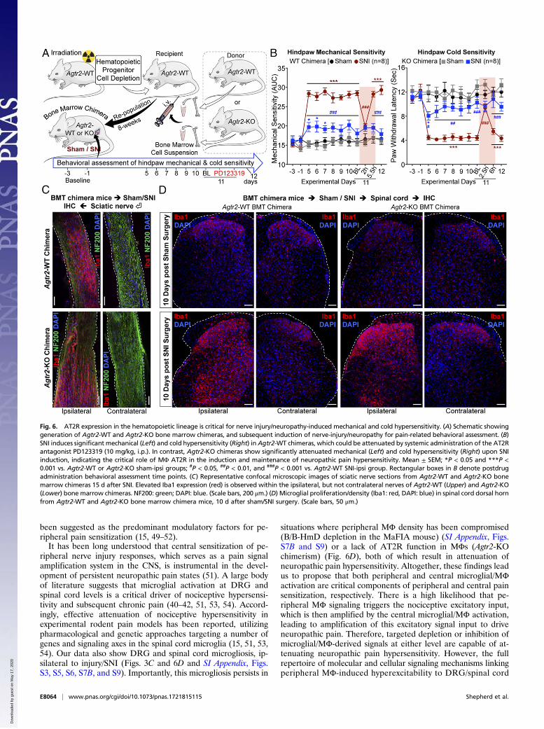
signaling resides within hematopoietic cells, we irradiated Agtr2-WT recipients and transplanted bone marrow hematopoieticprogenitors from Agtr2-WT or Agtr2-KO donors 8 wk before SNI(Fig. 6A). We verified the loss of Agtr2 mRNA expression in theisolated MΦs from the ipsilateral sciatic nerves of Agtr2-KOchimera mice with SNI by qRT-PCR (SI Appendix, Fig. S10A).Agtr2-WT chimeras display similar mechanical and cold hyper-sensitivity to that seen in Agtr2-WT mice, and retained respon-siveness to treatment with PD123319 on postoperative day 11(Fig. 6B and SI Appendix, Fig. S10 B–D). However, significantattenuation of mechanical and cold hypersensitivity was observedin Agtr2-KO chimeras (Fig. 6B), despite a similar increase in MΦinfiltration into the sciatic nerve and elevation of microglialdensity in the spinal cord of Agtr2-WT and Agtr2-KO chimeras(Fig. 6 C and D). Taken together, these observations suggest thatperipheral MΦ infiltration and the AT2R signaling therein arenecessary for SNI/Ang II-induced pain hypersensitivity.
DiscussionOur findings demonstrate that local Ang II-AT2R activation inperipheral MΦs constitutes a critical trigger for chronic pain
Fig. 2. Peripheral AT2R/TRPA1 inhibition attenuates neuropathic pain hypersensitivity. (A) TRPA1 antagonist A967079, but not TRPV1 antagonist AMG9810(30 mg/kg for each, i.p.), attenuates SNI-induced mechanical hypersensitivity. Rectangular boxes denote postdrug administration behavioral assessment timepoints. Mean ± SEM; *P < 0.05, **P < 0.01, and ***P < 0.001 vs. sham-A967079/AMG9810 groups; not significant (ns) vs. sham-A967079 group, n = 8 mice pergroup. (B) TRPA1 antagonist A967079 attenuates SNI-induced cold hypersensitivity. Rectangular box denotes postdrug administration behavioral assessmenttime point. Mean ± SEM; ***P < 0.001 vs. sham-A967079 group; ##P < 0.01 and ###P < 0.001 vs. SNI-saline group. (C) Coadministration of the AT2R antagonistPD123319 (3 mg/kg, i.p.) and the TRPA1 antagonist A967079 (10 mg/kg, i.p.) does not additively reverse SNI-induced mechanical (Left) or cold hypersensitivity(Right). Rectangular boxes denote postdrug administration behavioral assessment time points. Mean ± SEM; *P < 0.05, **P < 0.01, and ***P < 0.001 vs.respective SNI-contralateral groups; #P < 0.05 vs. 10d-BL time point.
E8060 | www.pnas.org/cgi/doi/10.1073/pnas.1721815115 Shepherd et al.
Dow
nloa
ded
by g
uest
on
May
17,
202
0

hypersensitivity associated with nerve injury/neuropathy. Priorreports suggested that Ang II acts directly on DRG neurons toinduce neurite outgrowth and PKA-mediated TRPV1 modula-tion via Gαs-coupled AT2R, resulting in peripheral pain sensi-tization (9, 10). Furthermore, activation of Gαi/o-coupled AT2Ron sensory neurons by a bacterial mycolactone toxin has beenreported to be analgesic in mice (13). Our findings indicate thatAT2R antagonism provides effective analgesia in neuropathic,but not inflammatory pain. However, our findings also suggestthat DRG neurons do not express AT2R. Instead, AT2R acti-vation in MΦs that infiltrate the site of injury induces persistentneuropathic mechanical and cold pain hypersensitivity. Ourfindings identify MΦ AT2R as the tissue/cell target underlyingthe analgesic action of AT2R antagonism for chronic neuro-pathic pain, and also uncover a translatable peripheral mecha-nism for such pain.We demonstrate that Ang II levels are elevated in injured sci-
atic nerve, and that an AT2R antagonist dose-dependently at-tenuates mechanical hypersensitivity induced by nerve injury/neuropathy, but not by chronic hindpaw inflammation. Attenua-tion of both heat and mechanical hypersensitivity by the sameAT2R antagonist in CFA-induced chronic inflammation has beenshown previously (46). Similar to MΦ infiltration in nerve injury/neuropathy, MΦs and other immune cell infiltration has been wellcharacterized in the CFA-induced model of inflammation (24).
Furthermore, accumulation of a wide variety of inflammatorymediators that sensitize multiple pain-transducing receptors/channels, such as TRPs and Nav, are considered to constitute in-flammatory thermal and mechanical pain mechanisms (32, 47).This, in combination with our observation that Ang II levels areunchanged in CFA versus saline-injected hindpaws, suggests a lackof AT2R activation at the site of CFA-induced inflammation,which would preclude the effectiveness of AT2R antagonism forinflammatory pain.With regard to the source of Ang II, mouse and human MΦs
have been shown to express the RAS genes Agt, renin, and ACE(48), raising the possibility that the entirety of the RAS requiredis supplied by MΦs. A scenario where the liver and vasculatureare the source of this Agt/Ang II is unlikely, because this wouldpresumably lead to changes in blood pressure, which we showremains unaltered following nerve injury. It is more likely thatinfiltrating MΦs at the site of nerve injury contribute to the localelevation of Ang II levels. Considerable levels of Agt mRNAhave also been detected in mouse and human DRGs, withoutany detectable renin mRNA, as revealed by RNA-seq data (14–16, 36, 37). Because renin serves as the first rate-limiting enzymefor the generation of Ang II, direct secretion of Ang II by neu-rons is implausible. One possible scenario is that following nerveinjury, sensory nerves secrete Agt, which is then processed bylocal MΦ-derived renin and ACE to produce Ang II. In depth
Fig. 3. Absence of AT2R gene expression in DRG sensory neurons and microglia/MΦs without or with nerve injury/neuropathy. (A) The Agtr2 gene (codingfor AT2R) is not expressed in neurons and nonneuronal cells in mouse DRG, as verified by lack of GFP signal in DRG sections from Agtr2GFP reporter mice, inwhich the Agtr2 promoter drives GFP expression. DRG sections are stained with CGRP and NF200 antibodies to mark peptidergic and myelinated sensoryneurons. (Scale bars, 50 μm.) (B) A subset of sciatic nerve fibers of Agtr2GFP mice are GFP+ (green). Such fibers are CGRP− (red; Upper), and NF200+ (red; Lower)DAPI: blue. (Scale bars, 200 μm.) (C) Seven days following SNI surgery, there is an appreciable increase in Iba1+ cells (red; Center) in ipsilateral vs. contralateralDRG, wherein GFP signal (green; Left) remains negligible. DAPI: blue. (Scale bars, 50 μm.)
Shepherd et al. PNAS | vol. 115 | no. 34 | E8061
NEU
ROSC
IENCE
Dow
nloa
ded
by g
uest
on
May
17,
202
0

studies utilizing tissue-specific expression/knockdown of RASgenes are thus needed to determine the precise source of Ang IIunder multiple experimental and disease-related neuropathicpain conditions.Prior studies have suggested AT2R expression in DRG neu-
rons, with AT2R antibody staining, Ang II-induced potentiationof capsaicin-mediated Ca2+ influx, and its attenuation by anAT2R antagonist (11, 12). However, our histological analysisutilizing Agtr2GFP show no detectable AT2R expression on sen-sory neurons under naïve or SNI conditions, clearly implicatingnonneuronal AT2R signaling in the development of neuropathicpain. It is important to note that we do observe AT2R expressionin a subset of spinal cord ventral horn neurons, possibly in motorneurons that send efferents to the periphery along the sciaticnerve. Because intrathecal administration of an AT2R antago-nist did not influence pain hypersensitivity in mice, we speculatethat AT2R function in these spinal cord ventral horn neurons isnot involved in neuropathic pain states. The Agtr2GFP reportermouse we utilized is a BAC-transgenic line, and it does not
employ expression from the endogenous Agtr2 locus. However,prior studies in the central nervous system detected a high de-gree of colocalization between GFP immunoreactivity andpresence of the Agtr2 transcript (21).In search of the mechanism underlying the analgesic action of
AT2R antagonism, we observed massive MΦ infiltration into theinjured sciatic nerve, as well as increased density of microglia inthe ipsilateral DRG and spinal cord, consistent with prior ob-servations (43, 49). Chemogenetic depletion of peripheral MΦs(while sparing DRG and spinal cord microglia) in mice attenu-ated nerve injury-induced mechanical and cold pain hypersen-sitivity, indicating that peripheral MΦs are an indispensablecomponent. Restoration of mechanical and cold hypersensitivityfollowing repopulation of MΦs at the site of nerve injurystrengthens this assertion. Infiltration of MΦs into peripheralnerves and DRGs, as well as microglial activation in spinal cord,have been implicated in multiple inflammatory, neuropathic, andcancer pain conditions. MΦ/microglia-derived inflammatorymediators, growth factors, and spinal modulatory signaling have
Fig. 4. Peripheral MΦ infiltration and AT2R expression therein are associated with nerve injury/neuropathy. (A) Experimental protocol for identification ofinjury markers in the sciatic nerve of mice subjected to sham or SNI surgery. (B–D) Massive MΦ infiltration (Iba1-red, Upper row in B; Iba1-green and F4/80-redin C) and considerable neutrophil infiltration (Ly6g-red, Lower row in B) accompany SNI-induced nerve fiber degeneration (decreased NF200 staining; green)in ipsilateral sciatic nerves, 5 and 15 d after SNI. Sections are costained with nuclear marker (DAPI; blue). (Scale bars, 200 μm.) MΦ density in sciatic nerves isquantified in B. Mean ± SEM; ***P < 0.001 vs. respective sham-ipsilateral groups; not significant (ns) vs. contralateral groups (n = 2 sections per mouse, 4 miceper group). Lower row images in C are magnified (630×) views of the areas marked with white dotted boxes in Upper row images. (D) Enhanced infiltration ofMΦs that express GFP (F4/80-red, GFP-green and DAPI-blue) in the ipsilateral sciatic nerves from Agtr2GFP is observed 7 d after SNI, indicating AT2R expressionin MΦs under nerve injury/neuropathy conditions. (Scale bars, 200 μm.) Right column images are magnified (630×) views of the areas marked with whitedotted boxes on Left column images.
E8062 | www.pnas.org/cgi/doi/10.1073/pnas.1721815115 Shepherd et al.
Dow
nloa
ded
by g
uest
on
May
17,
202
0

Fig. 5. Peripheral MΦ infiltration is critical for nerve injury/neuropathy-induced mechanical and cold hypersensitivity. (A–C) Chemogenetic depletion ofperipheral MΦs in MaFIA mice with B/B-HmD administration (2 mg/kg/d for 5 d, starting 6 d after SNI) (A), significantly attenuates SNI-induced ipsilateralhindpaw mechanical (B) and cold hypersensitivity (C), which subsequently returns to predepletion levels 3–4 d after the last B/B-HmD administration. Mean ±SEM; *P < 0.05, **P < 0.01, and ***P < 0.001 vs. respective baselines, sham – B/B-HmD-ipsi group or respective SNI – B/B-HmD-contra group; #P < 0.05, ##P <0.01, ###P < 0.001 and not significant (ns) vs. SNI – vehicle-ipsi group. (D) Histological confirmation of MΦ (Iba1-red) depletion at day 11 after SNI (after fifth B/B-HmD), and repopulation at day 16 after SNI (5 d after final B/B-HmD) in the sciatic nerves (NF200-green) of MaFIA mice, which are quantified in E. (Scalebars, 200 μm.) (E) Mean ± SEM; ***P < 0.001 vs. respective sham – B/B-HmD-ipsi group; ###P < 0.001 and not significant (ns) vs. respective SNI – vehicle-ipsigroups (n = 2 sections per mouse, 4 mice per group). (F and G) Following MΦ depletion, administration of PD123319 on day 10 (red box) has no additionaleffect on ipsilateral hindpaw mechanical (F) or cold (G) hypersensitivity. Aqua rectangular boxes in B, C, F, and G, and red rectangular boxes in D and E denotepostdrug administration behavioral assessment time points. *P < 0.05, and ***P < 0.001 vs. respective SNI-contra group.
Shepherd et al. PNAS | vol. 115 | no. 34 | E8063
NEU
ROSC
IENCE
Dow
nloa
ded
by g
uest
on
May
17,
202
0
been suggested as the predominant modulatory factors for pe-ripheral pain sensitization (15, 49–52).It has been long understood that central sensitization of pe-
ripheral nerve injury responses, which serves as a pain signalamplification system in the CNS, is instrumental in the devel-opment of persistent neuropathic pain states (51). A large bodyof literature suggests that microglial activation at DRG andspinal cord levels is a critical driver of nociceptive hypersensi-tivity and subsequent chronic pain (40–42, 51, 53, 54). Accord-ingly, effective attenuation of nociceptive hypersensitivity inexperimental rodent pain models has been reported, utilizingpharmacological and genetic approaches targeting a number ofgenes and signaling axes in the spinal cord microglia (15, 51, 53,54). Our data also show DRG and spinal cord microgliosis, ip-silateral to injury/SNI (Figs. 3C and 6D and SI Appendix, Figs.S3, S5, S6, S7B, and S9). Importantly, this microgliosis persists in
situations where peripheral MΦ density has been compromised(B/B-HmD depletion in the MaFIA mouse) (SI Appendix, Figs.S7B and S9) or a lack of AT2R function in MΦs (Agtr2-KOchimerism) (Fig. 6D), both of which result in attenuation ofneuropathic pain hypersensitivity. Altogether, these findings leadus to propose that both peripheral and central microglial/MΦactivation are critical components of peripheral and central painsensitization, respectively. There is a high likelihood that pe-ripheral MΦ signaling triggers the nociceptive excitatory input,which is then amplified by the central microglial/MΦ activation,leading to amplification of this excitatory signal input to driveneuropathic pain. Therefore, targeted depletion or inhibition ofmicroglial/MΦ-derived signals at either level are capable of at-tenuating neuropathic pain hypersensitivity. However, the fullrepertoire of molecular and cellular signaling mechanisms linkingperipheral MΦ-induced hyperexcitability to DRG/spinal cord
Fig. 6. AT2R expression in the hematopoietic lineage is critical for nerve injury/neuropathy-induced mechanical and cold hypersensitivity. (A) Schematic showinggeneration of Agtr2-WT and Agtr2-KO bone marrow chimeras, and subsequent induction of nerve-injury/neuropathy for pain-related behavioral assessment. (B)SNI induces significant mechanical (Left) and cold hypersensitivity (Right) inAgtr2-WT chimeras, which could be attenuated by systemic administration of the AT2Rantagonist PD123319 (10 mg/kg, i.p.). In contrast, Agtr2-KO chimeras show significantly attenuated mechanical (Left) and cold hypersensitivity (Right) upon SNIinduction, indicating the critical role of MΦ AT2R in the induction and maintenance of neuropathic pain hypersensitivity. Mean ± SEM; *P < 0.05 and ***P <0.001 vs. Agtr2-WT or Agtr2-KO sham-ipsi groups; #P < 0.05, ##P < 0.01, and ###P < 0.001 vs. Agtr2-WT SNI-ipsi group. Rectangular boxes in B denote postdrugadministration behavioral assessment time points. (C) Representative confocal microscopic images of sciatic nerve sections from Agtr2-WT and Agtr2-KO bonemarrow chimeras 15 d after SNI. Elevated Iba1 expression (red) is observed within the ipsilateral, but not contralateral nerves of Agtr2-WT (Upper) and Agtr2-KO(Lower) bone marrow chimeras. NF200: green; DAPI: blue. (Scale bars, 200 μm.) (D) Microglial proliferation/density (Iba1: red, DAPI: blue) in spinal cord dorsal hornfrom Agtr2-WT and Agtr2-KO bone marrow chimera mice, 10 d after sham/SNI surgery. (Scale bars, 50 μm.)
E8064 | www.pnas.org/cgi/doi/10.1073/pnas.1721815115 Shepherd et al.
Dow
nloa
ded
by g
uest
on
May
17,
202
0

microglial/MΦ activation, and how such processes are disrupted byinterfering with peripheral MΦ function, remains to be uncovered.We observed that AT2R is not expressed in DRG and spinal cordmicroglia under naïve or nerve injury conditions (Fig. 3C and SIAppendix, Figs. S3 and S6). Furthermore, it is important to notethat AT2R antagonists, including PD123319 and EMA401, do notcross the blood–brain barrier (11, 12). Recent findings suggest theinvolvement of MΦ infiltration in pain hypersensitivity in rodentmodels of experimental trigeminal neuropathy and chemotherapy-induced neuropathy (49, 52). This phenomenon, in combinationwith DRG/trigeminal/spinal cord sensitization pathways, likely main-tains persistent neuropathic pain. Furthermore, we observed no sex-specific differences in neuropathic pain hypersensitivity. Sex differ-ences in the contribution of immune cells, including spinal cordmicroglia, to chronic pain conditions in mice have recently beendetailed (55). The observation that Ang II-AT2R–dependent me-chanical and cold hypersensitivity does not appear to directly involvemicroglia in the spinal cord may explain the lack of any sex-relateddifferences in peripheral nerve injury-induced pain hypersensitivity.The mechanism linking MΦ AT2R activation to persistent ex-
citation of peripheral sensory nerves remains a matter of in depthinvestigation. Among the pain-transducing receptors, TRPV1 wasshown to be directly modulated by AT2R expressed on DRGneurons (11). However, our study did not find evidence of AT2Rexpression in DRG neurons. Furthermore, TRPA1—but notTRPV1—has been suggested to be involved in nociceptor sensi-tization underlying neuropathic mechanical and cold pain hyper-sensitivity in rodent models (32–35). TRPA1 is also shown to be asensor of cell damage products, including reactive oxygen/nitrogenspecies (ROS/RNS), which activate the channel in sensory neu-rons (56–58). Our findings suggest that TRPA1 is involved inAT2R-dependent neuropathic mechanical and cold pain hyper-sensitivity (Fig. 2). Because peripheral MΦs infiltrate the site ofnerve injury in neuropathy, it is plausible that AT2R activation inMΦs serves as a cell damage signal, which subsequently providespathological activators/modulators of TRPA1. Our parallel studyhas recently identified such macrophage-to-sensory neuron celldamage signaling. This involves MΦ AT2R activation followed byROS/RNS production, which then transactivate TRPA1 on sen-sory neurons to elicit sustained nociceptor excitation (17). Pre-viously, ROS activation of TRPA1 has been shown to sensitizechannel activation to mild cold temperatures (59), which pre-sumably constitutes a mechanism for MΦ AT2R-mediated coldhypersensitivity in nerve injury/neuropathy.Interestingly, a recent study utilizing MΦ depletion in clodro-
nate liposome-treated mice showed a significant delay in the de-velopment of SNI-induced tactile hypersensitivity, with only asmall/transient delay in cold hypersensitivity, suggesting no in-volvement of MΦs in neuropathic cold hypersnsitivity (40).Clodronate liposome-treatment leads to depletion of monocytes/MΦs in blood and DRGs (40). However, in our chemogeneticmonocyte/MΦ depletion, utilizing MaFIA mice, the DRG micro-glia/MΦs remain unaffected (SI Appendix, Fig. S7B), and AT2R isexpressed only in peripheral monocyte/MΦs that infiltrate the in-jured sciatic nerve, but not in DRG microglia/MΦs (Figs. 3C and4D). Additionally, in the above-mentioned study, clodronateliposome-mediated monocyte/MΦ depletion was initiated beforethe induction of neuropathic injury (SNI), whereas we performedmonocyte/MΦ depletion after the establishment of sustained me-
chanical and cold hypersensitivity (Fig. 5). This may explain thedifferences in our observation on attenuation of both mechanical andcold hypersensitivity in SNI by peripheral monocyte/MΦ deple-tion. AT2R has previously been implicated in injury/inflammatoryresponses, albeit in a largely antiinflammatory capacity (60). Fur-thermore, increased expression of RAS components, includingAT2R, has been shown to accompany the differentiation of MΦsfrom monocytes (48). Therefore, future studies are needed toidentify the role of AT2R activation in MΦ infiltration at the site ofnerve injury, and its involvement in the induction versus mainte-nance of mechanical and cold pain hypersensitivity under specificdisease-related neuropathies.Our findings raise some intriguing possibilities that warrant
further exploration. Conditions in which local or circulating RAScomponents are elevated may be associated with mechanical andcold pain hypersensitivity. An association between hypertensionand neuropathy has been observed in diabetes mellitus (61, 62).Furthermore, ACE inhibitors have been demonstrated to impactnerve conduction in human diabetic neuropathy (63, 64). Whetherthis effect could be ascribed to a reduction in Ang II levels or AngII-AT2R–mediated pain sensitization remains unclear, but it maybe contingent on MΦ accumulation in the vicinity of damagednerves. A recent report showed an increase in MΦ density in theskin biopsies of diabetic neuropathic patients (65). Consistent withthis speculation on RAS involvement in pain, our study definesAng II-AT2R signaling on peripheral MΦs as an indispensablecomponent for the development of chronic neuropathic pain.The performance of existing analgesics for neuropathic pain
and the success rate of new-generation analgesic developmenthave been suboptimal (66). This necessitates a comprehensiveunderstanding of the pathophysiology and mechanisms un-derlying neuropathic pain. With the recent success of the AT2Rantagonist EMA401 for treatment of neuropathic pain associ-ated with PHN (8), our discovery of MΦ AT2R as a criticalperipheral trigger for neuropathic pain sensitization provides atranslatable mechanism for pharmacotherapeutic targeting.
Materials and MethodsAll experiments involving the use of mice and the procedures followed thereinwere approved by Institutional Animal Studies Committees of WashingtonUniversity in St. Louis and The University of Iowa, in strict accordance with theNIH Guide for the Care and Use of Laboratory Animals (67). Details of mice,experimental models of neuropathy and inflammation, behavior tests, bloodpressure monitoring, plasma extravasation assay, Ang II enzyme immunoassay,immunohistochemistry and image quantification, irradiation and mouse bonemarrow transplantation, FACS, flow cytometry, qPCR, chemicals and reagents,and statistical analyses are provided in SI Appendix, SI Materials and Methods.
ACKNOWLEDGMENTS. We thank Erica Lantelme, Samantha Kelly, and SherriVogt for technical assistance; Drs. Justin Grobe and Nicole Littlejohn for helpwith Agtr2-WT and Agtr2-KO mouse breeding; Dr. Mathias Leinders for helpwith intravenous injections in mice and advice on plasma extravasation assays;Drs. Curt D. Sigmund and Justin Grobe for generously providing Agtr2-KO mice(originally generated by Drs. Victor J. Dzau and Richard E. Pratt); andProfs. Donna L. Hammond and Alex S. Evers for their continued support andencouragement for this work. The majority of this study was supported byfunds from the Washington University Pain Center and Washington UniversitySchool of Medicine, Department of Anesthesiology. Additional funding sourcesthat supported this study are: pilot and feasibility NIH Grant P30DK056341 fromthe Washington University Nutrition Obesity Research Center (to A.J.S.); NIHGrant NS069898 (to D.P.M.); NIH Grant CA171927 (to A.D.M.); NIH GrantHL125805 (to A.D.d.K.); and NIH Grant NS42595 (to R.W.G.).
1. van Hecke O, Austin SK, Khan RA, Smith BH, Torrance N (2014) Neuropathic pain in thegeneral population: A systematic review of epidemiological studies. Pain 155:654–662.
2. Colloca L, et al. (2017) Neuropathic pain. Nat Rev Dis Primers 3:17002.3. Meacham K, Shepherd A, Mohapatra DP, Haroutounian S (2017) Neuropathic pain:
Central vs. peripheral mechanisms. Curr Pain Headache Rep 21:28.4. Moore RA, Wiffen PJ, Derry S, Toelle T, Rice AS (2014) Gabapentin for chronic neu-
ropathic pain and fibromyalgia in adults. Cochrane Database Syst Rev (4):CD007938.5. Woolf CJ, Mannion RJ (1999) Neuropathic pain: Aetiology, symptoms, mechanisms,
and management. Lancet 353:1959–1964.
6. Mathieson S, et al. (2017) Trial of pregabalin for acute and chronic sciatica. N Engl JMed 376:1111–1120.
7. Smith MT, Anand P, Rice AS (2016) Selective small molecule angiotensin II type 2 re-ceptor antagonists for neuropathic pain: Preclinical and clinical studies. Pain 157:S33–S41.
8. Rice ASC, et al.; EMA401-003 study group (2014) EMA401, an orally administeredhighly selective angiotensin II type 2 receptor antagonist, as a novel treatment forpostherpetic neuralgia: A randomised, double-blind, placebo-controlled phase 2 clin-ical trial. Lancet 383:1637–1647.
Shepherd et al. PNAS | vol. 115 | no. 34 | E8065
NEU
ROSC
IENCE
Dow
nloa
ded
by g
uest
on
May
17,
202
0
9. Danser AHJ, Anand P (2014) The angiotensin II type 2 receptor for pain control. Cell157:1504–1506.
10. Anand U, et al. (2015) Mechanisms underlying clinical efficacy of angiotensin II type2 receptor (AT2R) antagonist EMA401 in neuropathic pain: Clinical tissue and in vitrostudies. Mol Pain 11:38.
11. Anand U, et al. (2013) Angiotensin II type 2 receptor (AT2 R) localization andantagonist-mediated inhibition of capsaicin responses and neurite outgrowth inhuman and rat sensory neurons. Eur J Pain 17:1012–1026.
12. Smith MT, Woodruff TM, Wyse BD, Muralidharan A, Walther T (2013) A small mol-ecule angiotensin II type 2 receptor (AT2R) antagonist produces analgesia in a ratmodel of neuropathic pain by inhibition of p38 mitogen-activated protein kinase(MAPK) and p44/p42 MAPK activation in the dorsal root ganglia. Pain Med 14:1557–1568.
13. Marion E, et al. (2014) Mycobacterial toxin induces analgesia in Buruli ulcer by tar-geting the angiotensin pathways. Cell 157:1565–1576.
14. Goswami SC, et al. (2014) Molecular signatures of mouse TRPV1-lineage neurons re-vealed by RNA-Seq transcriptome analysis. J Pain 15:1338–1359.
15. Guan Z, et al. (2016) Injured sensory neuron-derived CSF1 induces microglial pro-liferation and DAP12-dependent pain. Nat Neurosci 19:94–101.
16. Sapio MR, Goswami SC, Gross JR, Mannes AJ, Iadarola MJ (2016) Transcriptomicanalyses of genes and tissues in inherited sensory neuropathies. Exp Neurol 283:375–395.
17. Shepherd AJ, et al. (2018) Angiotensin II triggers peripheral macrophage-to-sensoryneuron redox crosstalk to elicit pain. J Neurosci 3542-17.
18. Yin K, Baillie GJ, Vetter I (2016) Neuronal cell lines as model dorsal root ganglionneurons: A transcriptomic comparison. Mol Pain 12:1–17.
19. de Gasparo M, Catt KJ, Inagami T, Wright JW, Unger T (2000) International union ofpharmacology. XXIII. The angiotensin II receptors. Pharmacol Rev 52:415–472.
20. Hein L, Barsh GS, Pratt RE, Dzau VJ, Kobilka BK (1995) Behavioural and cardiovasculareffects of disrupting the angiotensin II type-2 receptor in mice. Nature 377:744–747.
21. de Kloet AD, et al. (2016) Reporter mouse strain provides a novel look at angiotensintype-2 receptor distribution in the central nervous system. Brain Struct Funct 221:891–912.
22. Anand U, et al. (2016) Mycolactone-mediated neurite degeneration and functionaleffects in cultured human and rat DRG neurons: Mechanisms underlying hypoalgesiain Buruli ulcer. Mol Pain 12:1–11.
23. Decosterd I, Woolf CJ (2000) Spared nerve injury: An animal model of persistentperipheral neuropathic pain. Pain 87:149–158.
24. Ghasemlou N, Chiu IM, Julien JP, Woolf CJ (2015) CD11b+Ly6G- myeloid cells mediatemechanical inflammatory pain hypersensitivity. Proc Natl Acad Sci USA 112:E6808–E6817.
25. Decosterd I, Allchorne A, Woolf CJ (2002) Progressive tactile hypersensitivity after aperipheral nerve crush: Non-noxious mechanical stimulus-induced neuropathic pain.Pain 100:155–162.
26. Shields SD, Eckert WA, 3rd, Basbaum AI (2003) Spared nerve injury model of neuro-pathic pain in the mouse: A behavioral and anatomic analysis. J Pain 4:465–470.
27. Banik RK, Woo YC, Park SS, Brennan TJ (2006) Strain and sex influence on pain sen-sitivity after plantar incision in the mouse. Anesthesiology 105:1246–1253.
28. Mickle AD, Shepherd AJ, Loo L, Mohapatra DP (2015) Induction of thermal and me-chanical hypersensitivity by parathyroid hormone-related peptide through upregu-lation of TRPV1 function and trafficking. Pain 156:1620–1636.
29. Shepherd AJ, Mickle AD, Kadunganattil S, Hu H, Mohapatra DP (2018) Parathyroidhormone-related peptide elicits peripheral TRPV1-dependent mechanical hypersen-sitivity. Front Cell Neurosci 12:38.
30. Shepherd AJ, Mohapatra DP (2018) Pharmacological validation of voluntary gait andmechanical sensitivity assays associated with inflammatory and neuropathic pain inmice. Neuropharmacology 130:18–29.
31. Yamamoto K, et al. (2015) Transient receptor potential ankyrin 1 that is induced indorsal root ganglion neurons contributes to acute cold hypersensitivity after ox-aliplatin administration. Mol Pain 11:69.
32. Mickle AD, Shepherd AJ, Mohapatra DP (2016) Nociceptive TRP channels: Sensorydetectors and transducers in multiple pain pathologies. Pharmaceuticals (Basel) 9:E72.
33. Eid SR, et al. (2008) HC-030031, a TRPA1 selective antagonist, attenuates in-flammatory- and neuropathy-induced mechanical hypersensitivity. Mol Pain 4:48.
34. Chen J, et al. (2011) Selective blockade of TRPA1 channel attenuates pathologicalpain without altering noxious cold sensation or body temperature regulation. Pain152:1165–1172.
35. del Camino D, et al. (2010) TRPA1 contributes to cold hypersensitivity. J Neurosci 30:15165–15174.
36. Khoury-Hanold W, et al. (2016) Viral spread to enteric neurons links genital HSV-1 infection to toxic megacolon and lethality. Cell Host Microbe 19:788–799.
37. Yin K, Deuis JR, Lewis RJ, Vetter I (2016) Transcriptomic and behavioural character-isation of a mouse model of burn pain identify the cholecystokinin 2 receptor as ananalgesic target. Mol Pain 12:1–13.
38. Altmann C, et al. (2016) Progranulin promotes peripheral nerve regeneration andreinnervation: Role of notch signaling. Mol Neurodegener 11:69.
39. Bonanomi D, et al. (2012) Ret is a multifunctional coreceptor that integrates diffusible-and contact-axon guidance signals. Cell 148:568–582.
40. Cobos EJ, et al. (2018) Mechanistic differences in neuropathic pain modalities re-vealed by correlating behavior with global expression profiling. Cell Rep 22:1301–1312.
41. Hu P, Bembrick AL, Keay KA, McLachlan EM (2007) Immune cell involvement in dorsalroot ganglia and spinal cord after chronic constriction or transection of the rat sciaticnerve. Brain Behav Immun 21:599–616.
42. Kim CF, Moalem-Taylor G (2011) Detailed characterization of neuro-immune re-sponses following neuropathic injury in mice. Brain Res 1405:95–108.
43. Scholz J, et al. (2008) Low-dose methotrexate reduces peripheral nerve injury-evokedspinal microglial activation and neuropathic pain behavior in rats. Pain 138:130–142.
44. Burnett SH, et al. (2004) Conditional macrophage ablation in transgenic mice ex-pressing a Fas-based suicide gene. J Leukoc Biol 75:612–623.
45. Chow A, et al. (2011) Bone marrow CD169+ macrophages promote the retention ofhematopoietic stem and progenitor cells in the mesenchymal stem cell niche. J ExpMed 208:261–271.
46. Chakrabarty A, Liao Z, Smith PG (2013) Angiotensin II receptor type 2 activation isrequired for cutaneous sensory hyperinnervation and hypersensitivity in a rat hindpaw model of inflammatory pain. J Pain 14:1053–1065.
47. Mickle AD, Shepherd AJ, Mohapatra DP (2015) Sensory TRP channels: The keytransducers of nociception and pain. Prog Mol Biol Transl Sci 131:73–118.
48. Okamura A, et al. (1999) Upregulation of renin-angiotensin system during differen-tiation of monocytes to macrophages. J Hypertens 17:537–545.
49. Old EA, et al. (2014) Monocytes expressing CX3CR1 orchestrate the development ofvincristine-induced pain. J Clin Invest 124:2023–2036.
50. Weaver JL, et al. (2017) Hematopoietic pannexin 1 function is critical for neuropathicpain. Sci Rep 7:42550.
51. Kuner R (2010) Central mechanisms of pathological pain. Nat Med 16:1258–1266.52. Trevisan G, et al. (2016) TRPA1 mediates trigeminal neuropathic pain in mice
downstream of monocytes/macrophages and oxidative stress. Brain 139:1361–1377.53. Basbaum AI, Bautista DM, Scherrer G, Julius D (2009) Cellular and molecular mecha-
nisms of pain. Cell 139:267–284.54. Beggs S, Salter MW (2013) The known knowns of microglia-neuronal signalling in
neuropathic pain. Neurosci Lett 557:37–42.55. Sorge RE, et al. (2015) Different immune cells mediate mechanical pain hypersensi-
tivity in male and female mice. Nat Neurosci 18:1081–1083.56. Macpherson LJ, et al. (2007) Noxious compounds activate TRPA1 ion channels through
covalent modification of cysteines. Nature 445:541–545.57. Macpherson LJ, et al. (2007) An ion channel essential for sensing chemical damage.
J Neurosci 27:11412–11415.58. Viana F (2016) TRPA1 channels: Molecular sentinels of cellular stress and tissue
damage. J Physiol 594:4151–4169.59. Miyake T, et al. (2016) Cold sensitivity of TRPA1 is unveiled by the prolyl hydroxylation
blockade-induced sensitization to ROS. Nat Commun 7:12840.60. Matavelli LC, Siragy HM (2015) AT2 receptor activities and pathophysiological impli-
cations. J Cardiovasc Pharmacol 65:226–232.61. Teunissen LL, et al. (2002) Is cardiovascular disease a risk factor in the development of
axonal polyneuropathy? J Neurol Neurosurg Psychiatry 72:590–595.62. Zarrelli MM, et al.; Italian General Practitioner Study Group (2001) Arterial hyper-
tension as a risk factor for chronic symmetric polyneuropathy. J Epidemiol Biostat 6:409–413.
63. Malik RA, et al. (1998) Effect of angiotensin-converting-enzyme (ACE) inhibitortrandolapril on human diabetic neuropathy: Randomised double-blind controlledtrial. Lancet 352:1978–1981.
64. Suresha RN, et al. (2014) Evaluation of analgesic activity of perindopril in albino mice.J Adv Pharm Technol Res 5:129–133.
65. Üçeyler N, et al. (2017) Cellular infiltrates in skin and sural nerve of patients withpolyneuropathies. Muscle Nerve 55:884–893.
66. Percie du Sert N, Rice ASC (2014) Improving the translation of analgesic drugs to theclinic: Animal models of neuropathic pain. Br J Pharmacol 171:2951–2963.
67. Committee on Care and Use of Laboratory Animals (2011) Guide for the Care and Useof Laboratory Animals (The National Academies Press, Washington, DC), 8th Ed.
E8066 | www.pnas.org/cgi/doi/10.1073/pnas.1721815115 Shepherd et al.
Dow
nloa
ded
by g
uest
on
May
17,
202
0