machine learning approaches to drug response prediction
TRANSCRIPT

REVIEW ARTICLE OPEN
Machine learning approaches to drug response prediction:challenges and recent progressGeorge Adam 1,2,3, Ladislav Rampášek 2,3,4, Zhaleh Safikhani1,3,5, Petr Smirnov 1,3,6, Benjamin Haibe-Kains1,2,3,5,6✉ andAnna Goldenberg2,3,4✉
Cancer is a leading cause of death worldwide. Identifying the best treatment using computational models to personalize drugresponse prediction holds great promise to improve patient’s chances of successful recovery. Unfortunately, the computational taskof predicting drug response is very challenging, partially due to the limitations of the available data and partially due to algorithmicshortcomings. The recent advances in deep learning may open a new chapter in the search for computational drug responseprediction models and ultimately result in more accurate tools for therapy response. This review provides an overview of thecomputational challenges and advances in drug response prediction, and focuses on comparing the machine learning techniquesto be of utmost practical use for clinicians and machine learning non-experts. The incorporation of new data modalities such assingle-cell profiling, along with techniques that rapidly find effective drug combinations will likely be instrumental in improvingcancer care.
npj Precision Oncology (2020) 4:19 ; https://doi.org/10.1038/s41698-020-0122-1
INTRODUCTIONCancer is a leading cause of death worldwide and the mostimportant impediment to increasing life expectancy in everycountry of the world in the 21st century1. Fortunately, from 2011to 2015, there has been a small but prominent decrease in deathrates for all races/ethnicities combined for 11 out of 18 mostcommon cancers among men and 14 of the 20 most commoncancers among women. The continued decreases in death ratesfor colorectal cancer, prostate cancer and female breast cancer arelargely due to advances in early detection and more effectivetreatments2. In this review, we will focus on the computationalchallenges of identifying the best treatment that improveschances of successful recovery.Until recently, treatments were chosen based on the type of
cancer in a one-size-fits-all manner. We are now witnessing theadvent of precision oncology3–5 that takes into account patients’genomic makeup for treatment decisions3,6,7. Treatment approvalbased on tumor-site agnostic molecular aberration biomarkers hasbecome reality. The year 2017 marked the first FDA approval ofsuch a treatment8. Based on clinical trials in 15 types of cancer,pembrolizumab was approved for treatment of solid tumors withmismatch repair deficiency or high microsatellite instability9.Larotrectinib is another promising treatment, targeting thetropomyosin receptor kinase gene fusion in a variety of cancers10.Unfortunately, there are no established biomarkers for majority ofthe anticancer drug compounds. Identification of reliable biomar-kers is a challenge not only for the most commonly used cytotoxicdrugs, but also in the case of targeted therapies as the drugtargets alone are generally poor therapeutic indicators11,12.Discovery of biomarkers predictive of drug response and
development of multivariate companion diagnostics requireefficient computational tools and substantial number of samples.Traditional statistical models and more sophisticated machinelearning approaches have been used to build predictors of drug
response and resistance both in the clinical13 and preclinical14
settings. As predictive models increase in complexity, the numberof observations required to train these models increases as well.While omic profiles and clinical outcomes of patients are the mostrelevant data sources for the development of clinically relevantpredictors, these datasets are often limited in size due to manyfactors including high costs, limited accrual rates, and complexregulatory landscape. In addition, by the nature of the experiment,unbiased testing of multiple therapeutic strategies for the samepatient in the patient itself is practically infeasible. Cancer modelsprovide access to patient tumors in preclinical models, bothin vivo and in vitro, allowing researchers to test multiple drugs andcombinations in parallel14. Although these preclinical modelsrecapitulate patient therapy response to varying degrees, theyprovide massive amounts of pharmacogenomic data for drugresponse prediction. Here we review the recent applications ofmachine learning to prediction of response to monotherapies andidentification of combination therapies (Fig. 1).
PREDICTION OF RESPONSE TO MONOTHERAPIESIn vitro and ex vivo tumor modelsLarge-scale efforts to associate molecular profiles with drugresponse phenotypes in preclinical models date back to the late90s when the National Cancer Institute Developmental Therapeu-tics Program released large-scale pharmacogenomic data of 60cancer cell lines (NCI60) screened with tens of thousands ofchemical compounds, including a large panel of FDA-approveddrugs15. NCI60 facilitated several drug discoveries, notably a 26Sproteasome inhibitor bortezomib that is now used in multiplemyeloma treatment15. Since then, high-throughput in vitro drugscreens of cancer cell lines (CCLs) derived by immortalization ofhuman cancer cells became popular experimental bases fordiscovery of multi-omic underpinnings of drug sensitivity and
1Princess Margaret Cancer Centre, University Health Network, Toronto, ON, Canada. 2Department of Computer Science, University of Toronto, Toronto, ON, Canada. 3VectorInstitute, Toronto, ON, Canada. 4Genetics and Genome Biology, Hospital for Sick Children, Toronto, ON, Canada. 5Department of Medical Biophysics, University of Toronto,Toronto, ON, Canada. 6Ontario Institute for Cancer Research, Toronto, ON, Canada. ✉email: [email protected]; [email protected]
www.nature.com/npjprecisiononcology
Published in partnership with The Hormel Institute, University of Minnesota
1234567890():,;

resistance16. Since this seminal study, multiple large-scaledatabases have been publicly released to the cancer researchcommunity17,18. More recently, advances in growing tumors inanimal models enabled the generation of large collection ofpatient-derived xenografts (PDX) to monitor tumor growth withand without drug treatment in mice19. Novartis published thelargest PDX-based pharmacogenomic dataset to date, referred toas the PDX Encyclopedia20. The NCI recently announced thePatient-Derived Models Repository (PDMR) with comprehensivemolecular profiling and commitment to release pharmacologicalprofiles in the future. A series of databases and tools have beendeveloped recently to harmonize and make easily availablemultiple pharmacogenomic studies investigating anticancermonotherapies (Table 1).
Methods for monotherapy predictionThe availability of commercial drug response predictionapproaches is limited. In fact, publicly available methods mainlyconsist of biomarker assays which measure quantities such asgene expression and determine whether or not a specific therapylinked to the biomarker assay would be effective for a givenpatient. Most of these assays and predictive models are univariate,with only a few multivariate assays that are based on simplestatistical and machine learning approaches (the OncotypeDx21
and MAMMAPRINT22 models for breast cancer are based on alinear regression model and a nearest centroid model,
respectively). Thus, this review focuses on academic approachesto drug response prediction since they significantly outnumbercommercial approaches, are more transparent, and address themore difficult task of predicting the efficacy of multiple drugswithout knowing ahead of time the useful features for the task.The most typical computational approaches to drug response
prediction, specifically in preclinical models, consist of (1)quantification of drug response; (2) molecular feature selectionor dimensionality reduction of the cellular measurements; (3)machine learning model fitting to predict drug response; and (4)model evaluation23,24. Multiple studies explored which genomicmodalities harbor the most predictive signal of drug response byanalyzing performance of predictive models. The most commonlyutilized modalities include single nucleotide variations, copynumber variations, RNA expression, methylation, and proteomics.Despite their widespread use in clinical settings, mutations andcopy number variations have been shown to account for only asmall subset of candidate biomarkers, while gene expression,methylation and protein abundance are regarded as the mostpredictive modalities25–27, each can be complemented by themulti-omic view of the cancer28–30. Perhaps the main obstacle ineffectively leveraging all data modalities is fusing them whileignoring redundancies. A combined set of measurements canreach hundreds of thousands of features, while the number ofavailable patients or cell lines remains in the hundreds. Such ahigh feature to sample ratio is bound to lead to overfitting where
Fig. 1 Graphical abstract. Patient data are limited, so to predict drug response, much of the existing literature use model system data, e.g.immortalized cell lines and PDX. a Currently most patients in cancer are still treated in a one-size-fits-all manner according to the type (orsubtype) of cancer they have. b There is a growing number of examples of personalizing monotherapy in practice, where depending on themutations in the tumor, the patient can be prescribed a targeted drug. This approach is applicable to fewer than 20% of the patients. Thecomputational contribution is to take a large number of model systems and patients, when available and construct a predictive model toidentify the best drug for majority of the patients. c Due to tumor heterogeneity and acquired drug resistance, monotherapies may not beeffective, there is currently a growing body of work predicting drug synergy and effective drug combinations. Originally these models weretrained using bulk data, but more recently, single-cell data-based approaches are starting to show promise. The person symbol in the figurewas obtained from dryicons.com. The black magnifying glass is courtesy of Stanislav Tischenko under the Creative Commons Attribution 3.0License.
G Adam et al.
2
npj Precision Oncology (2020) 19 Published in partnership with The Hormel Institute, University of Minnesota
1234567890():,;

a model can perfectly fit the limited size training set, yet will havepoor generalization performance when tested on new data. Thislimits the class of applicable predictive models to those with lowcomplexity such as support vector machines or logistic regressionsince high complexity models like deep neural networks requiremany samples to avoid overfitting. Successful applications of deeplearning in domains such as image classification or machinetranslation have worked due to a more favorable measurement tosample ratio (N > D) in addition to architectures that mimic thehuman brain and limit overfitting such as convolutional neuralnetworks. Developing neural network architectures with aneffective inductive bias for genomics will allow the complexunderlying cancer biology to be better modeled compared tolinear models which reduce risk of overfitting at the cost ofintroducing significant modeling bias. Another approach to dealwith the limited number of samples typically available in drugresponse prediction experiments is feature selection. Featureselection removes features such as the gene expression of geneswhich are determined to be uninformative for the phenotypebeing predicted. This improves the ratio of features to samples,and a common to feature selection is univariate feature selectionwhere only features highly corrected with the phenotype are kept.Multivariate approaches to feature also exist and consider sets offeatures at a time since any single feature individually might notbe predictive of the outcome, but that does not imply that acollection of features is uninformative as well. Papillon-Cavanaghet al.31 identified univariate feature selection as a robust selectionapproach, later improved by minimum Redundancy, MaximumRelevance (mRMR) Ensemble feature selection32. Costello et al.and Jang et al. performed extensive comparative analyses ofmachine learning methods for drug response prediction in cancercell lines, recommending using elastic net or ridge regression withinput features from all genomic profiling platforms27,29. Costelloet al. summarized a crowdsourced DREAM drug predictionchallenge29, revealing two leading trends among the mostsuccessful methods. First, the importance of the ability to modelnonlinear relationships between data and outcomes, and second,the incorporation of prior knowledge, e.g. biological pathways.The challenge winning model, Bayesian multitask multiple kernellearning method33, incorporated both of these approachestogether with multi-drug learning34. Such multitask framing ofthe prediction problem is highly effective as it enables a moreefficient use of available data when tuning parameters. Specifi-cally, instead of building separate prediction models for each drugthereby using just a subset of the data, a single model trained withall the data that has some parameters shared amongst all thedrugs, and some drug-specific parameters is the better choice.Nonlinear relationships are of utmost importance since many
cellular processes follow nonlinear dose-response relationshipssuch as the activation of MAPK via Progesterone in oocytes35.Furthermore, models encoding prior biological knowledge haveimproved and more stable feature selection since noisy gene-levelmeasurements can be abstracted into gene sets that have been
experimentally validated to be involved in cancer-related pro-cesses. Lee et al.36 developed a method that integrates diseaserelevant multi-omic prior information to prioritize gene-drugassociations. Most recently, Zhang et al.37 and Wang et al.38
introduced methods based on similarity network fusion andsimilarity-regularized matrix factorization, respectively, that takeinto account similarity among cell lines, drugs and targets. Drugchemical features and similarities were shown to be a promisingadditional information that can improve drug response predictionperformance. There is no canonical way of incorporating drugfeatures into most predictive models since it is difficult to encodehow the drug features and omics features interact. Future modelsthat address this shortcoming are likely to outperform competitorsthat do not, due to the highly informative content of molecularfingerprints. Specifically, a predictive model in a multitask settingcan take compounds with known molecular targets, use thesimilarity computed between the molecular fingerprints, andmore effectively tune parameters using similarity betweencompounds for parameter regularization.
Deep learning methods for monotherapy predictionThe use of neural networks for drug response prediction datesback to the 90s. El-Deredy et al. showed that a neural networktrained on tumor nuclear magnetic resonance (NMR) spectra datahas potential as a drug response predictor in gliomas, and may beused to provide information about the metabolic pathwaysinvolved in drug response39. Neural networks, however, did notbecome a method of choice for monotherapy prediction yet. Infact, despite the recent prevalence of deep neural network (DNN)methods across many areas and industries, including relatedfields, such as computational chemistry40–45, DNNs have only fairlyrecently found their way into the drug response prediction. Thereason for this is the typically low ratio of the number of samplesto the number of measurements per sample that does not favortraditional feedforward neural architectures. Overparameterizationin these models easily leads to overfitting and poor generalizationto new datasets. However, in recent years, more public data hasbecome available and newly developed deep neural networkmodels are showing promise. For example, Chang et al.46
developed the CDRscan model, featuring a convolutional neuralnetwork architecture trained on a dataset of ~1000 drug responseexperiments per compound. Their model achieved significantlyimproved performance compared to other classical machinelearning approaches such as Random Forests and SVM. Part ofwhy CDRscan performed better than these baseline modelsresides in its ability to integrate genomic data and molecularfingerprints. In addition, its convolutional architecture has shownto be effective in many machine learning domains. Takinginspiration from already well-established neural architectures,and modifying their structure to properly handle genomic data iscertainly a promising future direction.
Table 1. Platforms harmonizing preclinical pharmacogenomic datasets and providing basic processing functions for biomarker discovery.
Platforms Cancer models # Models # Drugs URL References
PharmacoGx PharmacoDB Cell lines 1691 759 https://bioconductor.org/packages/PharmacoGx/http://pharmacodb.ca/
17,111
GDSCTools Cell lines 1001 265 https://gdsctools.readthedocs.io 112
CellminerCDB Cell lines ∼1000 ~50,000 https://discover.nci.nih.gov/cellminercdb/ 113
CancerDP Cell lines 1061 24 http://crdd.osdd.net/raghava/cancerdp/index.php 114
PDXFinder PDX 567 33 https://www.pdxfinder.org/ Unpublished
Xeva PDX 277 61 https://github.com/bhklab/Xeva 115
Cancer-Drug eXplorer 2D cell cultures 462 60 http://cancerdrugexplorer.org/ 116
G Adam et al.
3
Published in partnership with The Hormel Institute, University of Minnesota npj Precision Oncology (2020) 19
Another promising direction is autoencoders that are able tolearn from smaller datasets. An autoencoder is a neural networkthat compresses its input and tries to reconstruct the original datafrom the compressed representation. This is quite useful forfeature extraction as shown by Way and Greene47 where a 5000dimensional gene expression profile was compressed into just 100dimensions, some of which represented phenotypically relevantfeatures such as patient sex or melanoma status. Rampášek et al.48
evaluated semi-supervised variational autoencoders on mono-therapy response prediction and developed an extension—a jointdrug response prediction model, Dr.VAE, that leveraged pre- andpost-treatment gene expression in cell lines, showing improvedperformance in drug response prediction on a variety of FDA-approved drugs compared comprehensively to many classicalmachine learning approaches. This improvement could potentiallyhave been even greater if the model was setup in a multitaskfashion in combination with molecular fingerprints. Dincer et al.49
developed DeepProfile, a method that combined variationalautoencoders to learn 8-dimensional representation of geneexpression in AML patients and then used this representation tofit a Lasso linear model for drug response prediction withimproved performance compared to no feature extraction.Similarly, Chiu et al.50 pretrained autoencoders on mutation dataand expression features on TCGA dataset and subsequentlytrained a deep drug response predictor. What differentiates theirmethod from others is the use of pretraining. Pretraining allowsfor using unlabeled data from other sources such as TCGA, insteadof just the gene expression profiles available from the drugresponse experiments, thereby significantly increasing the numberof samples available and improving performance compared tousing just the labeled data. The brief summary of methods isavailable in Table 2. The trend of model development shows that asmore data become available and deep learning methods becomebetter adapted to high dimensional/low sample size data, there ishope for convergence and creation of sophisticated models thatwill likely push the field of computational drug response predictionforward to eventually become clinically relevant.
RESISTANCE TO MONOTHERAPYWhile drug response prediction can help pick an optimal therapygiven the current molecular characteristics of the cancer cells,tumors often exhibit drug resistance over the course of thetreatment. Consequently, patients that respond initially to therapyregress as their cancer either adapts to overcome the chosen
treatment, or an existing resistant subclone repopulates thetumor51. Understanding the common mechanisms cancers use todevelop resistance can help inform treatment approaches tocounteract this phenomena.For therapies inhibiting the activity or signaling of their target, a
common mechanism towards resistance is feedback selecting forupregulated expression of the target protein. For example,resistance to 5-FU has been demonstrated to arise from theamplification of its target thymidylate synthase (TS)52, withcorresponding overproduction of TS enzyme and mRNA tran-scripts53. Furthermore, especially for tyrosine kinase inhibitors,tumors will evolve to re-activate pathways downstream of thetargeted protein. A classical example is the resistance to the EGFRinhibitor Gefitinib which can often be explained by an acquiredT790M mutation reducing drug binding affinity54.For DNA damaging compounds or compounds inhibiting DNA
repair, altered DNA damage response can lead to resistance.Studies have shown that treatment with cisplatin, a DNAdamaging agent usually effective against BRCA deficient cancers,can lead to mutations restoring BRCA function and subsequentlythe activity of the Homologous Repair (HR) pathway55,56.Furthermore, studies suggest that secondary alterations to DNAdamage response proteins can shift the response from the error-prone Non-Homologous End Joining pathway to HR, reducingsensitivity to DNA damaging agents57. Other mechanisms ofresistance include modifications to enzymes involved in drugmetabolism to either reduce conversion of drugs to active formsor deactivate the compound58,59, and more recently, intra-tumorheterogeneity (ITH)60. As this review is focuses on drug responseprediction, not enough depth is provided to discussing howtumors acquire resistance to therapies, or how therapies work.Readers are referred to work by Holohan et al.51, Housman et al.59,and Malhotra and Perry61 for a comprehensive discussion on thistopic. For more details on the biological complexity of cancer ingeneral, readers are referred to the review articles by Blackadar62,and Bertram63.
COMBINATION THERAPIESDrug combinations are crucial for addressing the issue of drugresistance and preventing recurrence caused by a negligibleamount of remaining cancer cells. Synergistic combinations canalso reduce toxicity by allowing for lower doses of either drug tobe used. By enabling reduced doses, drug combinations canfurther increase the feasibility of drug repurposing by increasing
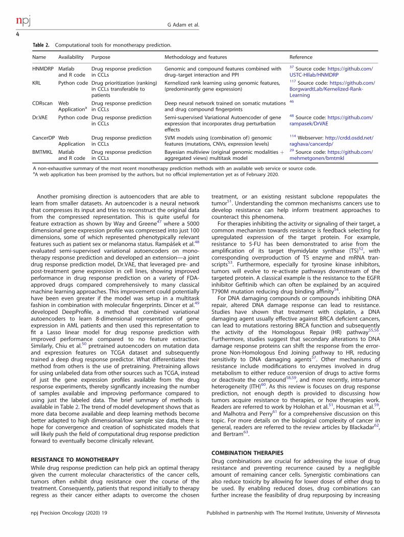
Table 2. Computational tools for monotherapy prediction.
Name Availability Purpose Methodology and features Reference
HNMDRP Matlaband R code
Drug response predictionin CCLs
Genomic and compound features combined withdrug–target interaction and PPI
37 Source code: https://github.com/USTC-HIlab/HNMDRP
KRL Python code Drug prioritization (ranking)in CCLs transferable topatients
Kernelized rank learning using genomic features,(predominantly gene expression)
117 Source code: https://github.com/BorgwardtLab/Kernelized-Rank-Learning
CDRscan WebApplicationa
Drug response predictionin CCLs
Deep neural network trained on somatic mutationsand drug compound fingerprints
46
Dr.VAE Python code Drug response predictionin CCLs
Semi-supervised Variational Autoencoder of geneexpression that incorporates drug perturbationeffects
48 Source code: https://github.com/rampasek/DrVAE
CancerDP WebApplication
Drug response predictionin CCLs
SVM models using (combination of) genomicfeatures (mutations, CNVs, expression levels)
114 Webserver: http://crdd.osdd.net/raghava/cancerdp/
BMTMKL Matlaband R code
Drug response predictionin CCLs
Bayesian multiview (original genomic modalities +aggregated views) multitask model
29 Source code: https://github.com/mehmetgonen/bmtmkl
A non-exhaustive summary of the most recent monotherapy prediction methods with an available web service or source code.aA web application has been promised by the authors, but no official implementation yet as of February 2020.
G Adam et al.
4
npj Precision Oncology (2020) 19 Published in partnership with The Hormel Institute, University of Minnesota

the potency of compounds that are only effective at clinicallydangerous doses64.Trial and error combination design has limited applicability in
the clinic due to time constraints and potential hazardousexposure to toxic combinations without improving efficacy. Forexample, Hecht et al.65 performed a clinical trial for metastaticcolorectal cancer (mCRC) patients involving the targeted com-pound bevacizumab, either oxaliplatin or irinotecan as achemotherapeutic agent, and an optional addition of a humanantibody panitumumab. The purpose of the trial was to evaluatebenefit conferred by panitumumab. It was revealed that for thecohort that used oxaliplatin as a chemotherapeutic agent, survivalwas 5 months lower for patients that also received panitumumab,and there was a significant increase in adverse effects such asinfections and pulmonary embolism compared to patients thatdid not receive panitumumab. Tol et al.66 also performed a clinicaltrial for mCRC, using combination of capecitabine, oxaliplatin, andbevacizumab, as the baseline treatment to investigate cetuximab.Patients that received cetuximab had a shorter progression-freesurvival and reported significantly more adverse effects comparedto patients that did not receive cetuximab.One promising direction for a setting where the goal is to study
a constrained set of options to design an optimal treatment planfor a patient is adaptive trials via reinforcement learning67. Theprobabilistic ranking given by their method potentially allows foridentifying when tumors develop drug resistance by analyzingwhen drug combinations are given priority over individualtreatments. While this work, performed on PDX, learns morecomplex yet more effective policies in terms of survival thancurrently offered in the clinic, it is not clear how to mitigate thepotential risks of exploration needed for reinforcement learning.We do hope that this direction is given its due consideration in theclinic since these early results appear to be very promising.The limits of trial and error in the clinic can also be overcome
in vitro with the use of preclinical models in the form ofimmortalized cancer cell lines or cell lines derived from patientbiopsies. Patient-derived cancer models allow screening drugcombinations in parallel without subjecting patients to serioustoxicity risk (Table 4). Unfortunately, due to the sheer number ofpossible drug combinations, it is not possible to explore theirpotential antagonism, additive or synergistic effects68, so there is aneed for methods that can predict combination therapy responseprior to experimentally validating it.
Methods for combination therapy predictionMany computational methods have been developed to predictanticancer drug combination synergy based on a variety ofgenomic, drug structure, and biological network data. Thesemethods vary in how much drug combination screening data isrequired, if used at all. Drug combination screening data refers totesting cancer models with combinations of two or more drugsrather than a single drug. A typical combination experiment setup
involves testing two drugs at 8 different half-log dilutionconcentrations each including the null concentration as acontrol69. This gives rise to an 8×8 dose-response matrix. Usinga 384-well assay plate, six pairs of drugs can be screened at oncein this arrangement. Once cells are incubated in the wells for asufficient amount of time, usually 72 h, a cell viability readout isconducted to determine the number of viable cells in each well.The collected data is then processed using a tool such asSynergyFinder70 to quantify the drug combination responsecompared to individual compound response based on a varietyof models. As an example, the Bliss independence model71
provides a score under the assumption that the two drugs actindependently, so measurement above this score indicatessynergy. For more details on different synergy scores as well asexperimental design of drug combination studies, the reader isreferred to the experimental design guide by He et al.69. Thenumber of experiments increases exponentially with the numberof drugs tested in combination, making these combinationscreens both logistically complex and expensive. It is thereforefavorable to have a method which does not require significantamounts of combination screens. Several approaches for drugsynergy prediction described in the literature instead use acombination of either perturbation experiments or sensitivityexperiments coupled with drug target and drug structure data. Forexample, the work done by Li et al.72 leverages gene expressionperturbation data, measured as the difference in gene expressionbefore and after treatment, to compute various statistics aboutdifferentially expressed genes as the main pharmacogenomicfeatures. Additionally, the authors extracted drug physicochemicalproperties, distance between drug targets in PPI networks, andJaccard similarity between targeted pathways to representbiological and chemical prior knowledge. These features werethen used to train a random forest model to perform the binaryprediction task of whether a drug combination is synergistic ornot. Gayvert et al.73 also made predictions with random forests byusing both single-drug response values and combination therapyresponse values when available. Interestingly, they did notleverage drug structure information nor gene expression profileswhen making predictions. This is a drawback since drug structureinformation is easily available, and including it may improveperformance, but it provides flexibility in not having to measuregene expression. However, their framework is broadly applicable,and their results indicate that even a small number of drugcombination experiments can have a great performance benefitwhen used to train a model that makes predictions using primarilysingle-drug response data.There is a class of drug combination optimization approaches
that interacts with the user by suggesting promising combinationsto test. Both Weiss et al.74 and Nowak-Sliwinska et al.75 useFeedback System Control (FSC) to iteratively refine drugcombinations and suggest new ones to test in vitro. The processworks by first starting with some randomly selected drugcombinations for some range of doses. This group of
Table 3. Methods to infer tumor clonal composition from bulk DNA sequencing data.
Name Using SSM or CNV for phylogeny reconstruction Joint Deconvolution and Phylogeny inference? Reference
PhyloWGS SSM and percomuted CNV mixing proportion estimates Joint Inference 118
Canopy Both Joint Inference 119
SPRUCE SSM and percomuted CNV mixing proportion estimates Joint Inference 120
PASTRI SSM only Two step clustering and Phylogeny Inference 121
PyClone SSM only, corrects VAFs for CNV, does not use in reconstruction explicitly Clustering and Identifying clonal genotypes only 122
SciClone SSM only Clustering and Identifying clonal genotypes only 123
THetA2 CNV only Clustering and Identifying clonal genotypes only 124
G Adam et al.
5
Published in partnership with The Hormel Institute, University of Minnesota npj Precision Oncology (2020) 19

combinations is then mutated using Differential Evolution (DE) topropose new drug combinations that are to be tested in vitro, andwhose efficacy will be compared against the original randomlyselected combinations. For each mutated combination, if thatcombination had higher efficacy than the original randomcombination that it was created from, then the new combinationis kept, otherwise the original combination is kept. This procedureis repeated until some convergence criterion is met. This approachseems to be very effective in practice because the efficacy versusdrug combination surface is smooth thereby allowing FSC toconverge in 10–15 iterations. Lastly, the optimal drug combinationidentified by DE and evaluated in vitro is further optimized toeliminate redundant compounds or compounds having anantagonistic effects. Importantly, FSC based approaches are notlimited in the number of drugs used in a given combination,unlike many methods that are created specifically for pairs ofdrugs. It might be possible to accelerate the convergence of FSCmethods by including genomic or chemical data since bothmethods described above perform the optimization withoutconsidering drug targets or drug similarities.
Deep learning methods for combination therapy predictionThe most extreme prediction scenario is to not use drug responsedata at all when building a model. This is done by Preuer et al.76
where the authors only leverage transcriptomic data and drugstructure data to predict Loewe score which quantifies the excessover the expected response if the two drugs used in acombination were the same compound. What further differenti-ates this work from previous works is that the authors use deeplearning to achieve state-of-the-art performance compared tobaseline models such as gradient boosting machines, randomforests, and support vector machines. Xia et al.77 used deeplearning as a means of simultaneously extracting and integratingfeatures from multiple data types to predict the efficacy of drugpairs. Combination response data as well as gene expression,microRNA, and protein abundance from the NCI-ALMANACdataset was used78. Additionally, drug features were obtainedusing Dragon software79 which provides chemical fingerprints andother properties. Each data type was passed through its ownsubmodel where a submodel is just a deep fully connected neuralnetwork in order to obtain useful features and performdimensionality reduction. Then, these features for the differentdata types were concatenated and passed through a finalsubmodel that uses residual connections in order to predict thedrug combination score. Ultimately, the authors were able toobtain impressive results with R^2 of 0.92, and much of thatexplained variance was due to the drug descriptors. Theseapproaches reinforce the importance of newer deep learningmethods such as molecular graph convolution to extract task-specific molecular fingerprints. A summary of tools related to drugcombinations is provided in Table 5. In terms of the availability,there are more synergy visualization tools rather than synergy
prediction tools available to date. We hope that this trend willchange as more researchers work on this important area andprovide their tools in publicly available packages.
DRUG COMBINATION DISCOVERY USING SINGLE-CELLSEQUENCINGThe development of single-cell sequencing technologies hasgiven researchers a new set of tools to interrogate tumorheterogeneity. Single-cell DNA sequencing (scDNAseq), can beused to more directly investigate the clonal structure of a tumor. Itworks by isolating individual cells and performing whole genomeamplification to increase the amount of DNA present in order tobe detectable by a DNA sequencer80. These data can be used todirectly reconstruct the unique genotypes as well as to estimatethe clonal fraction within the sample. Bulk DNA sequencing doesnot have these abilities, so simply identifying populations of cellswith different mutations can already significantly improvetreatment plans (Table 3). However, scDNAseq data suffers fromincreased noise—each cell has only two copies of each genomiclocus, requiring amplification before sequencing81. The amplifica-tion process can introduce errors into the sequenced reads, andamplification can be uneven across the genome as well asbetween cells, introducing bias into the observed reads.Computational approaches estimating tumor clonal compositionwhile taking into account these sources of error have beendeveloped82–84. For a thorough discussion of the methods used toanalyze snDNAseq data, we refer the reader to the work by Qiet al.85. Interestingly, single-cell RNA sequencing (scRNAseq) isstarting to be used to design novel drug combinations throughidentifying druggable subclones86,87. Unlike DNA, where each cellcontains only one copy of each allele (to a total of 6 pg of DNA),there is approximately 30 pg of RNA in a single cell. With theadvent of the Chromium platform it is also now possible tosequence the RNA across 100,000s of cells in a single experimentalrun88. Predictive models of drug response could be developed andtrained using high-throughput preclinical pharmacogenomic data,and an optimization framework to predict the most efficient andthe least toxic combination treatment could be established.One of the first analyses to examine the influence of treatment
on the transcriptome of cancer cells at single-cell resolution wasconducted by Suzuki et al.89. They first performed single-cellsequencing on four different cell lines derived from lungadenocarcinoma to compare the relative divergence in their geneexpression profiles. Even though the average gene expressionlevels were generally similar, the relative divergences between celltypes were pronounced. To investigate how targeted therapyaffects individual cells, they treated LC2/ad cell line and thederived resistant version of it with vandetanib, a multi-tyrosinekinase inhibitor. The comparison of single-cell profiles of treatedcells versus parental cells identified a wide variety of genesoverexpressed by drug stimulation. Particularly in case of LC2/ad,the diversity level of gene abundances between cells was
Table 4. Drug combination datasets.
Dataset Name Type #Combinations
# Drugs # Patients/cell Lines
URL Ref
Drug CombinationDatabase
Clinical 1363 904 ~140,000 http://www.cls.zju.edu.cn/dcdb/ 125
Merck In vitro 583 38 39 http://mct.aacrjournals.org/highwire/filestream/53222/field_highwire_adjunct_files/3/156849_1_supp_1_w2lrww.xls
126
AstraZeneca-Sanger DrugCombination Dataset
In vitro 910 118 85 https://www.synapse.org/#!Synapse:syn4231880/wiki/235645 30
NCI ALMANAC In vitro 5,000+ 105 60 https://wiki.nci.nih.gov/display/NCIDTPdata/NCI-ALMANAC 78
G Adam et al.
6
npj Precision Oncology (2020) 19 Published in partnership with The Hormel Institute, University of Minnesota

significantly reduced after treatment so authors hypothesized thatcells lose diversity in response to treatment. Interestingly, targetgenes of vandetanib, EGFR and RET, were not as affected by thetreatment as some of the other off-target genes possibly due tothe rigid transcriptional controls over these targets.Kim et al.90 sequenced the transcriptome at single-cell
resolution of a primary renal cell carcinoma (pRCC) and its lungmetastasis (mRCC) from a patient and paired PDX models todesign a combination therapy that would address the hetero-geneous nature of the tumor. Whole exome sequencing of themetastatic sample and its PDX model indicated the preservationof major tumor features in the PDX model. In order to predictsingle-cell response of the RCC to the clinically approved drugs,activity of drug target pathways was estimated by conductinggene set enrichment analysis. Subsequently, cell lines derivedfrom the PDX models were screened with the drugs. Predictivedrug response models, based on ridge regression, were built usingexpression profiles of cancer cell lines from a publicly availabledrug screening dataset91,92 to predict response to the drugs.Authors used ComBat to remove the technical variation betweenthe cell line dataset used for training, the drug responsepredictors, and single-cell RNA-seq data. Predicted drug responsevalues were substantially correlated with measured sensitivityvalues (0.65). Accordingly, by considering high sensitivity predic-tion of cells to Afatinib and Dasatinib and mutually exclusivepatterns in the activation status of their signaling pathways incells, the authors suggested a combination of these twocompounds as an efficient therapeutic strategy. In vitro validationin 2D and 3D cultured mRCC cells and in vivo validation insubcutaneous xenografts validated the expected additive effect ofthe drug combination over monotherapy responses. The admin-istration of this combinatorial therapy is inducing superior growthinhibition by co-targeting mutually exclusive EGFR and Srcsignaling pathways.One of the major weaknesses of Kim et al.’s90 work is the low
number of single cells sequenced. The captured cells may notreflect the true clonality of the patient tumor and might even leadto false discoveries. Recent technological advances in single-cellsequencing made it feasible to capture large numbers of singlecells in one experiment. New computational pipelines andapproaches have been developed to improve all the steps inprocessing of the single-cell sequencing data93,94, includingtackling noise and dropout in these experiments, normalizationtechniques, dimensionality reduction95–97 and clusteringapproaches98,99. These rapidly evolving methodologies provideremarkable opportunities for the discovery of biomarkers, predic-tion of efficient therapies, and the study of mechanisms ofacquiring resistance to treatments.
Anchang et al.100 were the first to use single-cell perturbationexperiments to optimize drug combinations. Their model DRUG-NEM required the specification of lineage, intracellular commu-nication, and apoptosis markers that were measured in drugperturbation experiments using Mass Cytometry Time-of-Flight(CyTOF). The objective of the model is to select the minimumnumber of drugs that creates the maximum perturbation effect onthe markers of interest using perturbation data from single-drugexperiments. Drug effects were measured using a Bayesian linearmodel to compute the probability that an intracellular commu-nication marker is differentially expressed between treatment andcontrol. A graphical model is then created from these probabilitiesusing a nested effects model, and all the possible drugcombinations are ranked. This approach is limited by having toknow ahead of time which markers to use, and this in turnrequires knowing the mechanisms of action for the drugs, whichin many cases is not available. Nevertheless, this direction for drugresponse prediction is very promising and will be greatly aided bythe burgeoning single cell and drug clonality research.
OPPORTUNITIES AND CHALLENGES: DATA AND DEEPLEARNINGThe only standardized metric to date for cancer response isRECIST, and it relies on imaging data, mainly CT and MRI, todetermine how tumors grow or shrink in patients. RECIST canhandle up to 10 lesions in the patient, prioritized based on thelargest lesions, and uses the sum of the lesion diameters (LD)when first measured as the baseline value. In subsequent scans,response is categorized into 4 different categories based on howmuch the sum of LDs has changed: complete response, partialresponse, stable disease, and progressive disease. There is no suchinternational standard used to measure response for in vitropreclinical models and RECIST is usually not used in in vivopreclinical models due to costs, thus prohibiting fair comparisonsbetween response prediction methods. Furthermore, some drugresponse prediction studies frame the task as regression wherecontinuous values such as IC50 are predicted, and others framethe task as classification where a binary value which indicatesinhibition or growth is predicted. Reproducibility between cell linebased drug response studies remains a challenge due todifferences in viability assays, drug concentrations, and cellseeding density101. There is also a need for better data sharingas technical replicates are necessary for estimating within-studyvariability, yet are sometimes not publicly released102. Addition-ally, the studies use a variety of datasets, thereby makingquantitative comparisons even less feasible. Instead, qualitativecomparisons are made between the methods that consider data
Table 5. Tools for visualizing, evaluating, and predicting synergistic drug combinations.
Name Implementation Purpose Features URL
SynergyFinder WebApplication
EvaluatingCombo Efficacy
Has 4 different drug interactivity models Computes single-agent effects Computes synergy scores
https://synergyfinder.fimm.fi/
Combenefit DesktopApplication
EvaluatingCombo Efficacy
Has 3 different drug interactivity models Meant to handlelarge batch experiments
https://www.cruk.cam.ac.uk/research-groups/jodrell-group/combenefit
CImbinator WebApplication
EvaluatingCombo Efficacy
Has 1 drug interactivity model http://cimbinator.bioinfo.cnio.es/CombinationIndex
DIGREM WebApplication
EvaluatingCombo Efficacy
Models response curve and gene expression changes aftertreatment
http://lce.biohpc.swmed.edu/drugcombination/
RACS R Package In-Silico SynergyPrediction
Leverages drug target networks and transcriptomic profiles https://github.com/DrugCombination/RACS
DeepSynergy WebApplication
PredictsSynergy Scores
Selects novel synergistic drug combinations http://www.bioinf.jku.at/software/DeepSynergy/
G Adam et al.
7
Published in partnership with The Hormel Institute, University of Minnesota npj Precision Oncology (2020) 19

requirements, generalization ability, and capacity to modelcomplex biological interactions and chemical interactions. Thesecomparisons are of great practical use as they provide context andscenarios in which one method is likely better than another.The success of deep learning across scientific fields followed the
collection of large standardized datasets. An additional factorimportant to broad utilization of deep learning was the growth inavailable computational power for training these models. Simi-larly, successful applications of deep learning in predictiveoncology followed the growth of high-throughput preclinicaldatasets. This suggests that with additional data from studies thatare more reproducible, deep learning could provide significantimprovements over traditional machine learning methods in drugresponse prediction and drug combination prioritization. Specifi-cally, the end-to-end nature of deep learning allows for extremelyeffective feature extraction and also enables the integration ofmultiple distinct data modalities. Additionally, encoding priorbiological knowledge in neural networks can be done via severalmechanisms such as graph-convolution networks103, or condi-tional scaling which allows for multiplicative relations betweenfeatures such as a mutation being required for gene expressionlevels to be relevant. The nonlinear nature of deep neuralnetworks, combined with their inductive bias that allows themto generalize even though they have many more parameters thansamples, suggests that promising applications are possible inpharmacogenomics where complex correlation structures existamong features and between features and labels. For example,graph convolutional networks are a promising new way ofencoding structural information from molecular graphs104 andcan give application-specific chemical fingerprints that are morespecialized for drug response or combination therapy discovery.Another fruitful direction is the use of transfer learning to leveragean abundance of omics data already available. The main obstaclefor transfer learning is the large discrepancies between thetechniques and experimental protocols used for different studieswhich lead to batch effects that violate the assumptions on whichdeep learning relies to generalize to new datasets. The creation ofdomain adaptation techniques, similar to computer vision105,specific for omics data will be of immense help in enablingtransfer learning. Still, creating architectures with an effectiveinductive bias for processing omics data is difficult since it is notpossible to just rely on the human brain for inspiration like inimage analysis. Thus, neural architecture search techniques whichremove humans from the design loop by automating the creationand testing of architectures are of key importance in making deeplearning more successful in drug response prediction106. It hasrecently been shown that the success of architecture searchtechniques depends significantly on careful design of the searchspace107. This requires encoding prior knowledge about poten-tially effective architecture choices which is certainly less difficultthan specifying an entire architecture, but still remains challen-ging. Deep learning can certainly help in better understandingcancer biology by predicting binding sites or discovering newbiomarkers by analyzing RNA transcripts47,108,109. In fact, deeplearning has also been used to predict protein-protein interac-tions109 which are of increasing interest as potential targets forcancer therapies110, so deep learning will have an impact on bothdrug discovery and drug response prediction.The problem of predicting the optimal treatment or combina-
tion of treatments for a cancer patient remains unsolved. Theapproaches reviewed above seek to bring recent advances inmachine learning to bear on this challenge, leveraging thegrowing high-throughput preclinical screening data and newtechnologies allowing the profiling of tumors on a single-cell level.Promising results in this area should encourage both theinvestigators working on developing cheaper and more precisehigh-throughput screens to enable further data collection as wellas ML method developers to develop novel tools incorporating
peculiarities of cancer biology. While there remains much work tobe done, the field is nascent and offers a path to a trulypersonalized approach to oncology.
Received: 23 August 2019; Accepted: 17 April 2020;
REFERENCES1. Bray, F. et al. Global cancer statistics 2018: GLOBOCAN estimates of incidence
and mortality worldwide for 36 cancers in 185 countries. CA Cancer J. Clin.https://doi.org/10.3322/caac.21492 (2018).
2. Cronin, K. A. et al. Annual report to the nation on the status of cancer, part I:National cancer statistics. Cancer 124, 2785–2800 (2018).
3. Garraway, L. A., Verweij, J. & Ballman, K. V. Precision oncology: an overview. J.Clin. Oncol. 31, 1803–1805 (2013).
4. Doherty, M., Metcalfe, T., Guardino, E., Peters, E. & Ramage, L. Precision medicineand oncology: an overview of the opportunities presented by next-generationsequencing and big data and the challenges posed to conventional drugdevelopment and regulatory approval pathways. Ann. Oncol. 27, 1644–1646(2016).
5. Heymach, J. et al. Clinical Cancer Advances 2018: annual report on progressagainst cancer from the American Society of Clinical Oncology. J. Clin. Oncol. 36,1020–1044 (2018).
6. Twomey, J. D., Brahme, N. N. & Zhang, B. Drug-biomarker co-development inoncology—20 years and counting. Drug Resist. Updat 30, 48–62 (2017).
7. Johnson, A. et al. The right drugs at the right time for the right patient: the MDAnderson precision oncology decision support platform. Drug Discov. Today 20,1433–1438 (2015).
8. Prasad, V., Kaestner, V. & Mailankody, S. Cancer drugs approved based on bio-markers and not tumor type—FDA approval of pembrolizumab for mismatchrepair-deficient solid cancers. JAMA Oncol. 4, 157–158 (2018).
9. Le, D. T. et al. Mismatch repair deficiency predicts response of solid tumors toPD-1 blockade. Science 357, 409–413 (2017).
10. Drilon, A. et al. Efficacy of larotrectinib in TRK fusion–positive cancers in adultsand children. N. Engl. J. Med. 378, 731–739 (2018).
11. De Roock, W., De Vriendt, V., Normanno, N., Ciardiello, F. & Tejpar, S. KRAS, BRAF,PIK3CA, and PTEN mutations: implications for targeted therapies in metastaticcolorectal cancer. Lancet Oncol. 12, 594–603 (2011).
12. Ding, M. Q., Chen, L., Cooper, G. F., Young, J. D. & Lu, X. Precision oncologybeyond targeted therapy: combining omics data with machine learning mat-ches the majority of cancer cells to effective therapeutics. Mol. Cancer Res. 16,269–278 (2018).
13. Perez-Gracia, J. L. et al. Strategies to design clinical studies to identify predictivebiomarkers in cancer research. Cancer Treat. Rev. 53, 79–97 (2017).
14. Dhandapani, M. & Goldman, A. Preclinical cancer models and biomarkers fordrug development: new technologies and emerging tools. J. Mol. Biomark.Diagn. 8, 356 (2017).
15. Shoemaker, R. H. The NCI60 human tumour cell line anticancer drug screen. Nat.Rev. Cancer 6, 813–823 (2006).
16. Macarron, R. et al. Impact of high-throughput screening in biomedical research.Nat. Rev. Drug Discov. 10, 188–195 (2011).
17. Smirnov, P. et al. PharmacoDB: an integrative database for mining in vitroanticancer drug screening studies. Nucleic Acids Res. 46, D994–D1002 (2018).
18. Ling, A., Gruener, R. F., Fessler, J. & Huang, R. S. More than fishing for a cure: thepromises and pitfalls of high throughput cancer cell line screens. Pharmacol.Ther. https://doi.org/10.1016/j.pharmthera.2018.06.014 (2018).
19. Aparicio, S., Hidalgo, M. & Kung, A. L. Examining the utility of patient-derivedxenograft mouse models. Nat. Rev. Cancer 15, 311–316 (2015).
20. Gao, H. et al. High-throughput screening using patient-derived tumor xeno-grafts to predict clinical trial drug response. Nat. Med. 21, 1318–1325 (2015).
21. McVeigh, T. P. et al. The impact of Oncotype DX testing on breast cancermanagement and chemotherapy prescribing patterns in a tertiary referralcentre. Eur. J. Cancer 50, 2763–2770 (2014).
22. Slodkowska, E. A. & Ross, J. S. MammaPrintTM 70-gene signature: anothermilestone in personalized medical care for breast cancer patients. Expert Rev.Mol. Diagn. 9, 417–422 (2009).
23. Azuaje, F. Computational models for predicting drug responses in cancerresearch. Brief. Bioinform. 18, 820–829 (2017).
24. De Niz, C., Rahman, R., Zhao, X. & Pal, R. Algorithms for drug sensitivity pre-diction. Algorithms 9, 77 (2016).
25. Iorio, F. et al. A landscape of pharmacogenomic interactions in cell. Cell 166,740–754 (2016).
G Adam et al.
8
npj Precision Oncology (2020) 19 Published in partnership with The Hormel Institute, University of Minnesota

26. Safikhani, Z. et al. Gene isoforms as expression-based biomarkers predictive ofdrug response in vitro. Nat. Commun. 8, 1126 (2017).
27. Jang, I. S., Neto, E. C., Guinney, J., Friend, S. H. & Margolin, A. A. Systematicassessment of analytical methods for drug sensitivity prediction from cancer cellline data. Pac. Symp. Biocomput. 19, 63–74 (2014).
28. Stetson, L. C., Pearl, T., Chen, Y. & Barnholtz-Sloan, J. S. Computational identifi-cation of multi-omic correlates of anticancer therapeutic response. BMC Geno-mics 15(Suppl. 7), S2 (2014).
29. Costello, J. C. et al. A community effort to assess and improve drug sensitivityprediction algorithms. Nat. Biotechnol. 32, 1202–1212 (2014).
30. Menden, M. P. et al. A cancer pharmacogenomic screen powering crowd-sourced advancement of drug combination prediction. bioRxiv 200451. https://doi.org/10.1101/200451 (2018).
31. Papillon-Cavanagh, S. et al. Comparison and validation of genomic predictorsfor anticancer drug sensitivity. J. Am. Med. Inform. Assoc. 20, 597–602 (2013).
32. De Jay, N. et al. mRMRe: an R package for parallelized mRMR ensemble featureselection. Bioinformatics 29, 2365–2368 (2013).
33. Gönen, M. & Margolin, A. A. Drug susceptibility prediction against a panel of drugsusing kernelized Bayesian multitask learning. Bioinformatics 30, i556–i563 (2014).
34. Ammad-Ud-Din, M., Khan, S. A., Wennerberg, K. & Aittokallio, T. Systematicidentification of feature combinations for predicting drug response with Baye-sian multi-view multi-task linear regression. Bioinformatics 33, i359–i368 (2017).
35. Andersen, M. E., Yang, R. S. H., French, C. T., Chubb, L. S. & Dennison, J. E.Molecular circuits, biological switches, and nonlinear dose–response relation-ships. Environ. Health Perspect. 110(Suppl. 6), 971–978 (2002).
36. Lee, S.-I. et al. A machine learning approach to integrate big data for precisionmedicine in acute myeloid leukemia. Nat. Commun. 9, 42 (2018).
37. Zhang, F., Wang, M., Xi, J., Yang, J. & Li, A. A novel heterogeneous network-basedmethod for drug response prediction in cancer cell lines. Sci. Rep. 8, 3355 (2018).
38. Wang, L., Li, X., Zhang, L. & Gao, Q. Improved anticancer drug response pre-diction in cell lines using matrix factorization with similarity regularization. BMCCancer 17, 513 (2017).
39. El-Deredy, W. et al. Pretreatment prediction of the chemotherapeutic responseof human glioma cell cultures using nuclear magnetic resonance spectroscopyand artificial neural networks. Cancer Res. 57, 4196–4199 (1997).
40. Dahl, G. E., Jaitly, N. & Salakhutdinov, R. Multi-task neural networks for QSARpredictions. Preprint at https://arxiv.org/abs/1406.1231 (2014).
41. Unterthiner, T. et al. Deep learning as an opportunity in virtual screening. inProc. Deep Learning Workshop at NIPS, NeurIPS workshop, Vol. 27, 1–9 (2014).
42. Aliper, A. et al. Deep learning applications for predicting pharmacologicalproperties of drugs and drug repurposing using transcriptomic data. Mol.Pharm. 13, 2524–2530 (2016).
43. Gilmer, J., Schoenholz, S. S., Riley, P. F., Vinyals, O. & Dahl, G. E. Neural messagepassing for Quantum chemistry. in Proceedings of the 34th International Con-ference on Machine Learning - Vol. 70, 1263–1272 (JMLR.org, 2017).
44. Gómez-Bombarelli, R. et al. Automatic chemical design using a data-drivencontinuous representation of molecules. ACS Cent. Sci. 4, 268–276 (2018).
45. Menden, M. P. et al. Machine learning prediction of cancer cell sensitivity todrugs based on genomic and chemical properties. PLoS ONE 8, e61318 (2013).
46. Chang, Y. et al. Cancer drug response profile scan (CDRscan): a deep learningmodel that predicts drug effectiveness from cancer genomic signature. Sci. Rep.8, 8857 (2018).
47. Way, G. P. & Greene, C. S. Extracting a biologically relevant latent space fromcancer transcriptomes with variational autoencoders. Pac. Symp. Biocomput 23,80–91 (2018).
48. Rampášek, L. et al Improving drug response prediction via modeling of drugperturbation effects. Bioinformatics. https://doi.org/10.1093/bioinformatics/btz158 (2019).
49. Dincer, A. B., Celik, S., Hiranuma, N. & Lee, S.-I. DeepProfile: deep learning ofcancer molecular profiles for precision medicine. bioRxiv 278739. https://doi.org/10.1101/278739 (2018).
50. Chiu, Y.-C. Predicting drug response of tumors from integrated genomic profilesby deep neural networks. BMC Med. Genomics 12, 119 (2019).
51. Holohan, C., Van Schaeybroeck, S., Longley, D. B. & Johnston, P. G. Cancer drugresistance: an evolving paradigm. Nat. Rev. Cancer 13, 714–726 (2013).
52. Jenh, C. H., Geyer, P. K., Baskin, F. & Johnson, L. F. Thymidylate synthase geneamplification in fluorodeoxyuridine-resistant mouse cell lines. Mol. Pharmacol.28, 80–85 (1985).
53. Berger, S. H., Jenh, C. H., Johnson, L. F. & Berger, F. G. Thymidylate synthaseoverproduction and gene amplification in fluorodeoxyuridine-resistant humancells. Mol. Pharmacol. 28, 461–467 (1985).
54. Kobayashi, S. et al. EGFR mutation and resistance of non–small-cell lung cancerto gefitinib. N. Engl. J. Med. 352, 786–792 (2005).
55. Sakai, W. et al. Secondary mutations as a mechanism of cisplatin resistance inBRCA2-mutated cancers. Nature 451, 1116–1120 (2008).
56. Edwards, S. L. et al. Resistance to therapy caused by intragenic deletion inBRCA2. Nature 451, 1111–1115 (2008).
57. Bouwman, P. & Jonkers, J. The effects of deregulated DNA damage signalling oncancer chemotherapy response and resistance. Nat. Rev. Cancer 12, 587–598 (2012).
58. Meijer, C. et al. Relationship of cellular glutathione to the cytotoxicity andresistance of seven platinum compounds. Cancer Res. 52, 6885–6889 (1992).
59. Housman, G. et al. Drug resistance in cancer: an overview. Cancers 6, 1769–1792(2014).
60. Sun, X.-X. & Yu, Q. Intra-tumor heterogeneity of cancer cells and its implicationsfor cancer treatment. Acta Pharmacol. Sin. 36, 1219–1227 (2015).
61. Malhotra, V. & Perry, M. C. Classical chemotherapy: mechanisms, toxicities andthe therapeutc window. Cancer Biol. Ther. 2, 1–3 (2003).
62. Blackadar, C. B. Historical review of the causes of cancer. World J. Clin. Oncol. 7,54–86 (2016).
63. Bertram, J. S. The molecular biology of cancer. Mol. Asp. Med. 21, 167–223 (2000).64. Sun, W., Sanderson, P. E. & Zheng, W. Drug combination therapy increases
successful drug repositioning. Drug Discov. Today 21, 1189–1195 (2016).65. Hecht, J. R. et al. A randomized phase IIIB trial of chemotherapy, bevacizumab,
and panitumumab compared with chemotherapy and bevacizumab alone formetastatic colorectal cancer. J. Clin. Oncol. 27, 672–680 (2009).
66. Tol, J. et al. Chemotherapy, bevacizumab, and cetuximab in metastatic color-ectal cancer. N. Engl. J. Med 360, 563–572 (2009).
67. Durand, A. et al. Contextual bandits for adapting treatment in a mouse model ofde novo carcinogenesis. In Proc. 3rd Machine Learning for Healthcare Conference(eds. Doshi-Velez, F. et al.) Vol. 85, 67–82 (PMLR, 2018).
68. Rationalizing combination therapies. Nat. Med. 23, 1113 (2017).69. He, L. et al. Methods for high-throughput drug combination screening and
synergy scoring. Methods Mol. Biol. 1711, 351–398 (2018).70. Ianevski, A., He, L., Aittokallio, T. & Tang, J. SynergyFinder: a web application for
analyzing drug combination dose–response matrix data. Bioinformatics 33,2413–2415 (2017).
71. Bliss, C. I. The toxicity of poisons applied jointly 1. Ann. Appl. Biol. 26, 585–615(1939).
72. Li, X. et al. Prediction of synergistic anti-cancer drug combinations based ondrug target network and drug induced gene expression profiles. Artif. Intell. Med.83, 35–43 (2017).
73. Gayvert, K. M. et al. A computational approach for identifying synergistic drugcombinations. PLoS Comput. Biol. 13, e1005308 (2017).
74. Weiss, A. et al. Rapid optimization of drug combinations for the optimalangiostatic treatment of cancer. Angiogenesis 18, 233–244 (2015).
75. Nowak-Sliwinska, P. et al. Optimization of drug combinations using FeedbackSystem Control. Nat. Protoc. 11, 302–315 (2016).
76. Preuer, K. et al. DeepSynergy: predicting anti-cancer drug synergy with DeepLearning. Bioinformatics 34, 1538–1546 (2018).
77. Fangfang Xia et al. Predicting tumor cell line response to drug pairs with deeplearning. In Computational Approaches for Cancer Workshop at SC17. Available at:http://www.scworkshops.net/cancer2017/ (2017). (Accessed 20 Nov 2018).
78. Holbeck, S. L. et al. The National Cancer Institute ALMANAC: a comprehensivescreening resource for the detection of anticancer drug pairs with enhancedtherapeutic activity. Cancer Res. 77, 3564–3576 (2017).
79. Mauri, A., Consonni, V., Pavan, M. & Todeschini, R. Dragon software: an easyapproach to molecular descriptor calculations. Match 56, 237–248 (2006).
80. Hwang, B., Lee, J. H. & Bang, D. Single-cell RNA sequencing technologies andbioinformatics pipelines. Exp. Mol. Med. 50, 96 (2018).
81. Ortega, M. A. et al. Using single-cell multiple omics approaches to resolve tumorheterogeneity. Clin. Transl. Med. 6, 46 (2017).
82. Ross, E. M. & Markowetz, F. OncoNEM: inferring tumor evolution from single-cellsequencing data. Genome Biol. 17, 69 (2016).
83. Jahn, K., Kuipers, J. & Beerenwinkel, N. Tree inference for single-cell data. Gen-ome Biol. 17, 86 (2016).
84. Roth, A. et al. Clonal genotype and population structure inference from single-cell tumor sequencing. Nat. Methods 13, 573–576 (2016).
85. Qi, R., Ma, A., Ma, Q. & Zou, Q. Clustering and classification methods for single-cell RNA-sequencing data. Brief. Bioinform. https://doi.org/10.1093/bib/bbz062(2019).
86. Shalek, A. K. & Benson, M. Single-cell analyses to tailor treatments. Sci. Transl.Med. 9, eaan4730 (2017).
87. Zhu, S., Qing, T., Zheng, Y., Jin, L. & Shi, L. Advances in single-cell RNAsequencing and its applications in cancer research. Oncotarget 8, 53763–53779(2017).
88. Baran-Gale, J., Chandra, T. & Kirschner, K. Experimental design for single-cell RNAsequencing. Brief. Funct. Genomics 17, 233–239 (2018).
89. Suzuki, A. et al. Single-cell analysis of lung adenocarcinoma cell lines revealsdiverse expression patterns of individual cells invoked by a molecular targetdrug treatment. Genome Biol. 16, 66 (2015).
G Adam et al.
9
Published in partnership with The Hormel Institute, University of Minnesota npj Precision Oncology (2020) 19
90. Kim, K.-T. et al. Application of single-cell RNA sequencing in optimizing acombinatorial therapeutic strategy in metastatic renal cell carcinoma. GenomeBiol. 17, 80 (2016).
91. Garnett, M. J. et al. Systematic identification of genomic markers of drug sen-sitivity in cancer cells. Nature 483, 570–575 (2012).
92. Yang, W. et al. Genomics of drug sensitivity in cancer (GDSC): a resource fortherapeutic biomarker discovery in cancer cells. Nucleic Acids Res. 41,D955–D961 (2013).
93. Butler, A., Hoffman, P., Smibert, P., Papalexi, E. & Satija, R. Integrating single-celltranscriptomic data across different conditions, technologies, and species. Nat.Biotechnol. 36, 411–420 (2018).
94. Lun, A. T. L., McCarthy, D. J. & Marioni, J. C. A step-by-step workflow for low-levelanalysis of single-cell RNA-seq data with Bioconductor. F1000Res 5, 2122 (2016).
95. Amodio, M. et al. Exploring single-cell data with deep multitasking neural net-works. bioRxiv 237065. https://doi.org/10.1101/237065 (2018).
96. Ding, J., Condon, A. & Shah, S. P. Interpretable dimensionality reduction of singlecell transcriptome data with deep generative models. Nat. Commun. 9, 2002(2018).
97. Wang, D. & Gu, J. VASC: dimension reduction and visualization of single cell RNAsequencing data by deep variational autoencoder. bioRxiv 199315. https://doi.org/10.1101/199315 (2017).
98. Risso, D. et al. clusterExperiment and RSEC: A Bioconductor package and fra-mework for clustering of single-cell and other large gene expression datasets.PLoS Comput. Biol. 14, e1006378 (2018).
99. Li, H. et al. Reference component analysis of single-cell transcriptomes eluci-dates cellular heterogeneity in human colorectal tumors. Nat. Genet. 49,708–718 (2017).
100. Anchang, B. et al. DRUG-NEM: Optimizing drug combinations using single-cellperturbation response to account for intratumoral heterogeneity. Proc. NatlAcad. Sci. USA 115, E4294–E4303 (2018).
101. Haverty, P. M. et al. Reproducible pharmacogenomic profiling of cancer cell linepanels. Nature 533, 333–337 (2016).
102. Hatzis, C. et al. Enhancing reproducibility in cancer drug screening: how do wemove forward? Cancer Res. 74, 4016–4023 (2014).
103. Hamilton, W. et al. Inductive representation learning on large graphs. In NeuralInformation Processing Systems 1024–1034 (Curran Associates, Inc., 2017).
104. Kearnes, S., McCloskey, K., Berndl, M., Pande, V. & Riley, P. Molecular graphconvolutions: moving beyond fingerprints. J. Comput. Aided Mol. Des. 30,595–608 (2016).
105. Hertel, L., Barth, E., Kaster, T. & Martinetz, T. Deep convolutional neural networksas generic feature extractors. In 2015 International Joint Conference on NeuralNetworks (IJCNN). https://doi.org/10.1109/ijcnn.2015.7280683 (2015).
106. Zoph, B. & Le, Q. V. Neural architecture search with reinforcement learning.Preprint at https://arxiv.org/abs/1611.01578 (2016).
107. Li, L. & Talwalkar, A. Random search and reproducibility for neural architecturesearch. Preprint at https://arxiv.org/abs/1902.07638 (2019).
108. Rampášek, L., Hidru, D., Smirnov, P., Haibe-Kains, B. & Goldenberg, A. Dr.VAE:Improving drug response prediction via modeling of drug perturbation effects.Bioinformatics. https://doi.org/10.1093/bioinformatics/btz158 (2019).
109. Zhang, Z. et al. Deep learning in omics: a survey and guideline. Brief. Funct.Genomics 18, 41–57 (2019).
110. Ivanov, A. A., Khuri, F. R. & Fu, H. Targeting protein–protein interactions as ananticancer strategy. Trends Pharmacol. Sci. 34, 393–400 (2013).
111. Smirnov, P. et al. PharmacoGx: an R package for analysis of large pharmaco-genomic datasets. Bioinformatics 32, 1244–1246 (2016).
112. Cokelaer, T. et al. GDSCTools for mining pharmacogenomic interactions incancer. Bioinformatics. https://doi.org/10.1093/bioinformatics/btx744 (2017).
113. Rajapakse, V. N., Luna, A., Yamade, M., Loman, L. & Varma, S. Integrative analysisof pharmacogenomics in major cancer cell line databases using CellMinerCDB.bioRxiv https://doi.org/10.1101/292904 (2018).
114. Gupta, S. et al. Prioritization of anticancer drugs against a cancer using genomicfeatures of cancer cells: A step towards personalized medicine. Sci. Rep. 6, 23857(2016).
115. Mer, A. S. et al. Integrative pharmacogenomics analysis of patient derivedxenografts. Cancer Res. 471227. https://doi.org/10.1101/471227 (2019).
116. Lee, J.-K. et al. Pharmacogenomic landscape of patient-derived tumor cellsinforms precision oncology therapy. Nat. Genet. 50, 1399–1411 (2018).
117. He, X., Folkman, L. & Borgwardt, K. Kernelized rank learning for personalizeddrug recommendation. Bioinformatics 34, 2808–2816 (2018).
118. Deshwar, A. G. et al. PhyloWGS: reconstructing subclonal composition and evo-lution from whole-genome sequencing of tumors. Genome Biol. 16, 35 (2015).
119. Jiang, Y., Qiu, Y., Minn, A. J. & Zhang, N. R. Assessing intratumor heterogeneityand tracking longitudinal and spatial clonal evolutionary history by next-generation sequencing. Proc. Natl Acad. Sci. USA 113, E5528–E5537 (2016).
120. El-Kebir, M., Satas, G., Oesper, L. & Raphael, B. J. Inferring the mutational historyof a tumor using multi-state perfect phylogeny mixtures. Cell Syst. 3, 43–53(2016).
121. Satas, G. & Raphael, B. J. Tumor phylogeny inference using tree-constrainedimportance sampling. Bioinformatics 33, i152–i160 (2017).
122. Roth, A. et al. PyClone: statistical inference of clonal population structure incancer. Nat. Methods 11, 396–398 (2014).
123. Miller, C. A. et al. SciClone: inferring clonal architecture and tracking the spatial andtemporal patterns of tumor evolution. PLoS Comput. Biol. 10, e1003665 (2014).
124. Oesper, L., Satas, G. & Raphael, B. J. Quantifying tumor heterogeneity in whole-genome and whole-exome sequencing data. Bioinformatics 30, 3532–3540(2014).
125. Liu, Y. et al. DCDB 2.0: a major update of the drug combination database.Database 2014, bau124–bau124 (2014).
126. O’Neil, J. et al. An unbiased oncology compound screen to identify novelcombination strategies. Mol. Cancer Ther. 15, 1155–1162 (2016).
AUTHOR CONTRIBUTIONSG.A. wrote section on combination therapies, opportunities and challenges for deeplearning, edited manuscript. L.R. wrote section on monotherapy prediction. Z.S. wrotesection on single-cell drug combination discovery. P.S. wrote section on resistance tomonotherapy. B.H.-K. co-wrote introduction and abstract, edited manuscript. A.G. co-wrote introduction and abstract, edited manuscript.
COMPETING INTERESTSThe authors declare no competing interests.
ADDITIONAL INFORMATIONSupplementary information is available for this paper at https://doi.org/10.1038/s41698-020-0122-1.
Correspondence and requests for materials should be addressed to B.H.-K. or A.G.
Reprints and permission information is available at http://www.nature.com/reprints
Publisher’s note Springer Nature remains neutral with regard to jurisdictional claimsin published maps and institutional affiliations.
Open Access This article is licensed under a Creative CommonsAttribution 4.0 International License, which permits use, sharing,
adaptation, distribution and reproduction in anymedium or format, as long as you giveappropriate credit to the original author(s) and the source, provide a link to the CreativeCommons license, and indicate if changes were made. The images or other third partymaterial in this article are included in the article’s Creative Commons license, unlessindicated otherwise in a credit line to the material. If material is not included in thearticle’s Creative Commons license and your intended use is not permitted by statutoryregulation or exceeds the permitted use, you will need to obtain permission directlyfrom the copyright holder. To view a copy of this license, visit http://creativecommons.org/licenses/by/4.0/.
© The Author(s) 2020
G Adam et al.
10
npj Precision Oncology (2020) 19 Published in partnership with The Hormel Institute, University of Minnesota