m6a modification in rna: biogenesis, functions and roles
TRANSCRIPT

REVIEW Open Access
m6A modification in RNA: biogenesis,functions and roles in gliomasYuhao Zhang1†, Xiuchao Geng2†, Qiang Li3, Jianglong Xu1, Yanli Tan4, Menglin Xiao1, Jia Song5, Fulin Liu6*,Chuan Fang1* and Hong Wang1,2,7*
Abstract
The chemical modification of RNA is a newly discovered epigenetic regulation mechanism in cells and plays acrucial role in a variety of biological processes. N6-methyladenine (m6A) mRNA modification is the most abundantform of posttranscriptional RNA modification in eukaryotes. Through the development of m6A RNA sequencing, therelevant molecular mechanism of m6A modification has gradually been revealed. It has been found that the effectof m6A modification on RNA metabolism involves processing, nuclear export, translation and even decay. As themost common malignant tumour of the central nervous system, gliomas (especially glioblastoma) have a very poorprognosis, and treatment efficacy is not ideal even with the application of high-intensity treatment measures ofsurgery combined with chemoradiotherapy. Exploring the origin and development mechanisms of tumour cellsfrom the perspective of tumour biogenesis has always been a hotspot in the field of glioma research. Emergingevidence suggests that m6A modification can play a key role in gliomas through a variety of mechanisms,providing more possibilities for early diagnosis and targeted therapy of gliomas. The aim of the present review is tofocus on the research progress regarding the association between m6A modification and gliomas. And to provide atheoretical basis according to the currently available literature for further exploring this association. This review mayprovide new insights for the molecular mechanism, early diagnosis, histologic grading, targeted therapy andprognostic evaluation of gliomas.
Keywords: m6A modification, RNA, Central nervous system, Glioma, Glioblastoma, GBM, Tumourigenesis
BackgroundGliomas are the most common malignancy in the cen-tral nervous system. Glioblastoma (GBM) has the high-est malignancy rate and account for 50% of all braintumours. The average survival time of patients withGBM is only 14.6 months [1]. GBM originate frompoorly differentiated glial cells and have the characteris-tics of nuclear atypia, cellular polymorphism, and a high
degree of mitotic activity. Given the aggressiveness ofGBM, surgical resection to resolve all intracranial lesionsin clinical practice is challenging. Therefore, mostpatients receive radiotherapy and temozolomide (TMZ)combined with chemotherapy after surgery. Althoughthe treatment intensity is very high, the outcomes arestill not satisfactory [2–5]. Therefore, there is an urgentneed for new treatment strategies or regimens.To improve the efficacy of GBM treatment, it is neces-
sary to understand the occurrence and development ofGBM and determine the molecular biological character-istics of GBM. In recent years, with the in-depth studyof the epigenetics, metabolism, and immunology ofGBM, our knowledge of GBM has greatly expanded,which provides new clues for the treatment of GBM.
© The Author(s). 2020 Open Access This article is licensed under a Creative Commons Attribution 4.0 International License,which permits use, sharing, adaptation, distribution and reproduction in any medium or format, as long as you giveappropriate credit to the original author(s) and the source, provide a link to the Creative Commons licence, and indicate ifchanges were made. The images or other third party material in this article are included in the article's Creative Commonslicence, unless indicated otherwise in a credit line to the material. If material is not included in the article's Creative Commonslicence and your intended use is not permitted by statutory regulation or exceeds the permitted use, you will need to obtainpermission directly from the copyright holder. To view a copy of this licence, visit http://creativecommons.org/licenses/by/4.0/.The Creative Commons Public Domain Dedication waiver (http://creativecommons.org/publicdomain/zero/1.0/) applies to thedata made available in this article, unless otherwise stated in a credit line to the data.
* Correspondence: [email protected]; [email protected];[email protected]†Yuhao Zhang and Xiuchao Geng contributed equally to this work.6Office of Academic Research, Affiliated Hospital of Hebei University, 071000Baoding, China1Department of Neurosurgery, Affiliated Hospital of Hebei University, 071000Baoding, ChinaFull list of author information is available at the end of the article
Zhang et al. Journal of Experimental & Clinical Cancer Research (2020) 39:192 https://doi.org/10.1186/s13046-020-01706-8
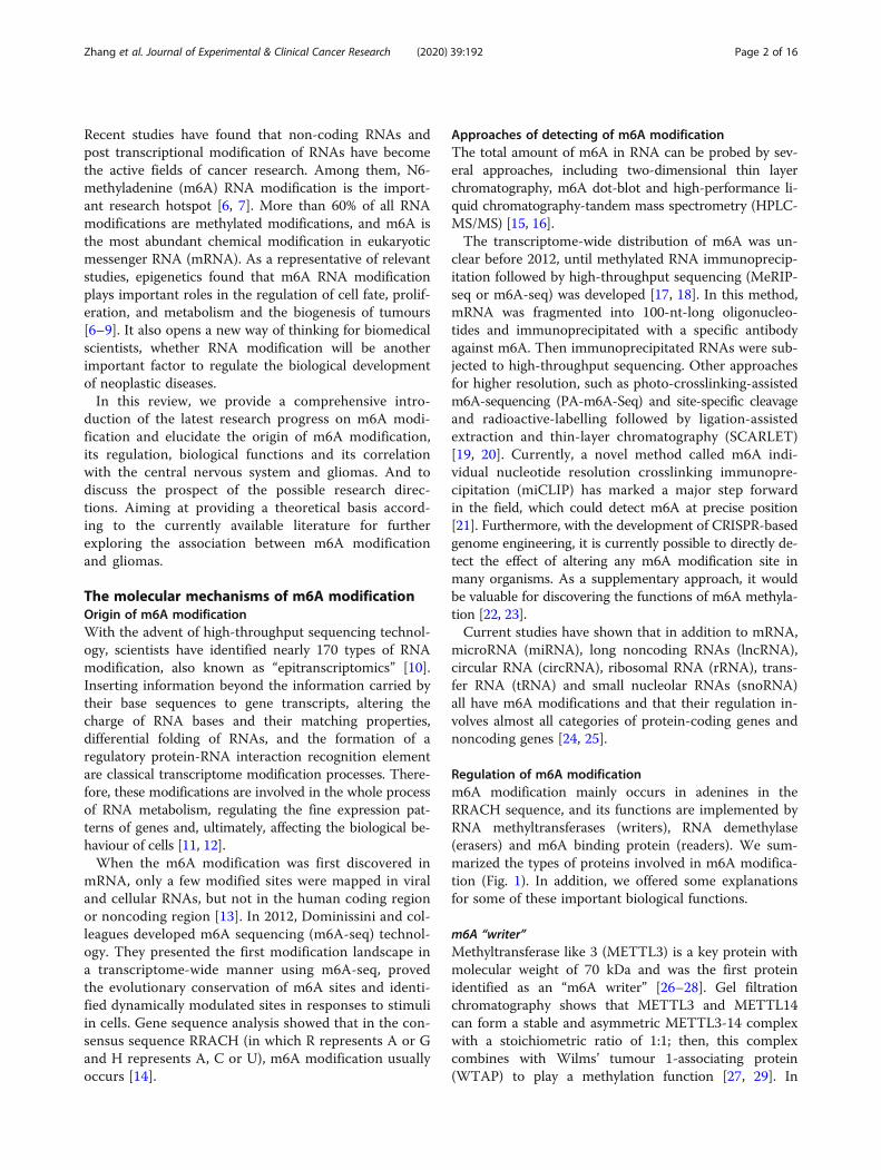
Recent studies have found that non-coding RNAs andpost transcriptional modification of RNAs have becomethe active fields of cancer research. Among them, N6-methyladenine (m6A) RNA modification is the import-ant research hotspot [6, 7]. More than 60% of all RNAmodifications are methylated modifications, and m6A isthe most abundant chemical modification in eukaryoticmessenger RNA (mRNA). As a representative of relevantstudies, epigenetics found that m6A RNA modificationplays important roles in the regulation of cell fate, prolif-eration, and metabolism and the biogenesis of tumours[6–9]. It also opens a new way of thinking for biomedicalscientists, whether RNA modification will be anotherimportant factor to regulate the biological developmentof neoplastic diseases.In this review, we provide a comprehensive intro-
duction of the latest research progress on m6A modi-fication and elucidate the origin of m6A modification,its regulation, biological functions and its correlationwith the central nervous system and gliomas. And todiscuss the prospect of the possible research direc-tions. Aiming at providing a theoretical basis accord-ing to the currently available literature for furtherexploring the association between m6A modificationand gliomas.
The molecular mechanisms of m6A modificationOrigin of m6A modificationWith the advent of high-throughput sequencing technol-ogy, scientists have identified nearly 170 types of RNAmodification, also known as “epitranscriptomics” [10].Inserting information beyond the information carried bytheir base sequences to gene transcripts, altering thecharge of RNA bases and their matching properties,differential folding of RNAs, and the formation of aregulatory protein-RNA interaction recognition elementare classical transcriptome modification processes. There-fore, these modifications are involved in the whole processof RNA metabolism, regulating the fine expression pat-terns of genes and, ultimately, affecting the biological be-haviour of cells [11, 12].When the m6A modification was first discovered in
mRNA, only a few modified sites were mapped in viraland cellular RNAs, but not in the human coding regionor noncoding region [13]. In 2012, Dominissini and col-leagues developed m6A sequencing (m6A-seq) technol-ogy. They presented the first modification landscape ina transcriptome-wide manner using m6A-seq, provedthe evolutionary conservation of m6A sites and identi-fied dynamically modulated sites in responses to stimuliin cells. Gene sequence analysis showed that in the con-sensus sequence RRACH (in which R represents A or Gand H represents A, C or U), m6A modification usuallyoccurs [14].
Approaches of detecting of m6A modificationThe total amount of m6A in RNA can be probed by sev-eral approaches, including two-dimensional thin layerchromatography, m6A dot-blot and high-performance li-quid chromatography-tandem mass spectrometry (HPLC-MS/MS) [15, 16].The transcriptome-wide distribution of m6A was un-
clear before 2012, until methylated RNA immunoprecip-itation followed by high-throughput sequencing (MeRIP-seq or m6A-seq) was developed [17, 18]. In this method,mRNA was fragmented into 100-nt-long oligonucleo-tides and immunoprecipitated with a specific antibodyagainst m6A. Then immunoprecipitated RNAs were sub-jected to high-throughput sequencing. Other approachesfor higher resolution, such as photo-crosslinking-assistedm6A-sequencing (PA-m6A-Seq) and site-specific cleavageand radioactive-labelling followed by ligation-assistedextraction and thin-layer chromatography (SCARLET)[19, 20]. Currently, a novel method called m6A indi-vidual nucleotide resolution crosslinking immunopre-cipitation (miCLIP) has marked a major step forwardin the field, which could detect m6A at precise position[21]. Furthermore, with the development of CRISPR-basedgenome engineering, it is currently possible to directly de-tect the effect of altering any m6A modification site inmany organisms. As a supplementary approach, it wouldbe valuable for discovering the functions of m6A methyla-tion [22, 23].Current studies have shown that in addition to mRNA,
microRNA (miRNA), long noncoding RNAs (lncRNA),circular RNA (circRNA), ribosomal RNA (rRNA), trans-fer RNA (tRNA) and small nucleolar RNAs (snoRNA)all have m6A modifications and that their regulation in-volves almost all categories of protein-coding genes andnoncoding genes [24, 25].
Regulation of m6A modificationm6A modification mainly occurs in adenines in theRRACH sequence, and its functions are implemented byRNA methyltransferases (writers), RNA demethylase(erasers) and m6A binding protein (readers). We sum-marized the types of proteins involved in m6A modifica-tion (Fig. 1). In addition, we offered some explanationsfor some of these important biological functions.
m6A “writer”Methyltransferase like 3 (METTL3) is a key protein withmolecular weight of 70 kDa and was the first proteinidentified as an “m6A writer” [26–28]. Gel filtrationchromatography shows that METTL3 and METTL14can form a stable and asymmetric METTL3-14 complexwith a stoichiometric ratio of 1:1; then, this complexcombines with Wilms’ tumour 1-associating protein(WTAP) to play a methylation function [27, 29]. In
Zhang et al. Journal of Experimental & Clinical Cancer Research (2020) 39:192 Page 2 of 16

addition, METTL3 can play a central catalytic role to-wards methyl groups, allowing these groups to partiallytransfer from the S-adenosylmethionine (SAM) moietyto the receptor adenine. At the same time, METTL14plays an important role in promoting substrate binding[30]. WTAP interacts with the METTL3-14 complex toaffect m6A methyltransferase activity and localization innuclear speckles [26, 27].
m6A “eraser”After fat mass and obesity-associated gene (FTO) knock-down, the m6A level in mRNA increases [31]. Meyer Cet al. noted that FTO has a high affinity for m6A modifica-tions; because m6A is a reversible RNA modification,FTO can affect the fate of cellular mRNA. In this process,FTO preferentially demethylates m6A and reduces thestability of the m6A-related mRNA [32]. AlkB homologue5, RNA demethylase (ALKBH5) resides in the nucleus,
and the m6A level in mRNA is significantly reduced incells that overexpress ALKBH5. ALKBH5’s depletion af-fects mRNA export and assembly processes [33].
m6A “reader”YTH N6-methyladenosine RNA binding protein 1 (YTHDF1) promotes the translation of m6A-modified mRNA,YTHDF2 accelerates RNA decay, and YTHDF1/2 andYTHDF3 synergistically promote RNA metabolism in thecytoplasm [34–36]. In the localization of nuclear speckles,under the action of YTH domain containing 1 (YTHDC1),serine-rich splicing factor 3 (SRSF3) expression is pro-moted, and SRSF10 expression is inhibited, thereby regulat-ing the splicing of mRNA [37]. In addition, YTHDC2preferentially binds to m6A-containing transcripts, therebyreducing mRNA abundance and improving translation effi-ciency through interactions with translation initiation anddecay mechanisms [38].
Fig. 1 The molecular mechanism of RNA m6A modification. m6A is installed by “Writers” (METTL3/14/16, WTAP, KIAA1429, RBM15/15B andZC3H13), removed by “Erasers” (FTO, ALKBH3 and ALKBH5), and recognized by “Readers” (YTHDC1/2, YTHDF1/2/3, IGF2BPs, HNRNPA2/B1 and eIF3)
Zhang et al. Journal of Experimental & Clinical Cancer Research (2020) 39:192 Page 3 of 16

Biological functions of m6A modificationm6A modification involved in RNA metabolismm6A modification is involved in the regulation of almostall processes of RNA metabolism. For mRNA, m6A isinvolved in the regulation of the processing and expres-sion of pre-mRNA (precursor RNA) in the nucleus andthe translation and decay of mature mRNA in the cyto-plasm (Fig. 2) [39]. The regulation of the alternative spli-cing of precursor mRNA [35] is achieved through theactivation of the SRSF3 pathway by the binding of m6Ato YTHDC1 in the nucleus, which mainly involves theexons at the ends of mRNA. This process is also theregulatory mechanism for the diversity of adenosinepolymer [40, 41]. METTL3, ALKBH5, and YTHDC1 alsoplay important roles in the regulation of mRNA nuclearexport [33, 37]. In addition, the diverse mechanism oftranslation regulation is a major function of m6A in thecytoplasm. m6A can not only regulate the translation ef-ficiency of mRNA through the YTHDF1-eIF3 pathway[34] but can also mediate related processes through
insulin-like growth factor-II mRNA binding protein(IGF2BP) [42]. During cellular stress responses, signal-ling pathways regulating 5’-end cap structure independ-ent translation is also dependent on m6A [43]. Second,m6A also plays an important role in maintaining mRNAstability [42], of which the mechanism mainly involvesthe recruitment of mRNA into the processing bodies (Pbody) through YTHDF2, followed by biological degrad-ation [44]. Liu et al. also found that m6A can fully par-ticipate in the entire RNA metabolism process throughthe regulation of the RNA secondary structure; further-more, this function of m6A modification is also closelyrelated to the biogenesis and development of neoplasticdiseases [45, 46].
m6A modification regulates the directed differentiation ofhaematopoietic stem cellsMETTL3-mediated m6A modification is involved in theregulation of multiple biological functions in eukaryoticorganisms. In recent years, relevant studies have found
Fig. 2 The functions of RNA m6A modification related proteins. “Writers”, “Erasers” and “Readers” relay on some important factors install, removeand recognize m6A modification and participate in a variety of steps in RNA metabolism, including splicing, export, translation, degradation,decay and so on
Zhang et al. Journal of Experimental & Clinical Cancer Research (2020) 39:192 Page 4 of 16

that it is closely related to biological rhythm, stem cellself-renewal, maternal-to-zygotic transition, DNA dam-age response, neurological function regulation and sexdetermination in Drosophila and early mouse embryonicdevelopment [47–56].Zhang et al. completed the m6A modification profile
for zebrafish embryogenesis and found that in theprocess of EHT, as a key factor, neurogenic locus Notchhomologue protein 1 (Notch1a) under m6A-specificmodification initiates the binding of YTHDF2 andmRNA decay, resulting in the inhibition of the Notchpathway, thereby allowing the programmed occurrenceof aforementioned biological behaviours and ultimatelyendothelial cell transition into haematopoietic stem cells(HSPCs) [57]. In addition, after METTL3-specific knock-out in mouse embryos, similar functional phenotypeswere observed [58]. In general, m6A modification playsan important regulatory role in the process of vertebrateHSPC differentiation and even in the blood developmentprocess.
m6A modification and the maintenance of embryonicstem cell (ESC) pluripotencyEpigenetic and epitranscriptomic networks play import-ant roles in somatic cell reprogramming and in themaintenance of ESC pluripotency. Recent studies haveshown that zinc finger protein 217 (ZFP217) activatesthe transcription of key pluripotency genes and regulatesm6A deposition on transcripts. Zfp217 depletion gener-ally enhances the m6A modification of Nanog, sex deter-mining region Y box 2 (Sox2), Kruppel-like factor 4(Klf4) and c-Myc mRNAs, thereby accelerating theirdecay and directly destroying ESC self-renewal and som-atic cell reprogramming [51, 52]. This finding reveals theclose relationship between m6A and somatic cell repro-gramming and the maintenance of ESC pluripotency.
m6A modification and the regulation of spermatogenesisGeula et al. found that METTL3-specific knockout inmice leads to early embryonic lethality [52]. Using theCRISPR/Cas9 and Cre-loxP systems, Xu et al. constructeda mouse model based on homologous recombinationtechnology, specifically the knockdown of METTL3 ingerm cells (Vasa-Cre). Haematoxylin-eosin (HE) stainingand immunofluorescence staining showed that the differ-entiation and meiosis of spermatogonial stem cells in malemice were significantly inhibited, leading to infertility. Inaddition, the researchers also found that after METTL3knockout, not only was the m6A RNA modification levelreduced but also altered RNA alternative splicing and ex-pression profiles were observed in the genes functioningin the maintenance of spermatogonial stem cell differenti-ation and cell meiosis, seriously affecting the biologicalformation of gametes [59].
In addition, using Vasa-Cre technology, Lin et al. spe-cifically knocked out METTL14 in the germ cells ofmice, significant disruption of the translation function oftranscription products of spermatogonial stem cell pro-liferation and differentiation was also observed, eventu-ally leading to the depletion of spermatogonial stemcells. Stra8-GFP Cre technology was used for the simul-taneous knockout of METTL3 and METTL14, thecoding and translation of haploid-specific genes was in-correct, and sperm production was inhibited [60]. Onthe other hand, animal experiments showed that modelmice presented testicular shrinkage, spermatogenesis ab-normalities, and poor sperm motility after ALKBH5overexpression [33]. After YTHDC2 knockout, apoptosisoccurred in spermatogonial cells at the early stage ofmeiosis, resulting in testicular shrinkage and spermato-genesis disorder [36, 61]. In summary, maintaining m6Amodification homeostasis is critical for the regulationand protection of normal sperm development.
m6A modification and the regulation of braindevelopmentAnimal experiments showed that the abundance of m6Awas significantly higher in the cerebellum than in thecerebral cortex of mice, suggesting that m6A may play acritical role in this anatomical region [62]. Wang et al.studied the role of m6A modification in the develop-ment of the central nervous system. Scientists used theCre-loxP system to silent the METTL3 gene in thebrains of mice. The study found that the model micehad severe movement disorders during the lactationperiod; some mice even died. Morphological observationof dead mice revealed severe dysplasia in both the cere-bral cortex and cerebellar area. Mice in which theMETTL3 gene was silenced showed severe cerebellar hy-poplasia, which might be associated with the rapid apop-tosis of cerebellar granule cells (CGCs) in the externalgranular layer (EGL) of new-born mice [63].Studies have shown that the dynamic process of m6A
modification occurs throughout the entire process ofcerebellum development after a mouse is born. In thecase of hypobaric hypoxia, the presence of ALKBH5gene deletion directly lead to altered m6A levels duringcerebellar development and accelerated RNA nuclear ex-port, seriously affecting the developmental process ofthe cerebellum [64]. In addition, after METTL14-specificknockout, the development of the mouse cerebral cortexwas also severely impaired [65]. Li et al. found that thedeletion of the YTHDF2 gene in mice not only improvedthe level of m6A modification but also inhibited the nor-mal RNA decay of the genes and proteins involved inneural stem cell differentiation and neuron dendrite for-mation; therefore, neural stem cells were unable to div-ide, the number of neural precursor cells was seriously
Zhang et al. Journal of Experimental & Clinical Cancer Research (2020) 39:192 Page 5 of 16
insufficient, and the differentiation of mature neuronswas severely affected, thereby affecting the developmentof brain tissues [66]. The above studies revealed thatmethylation and demethylase-mediated posttranscrip-tional m6A modification play key roles in the develop-ment of the mammalian central nervous system,especially cerebellar development.
m6A modification involvement in the development ofnerve cells and neural regulation in adultsAfter axon injury, the level of m6A modification in nervecells increases, and the protein translation efficiency ofmany related genes, including axonal regeneration-related genes, significantly improves [63]. Studies haveshown that the expression of the important “eraser” ofm6A, FTO, is highest in the cerebral cortex and is highlyexpressed in mature neural stem cells and neurons[67] and that FTO deletion seriously damages the neuro-genesis and cognitive function of adults [68]. Further-more, FTO has a highly dynamic expression profile, andresearchers suspect that FTO may play a key role in thecentral nervous system by regulating the expression ofm6A-modified nerve-related genes. In addition, RNAmethylation also plays a role in the regulation of synap-togenesis in some areas to participate in learning andmemory [69–72]. Generally, the dynamic equilibrium ofRNA chemical modification is the most important fea-ture in nervous system function. m6A RNA modificationalso plays an important role in the formation, differenti-ation, and self-renewal process of mature neurons [67].
Other important functions of m6A modificationStudies have shown that if the m6A modification level ofmRNAs of suppressor of cytokine signalling (SOCS) fam-ily genes declines, mRNA decay of naïve T cells can be de-layed, and the protein expression levels of SOCS1/3 andcytokine-inducible SH2-containing protein (CISH) can beincreased; however, SOCS1/3 and CISH overexpressiondirectly leads to the inhibition of the downstream IL-7/STAT5 signalling pathway and the suppression of the pro-liferation and differentiation of naïve T cells while main-taining the activity of T cells [73]. Therefore, m6A RNAmodification plays an important role in T-cell homeosta-sis. In addition, a new study showed that m6A preferen-tially deposited on the 5’ end of nascent transcripts underthe action of heat shock proteins. In the UTR, e.g., inHSPH1, m6A modification was increased at the 5’-un-translated region (5’-UTR) to enhance cap-independenttranslation initiation [74]. In general, the association be-tween 5’-UTR methylation and independent translationreveals the potential relationship between the heat shockresponse and m6A. We’ve sorted it out some m6A modifi-cation regulators and their major biological functions(Table 1).
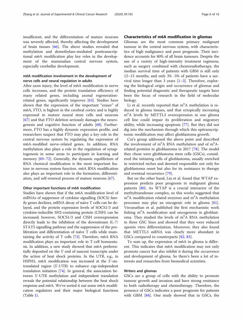
Characteristics of m6A modification in gliomasGliomas are the most common primary malignanttumour in the central nervous system, with characteris-tics of high malignancy and poor prognosis. Their inci-dence accounts for 80% of all brain tumours. Despite theuse of a variety of high-intensity treatment regimens,such as surgery combined with chemoradiotherapy, themedian survival time of patients with GBM is still only12–15 months, and only 3% -5% of patients have a sur-vival time longer than 3 years [1–3]. Therefore, explor-ing the biological origin and occurrence of gliomas andfinding potential diagnostic and therapeutic targets havebeen the focus of research in the field of molecularbiology.Li et al. recently reported that m6A methylation is re-
duced in glioma tissues, and that ectopically increasingm6A levels by METTL3 overexpression in one gliomacell line could impair its proliferation and migratoryability, while increasing apoptosis [77]. But they did notdig into the mechanism through which this epitranscrip-tomic modification may affect glioblastoma growth.Cui’s group addressed the above point and described
the involvement of m6A RNA methylation and of m6A-related proteins in glioblastoma in 2017 [78]. The modelthey chose were glioblastoma stem cells (GSCs), consid-ered the initiating cells of glioblastoma, usually enrichedin restricted niches and deemed responsible not only forglioblastoma onset but also for its resistance to therapyand eventual recurrence [79].But on the other hand, Liu et al. found that WTAP ex-
pression predicts poor prognosis in malignant gliomapatients [80]. As WTAP is a crucial interactor of themethyltransferase complex, so this works suggested thatm6A modification related enzymes and m6A methylationprocesses may play an oncogenic role in glioma [81].Visvanathan et al. published the first mechanistic worklinking m6A modification and oncogenesis in glioblast-oma. They studied the levels of m6A RNA methylationin three GSC lines and showed that they were reduceduponin vitro differentiation. Moreover, they also foundthat METTL3 mRNA was clearly more abundant inGSCs compared to counterparts [82, 83].To sum up, the expression of m6A in glioma is differ-
ent. This indicates that m6A modification may not onlypromote cancer but also inhibit it during the occurrenceand development of glioma. So there’s been a lot of in-terests and researches from biomedical scientists.
Writers and gliomasGSCs are a group of cells with the ability to promotetumour growth and invasion and have strong resistanceto both radiotherapy and chemotherapy. Therefore, thepresence of GSCs indicates a poor prognosis for patientswith GBM [84]. One study showed that in GSCs, the
Zhang et al. Journal of Experimental & Clinical Cancer Research (2020) 39:192 Page 6 of 16

expression levels of METTL3 increased, and the expres-sion levels of METTL14 and ALKBH5 decreased, whileFTO did not show significant changes. By installingm6A on the SOX2 3’-untranslated region (3’-UTR),METTL3 mediates GSC maintenance and dedifferenti-ation by regulating the stability of SOX2 mRNA. Thecomplete structure of METTL3 and human antigen R(HuR) is critical for maintaining this process. Inaddition, METTL3 knockdown inhibited GSC growthand neurosphere formation and reduced the expressionlevels of stem cell-specific markers, stage-specific embry-onic antigen-1 (SSEA1), and glioma reprogramming fac-tors (including POU class 3 homeobox 2 (POU3F2),oligodendrocyte transcription factor 2 (OLIG2), spaltlike transcription factor 2 (SALL2) and SOX2). Ofwhich, SOX2 has a high affinity for METTL3 [82].The deep sequencing of m6A and mRNA showed that
the knockdown of METTL3 and/or METTL14 led to theupregulation of oncogenes and genes coding down-stream proteins, including ADAM metallopeptidase do-main 19 (ADAM19), EPH receptor A3 (EPHA3),Kruppel-like factor 4 (KLF4) and tumour-inhibiting fac-tors, resulting in the inhibition of GSC growth and self-renewal [85]. METTL3 overexpression or treatment withthe FTO inhibitor MA2, the ethyl ester form of
meclofenamic acid (MA), can cause an increase in m6Alevels in GBM cells [78]. However, another study re-ported the opposite effect of METTL3 in GBM; this ef-fect was related to a decrease in m6A levels duringdifferentiation. Silencing METTL3 expression in GBMcan significantly inhibit tumour growth and prolongmouse survival time, which is consistent with clinical ob-servations that an increase in METTL3 expression isconsistent with the poor survival of patients with GBM.Further studies on the mechanisms of action have shownthat METTL3 is involved in the RNA processing andcarcinogenic pathways of GSCs and has a variety ofcomplexities. METTL3 plays a major role in m6A modi-fication in GSCs and participates in the expression andalternative splicing of GSC-specific genes. In addition,METTL3 reduced A-to-I RNA editing by downregulat-ing ADAR and ADAMRB1 and increased the editingabundance of C-U RNA by upregulating apolipoproteinB mRNA editing enzyme catalytic subunit 1 (APOBEC1)and APOBEC3A [59].METTL3 expression is upregulated in GSCs and
weakens during differentiation. SOX2 was identified asan important target of METTL3-mediated m6A, whereasMETTL3 promoted the recruitment of HuR to m6A-modified SOX2 mRNA and enhanced SOX2 stability
Table 1 m6A modification regulators and their major biological functions
Category m6A regulator Main functions References
Writers METTL3 miRNA regulates mRNA methylation through sequence complementation and cellreprogramming
[47]
METTL3 Mediates m6A modification involved in the regulation of spermatogenesis in mouse [59]
METTL3 Mediates m6A modification involved in the regulation of mouse cerebellar development [63]
METTL14, WTAP, VIRMA,RBM15, ZC3H13, METTL16
m6A methyltransferase complex component identification [9]
WTAP, METTL3, METTL14 WTAP, as a regulatory subunit, regulates the localization and substrate binding capacityof the catalytic subunits of the METTL3/METTL14 complex
[26]
Erasers FTO As the first discovered demethylase, FTO can catalyse the demethylation of m6Am and m1A [75]
FTO Mediates m6A modification that can serve as a novel cis element to regulate mRNA splicingand adipocyte precursor cell differentiation
[48]
ALKBH5 The second discovered demethylase; participates in the regulation of mRNA nuclear exportand mouse sperm development
[33]
Readers YTHDC1 YTHDC1 directly interacts with SRSF3 and SRSF10 to regulate alternative mRNA splicing [35]
YTHDC1 YTHDC1 interacts with SRSF3 and RNA nuclear export factor 1 (NXF1) to regulate mRNAnuclear export
[37]
YTHDF1 YTHDF1 directly interacts with the translation initiation complex to promote the translationefficiency of m6A-modified RNA substrate
[34]
YTHDF2 Mediates m6A modification involved in the regulation of mRNA decay [44]
YTHDF2, METTL3 Mediates m6A modification involved in the regulation of the differentiation of haematopoieticstem cells
[57]
YTHDF1 and YTHDF3 YTHDF1 synergizes with YTHDF3 to regulate mRNA translation [76]
YTHDF2 and YTHDF3 YTHDF2 synergizes with YTHDF3 to mediate mRNA decay [36]
YTHDC2 YTHDC2 regulates mRNA translation or decay and mouse spermatogenesis [38]
IGF2BP1/2/3 Participates in m6A modification-mediated mRNA stability and translation [42]
Zhang et al. Journal of Experimental & Clinical Cancer Research (2020) 39:192 Page 7 of 16

[85]. In addition, after the downregulation of METTL3expression, GSCs showed strong radiosensitivity and aweak DNA repair capacity [82]. Therefore, the abovestudies also revealed that METTL3-mediated m6Amodification was important in GSC maintenance andradiotherapy resistance. As a zinc finger protein, zincfinger CCCH-type containing 13 (ZC3H13), is also animportant regulator in the m6A-METTL-associatedcomplex (MACOM) and can anchor WTAP, virilizerand Hakai in the nucleus [78]. A recent study showedthat the ZC3H13 mutation and retinoblastoma 1 (RB1)mutation could replicate human GBM in a mousemodel. In addition, the ZC3H13 mutation also changedthe gene expression profile of the RB1 mutant to en-hance the resistance of GBM tumours to TMZ [86].In addition, WTAP is overexpressed in GBM, and
WTAP enhances the proliferation, migration, invasion,and tumourigenicity of GBM cells in xenografts by me-diating the phosphorylation of epidermal growth factorreceptor (EGFR) and protein kinase B (AKT). Inaddition, WTAP also regulates the expression of certaingenes associated with cancer cell movement, such aschemokine ligand 2 (CCL2), CCL3, matrix metallopro-teinase 3 (MMP3), lysyl oxidase like 1 (LOXL1), hyalur-onic acid synthase 1 (HAS1) and thrombospondin 1(THBS1) [81]. High WTAP expression is an independentprognostic factor that is positively correlated with ageand World Health Organization (WHO) classificationand indicates poor overall survival in GBM patients [80].Cell-based experiments have shown that WTAP plays animportant role in the miR-29a/Quaking isoform 6 (QKI-6) axis-mediated inhibition of cell proliferation, migra-tion, and invasion as well as a downstream target forpromoting GSC apoptosis [87].In addition to the direct impact on pluripotent genes,
MeRIP-seq analyses based on m6A-Seq techniquesshowed that m6A-modification peaks tend to beenriched in metabolic pathway-related transcripts [88].METTL3 can cause changes through the downregulationof adenosine deaminase 1 (ADAR1) and apolipoproteinB mRNA expression, e.g., a reduction in editing eventssuch as adenosine to inosine (A to I) and cytidine to uri-dine (C to U) (such as APOBEC3A) in GSCs [89]. Inaddition, gene ontology analysis indicated that the directtarget of METTL3 seems to be enriched in some majoroncogenic pathways, including the Notch signallingpathway, vascular endothelial growth factor (VEGF) sig-nalling pathway, angiogenesis, glycolysis and the Hedge-hog signalling pathway; the indirect target is enriched inthe RAS pathway, mitogen-activated protein kinase(MAPK) pathway, G-protein coupled receptor (GPCR)pathway, cadherin signalling pathway and cell cycle [89].In addition, in GSCs, METTL3-mediated m6A modifica-tion can also affect expression levels of serine and
arginine rich splicing factors (SRSF) by upregulatingBCL-X or nuclear receptor corepressor 2 and can pre-vent YTHDC1-dependent nonsense-mediated mRNAdecay (NMD) [88]. Compared with protein-codinggenes, METTL3-mediated m6A-tagged lncRNAs are alsohighly expressed in GSCs. Furthermore, the m6A markerin the 3’-UTR appears to block the binding process ofmicroRNA-related genes in GSCs [89].In summary, m6A writers are critical for the occur-
rence and development of GBM, and most are upregu-lated in GBM and show carcinogenic effects byregulating specific signalling pathways, especially helpingto maintain cell stemness. However, some opposite find-ings indicate that the expression of some writers inGBM is downregulated and that some writers may haveanticancer properties. Therefore, the existence of thisconflict provides more research possibilities on the roleof m6A modification-related methylation in the bio-logical pathogenesis of gliomas.
Erasers and gliomasSimilar to writers, m6A erasers also play vital roles inGBM. The latest research shows that ALKBH5 is ele-vated in GSCs, enhancing cell self-renewal, proliferationand tumourigenicity [78]. In terms of a mechanism,ALKBH5 demethylates m6A-modified bases and en-hances the expression level of the key target gene fork-head box protein M1 (FOXM1) in GBM patients byreducing the abundance of m6A in the target mRNAtranscript (especially in the 3’-UTR) [90].As an important functional target of ALKBH5,
FOXM1 overexpression can reverse the function ofALKBH5 or inhibit FOXM1 long noncoding RNA anti-sense (FOXM1-AS) and restore GSC tumour growth.FOXM1-AS is a lncRNA on human chromosome 12 thatis opposite to and partially overlaps with FOXM1.FOXM1-AS can promote the interaction betweenALKBH5 and FOXM1 nascent transcripts, thereby pro-moting the recruitment of HuR. In general, under thecombined action of FOXM1-AS, ALKBH5 enhances theself-renewal and proliferation of GSCs by regulating theexpression of FOXM1 and promoting the occurrenceand development of GBM [90]. On the other hand,ALKBH5 knockdown inhibits the proliferation of GSCs,while wild-type ALKBH5 rescues the proliferation ofGSCs. After ALKBH5 knockdown, the m6A level in nas-cent FOXM1 transcripts is elevated, and the binding ofFOXM1 pre-mRNA to HuR is reduced; therefore, the re-cruitment of HuR to m6A-modified RNA is crucial forstabilizing FOXM1 mRNA [90].Su’s study showed that the inhibition of FTO hindered
the self-renewal ability and carcinogenicity of GBM stemcells in vitro and in mouse models. FTO plays carcino-genic roles through maintaining the stability of gliomas,
Zhang et al. Journal of Experimental & Clinical Cancer Research (2020) 39:192 Page 8 of 16

especially the stability of oncogene homologues (c-Myc)and CCAAT-enhancer-binding protein-α (CEBPA) tran-scripts in IDH1/2 mutant gliomas. In addition, the in-hibitory effect of MA2 on FTO significantly increasesthe tumourigenicity of GSC-transplanted mice [75, 78].The above evidence also potentially reveals that FTOmay be a promising target for the drug treatment ofGBM.
Readers and gliomasYTHDF and YTHDC familyThe YTHDF and YTHDC series are the most importantcode readers for m6A modification, as they include aYTH domain that can bind to RNA. They exhibit differ-ent functions: YTHDC1 mediates mRNA splicing;YTHDF2, YTHDF3 and YTHDC2 mediate mRNAdecay; YTHDF1, YTHDF3 and YTHDC2 mediatemRNA translation; and YTHDC2 and YTHDC2 mediateRNA structure [91]. Recently, a study by Li et al. showedthat these important m6A-related proteins are involvedin the development of GBM. After YTHDC1 knock-down, proliferation was significantly reduced in U87cells with METTL3 overexpression but not in controlcells. In addition, for the W377A/W428A mutant withMETTL3 overexpression, YTHDC1 failed to promotethe ability of U87 cells to form spheres, indicating thatYTHDC1 relies on its m6A binding activity to promotethe functional phenotype of GBM [88].
IGF2BP familyAnother m6A reader protein family, the IGF2BP family(IGF2BP1/2/3), inhibits the decay of m6A-modified tran-scripts and promotes their translation [42]. For a longtime, these proteins have been considered importantregulatory factors in the pathogenesis of GBM, eventhough some functions are not directly related to car-cinogenesis. For example, IGF2BP1 can promote theproliferation and invasion of GBM cells by stabilizingthe mRNA transcripts of its target genes, including c-Myc, Ki-67, phosphatases and tensin homologue (PTEN)and CD44 (cell-surface glycoprotein 44) [92, 93].Studies have shown that IGF2BP2 is upregulated in
GBM tissues, promoting the proliferation, migration, in-vasion, and epithelial-mesenchymal transition (EMT) ofGBM cells through the regulation of insulin-like growthfactor 2 (IGF2) activity while further activating the phos-phoinositide 3-kinases (PI3K)/AKT signalling pathway.In addition, IGF2BP2 inhibition can cause an increase inthe sensitivity of GBM to TMZ [94]. Another studyshowed that IGF2BP2 binds to the miRNA recognitionelements (MREs) of lethal-7 (let-7) and blocks let-7 tar-get gene silencing, which is LIN28-independent, includ-ing high mobility group AT-hook 1 (HMGA1), HMGA2,cyclin D2 (CCND2) and ribonucleotide reductase
regulatory subunit M2 (RRM2), thus maintaining thestemness of GSCs [95]. During this process, IGF2BP2 isresponsible for miRNA maturation, and it also interactswith lncRNAs such as hypoxia inducible factor 1 alpha-antisense RNA 2 (HIF1A-AS2), which can specifically in-duce hypoxia to maintain the expression level of its targetgene HMGA1, eventually promoting GSC proliferation,self-renewal and the reprogramming of hypoxia-dependent molecules [96]. In addition, IGF2BP2 also in-teracts with mRNAs and proteins. For example, IGF2BP2can bind to several mRNAs, including mitochondrial cyto-chrome C oxidase subunit 7B (COX7B), NADH dehydro-genase iron-sulfur protein 7 (NDUS7), and NADHdehydrogenase, to promote oxidative phosphorylation inGSCs [97].The mRNA and protein expression levels of IGF2BP3
are upregulated in GBM but not significantly upregu-lated in low-grade astrocytomas [98]. According to thegene expression microarray analysis of 9 pilomyxoid as-trocytoma (PMA) and 13 pilocytic astrocytoma (PA)from lower and upper loci, the expression level ofIGF2BP3 in malignant astrocytoma is significantly in-creased [99]. In addition, gene chip analysis in gliomacells indicated that IGF2BP3 mediates the associationbetween direct targets at the transcriptome level andprocesses related to the cell cycle as well as the associ-ation between direct targets at the translatome level andapoptosis-related pathways [100]. IGF2BP3 also inducedEMT by downregulating the expression of E-cadherinand upregulating the expression levels of N-cadherin,vimentin and MMP-9, thereby promoting cell prolifera-tion, migration and invasion [101]. In addition, IGF2BP3activates the PI3K/MAPK pathway by binding to the 5’-UTR of IGF-2 mRNA to activate its translation, therebypromoting cell proliferation, anchorage-independentgrowth, invasion, and chemoresistance [98]. IGF2BP3also stimulates the migration of glioma cells by enhan-cing the translation of p65 (RELA), which is a subunit ofthe nuclear factor-kappa B (NF-κB) heterodimer, andp65 can also transcriptionally activate IGF2BP3 to forma feedback loop [102].
hnRNPA2B1 and hnRNPC familyA study of SOX2 protein interactions showed thathnRNPA2B1 and hnRNPC could interact with SOX2 inGBM, suggesting that they might play a key role inmaintaining the stemness of GSCs [103]. Heterogeneousnuclear ribonucleoproteins A2/B1 (hnRNPA2B1) is animportant regulator of mRNA metabolism and transportin cells. Its downstream protein is highly expressed inglioma tissues and is associated with the histologic gli-oma grade and a poor prognosis [104]. hnRNPA2B1may promote the proliferation, migration, and invasionof GBM cells through the downregulation of tumour
Zhang et al. Journal of Experimental & Clinical Cancer Research (2020) 39:192 Page 9 of 16

suppressor factors, enhance chemoresistance to TMZ,and protect cells from apoptosis and damage caused byreactive oxygen species (ROS) [105].In addition, hnRNPC is an important physiological
modulator for 3’-UTR processing and miRNA matur-ation as well as a modulator for neoplastic disease [106].hnRNPC has higher expression levels in higher-gradeGBM; hnRNPC directly binds to miR-21 (mainly pri-miR-21) and promotes miR-21 processing against pro-grammed cell death 4 (PDCD4), which is an importantregulator of cell apoptosis and survival. PDCD4 subse-quently promotes the activation of AKT and p70 S6 kin-ase (p70S6K) and then enhances the migration andinvasion activity of tumour cells, increases cell prolifera-tion, and protects GBM cells from apoptosis [107].These results indicate that hnRNPA2B1 and hnRNPCmay be important m6A “readers” that are closely relatedto the biogenesis of GBM. Finally, we mapped the mech-anism of action of m6A-modified important proteins as-sociated with the biological behavior of glioma cells(Fig. 3).
Clinical significance of m6A modification ingliomasm6A modification and disease diagnosisEpigenetic alterations are considered promising markersfor GBM diagnosis. In addition, some epigenetic statusesindeed explain the outcome of GBM [108–111]. For ex-ample, the epigenetic silencing of O-6-methylguanine-DNA methyltransferase (MGMT) significantly affectedthe TMZ treatment effect in GBM patients. Therefore,the methylation status of the MGMT promoter is an im-portant biological marker used to predict GBM patientsurvival and GBM response to TMZ [112, 113].As an important chemical modification to RNA, regu-
lar changes in m6A can also predict the prognosis of pa-tients with GBM or be used for the diagnosis of GBM.Related studies have shown that m6A in RNA from per-ipheral blood is a biomarker of gastric cancer [114]. Themethylation level of miRNAs is also a potential diagnos-tic biomarker for early gastrointestinal cancer [115].ALKBH5 and FTO, which act as m6A erasers, have alsobeen proven to be prognostic biomarkers for patients
Fig. 3 The potential roles of RNA m6A modification in glioma progression. They are reflected in the regulation of tumor-associated factors. m6Apromotes glioma progression by enhancing oncogene expression and inhibiting tumor suppressor gene expression. m6A hampers gliomaprogression by inhibiting oncogene expression and enhancing tumor suppressor gene expression
Zhang et al. Journal of Experimental & Clinical Cancer Research (2020) 39:192 Page 10 of 16

with renal clear cell carcinoma [116]. Therefore, for cen-tral nervous system tumours, the identification of modi-fications to RNA from peripheral blood or cerebrospinalfluid may be a promising method for GBM diagnosis.
Expression of the m6A modifier is related to theclinicopathological features of gliomasConsidering the important biological functions of m6Amodification-related proteins involved in the occurrenceand development of tumours, some researchers con-ducted genomic profiling-based data mining and bio-informatics analysis to systematically investigate therelationship between each type of m6A modulator andpathological features of gliomas, including WHO classifi-cation, isocitrate dehydrogenase (IDH) classification and1p/19q status. The results showed that the expressionlevel of most m6A modulators was significantly corre-lated with the WHO histologic grade and correspondingclassification. Through quantitative analysis, the expres-sion levels of WTAP, YTHDF, ALKBH5 and FTO weresignificantly correlated with the histologic grade, the ex-pression abundances of WTAP, YTHDF and ALKBH5were positively correlated with the histologic grade, andthe expression abundance of FTO was negatively corre-lated with the histologic grade [117].
Potential therapeutic significance of m6A modification ingliomasThe covalent modification of DNA, histones and otherproteins has shown potential as a cancer treatment.Some epigenetic drugs, such as suberanilohydroxamicacid (SAHA), romidepsin, Belinostat and Panobinostat,have been approved by the U.S. Food and Drug Admin-istration (FDA). Moreover, chidamide has also been ap-proved by the Chinese National Medical ProductsAdministration for the treatment of certain T-celllymphomas or multiple myeloma. In addition, manyother clinical trials of epigenetic drugs are also underway[118]. Drugs targeting protein posttranslational modi-fiers (such as E3 ubiquitin-protein ligase), including S-phase kinase associated protein 2 (SKP2), speckle typeBTB/POZ protein (SPOP), cellular inhibitor of apoptosis2 (cIAP) and anaphase-promoting complex/cyclosome(APC/C), have also been evaluated in clinical practiceand preclinical applications [119].m6A is the most common covalent modification of
RNA at the posttranscriptional level. Scientists are explor-ing its therapeutic potential in malignant tumours such asGBM. Currently, some pharmaceutical companies areplanning to develop a METTL3-METTL14 complex and asmall molecule inhibitors of ADAR [120]. Several com-pounds with piperidine or piperazine rings have very highcooperative binding efficiency with METTL3-METTL14-WTAP complexes, thereby activating RNA methylation
[121]. Recently, METTL3 small molecule inhibitors havealso shown the ability to inhibit the progression of acutemyeloid leukaemia (AML) in the body [122]. Based on theabovementioned latest research, it is believed that m6Amodification-related targeted therapies for gliomas will bedeveloped.
m6A modification and prognosis of patients with gliomasSome researchers conducted a univariate Cox regressionanalysis on the expression levels of m6A-related proteinsin the Chinese Glioma Genome Atlas (CGGA) databaseand found that high-risk genes mainly include ALKBH5,YTHDF1, YTHDF2, HNRNPC, RNA binding motif pro-tein 15 (RBM15), KIAA1429 and WTAP and that theprotective genes are mainly FTO, YTHDC1, ZC3H13and METTL3. Among the genes with predictive value inadult low-grade gliomas (LGGs), the expression levels ofYTHDF2, WTAP, ALKBH5, RBM15, KIAA1429, HNRNPC, YTHDF1 and FTO were significantly correlated withthe overall survival (OS) of IDH-mutated glioma pa-tients. In summary, YTHDF2, KIAA1429, HNRNPC,and YTHDF1 had predictive value in LGB with wild-type IDH, and FTO, YTHDF2, and RBM15 had predict-ive value in GBM with wild-type IDH; FTO also hadpredictive value in GBM with mutated IDH [117].In addition, another univariate and multivariate Cox
regression analysis showed that risk score, 1p/19q se-quence, IDH status, age and WHO classification were allassociated with OS. After including these factors in themultivariate Cox regression, risk scores and WHO clas-sification were still significantly correlated with OS (P <0.001). Further multiple regression analysis showed thatrisk score (P = 0.027), IDH status (P < 0.001), age (P <0.001) and WHO classification (P = 0.005) were still sig-nificantly correlated with OS. In summary, the risk scorederived from the RNA m6A modulator can independ-ently predict the prognosis of glioma patients. Inaddition, for WHO grades II and III gliomas, the OS ofpatients with high risk scores was significantly shorterthan that of patients with lower risk scores. Throughcomprehensive analysis of the WHO classification, riskscore also had predictive value for gliomas. In addition,according to the CGGA and The Cancer Genome Atlas(TCGA) databases, GBM patients with high risk scoreswere also more sensitive to TMZ treatment and hadrelatively lower drug resistance [117].
Discussion and outlookThere is a correlation between the degree of m6A RNAmethylation and the differentiation of GSCs and betweenthe effect of METTL3 knockdown on in vitro self-renewal and in vivo tumourigenicity of glioblastoma inmice. GSCs and their derived tumours can be dividedinto subtypes based on different characteristics and
Zhang et al. Journal of Experimental & Clinical Cancer Research (2020) 39:192 Page 11 of 16

different aggressiveness and responsiveness to therapy.Research on m6A modification and oncology is still anemerging field. Therefore, all cited papers are based on avery limited number of cell models, and the results maybe affected by variability. One study analysed 7 GSClines using RNA-seq and compared the normal humanbrain with an established GBM cell line; the study didnot find any clear evidence (e.g., author, reader, eraser,etc.) to prove the differential expression of mRNA cod-ing proteins involved in m6A methylation. However, thecontroversial results were not discredited, and somegenes or proteins involved in mRNA expression werestill considered potential prognostic markers of GBM.However, for this mechanism, it seems more necessary to
clarify which mRNAs are hypermethylated or hypomethy-lated and which readers identify methylation under certaincircumstances. It has been demonstrated that IGF2BPs arecarcinogenic in glioblastoma, especially in GSCs, in whichtheir function as specific target mRNA stability enhancersis related to their ability to induce or maintain glioblastomagrowth. By correlating these data with currently knownm6A readers, we can gain a deeper understanding of othermolecular mechanisms by which these proteins may be in-volved in glioblastoma tumourigenesis.Wu et al. recently published a study demonstrating that
the m6A reader PRRC2a plays a key role in the formationof oligodendrocytes and myelination by binding to methyl-ated adenosine in glioblastoma cells. This binding canstabilize OLIG2 mRNA, thereby stabilizing Olig2 proteinproduction. In contrast, FTO demethylates OLIG2 mRNAat this site, thereby promoting its decay and Olig2 proteinconsumption. This study focused on oligodendrocytes inphysiological and pathologically low-myelination condi-tions, but it should be noted that in addition to playing amajor role in the development of oligodendrocytes, Olig2protein also plays a role in GBM cell reprogramming andthe genotoxicity and phenotypic plasticity of tumours. Inaddition, Olig2 expression is a recognized marker for theproneural subtype of GBM. More importantly, Olig2 pro-tein is synergistic with Sox2, Pou3f2 and Sall2 and is a keytranscription factor of glioma initiating cells [123]. This ob-servation is only an example that partially elucidates howthe proteins involved in transcriptome regulation are trans-ferred under specific pathological conditions, especially inGBM. This field is worthy of further in-depth studies. Basedon the information regarding transcriptome modificationsin gliomas obtained from recent studies, we may have onlyrevealed the tip of the iceberg, and in the future, we willdefinitely find more complex mechanisms of precise regula-tion of the transcriptional fate of glioma cells.
ConclusionsIn summary, although the correlation between m6Amodification and oncology, as a hotspot in the field of
biomedicine, has been extensively explored, most studiesconcentrated on gene sequencing analysis, differentialexpression analysis, and modification site analysis. Thereare few studies on the functional phenotypes and mech-anisms of action at the cell level, but studies in this fieldare likely to be key to revealing the origin of tumours,especially the origin and development of malignant tu-mours. With the rapid development of molecular biologytechnology, especially single-cell sequencing and third-generation sequencing, the answer of how RNA methy-lation modification affects the biological behaviour ofgliomas will gradually become clear. It provides new in-sights for the early diagnosis of, histologic grading of,and targeted therapy for gliomas. It also points to a newdirection for the study of other neoplastic diseases. As astar molecule in RNA post-transcription modificationresearch, exploring m6A modification might decipherthe molecular mechanism of many diseases, serving as anew medical breakthrough anticipated by biomedicalscientists.
AbbreviationsGBM: Glioblastoma; TMZ: Temozolomide; m6A: N6-methyladenine;mRNA: Messenger RNA; m6A-seq: M6A RNA immunoprecipitationsequencing; HPLC-MS/MS: High-performance liquid chromatography-tandemmass spectrometry; MeRIP-seq: Methylated RNA immunoprecipitationfollowed by high-throughput sequencing; PA-m6A-Seq: Photo-crosslinking-assisted m6A-sequencing; SCARLET: Site-specific cleavage and radioactive-labelling followed by ligation-assisted extraction and thin-layer chromatog-raphy; miCLIP: M6A individual nucleotide resolution crosslinkingimmunoprecipitation; miRNA: Micro RNA; lncRNA: Long noncoding RNA;circRNA: Circular RNA; rRNA: Ribosomal RNA; tRNA: Transfer RNA;snoRNA: Small nucleolar RNA; METTL3: Methyltransferase like 3; WTAP: Wilms'tumour 1-associating protein; SAM: S-adenosylmethionine; FTO: Fat mass andobesity-associated gene; ALKBH5: AlkB homologue 5; YTHDF1: YTH N6-methyladenosine binding protein 1; YTHDC1: YTH domain containing 1;SRSF3: Serine-rich splicing factor 3; IGF2BP: Insulin-like growth factor-II mRNAbinding protein ; P body: Processing body; EHT: Endothelial-to-haematopoietic transition; Notch1: Notch homologue protein 1;HSPC: Haematopoietic stem cell; ESC: Embryonic stem cell; ZFP217: Zincfinger protein 217; SOX2: Sex determining region Y box 2; KLF4: Kruppel-likefactor 4; HE: Haematoxylin-eosin; IL-7: Interleukin-7; STAT5: Signal transducerand activator of transcription 5; CGC: Cerebellar granule cell; EGL: Externalgranular layer; SOCS: Suppressor of cytokine signalling; CISH: Cytokine-inducible SH2-containing protein; 5ʹ-UTR: 5ʹ-untranslated region;GSC: Glioblastoma stem cell; 3ʹ-UTR: 3ʹ-untranslated region; HuR: Humanantigen R; SSEA1: Stage-specific embryonic antigen 1; POU3F2: POU class 3homeobox 2 ; OLIG2: Oligodendrocyte transcription factor 2; SALL2: Spalt liketranscription factor 2; ADAM19: ADAM metallopeptidase domain 19 ;EPHA3: EPH receptor A3; MA: Meclofenamic acid; APOBEC1: Apolipoprotein BmRNA editing enzyme catalytic subunit 1; ZC3H13: Zinc finger CCCH-typecontaining 13; MACOM: M6A-METTL-associated complex;RB1: Retinoblastoma 1; EGFR: Epidermal growth factor receptor;CCL2: Chemokine ligand 2; MMP3: Matrix metalloproteinase 3; LOXL1: Lysyloxidase like 1; HAS1: Hyaluronic acid synthase 1; THBS1: Thrombospondin 1 ;WHO: World Health Organization; QKI-6: Quaking isoform-6;ADAR1: Adenosine deaminase 1; VEGF: Vascular endothelial growth factor;MAPK: Mitogen-activated protein kinase; GPCR: G-protein coupled receptor;SRSF: Serine and arginine rich splicing factor; NMD: Nonsense-mediatedmRNA decay; FOXM1: Forkhead box protein M 1; CEBPA: CCAAT-enhancer-binding protein-α; PETN: Phosphatases and tensin homologue; CD44: Cell-surface glycoprotein 44; EMT: Epithelial-mesenchymal transition; IGF2: Insulin-like growth factor 2; PI3K: Phosphoinositide 3-kinases; MRE: MiRNArecognition element ; let-7: Lethal-7; HMGA1: High mobility group AT-hook 1;CCND2: Cyclin D 2; RRM2: Ribonucleotide reductase regulatory subunit M 2;
Zhang et al. Journal of Experimental & Clinical Cancer Research (2020) 39:192 Page 12 of 16

HIF1A-AS2: Factor 1 alpha-antisense RNA 2; COX7B: Cytochrome C oxidasesubunit 7 B; NDUS7: NADH dehydrogenase iron-sulfur protein 7;PMA: Pilomyxoid astrocytoma; PA: Pilocytic astrocytoma; NF-κB: Nuclearfactor-kappa B; hnRNPA2B1: Heterogeneous nuclear ribonucleoproteins A2/B1; ROS: Reactive oxygen species; PDCD4: Programmed cell death 4;p70S6K: P70 S6 kinase; MGMT: Methylguanine-DNA methyltransferase;IDH: Isocitrate dehydrogenase; SAHA: Suberanilohydroxamic acid ; FDA: Foodand drug administration; SKP2: S-phase kinase associated protein 2;SPOP: Speckle type BTB/POZ protein; cIAP: Cellular inhibitor of apoptosis;APC/C: Anaphase-promoting complex/cyclosome; AML: Acute myeloidleukaemia; CGGA: Chinese Glioma Genome Atlas; RBM15: RNA binding motifprotein 15; LGG: Adult low-grade glioma; OS: Overall survival; TCGA: TheCancer Genome Atlas
AcknowledgementsWe would like to give special thanks to Ruijuan Ren, the curator of HebeiUniversity library, for her guidance and assistance in the literature retrievalprocess of this paper.
Authors’ contributionsYHZ and XCG were the major contributors to the writing and revision of themanuscript. QL, JLX and YLT collected the related references and participated indiscussions. MLX and JS revised this article critically for important intellectualcontent. FLL, CF and HW gave approval for the final version of the manuscript.All authors have read and approved the final manuscript.
FundingThe present study was financially supported by grants from the NaturalScience Foundation of Hebei Province in 2020 (no.H2020201050); High-levelTalents Scientific Research Launched Project of Hebei Universi-ty(no.521000981301); Government-funded Clinical Medicine OutstandingTalent Training Project(Team) in 2019(no.360017); Science and TechnologyCapacity Improvement Projects of Hebei University of Chinese Medicine in2019 (no.KTZ2019019); Outstanding Student Scientific Research AbilityImprovement Projects of Hebei University of Chinese Medicine in 2019(no.YXZ2019002); Graduate Innovative Ability Training Projects of HebeiUniversity of Chinese Medicine in 2020 (no.XCXZZBS2020002); GraduateInnovative Ability Training Projects of Hebei University in 2020 (no.h-bu2020bs003); Foundation Project of Affiliated Hospital of Hebei University in2019 (no.2019Q009).
Availability of data and materialsThe datasets used and analysed during the present study are available fromthe corresponding author on reasonable request.
Ethics approval and consent to participateNot applicable.
Consent for publicationNot applicable.
Competing interestsThe authors declare that they have no competing interests
Author details1Department of Neurosurgery, Affiliated Hospital of Hebei University, 071000Baoding, China. 2Faculty of Integrated Traditional Chinese and WesternMedicine, Hebei University of Chinese Medicine, 050091 Shijiazhuang, China.3Faculty of Acupuncture-Moxibustion and Tuina, Hebei University of ChineseMedicine, 050200 Shijiazhuang, China. 4Department of Pathology, AffiliatedHospital of Hebei University, 071000 Baoding, China. 5School of BasicMedicine, Hebei University, 071000 Baoding, China. 6Office of AcademicResearch, Affiliated Hospital of Hebei University, 071000 Baoding, China.7Hebei Key Laboratory of Chinese Medicine Research onCardio-Cerebrovascular Disease, Hebei University of Chinese Medicine,050091 Shijiazhuang, China.
Received: 29 July 2020 Accepted: 10 September 2020
References1. Louis DN, Perry A, Reifenberger G, von Deimling A, Figarella-Branger D,
Cavenee WK, et al. The 2016 World Health Organization Classification ofTumors of the Central Nervous System: a summary. Acta Neuropathol. 2016;131(6):803–20.
2. Lapointe S, Perry A, Butowski NA. Primary brain tumours in adults. Lancet.2018;392(10145):432–46.
3. Kristensen BW, Priesterbach-Ackley LP, Petersen JK, Wesseling P. Molecularpathology of tumors of the central nervous system. Ann Oncol. 2019;30(8):1265–78.
4. Lim M, Xia Y, Bettegowda C, Weller M. Current state of immunotherapy forglioblastoma. Nat Rev Clin Oncol. 2018;15(7):422–42.
5. Touat M, Li YY, Boynton AN, Spurr LF, Iorgulescu JB, Bohrson CL, et al.Mechanisms and therapeutic implications of hypermutation in gliomas.Nature. 2020;580(7804):517–23.
6. Chen XY, Zhang J, Zhu JS. The role of m(6)A RNA methylation in humancancer. Mol Cancer. 2019;18(1):103.
7. Deng X, Su R, Weng H, Huang H, Li Z, Chen J. RNA N(6)-methyladenosinemodification in cancers: current status and perspectives. Cell Res. 2018;28(5):507–17.
8. Dawson MA, Kouzarides T. Cancer epigenetics: from mechanism to therapy.Cell. 2012;150(1):12–27.
9. Wimalasena VK, Wang T, Sigua LH, Durbin AD, Qi J. Using ChemicalEpigenetics to Target Cancer. Mol Cell. 2020;78(6):1086–95.
10. Saletore Y, Meyer K, Korlach J, Vilfan ID, Jaffrey S, Mason CE. The birth of theEpitranscriptome: deciphering the function of RNA modifications. GenomeBiol. 2012;13(10):175.
11. Wang T, Kong S, Tao M, Ju S. The potential role of RNA N6-methyladenosine in Cancer progression. Mol Cancer. 2020;19(1):88.
12. Frye M, Jaffrey SR, Pan T, Rechavi G, Suzuki T. RNA modifications: what havewe learned and where are we headed? Nat Rev Genet. 2016;17(6):365–72.
13. Shi H, Wei J, He C. Where, When, and How: Context-Dependent Functions ofRNA Methylation Writers, Readers, and Erasers. Mol Cell. 2019;74(4):640–50.
14. Dominissini D, Moshitch-Moshkovitz S, Schwartz S, Salmon-Divon M, UngarL, Osenberg S, et al. Topology of the human and mouse m6A RNAmethylomes revealed by m6A-sEq. Nature. 2012;485(7397):201–6.
15. Jia G, Fu Y, Zhao X, Dai Q, Zheng G, Yang Y, et al. N6-methyladenosine innuclear RNA is a major substrate of the obesity-associated FTO. Nat ChemBiol. 2011;7(12):885–7.
16. Bodi Z, Fray RG. Detection and Quantification of N (6)-Methyladenosine inMessenger RNA by TLC. Methods Mol Biol (Clifton NJ). 2017;1562:79–87.
17. Dominissini D, Moshitch-Moshkovitz S, Schwartz S, Salmon-Divon M, UngarL, Osenberg S, et al. Topology of the human and mouse m6A RNAmethylomes revealed by m6A-sEq. Nature. 2012;485(7397):201–6.
18. Meyer KD, Saletore Y, Zumbo P, Elemento O, Mason CE, Jaffrey SR.Comprehensive analysis of mRNA methylation reveals enrichment in 3’UTRs and near stop codons. Cell. 2012;149(7):1635–46.
19. Chen K, Lu Z, Wang X, Fu Y, Luo GZ, Liu N, et al. High-resolution N(6)-methyladenosine (m(6) A) map using photo-crosslinking-assisted m(6) Asequencing. Angewandte Chemie (International ed in English). 2015;54(5):1587–90.
20. Liu N, Pan T. Probing RNA Modification Status at Single-NucleotideResolution in Total RNA. Methods Enzymol. 2015;560:149–59.
21. Linder B, Grozhik AV, Olarerin-George AO, Meydan C, Mason CE, Jaffrey SR.Single-nucleotide-resolution mapping of m6A and m6Am throughout thetranscriptome. Nat Methods. 2015;12(8):767–72.
22. Gilbert WV, Bell TA, Schaening C. Messenger RNA modifications: Form,distribution, and function. Science (New York, NY). 2016;352(6292):1408-12.
23. Sun T, Wu R, Ming L. The role of m6A RNA methylation in cancer. BiomedPharmacother. 2019;112:108613.
24. Coker H, Wei G, Brockdorff N. m6A modification of non-coding RNA andthe control of mammalian gene expression. Biochim Biophys Acta GeneRegul Mech. 2019;1862(3):310–8.
25. Ma S, Chen C, Ji X, Liu J, Zhou Q, Wang G, et al. The interplay between m6ARNA methylation and noncoding RNA in cancer. J Hematol Oncol. 2019;12(1):121.
Zhang et al. Journal of Experimental & Clinical Cancer Research (2020) 39:192 Page 13 of 16

26. Ping XL, Sun BF, Wang L, Xiao W, Yang X, Wang WJ, et al. MammalianWTAP is a regulatory subunit of the RNA N6-methyladenosinemethyltransferase. Cell Res. 2014;24(2):177–89.
27. Liu J, Yue Y, Han D, Wang X, Fu Y, Zhang L. et al. A METTL3-METTL14complex mediates mammalian nuclear RNA N6-adenosine methylation. NatChem Biol. 2014;10(2):93–5.
28. Liu ZX, Li LM, Sun HL, Liu SM. Link Between m6A Modification and Cancers.Front Bioeng Biotechnol. 2018;6:89.
29. Śledź P, Jinek M. Structural insights into the molecular mechanism of them(6)A writer complex. Elife. 2016;5:e18434.
30. Wang P, Doxtader KA, Nam Y. Structural Basis for Cooperative Function ofMettl3 and Mettl14 Methyltransferases. Mol Cell. 2016;63(2):306–17.
31. Jia G, Fu Y, Zhao X, Dai Q, Zheng G, Yang Y, et al. N6-methyladenosine innuclear RNA is a major substrate of the obesity-associated FTO. Nat ChemBiol. 2011;7(12):885–7.
32. Mauer J, Luo X, Blanjoie A, Jiao X, Grozhik AV, Patil DP, et al. Reversiblemethylation of m(6)A(m) in the 5’ cap controls mRNA stability. Nature. 2017;541(7637):371–5.
33. Zheng G, Dahl JA, Niu Y, Fedorcsak P, Huang CM, Li CJ, et al. ALKBH5 is amammalian RNA demethylase that impacts RNA metabolism and mousefertility. Mol Cell. 2013;49(1):18–29.
34. Wang X, Zhao BS, Roundtree IA, Lu Z, Han D, Ma H, et al. N(6)-methyladenosine Modulates Messenger RNA Translation Efficiency. Cell.2015;161(6):1388–99.
35. Xiao W, Adhikari S, Dahal U, Chen YS, Hao YJ, Sun BF, et al. Nuclearm(6)A Reader YTHDC1 Regulates mRNA Splicing. Mol Cell. 2016;61(4):507–19.
36. Shi H, Wang X, Lu Z, Zhao BS, Ma H, Hsu PJ, et al. YTHDF3 facilitatestranslation and decay of N(6)-methyladenosine-modified RNA. Cell Res.2017;27(3):315–28.
37. Roundtree IA, Luo GZ, Zhang Z, Wang X, Zhou T, Cui Y, et al. YTHDC1mediates nuclear export of N(6)-methyladenosine methylated mRNAs. ELife.2017;6:e31311.
38. Hsu PJ, Zhu Y, Ma H, Guo Y, Shi X, Liu Y, et al. Ythdc2 is an N(6)-methyladenosine binding protein that regulates mammalianspermatogenesis. Cell Res. 2017;27(9):1115–27.
39. Sun T, Wu R, Ming L. The role of m6A RNA methylation in cancer. BiomedPharmacother. 2019;112:108613.
40. Ke S, Alemu EA, Mertens C, Gantman EC, Fak JJ, Mele A. et al. A majority ofm6A residues are in the last exons, allowing the potential for 3’ UTRregulation. Genes Dev. 2015;29(19):2037–53.
41. Molinie B, Wang J, Lim KS, Hillebrand R, Lu ZX, Van Wittenberghe N, et al.m(6)A-LAIC-seq reveals the census and complexity of the m(6)Aepitranscriptome. Nat Methods. 2016;13(8):692–8.
42. Huang H, Weng H, Sun W, Qin X, Shi H, Wu H, et al. Recognition of RNAN(6)-methyladenosine by IGF2BP proteins enhances mRNA stability andtranslation. Nat Cell Biol. 2018;20(3):285–95.
43. Zhou J, Wan J, Gao X, Zhang X, Jaffrey SR, Qian SB. Dynamic m(6)A mRNAmethylation directs translational control of heat shock response. Nature.2015;526(7574):591–4.
44. Wang X, Lu Z, Gomez A, Hon GC, Yue Y, Han D, et al. N6-methyladenosine-dependent regulation of messenger RNA stability. Nature. 2014;505(7481):117–20.
45. Liu N, Dai Q, Zheng G, He C, Parisien M, Pan T. N(6)-methyladenosine-dependent RNA structural switches regulate RNA-protein interactions.Nature. 2015;518(7540):560–4.
46. Liu N, Zhou KI, Parisien M, Dai Q, Diatchenko L, Pan T. N6-methyladenosinealters RNA structure to regulate binding of a low-complexity protein.Nucleic Acids Res. 2017;45(10):6051–63.
47. Chen T, Hao YJ, Zhang Y, Li MM, Wang M, Han W, et al. m(6)A RNAmethylation is regulated by microRNAs and promotes reprogramming topluripotency. Cell Stem Cell. 2015;16(3):289–301.
48. Zhao X, Yang Y, Sun BF, Shi Y, Yang X, Xiao W, et al. FTO-dependentdemethylation of N6-methyladenosine regulates mRNA splicing and isrequired for adipogenesis. Cell Res. 2014;24(12):1403–19.
49. Fustin JM, Doi M, Yamaguchi Y, Hida H, Nishimura S, Yoshida M, et al. RNA-methylation-dependent RNA processing controls the speed of the circadianclock. Cell. 2013;155(4):793–806.
50. Xiang Y, Laurent B, Hsu CH, Nachtergaele S, Lu Z, Sheng W, et al. RNAm(6)A methylation regulates the ultraviolet-induced DNA damage response.Nature. 2017;543(7646):573–6.
51. Aguilo F, Zhang F, Sancho A, Fidalgo M, Di Cecilia S, Vashisht A, et al.Coordination of m(6)A mRNA Methylation and Gene Transcription byZFP217 Regulates Pluripotency and Reprogramming. Cell Stem Cell. 2015;17(6):689–704.
52. Geula S, Moshitch-Moshkovitz S, Dominissini D, Mansour AA, Kol N, Salmon-Divon M, et al. Stem cells. m6A mRNA methylation facilitates resolution ofnaïve pluripotency toward differentiation. Science. 2015;347(6225):1002–6.
53. Zhao BS, Wang X, Beadell AV, Lu Z, Shi H, Kuuspalu A, et al. m(6)A-dependent maternal mRNA clearance facilitates zebrafish maternal-to-zygotic transition. Nature. 2017;542(7642):475–8.
54. Haussmann IU, Bodi Z, Sanchez-Moran E, Mongan NP, Archer N, Fray RG,et al. m(6)A potentiates Sxl alternative pre-mRNA splicing for robustDrosophila sex determination. Nature. 2016;540(7632):301–4.
55. Lence T, Akhtar J, Bayer M, Schmid K, Spindler L, Ho CH, et al. m(6)Amodulates neuronal functions and sex determination in Drosophila. Nature.2016;540(7632):242–7.
56. Shen L, Liang Z, Gu X, Chen Y, Teo ZW, Hou X, et al. N(6)-MethyladenosineRNA Modification Regulates Shoot Stem Cell Fate in Arabidopsis. Dev Cell.2016;38(2):186–200.
57. Zhang C, Chen Y, Sun B, Wang L, Yang Y, Ma D, et al. m(6)A modulateshaematopoietic stem and progenitor cell specification. Nature. 2017;549(7671):273–6.
58. Lv J, Zhang Y, Gao S, Zhang C, Chen Y, Li W, et al. Endothelial-specificm(6)A modulates mouse hematopoietic stem and progenitor celldevelopment via Notch signaling. Cell Res. 2018;28(2):249–52.
59. Xu K, Yang Y, Feng GH, Sun BF, Chen JQ, Li YF, et al. Mettl3-mediated m(6)Aregulates spermatogonial differentiation and meiosis initiation. Cell Res.2017;27(9):1100–14.
60. Lin Z, Hsu PJ, Xing X, Fang J, Lu Z, Zou Q, et al. Mettl3-/Mettl14-mediatedmRNA N(6)-methyladenosine modulates murine spermatogenesis. Cell Res.2017;27(10):1216–30.
61. Wojtas MN, Pandey RR, Mendel M, Homolka D, Sachidanandam R, Pillai RS.Regulation of m(6)A Transcripts by the 3’→5’ RNA Helicase YTHDC2 IsEssential for a Successful Meiotic Program in the Mammalian Germline. MolCell. 2017;68(2):374–87.e12.
62. Chang M, Lv H, Zhang W, Ma C, He X, Zhao S, et al. Region-specific RNAm(6)A methylation represents a new layer of control in the gene regulatorynetwork in the mouse brain. Open Biol. 2017;7(9): 170166.
63. Wang CX, Cui GS, Liu X, Xu K, Wang M, Zhang XX, et al. METTL3-mediatedm6A modification is required for cerebellar development. PLoS Biol. 2018;16(6):e2004880.
64. Ma C, Chang M, Lv H, Zhang ZW, Zhang W, He X, et al. RNA m(6)Amethylation participates in regulation of postnatal development of themouse cerebellum. Genome Biol. 2018;19(1):68.
65. Yoon KJ, Ringeling FR, Vissers C, Jacob F, Pokrass M, Jimenez-Cyrus D, et al.Temporal Control of Mammalian Cortical Neurogenesis by m(6)AMethylation. Cell. 2017;171(4):877–89.e17.
66. Li M, Zhao X, Wang W, Shi H, Pan Q, Lu Z, et al. Ythdf2-mediated m(6)AmRNA clearance modulates neural development in mice. Genome Biol.2018;19(1):69.
67. Engel M, Chen A. The emerging role of mRNA methylation in normal andpathological behavior. Genes Brain Behav. 2018;17(3):e12428.
68. Meyer KD, Saletore Y, Zumbo P, Elemento O, Mason CE, Jaffrey SR.Comprehensive analysis of mRNA methylation reveals enrichment in 3’UTRs and near stop codons. Cell. 2012;149(7):1635–46.
69. Walters BJ, Mercaldo V, Gillon CJ, Yip M, Neve RL, Boyce FM, et al. The Roleof The RNA Demethylase FTO (Fat Mass and Obesity-Associated) and mRNAMethylation in Hippocampal Memory Formation.Neuropsychopharmacology. 2017;42(7):1502–10.
70. Schwartz S, Mumbach MR, Jovanovic M, Wang T, Maciag K, Bushkin GG,et al. Perturbation of m6A writers reveals two distinct classes of mRNAmethylation at internal and 5’ sites. Cell Rep. 2014;8(1):284–96.
71. Engel M, Eggert C, Kaplick PM, Eder M, Röh S, Tietze L, et al. The Role ofm(6)A/m-RNA Methylation in Stress Response Regulation. Neuron. 2018;99(2):389-403.e9.
72. Nainar S, Marshall PR, Tyler CR, Spitale RC, Bredy TW. Evolving insights intoRNA modifications and their functional diversity in the brain. Nat Neurosci.2016;19(10):1292–8.
73. Li HB, Tong J, Zhu S, Batista PJ, Duffy EE, Zhao J, et al. m(6)A mRNAmethylation controls T cell homeostasis by targeting the IL-7/STAT5/SOCSpathways. Nature. 2017;548(7667):338–42.
Zhang et al. Journal of Experimental & Clinical Cancer Research (2020) 39:192 Page 14 of 16

74. Yu J, Li Y, Wang T, Zhong X. Modification of N6-methyladenosine RNAmethylation on heat shock protein expression. PLoS One. 2018;13(6):e0198604.
75. Su R, Dong L, Li C, Nachtergaele S, Wunderlich M, Qing Y, et al. R-2HGExhibits Anti-tumor Activity by Targeting FTO/m(6)A/MYC/CEBPA Signaling.Cell. 2018;172(1–2):90-105.e23.
76. Li A, Chen YS, Ping XL, Yang X, Xiao W, Yang Y, et al. Cytoplasmic m(6)Areader YTHDF3 promotes mRNA translation. Cell Res. 2017;27(3):444–7.
77. Li F, Zhang C, Zhang G. m6A RNA Methylation Controls Proliferation ofHuman Glioma Cells by Influencing Cell Apoptosis. Cytogenet Genome Res.2019;159(3):119–25.
78. Cui Q, Shi H, Ye P, Li L, Qu Q, Sun G, et al. m(6)A RNA MethylationRegulates the Self-Renewal and Tumorigenesis of Glioblastoma Stem Cells.Cell Rep. 2017;18(11):2622–34.
79. Suvà ML, Rheinbay E, Gillespie SM, Patel AP, Wakimoto H, Rabkin SD, et al.Reconstructing and reprogramming the tumor-propagating potential ofglioblastoma stem-like cells. Cell. 2014;157(3):580–94.
80. Xi Z, Xue Y, Zheng J, Liu X, Ma J, Liu Y. WTAP Expression Predicts PoorPrognosis in Malignant Glioma Patients. J Mol Neurosci. 2016;60(2):131–6.
81. Jin DI, Lee SW, Han ME, Kim HJ, Seo SA, Hur GY, et al. Expression and rolesof Wilms’ tumor 1-associating protein in glioblastoma. Cancer Sci. 2012;103(12):2102–9.
82. Visvanathan A, Patil V, Arora A, Hegde AS, Arivazhagan A, Santosh V, et al.Essential role of METTL3-mediated m(6)A modification in glioma stem-likecells maintenance and radioresistance. Oncogene. 2018;37(4):522–33.
83. Galardi S, Michienzi A, Ciafrè SA. Insights into the Regulatory Role of m(6)AEpitranscriptome in Glioblastoma. Int J Mol Sci. 2020;21(8): 2816.
84. Lee E, Yong RL, Paddison P, Zhu J. Comparison of glioblastoma (GBM)molecular classification methods. Sem Cancer Biol. 2018;53:201–11.
85. Deng X, Su R, Weng H, Huang H, Li Z, Chen J. RNA N(6)-methyladenosinemodification in cancers: current status and perspectives. Cell Res. 2018;28(5):507–17.
86. Chow RD, Guzman CD, Wang G, Schmidt F, Youngblood MW, Ye L, et al.AAV-mediated direct in vivo CRISPR screen identifies functional suppressorsin glioblastoma. Nat Neurosci. 2017;20(10):1329–41.
87. Xi Z, Wang P, Xue Y, Shang C, Liu X, Ma J, et al. Overexpression of miR-29areduces the oncogenic properties of glioblastoma stem cells bydownregulating Quaking gene isoform 6. Oncotarget. 2017;8(15):24949–63.
88. Li F, Yi Y, Miao Y, Long W, Long T, Chen S, et al. N(6)-MethyladenosineModulates Nonsense-Mediated mRNA Decay in Human Glioblastoma.Cancer Res. 2019;79(22):5785–98.
89. Visvanathan A, Patil V, Abdulla S, Hoheisel JD, Somasundaram K. N6-Methyladenosine Landscape of Glioma Stem-Like Cells: METTL3 Is Essentialfor the Expression of Actively Transcribed Genes and Sustenance of theOncogenic Signaling. Genes. 2019;10(2):141.
90. Zhang S, Zhao BS, Zhou A, Lin K, Zheng S, Lu Z, et al. m(6)A DemethylaseALKBH5 Maintains Tumorigenicity of Glioblastoma Stem-like Cells bySustaining FOXM1 Expression and Cell Proliferation Program. Cancer Cell.2017;31(4):591-606.e6.
91. Huang H, Weng H, Deng X, Chen J. RNA modifications in cancer: Functions,mechanisms, and therapeutic implications. Ann Rev Cancer. 2019;4:221–40.
92. Wang RJ, Li JW, Bao BH, Wu HC, Du ZH, Su JL, et al. MicroRNA-873 (miRNA-873) inhibits glioblastoma tumorigenesis and metastasis by suppressing theexpression of IGF2BP1. J Biol Chem. 2015;290(14):8938–48.
93. Luo Y, Sun R, Zhang J, Sun T, Liu X, Yang B. miR-506 inhibits theproliferation and invasion by targeting IGF2BP1 in glioblastoma. Am J TranslRes. 2015;7(10):2007–14.
94. Mu Q, Wang L, Yu F, Gao H, Lei T, Li P, et al. Imp2 regulates GBMprogression by activating IGF2/PI3K/Akt pathway. Cancer Biol Ther. 2015;16(4):623–33.
95. Degrauwe N, Schlumpf TB, Janiszewska M, Martin P, Cauderay A, Provero P,et al. The RNA Binding Protein IMP2 Preserves Glioblastoma Stem Cells byPreventing let-7 Target Gene Silencing. Cell Rep. 2016;15(8):1634–47.
96. Mineo M, Ricklefs F, Rooj AK, Lyons SM, Ivanov P, Ansari KI, et al. TheLong Non-coding RNA HIF1A-AS2 Facilitates the Maintenance ofMesenchymal Glioblastoma Stem-like Cells in Hypoxic Niches. Cell Rep.2016;15(11):2500–9.
97. Janiszewska M, Suvà ML, Riggi N, Houtkooper RH, Auwerx J, Clément-Schatlo V, et al. Imp2 controls oxidative phosphorylation and is crucialfor preserving glioblastoma cancer stem cells. Genes Dev. 2012;26(17):1926–44.
98. Suvasini R, Shruti B, Thota B, Shinde SV, Friedmann-Morvinski D, Nawaz Z,et al. Insulin growth factor-2 binding protein 3 (IGF2BP3) is a glioblastoma-specific marker that activates phosphatidylinositol 3-kinase/mitogen-activated protein kinase (PI3K/MAPK) pathways by modulating IGF-2. J BiolChem. 2011;286(29):25882–90.
99. Kleinschmidt-DeMasters BK, Donson AM, Vogel H, Foreman NK. PilomyxoidAstrocytoma (PMA) Shows Significant Differences in Gene Expression vs.Pilocytic Astrocytoma (PA) and Variable Tendency Toward Maturation to PA.Brain Pathol (Zurich, Switzerland). 2015;25(4):429–40.
100. Bhargava S, Patil V, Shah RA, Somasundaram K. IGF2 mRNA binding protein3 (IMP3) mediated regulation of transcriptome and translatome in gliomacells. Cancer Biol Ther. 2018;19(1):42–52.
101. Wu C, Ma H, Qi G, Chen F, Chu J. Insulin-like growth factor II mRNA-bindingprotein 3 promotes cell proliferation, migration and invasion in humanglioblastoma. Onco Targets Ther. 2019;12:3661–70.
102. Bhargava S, Visvanathan A, Patil V, Kumar A, Kesari S, Das S, et al. IGF2mRNA binding protein 3 (IMP3) promotes glioma cell migration byenhancing the translation of RELA/p65. Oncotarget. 2017;8(25):40469–85.
103. Fang X, Yoon JG, Li L, Tsai YS, Zheng S, Hood L, et al. Landscape of theSOX2 protein-protein interactome. Proteomics. 2011;11(5):921–34.
104. Golan-Gerstl R, Cohen M, Shilo A, Suh SS, Bakàcs A, Coppola L, et al. Splicingfactor hnRNP A2/B1 regulates tumor suppressor gene splicing and is anoncogenic driver in glioblastoma. Cancer Res. 2011;71(13):4464–72.
105. Deng J, Chen S, Wang F, Zhao H, Xie Z, Xu Z, et al. Effects of hnRNP A2/B1Knockdown on Inhibition of Glioblastoma Cell Invasion, Growth andSurvival. Mol Neurobiol. 2016;53(2):1132–44.
106. Gruber AJ, Schmidt R, Ghosh S, Martin G, Gruber AR, van Nimwegen E, et al.Discovery of physiological and cancer-related regulators of 3’ UTRprocessing with KAPAC. Genome Biol. 2018;19(1):44.
107. Park YM, Hwang SJ, Masuda K, Choi KM, Jeong MR, Nam DH, et al.Heterogeneous nuclear ribonucleoprotein C1/C2 controls the metastaticpotential of glioblastoma by regulating PDCD4. Mol Cell Biol. 2012;32(20):4237–44.
108. Klughammer J, Kiesel B, Roetzer T, Fortelny N, Nemc A, Nenning KH, et al.The DNA methylation landscape of glioblastoma disease progression showsextensive heterogeneity in time and space. Nat Med. 2018;24(10):1611–24.
109. Hyman G, Manglik V, Rousch JM, Verma M, Kinkebiel D, Banerjee HN.Epigenetic approaches in glioblastoma multiforme and their implication inscreening and diagnosis. Clifton NJ). 2015;1238:511–21.
110. Etcheverry A, Aubry M, de Tayrac M, Vauleon E, Boniface R, Guenot F, et al.DNA methylation in glioblastoma: impact on gene expression and clinicaloutcome. BMC Genomics. 2010;11:701.
111. Jia D, Lin W, Tang H, Cheng Y, Xu K, He Y, et al. Integrative analysis of DNAmethylation and gene expression to identify key epigenetic genes inglioblastoma. Aging. 2019;11(15):5579–92.
112. Hegi ME, Diserens AC, Gorlia T, Hamou MF, de Tribolet N, Weller M, et al.MGMT gene silencing and benefit from temozolomide in glioblastoma. NEngl J Med. 2005;352(10):997–1003.
113. Brandes AA, Franceschi E, Paccapelo A, Tallini G, De Biase D, Ghimenton C,et al. Role of MGMT Methylation Status at Time of Diagnosis andRecurrence for Patients with Glioblastoma: Clinical Implications. Oncologist.2017;22(4):432–7.
114. Ge L, Zhang N, Chen Z, Song J, Wu Y, Li Z, et al. Level of N6-Methyladenosine in Peripheral Blood RNA: A Novel Predictive Biomarker forGastric Cancer. Clin Chem. 2020;66(2):342–51.
115. Konno M, Koseki J, Asai A, Yamagata A, Shimamura T, Motooka D, et al.Distinct methylation levels of mature microRNAs in gastrointestinal cancers.Nat Commun. 2019;10(1):3888.
116. Strick A, von Hagen F, Gundert L, Klümper N, Tolkach Y, Schmidt D, et al.The N(6) -methyladenosine (m(6) A) erasers alkylation repair homologue 5(ALKBH5) and fat mass and obesity-associated protein (FTO) are prognosticbiomarkers in patients with clear cell renal carcinoma. BJU Int. 2020;125(4):617–24.
117. Chai RC, Wu F, Wang QX, Zhang S, Zhang KN, Liu YQ, et al. m(6)A RNAmethylation regulators contribute to malignant progression and haveclinical prognostic impact in gliomas. Aging. 2019;11(4):1204–25.
118. Cheng Y, He C, Wang M, Ma X, Mo F, Yang S, et al. Targeting epigeneticregulators for cancer therapy: mechanisms and advances in clinical trials.Signal Transduct Target Ther. 2019;4:62.
119. Senft D, Qi J, Ronai ZA. Ubiquitin ligases in oncogenic transformation andcancer therapy. Nat Rev Cancer. 2018;18(2):69–88.
Zhang et al. Journal of Experimental & Clinical Cancer Research (2020) 39:192 Page 15 of 16

120. Cully M. Chemical inhibitors make their RNA epigenetic mark. Nat Rev DrugDiscov. 2019;18(12):892–4.
121. Selberg S, Blokhina D, Aatonen M, Koivisto P, Siltanen A, Mervaala E, et al.Discovery of Small Molecules that Activate RNA Methylation throughCooperative Binding to the METTL3-14-WTAP Complex Active Site. Cell Rep.2019;26(13):3762–71.e5.
122. Tzelepis K, De Braekeleer E, Yankova E, Rak J, Aspris D, Domingues AF, et al.Pharmacological inhibition of the RNA m6A writer METTL3 as a noveltherapeutic strategy for acute myeloid leukemia. Blood. 2019;134:403.
123. Wu R, Li A, Sun B, Sun JG, Zhang J, Zhang T, et al. A novel m(6)A readerPrrc2a controls oligodendroglial specification and myelination. Cell Res.2019;29(1):23–41.
Publisher’s NoteSpringer Nature remains neutral with regard to jurisdictional claims inpublished maps and institutional affiliations.
Zhang et al. Journal of Experimental & Clinical Cancer Research (2020) 39:192 Page 16 of 16