lymphoid hyperplasia in the oral mucosa
TRANSCRIPT
38 Australian Dental Journal, February, I973
. . Lymphoid hyperplasia in the oral mucosa
K. F. Adkins, M.D.Sc., Ph.D.'
Responses of tissues to injuries are influenced by the type, intensity and duration of the stimuli and the potentials of the cells in the injured areas. Proliferation of tissues is induced by persistent stimuli of low intensity applied to cells which are capable of DNA synthesis and mitotic division. Proliferation which proceeds until equilibrium is re-established in irritated tissues is the most important component of hyperplastic reactions in the oral mucosa.
Hyperplasia is abnormal enlargement of a region due to increases in the numbers of those cells which are normally present there."' Excessive cellular proliferation in tissues may also occur in other pathological states, notably neoplasia, and it is necessary that diagnostic criteria allow distinction between the two. The essential differences bctween hyperplasia and neoplasia are qualitative : hyper- plastic cells do not display any biochemical superiority, atypical morphology or autonomy, whereas these are among the principal character istics of neoplastic cells.(2) I3J
Lymphoid tissue readily undergoes hyperplasia as a result of persistent local irritation.(a) Specific examples of those enlargements have been described in the wall of the colon,'4' the rectum and anal canal,(s) and the skin.(RJ t i ) However, no reference to the occurrence of lymphoid hyperplasia in oral tissues could be found in the literature.
Lymphoid tissues are widely distributed in the mucous membrane of the oral cavity, the oropharynx and tjhe nasopharynx. The locations and relation- ships of large lymphoid aggregations in the lingual, palatine and pharyngeal tonsils are well docu- mented.(@) However, smaller loss-well-organized lymphoid aggregations are also commonly present in t)he connectjive tissues of the vestibular mucosa, the posterior part of the hard palate, the soft palate and the lateral portions of the tongue in the region of t,he foliatc papillae. In these sites, lymphoid tissues are frequently subjected to persistent irritation and their hyperplastic enlargements provide difficulties in diagnosis and management. This paper reports the occurrence and characteristics of lymphoid hyperplasia in t>he oral mucosa.
* lleader, Oral Biology, I.iiiversity of' (~ueeiisl;riid. lteceived for publication June, 1971'.
' I ' Morehead, K. P.-Human pathology. New Tork, hlr(:r.;iw Hill Book Co., 1st ed., 1065 (1). 40).
Is' Rerenhlum, I.-The nature of tuirioiir growth. lit (+eiirr.al pathology. Edit. Florey, H. W. London, IJ&~l.Liike Ltd., 4th ed., 1970 (pp. 645-651).
lol Boyd, W.-A textbook of' p;itholoyy. phi lad el phi;^, Lrai ;iiid Febiger, 8th ed., 1970 (pp. 216-217, 248-249, 3013).
, I /
,i/
I<obbins, R. L.--Pathology. Philadelphia, W. B. Saundera Coy., 3rd ed., 1967 (p. 580):
hlorson, U. (>.--The large intestine. In Systemic patholopv. Edit. Payling-Wright, G., and Synim&s W. St. C. Londoii Longrrians Green and Co. Ltd. 1st ed."1966 (pp. 58C-587):
Lever, W. b.-Histopathology 'of the 'skin. Philadelphia, J. U. LinDincott Cov.. 4th ed.. 1967 hn. 761-762). - --,-
Haher, H.--The skh: -In, Systernic ~pathology, Edit. Payling-Wright, G . , and Symmers W. St. C. Loiidoir Longmans, Green and Co. Ltd.,' i s t ed., 1966 (1111; i6w-i finfij ---I -" _-,.
Ham, A. W.-Hirtolopy. I'hiladelphin, .I. 13. Lippinrott P o > . , 6th IYI., 1969 (pp. 313-319).
Australian Dental Journal. February, 1973
Observations Clinicnll?j ; Although, t,heorotically, lymphoitl
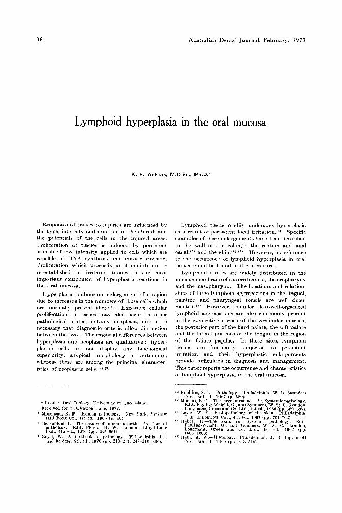
hyperplasia may occur in many areas of the oral mucosa, in this school it, has been seen most commonly in the palate (Fig. 1 ) and the tongue (Pig. 2) . Tho involved area is enlarged. The size of the wssilr enlargement, varies from a few milli- metres to several centimetres in lateral dimensions
3 9
t,ontls to support tht: history reported by h c : \ w a l paticnts that thv lrsions, although small, had hrcn presetit for prriotls of 111) to two yoars before advicc? was sought. A s with other typos of liyperplasia, these lesions appear to rmlarge until either equilibrium is established or the stimulus is withdrawn. There is no pain antl no tendency to 1i;emorrhagc. or ulccmtion has brrn noted.
Fig. I.-Two hro;rd, sinooth deeply colorired eiilar~eiiiriits Fig. 2.-YiidiiI:ir, (leepl~-i,oioiire(i &vation.; of the iiiiii'osii in the pxlat;il iiincosa separated by the mid-line :ind mi the Imtero-l:iter:tI hordw of the toiixiie. Earh norliile situated :it the poiterior of the hard pnl;ite. Srveral w:is siiiootli :nid soft. Hirips? slin\\rtl oiily w l l organized engorged sriperflcial vessels itre seen i i r the soft p:il:itr. I!.iiii~lioitl t iwieh.
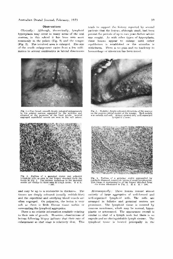
Big. S.-l'ortion of ;L germin:il centre and ;icljacent lymphoid cells as seen in the biopsy obt;iined frow the Vix. I.-Port ion of :I geriiiiri:il rrritre siuroiinded by lesioii in Fig. 1. The nncleiir variations in the geniiin:il rrgnl;irly disposed, conrriitrir 1;iyPrs of snl;ill lyrriphorytes. centre iirc siniilar t o those seen in lynipli iiodes. H & I.:. This tissue is representntive o f the biopsy olitained Irrnii
x 160. thr Icsim il1iistr;itrtl in Fie. 2 . 11 B E:. 100.
and may be up to a centimctre in thickncss. Thc tissucs are deeply coloured (usually retltlish-bluc) and the superficial and overlying blood vessels are often engorged. On palpation, the lesion is very soft as there is little fibrous tissue within o r surrounding the lymphoid aggregates.
There is no reliable informat>ion availablo rolating to their rate of growth. However, observat,ions of lesions following biopsy indicate that their rate of cmlargemenf at that, stage is rclativrsly slow. This
Microscopietrlly ; These losions consist almost entirely of large aggrogatw of well-formed antl wcll-organized lymphoid cclls. The cclls arc arranged in follicles and germinal centres art? prominent. The lymphoiil tissue is covered by mucous mctmbrano, which may be normal, hyper- plastic or attenuated. Tho appearance overall is similar to that of a lymph node but there is no capsule and no tlistingiiishablr lymph sinuses. The lymphoitl tissiw i s Iomtc~l principally in this

40 Australian Dental Journal, February, I973
small, rounded, compact structures which stain intrnsoly with basic dyes (Flc~ming's bodies) arc a constant and distirictive feature of tho gerrnitiitl centres in reactive hyperplasia. Evaluation of tho lesions diagnosed as lymphoid hypcrplasia in this laboratory has not consistently snpportctl this statement.
So recurrences have occurred in our experience. T11e lesion illustrated in Fig. 1 was sampled by incisioiinl biopsy but the patient elected to have no furthrr surgery. Tho lesion has since becri observctl without detectable change in colour, testure or sizv during the past four years.
In vicw of the aburidaiicr of lymphatic tissue in the posterior portions of t h r oral mucosa, in view of the constant low grade irritation arid stimrilatioii ( ~ f these tissues by a variety of physical, chemical ant1 microbial agents, and in vicw of thct rrc(~griizrtl rase with which lymphoid tissucv react in a pro- Iiferativc way, one woulcl cxpect hyperplasia of ttw lymphoid tissues in the oral mucosa to bc rq)ortc,cl more commonly.
The lesions respond well to surgical excision.
Summary Lymphoi(l tissuw readily untlcrgo hypc~pliisili
as a result of persistent local irritation. Such a responsc occurs occasionally iii tho oral I I I I IC~SD, particularly the palate antl t,hc tongur. Clinically, the lesions are raised, deeply colourrtl, very soft, slowly gruwing and painless. Microscopically, thcrc. is an abundance of lymphoid tissue with numerous prominent germinal crntrrs surrountlrtl by COII-
centric layers of lymphocytrs. Thtm is a strong resemblance to follicular lymphoma histologically. Similar lesionr, have bern reportrd in the colon. rectum antl skin. They rwpontl wrll to surgical c,scision.
University of Qurrnslantl Dental School,
Hrishancs. Q'laml, 4000. Trirbot, Strrct,
siibrnucosa an(1 thc t l e t p r parts of thtx lamina propria. which normally occur thrrcs, such as mucous glands, bnndles of periyhivral nwvcs ant1 Itlrgc. blood v(~ssc~ls.
Thc germinal ciwtros arc quito largcs aiicl may bc the most striking fraturra. 'I'hiy appear palcly stained and their compoiiciit, cc~lls art' similar t o those normally prwerit in such ccntrix (Pig. 3 ) . .-\tklitionally. frt'e macrophag(%. somr coiitiiiiinig ingestcd material, art' frcqucwtly s w n . ' ~ h c germinal crntrrs arv surrcJuntlixl h y ~~egrtlarly tlispoactl, conccntric Iayrrs of small lymphocyt(.s (Fig. 4).
I t is intwsporscd among tho
Discussion A niimhrr of tvrms httvr bccw usctl i n tlcwxibing
tho occurivncv of massi's of lymphoi(l tissrw in thr wall t ~ f tht. Iargt: intc%stinc, ant1 thv skin. 'I'hvsc. inclritlv benign lymphomas."l lymphoitl p ~ l y p , ' l ~ ~)sc~iidolympti(~ma,'"' ancl lyniphocytic granriloma.'hl [n all caws thcn. has hcvn argcvmrnt, that thv lesions are foci of srtbmricosal lymphoid hyp(~rp1asias rat,hcr than t,rric ricwplanms.
It may bc difficult, in sonic caws to tli,t,iriguish o i i
histological grounds brtwcon lymphoid hypwplasius and woll-tliffrrrntiatc~rI t ruc lymphomas which may involve similar areas. ' I ' h c a wc.ll-dixcil)linecl archi- tecture of the hyperplastic Iwions, tho ortlorly activit,y ancl appearanco of thv gorrnirial centrrs t,ogether with their lack ofcollular atypia arc usually dist)inctivc%. Howcvor, closc similarity mists bt!twcen lymphoid hyprrplasia arid f~illici~lar lymphoma ant1 distinction bc-twrm the two hist,nlogically frequcntly hinges on asswsmrnt of thc gvrminal centres. Symmers(I"l statetl thttt inacrophagw containing