lymphatic system: overview figure 19.1a. lymphatic system: overview consists of three parts –a...
TRANSCRIPT
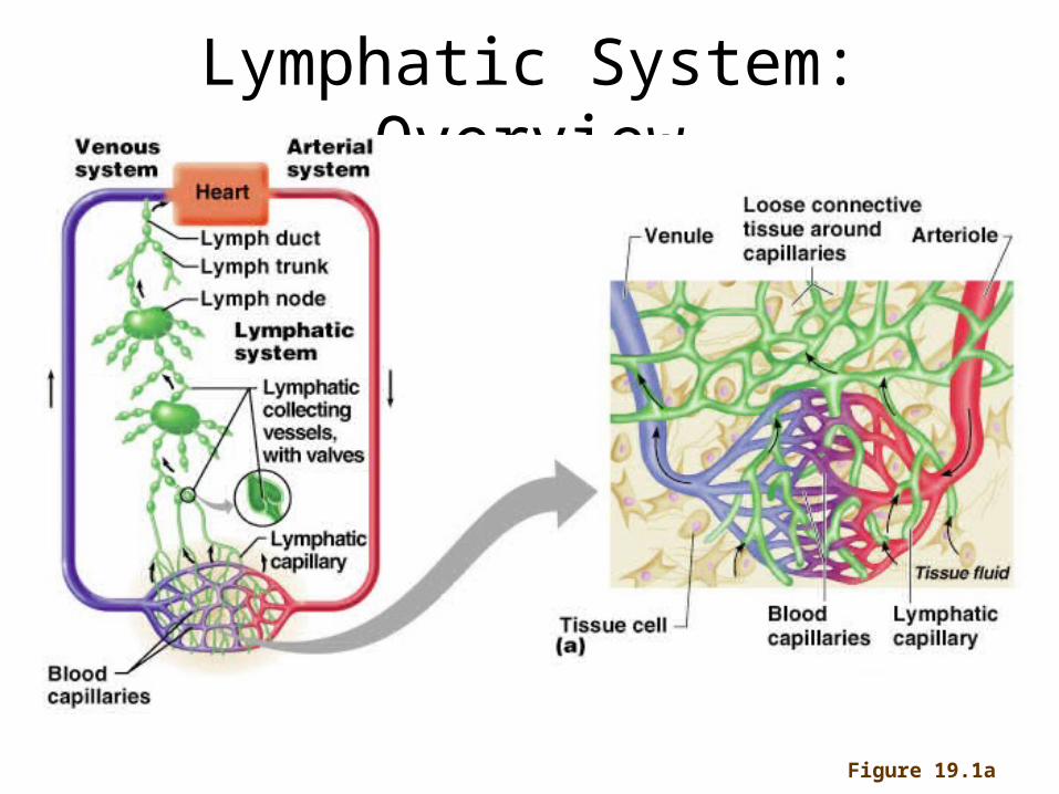
Lymphatic System: Overview
Figure 19.1a

Lymphatic System: Overview
• Consists of three parts– A network of lymphatic vessels– Lymph nodes scattered throughout the body– Lymph: interstitial fluid once it has entered
lymphatic vessels
• Returns interstitial fluid and leaked plasma proteins back to the blood
• Plays a role in the immune system• Absorption of fat from the digestive system

Lymphatic Vessels
• A one-way system in which lymph flows toward the heart
• Lymph vessels include:– Permeable, blind-ended capillaries– Lymphatic collecting vessels– Trunks and ducts (ducts are the largest)

Lymphatic Collecting Vessels
• Have the same three tunics as veins, but with thinner walls
• More internal valves than veins
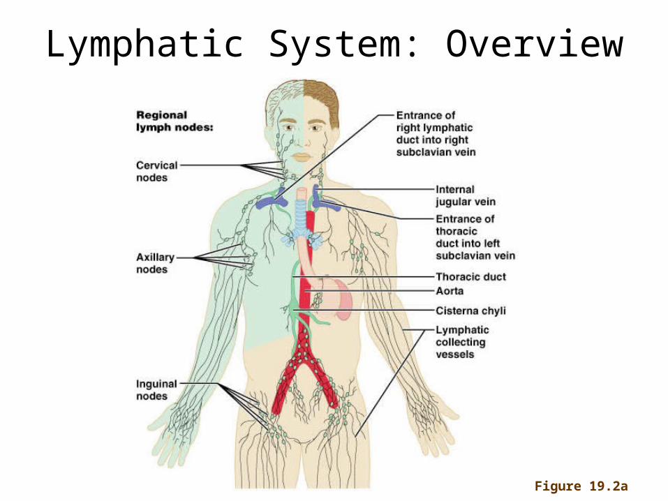
Lymphatic System: Overview
Figure 19.2a

Lymphatic Ducts
Entrance of thoracic duct into left subclavian vein
Internal jugular veins
Thoracic duct
Right lymphatic duct
Superior vena cava
Cisterna chyli
Right subclavian vein

Lymph Nodes
• Nodes are bean shaped and surrounded by a fibrous capsule
• Their two basic functions are:– Filtration: macrophages destroy microorganisms and debris
as lymph flows through the lymph nodes– Immune system activation: lymphocytes monitor antigen
presence and mount an attack against them

Lymph Node Structure DON’T NEED TO KNOW ALL DETAILS
Figure 19.4a, b

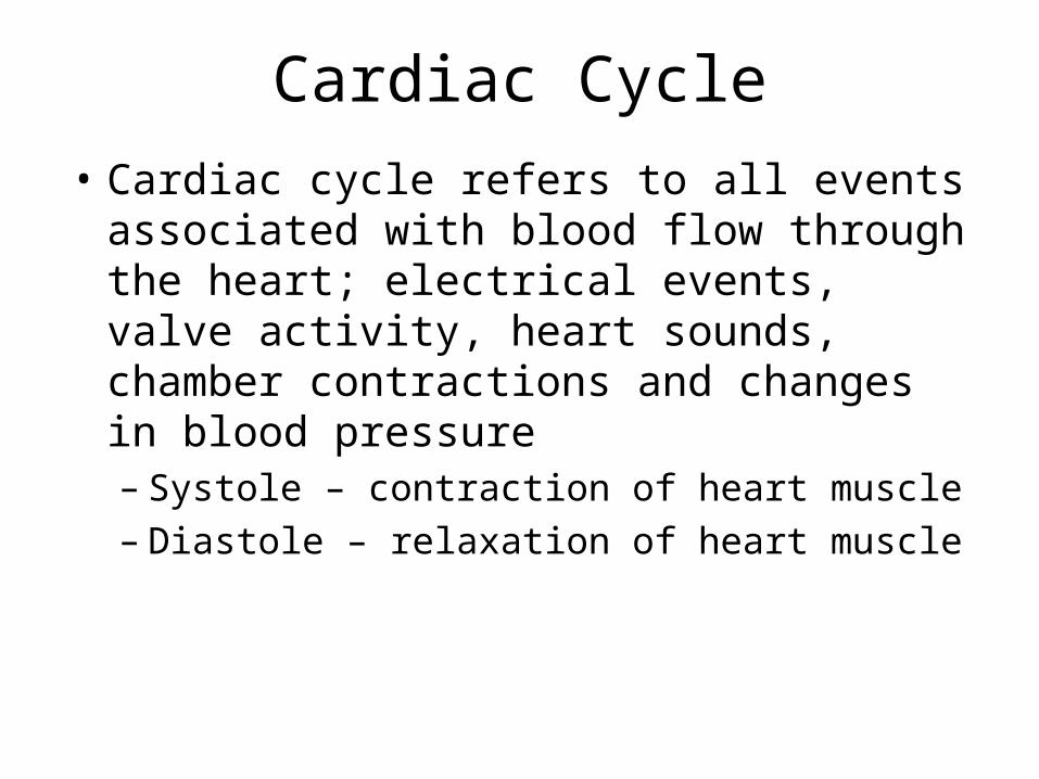
Cardiac Cycle
• Cardiac cycle refers to all events associated with blood flow through the heart; electrical events, valve activity, heart sounds, chamber contractions and changes in blood pressure– Systole – contraction of heart muscle– Diastole – relaxation of heart muscle
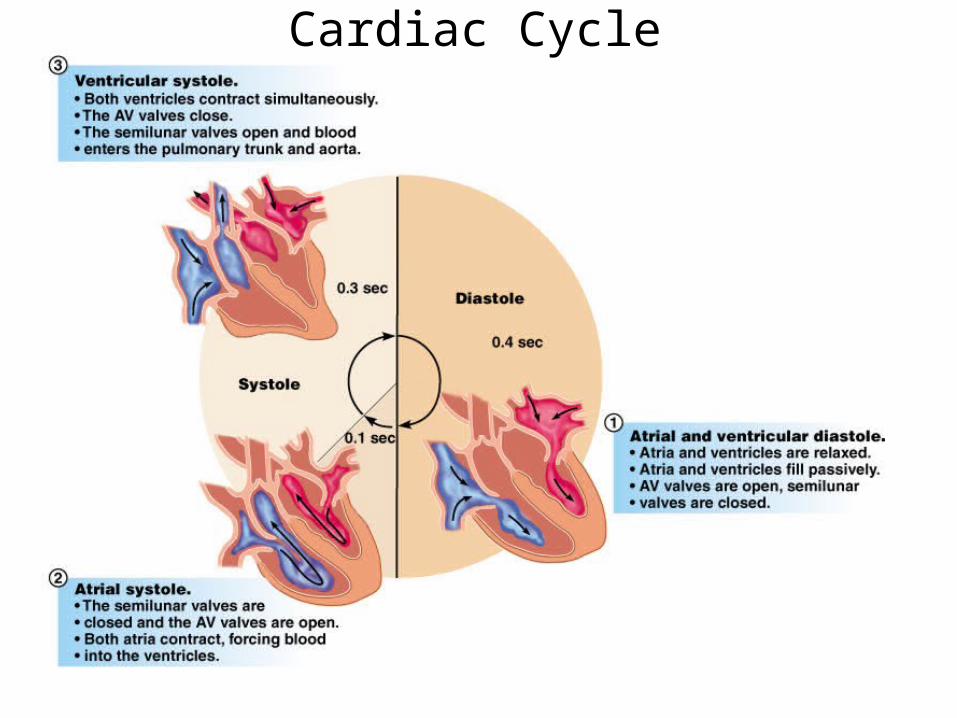
Cardiac Cycle
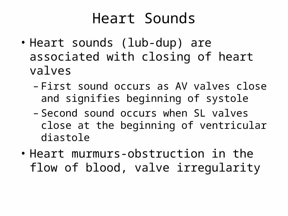
Heart Sounds
• Heart sounds (lub-dup) are associated with closing of heart valves– First sound occurs as AV valves close and
signifies beginning of systole– Second sound occurs when SL valves close
at the beginning of ventricular diastole
• Heart murmurs-obstruction in the flow of blood, valve irregularity
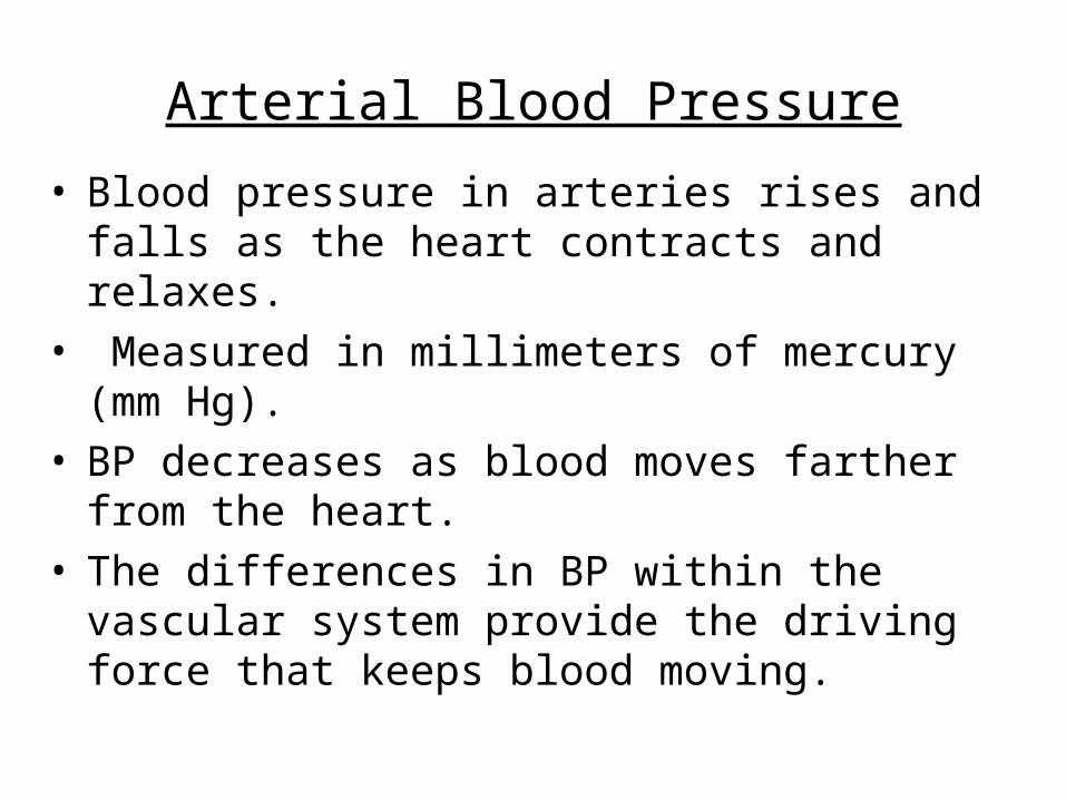
Arterial Blood Pressure
• Blood pressure in arteries rises and falls as the heart contracts and relaxes.
• Measured in millimeters of mercury (mm Hg).• BP decreases as blood moves farther from the
heart.• The differences in BP within the vascular system
provide the driving force that keeps blood moving.
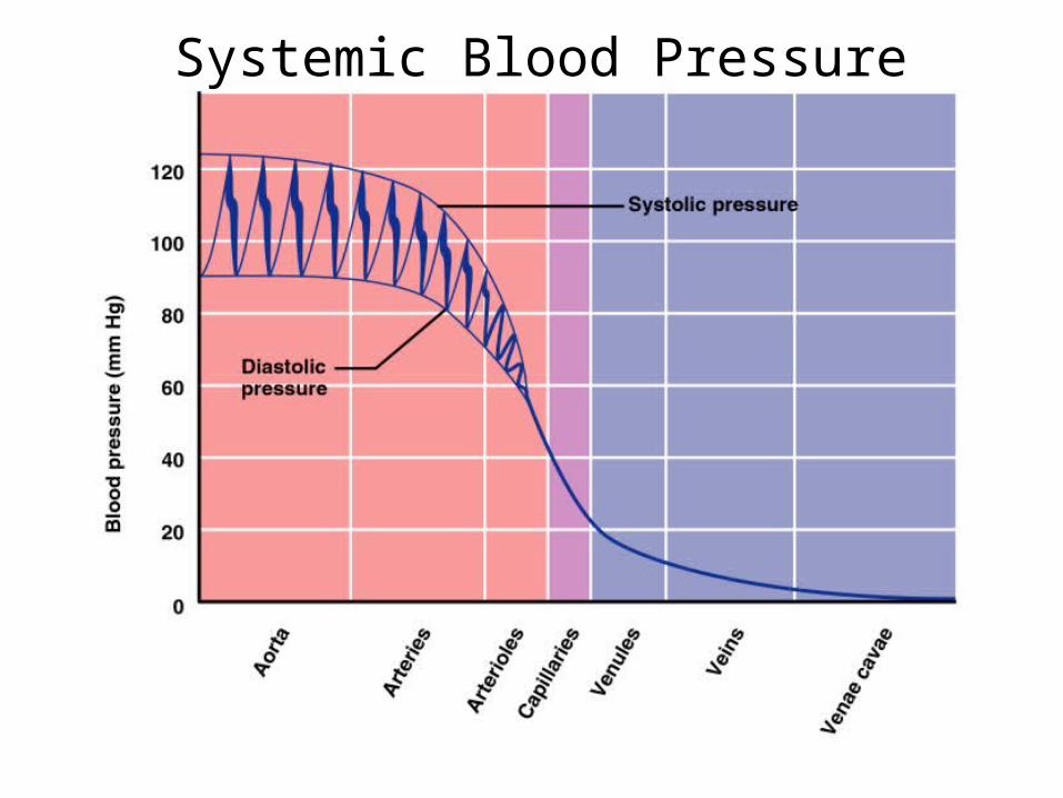
Systemic Blood Pressure

Phases of the Cardiac Cycle
EDV = end diastolic volume; ESV = end systolic volume; SV = stroke volume.
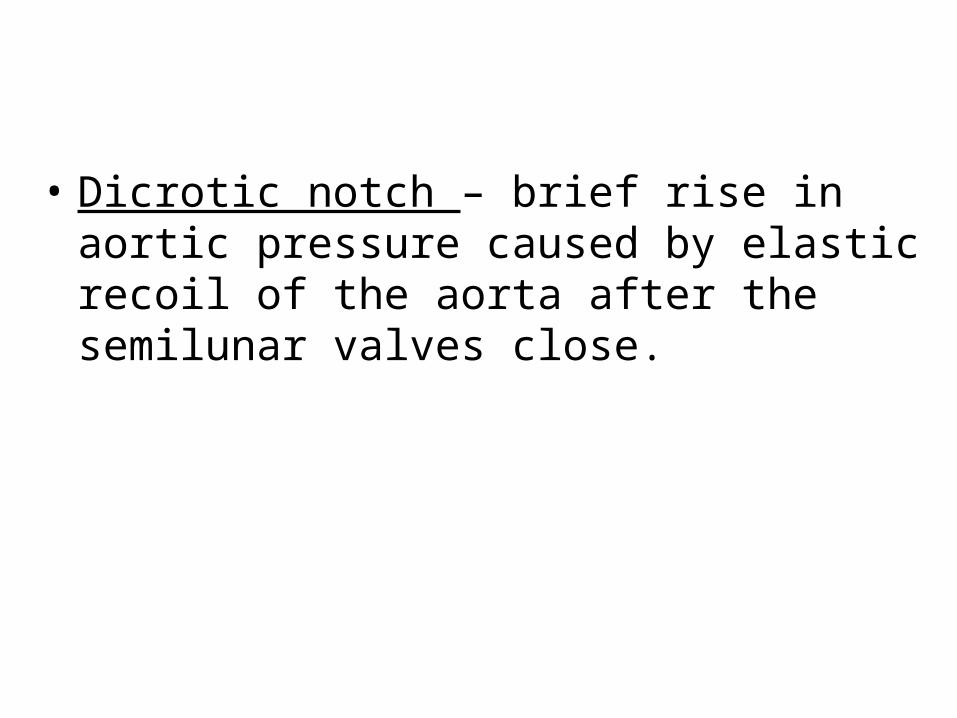
• Dicrotic notch – brief rise in aortic pressure caused by elastic recoil of the aorta after the semilunar valves close.

Measuring Blood Pressure• Measured indirectly with the auscultatory method• A sphygmomanometer’s inflatable cuff is placed on
the arm superior to the elbow• Pressure is increased in the cuff until it is greater
than systolic pressure in the brachial artery• Pressure is released slowly and the examiner
listens with a stethoscope• The first sound heard is recorded as the systolic
pressure• The pressure when sound disappears is recorded
as the diastolic pressure• What causes the sounds?

Copyright © 2009 Pearson Education, Inc.

Normal Variations in Blood Pressure
• Blood pressure cycles over a 24-hour period, and peaks in the morning.
• Factors such as age, sex, weight, race, mood, posture, and physical activity may also cause BP to vary.

Alterations in Blood Pressure
• Hypotension – low BP in which systolic pressure is below 100 mm Hg
• Hypertension – sustained arterial pressure of 140/90 or higher– Transient elevations are normal and can be
caused by fever, physical exertion, and emotional upset
– Chronic elevation is a major cause of heart failure, vascular disease, renal failure, and stroke
Maintaining Blood Pressure
• The main factors influencing blood pressure are:– Cardiac contraction– Peripheral resistance– Blood volume
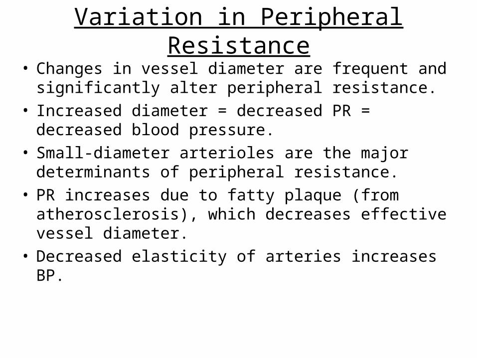
Variation in Peripheral Resistance
• Changes in vessel diameter are frequent and significantly alter peripheral resistance.
• Increased diameter = decreased PR = decreased blood pressure.
• Small-diameter arterioles are the major determinants of peripheral resistance.
• PR increases due to fatty plaque (from atherosclerosis), which decreases effective vessel diameter.
• Decreased elasticity of arteries increases BP.

Copyright © 2009 Pearson Education, Inc.
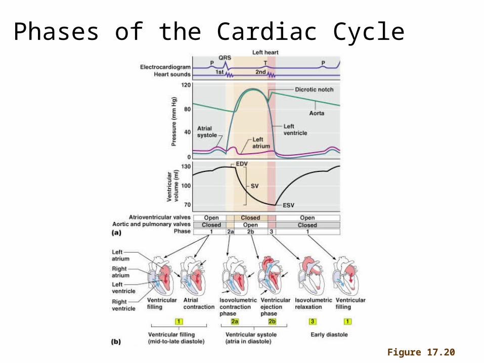
Phases of the Cardiac Cycle
Figure 17.20