lymphatic filariasis - …mansouramanchester2019grads.weebly.com/uploads/4/7/...filariasis results...
TRANSCRIPT

1

LYMPHATIC FILARIASIS (ELEPHANTIASIS)
Dr. Samar N. El-Beshbishi
Ass. Professor of Medical Parasitology
2

OBJECTIVES
1. Overview on lymphatic filariasis (L.F.)
2. Geographical distribution of L.F.
3. Species of filarial parasites
4. Morphology of Wuchereria bancrofti & Brugia malayi
5. Life cycle
6. Pathogenicity
7. Clinical manifestations of L.F.
8. Diagnosis of L.F.
9. Treatment
10. Control
3

1. OVERVIEW
-Infection by 2 Nematodes:
1. Wuchereria bancrofti
2. Brugia malayi
• Transmitted by the bite of infected mosquito.
• All the parasites have similar life cycle.
• Adults seen in lymphatic vessels.
• Microfilaria (pre-larva) seen in peripheral
blood during night.
4

2. DISTRIBUTION OF LYMPHATIC FILARIASIS
-World wide in tropical and sub-tropical regions of:
1. Africa
2. Asia
3. Western Pacific
4. Parts of central & south America
5

6

3. SPECIES OF FILARIAL PARASITE
7
Parasite Insect vector Disease
W. bancrofti Culex, Anopheles & Aedes L.F.
B. malayi Mansonia, Anopheles & Aedes L.F.
O. volvulus Simulium River
Blindness
L. Loa Chrysops flies S.c.
swellings
M. perstans Culicoides Serous
cavity
M. ozzardi Culicoides Serous
cavity

8
Adult worm Microfilaria
4. Morphology of Wuchereria bancrofti
300X10 µ
Male: 4 cm X 0.2 mm Female: 8 cm X 0.4 mm

9
5. Life Cycle

• Habitat: Adults in lymph nodes and lymph vessels draining the lower limbs. Microfilariae in the peripheral blood.
• Definitive host: Man.
• Intermediate host: female Culex, Anopheles, Aedes.
• Reservoir host: No.
• Infective stage: filariform larvae in the mouth of the infected mosquitoes.
• Mode of infection: through the skin during the bite of mosquito.
10

Pathology of Lymphatic Filariasis
• The pathology associated with lymphatic
filariasis results from a complex interplay of
the pathogenic potential of the parasite,
the tissue response of the host and external
bacterial and fungal infections. Most of the
pathology associated with LF is limited to
the lymphatics.

1.Microfilariae seem to be non-pathogenic, but may be associated with granuloma reactions.
2. Dilatation of lymphatic vessels: due to the
direct inflammatory reaction initiated by the toxic effect of worms or their products.
3. Lymphangitis: dilated, inflamed and thickened
lymphatic vessels associated with erythema, oedema and tender painful areas.
12
6. PATHOGENICITY

4. Lymphadenopathy: the lymph nodes affected are enlarged, tender, matted due to fibrosis, necrosis, with obstruction of the proximal lymphatic vessels.
5. Obstruction of lymph flow, caused by : a. Fibrosis of lymph nodes. b. Worms in the lumen of lymphatic vessels. c. Endothelial proliferation and thickening of
lymphatic vessels. d. Recurrent secondary bacterial lymphangitis.
13

6. Elephantiasis: due to obstruction and fibrosis of lymph nodes and lymph vessels. The affected tissues are oedematous and the covering skin becomes thickened and highly stretched.
7. Genital lesions: epididymo-orchitis and hydrocele. Microfilariae may be detected in the hydrocele fluid.
14

8. Rupture of lymphatic vessels results in:
a. Chyluria: Rupture of obstructed lymphatic vessels of the kidney leads to the passage of lymph into the urine.
b. Chylocele: When the varicose lymph vessels rupture in the tunica vaginalis.
c. Chylous ascites: When the varicose lymph vessels rupture in the peritoneal cavity.
15

9. Tropical pulmonary eosinophilia (TPE, diffuse filarial lung disease, occult filariasis): is due to hypersensitivity reaction to microfilarial antigens. The lung tissues show chronic interstitial fibrosis with destruction of microfilariae in the pulmonary vasculature.
-Microfilariae are not found in the peripheral blood and the classical clinical manifestations of filariasis are absent.
16

7. CLINICAL MANIFESTATIONS
• Clinical incubation period: 8-16 months
• Manifestations are 2 types:
1. Lymphatic Filariasis (Presence of Adult worms).
2. Occult Filariasis (Immuno- hyper responsiveness).
17

1. Lymphatic Filariasis
• There are 3 stages:
A. Asymptomatic microfilariaemic stage.
B. Stage of Acute manifestation.
C. Stage of Obstructive (Chronic) lesions.
18

A. Stage of asymptomatic microfilarimia:
• Occurs in endemic areas. Considerable proportions are asymptomatic for months and years.
• They have circulating microfilariae.
• They are an important source of infection.
• They can be detected by night blood survey.
19

B. Stage of acute manifestations:
• During initial months and years, there are recurrent episodes of acute inflammation in the lymph vessel/node of the limb & scrotum.
• Clinical manifestations are consisting of:
1. Fever
2. Lymphangitis
3. Lymphadenitis in the groin and axilla
4. Epididimo-orchitis
20

C. Stage of chronic manifestations:
Obstructive lesions takes 10-15 years. Permanent damage to the lymph vessels. Recurrent inflammatory episodes around the parasite.
Oedema of the affected part, it becomes hard, rough, tender with thick, rough, stretched and fissured overlying skin.
Elephantiasis of scrotum, penis, vulva, breast, leg, arm.
Hydrocele, chyluria, chylothorax, chylous ascites, chylocele, chylous diarrhea.
21
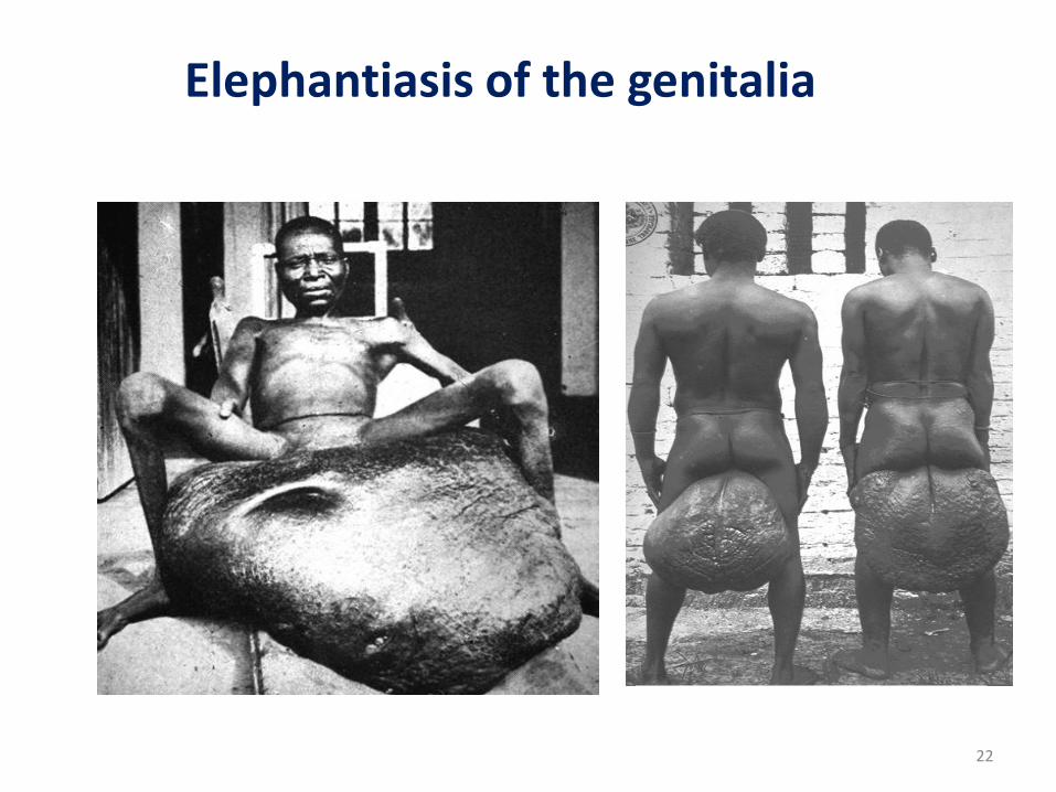
Elephantiasis of the genitalia
22

23
Elephantiasis of L.L.

Elephantiasis of breast and arm
24

Classification of Lymphoedema
• Lymphoedema is classified into 7 stages on the basis of the presence or absence of:
1. Oedema
2. Folds
3. Knobs
4. Mossy foot
5. Disability
25

Stage I:
• Swelling reverses at night
• Skin appearance:
normal
26
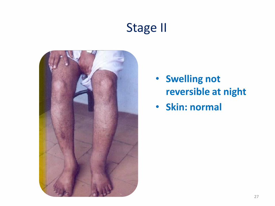
Stage II
• Swelling not reversible at night
• Skin: normal
27

Stage III:
• Swelling not reversible at night
• Skin folds: shallow
• Skin appearance: normal
28

Stage IV:
• Swelling not reversible at night
• Skin folds: shallow
• Appearance of skin:
- Irregular,
- Knobs & nodules
29

Stage V:
• Swelling not reversible at night
• Skin folds: deep
• Appearance of skin: smooth or irregular
30

Stage VI:
• Swelling not reversible at night
• Skin folds: absent, shallow or deep
• Appearance of skin Wart-like lesions on foot or top of the toes
31

Stage VII:
• Swelling not reversible at night
• Skin folds: deep
• Appearance of skin: irregular
• Needs help for daily activities as walking, bathing, using bathrooms
32

2. Occult Filariasis (TPE)
• Occult filariasis is believed to be the result of hyper responsiveness to microfilaria antigens.
• Patients present with paroxysmal cough, wheezes, low grade fever, scanty sputum with occasional haemoptysis.
• Eosinophilia.
• X-ray shows diffused nodular lesions.
33

8. DIAGNOSIS OF LYMPHATIC FILARIASIS
I. Clinical diagnosis.
II. Laboratory diagnosis:
A. Direct:
1.Demonstration of microfilariae in the peripheral blood:
a. Direct fresh smear.
b. Giemsa-stained thick blood film.
34

35
W. b. microfilaria

c. Concentration of microfilariae (Knott s method): 2 ml of blood are mixed with 10 ml of 2% formalin, let to stand for 10 minutes, centrifuge, and examine the sediment.
d. DEC provocative test (2mg/Kg): m.f. enters the peripheral blood in day time within 30 - 45 minutes.
36

Important points during examination:
-M.F. appear after a year or more after infection.
-M.F. are rarely found when lymphatics are
obstructed.
-Blood must be collected at night (10 PM-2 AM).
-M.F. are more in capillary than venous blood.
-M.F. are more in blood collected from the ear
lobe than finger.
37

2. Detection of microfilariae in the chylous urine or from fluid aspirated from hydrocele and peritoneal cavity: collect 10-20 ml of morning urine, add 2 ml ether, centrifuge and examine the sediment.
3. Detection of adult worms:
- Lymph node biopsy
- X-ray to detect dead calcified worm.
- Ultrasonography: can locate and visualize the movements of living adult worms in the lymphatics “Filaria dance sign”.
38

4.Immuno chromatographic test (ICT): Antigen detection assay can be done by Card test and through ELISA.
5. X-ray: is helpful in the diagnosis of Tropical pulmonary eosinophilia.
-Picture will show interstial thickening, diffused nodular lesions.
39

.B. Indirect diagnosis:
1. Immunodiagnosis:
-It is of value in incubation period, chronic infections and TPE.
-Serum antibodies like IgG & IgE will be extremely high and the presence of IgG4 antibodies indicate active infection.
-Test used are: IFAT, CFT and ELISA.
2. Haematology: increased eosinophil count.
40

9. TREATMENT OF LYMPHATIC FILARIASIS
A. Chemotherapy repeated every 6 months
till m.f. or symptoms dissapear.
B. Surgical management.
A. Chemotherapy of filariasis:
1. Diethyl Carbamazine citrate (DEC)
2. Ivermectin
3. Albendazole
41

1. Diethyl Carbamazine Citrate (Hetrazan, Banocide): • Very effective against m.f. (Microfilariacidal).
• Repeated courses can kill adult worms.
• Dose: 2mg/Kg/2 weeks.
• Adverse reactions are mostly due to the rapid destruction of m.f. which is characterised by fever, nausea, myalgia, sore throat, cough, headache.
• Drug of choice in the treatment of TPE.
42

2. Ivermectin:
• Very effective against m.f. (Microfilariacidal).
• Lowers m.f. level even in single dose of 200µg – 400µg/Kg.
• No action on TPE.
• Adverse reactions are less but similar to that of DEC.
• Microfilariae reappears faster than in treatment with DEC.
43

3. Albendazole:
• This antihelmenthic kills adult worms.
• No action on microfilariae.
• Dose: 400mg/twice daily for 2 weeks.
• With combination of DEC & Ivermectin, it enhances the action of the drugs.
• It induces severe adverse reactions due to the death of adult worms.
44

B. Surgical Treatment:
• Hydrocele: excision
• Scrotal elip: surgical removal of skin & tissue, preserving penis and testicles.
• Lymphoedema (Elephantiasis):
-Excision of redundant tissue, subcutaneous and fatty tissues.
-Postral drainage and physiotherapy.
45

• Treatment and prevention of lymphoedema and elephantiasis
-Early treatment may destroy the adult worms and prevent the later development of lymphoedema.
-Once lymphoedema is established there is no cure and the “foot care programme” may offer relief and prevent acute attacks thus preventing further progression of the swelling.

10. CONTROL OF LYMPHATIC FILARIASIS
1. Treatment of infected patients. 2. Vector control. 3. Health education in endemic areas. 4. Environmental sanitation.
47

Brugia malayi (Wuchereria malayi)
Morphology: 1. Adult: the same. 2. Microfilaria
48
B. malayi W. bancrofti
250 x 5 300 x 10 Size
Irregular Smooth Body curves
Loose Loose and redundant Sheath
Anterior end with 2 stylets
Blunt anterior end Ends
Swollen with 2 nuclei
Tapering without nuclei
Posterior end
8 pm-4 am 10 pm-2 am Nocturnal periodicity

49
W. bancrofti. and B. malayi microfilaria

Life cycle: the same except for
• Habitat: Adults in lymph nodes and lymph vessels draining the upper part of the body (upper limb, axilla, breasts and thorax).
• Intermediate host: female Mansonia, Anopheles, Aedes.
• Reservoir host: cats and monkeys.
50

Pathogenicity and clinical manifestations:
Malayan filariasis is the same as bancroftian filariasis except for:
-Elephantiasis affects the breasts and arms mainly.
-Genitalia are rarely affected and chyluria is
uncommon. Diagnosis, treatment and control: as for W.
bancrofti, plus control of reservoir hosts.
51

THANK YOU