lumbopelvic instability: a key in injury risk among pre ... · acknowledgment is made in the thesis...
TRANSCRIPT

Lumbopelvic instability: A key in injury risk among pre-
elite youth athletes?
A thesis submitted in fulfilment of the requirements
for the award of the degree
Doctorate of Philosophy
from
Charles Sturt University
by
Kerry Mann
BExSc (Rehab), BExSc (Hon)
School of Exercise Science, Sport and Health
March 2018

i
Dedication
I dedicate this thesis to my beautiful little boy, Kobey. Although you only came along half way
through my Ph.D., you were the light to keep me going through the hard times. You became
my motivation to finish to make the best possible life I could for you. I love you more than
words can explain! Love Mummy.

ii
Declaration
I Kerry Mann, hereby declare that this submission is my own work and that, to the best of my
knowledge and belief, it contains no material previously published or written by another person
nor material which to a substantial extent has been accepted for the award of any other degree
or diploma at Charles Sturt University or any other educational institution, except where due
acknowledgment is made in the thesis “Lumbopelvic stability: The primary cause of lower
limb injuries in pre-elite youth athletes?”. Any contribution made to the research by
colleagues with whom I have worked at Charles Sturt University or elsewhere during my
candidature is fully acknowledged. I agree that this thesis be accessible for the purpose of study
and research in accordance with the normal conditions established by the Executive Director,
Library Services or nominee, for the care, loan and reproduction of theses.
Kerry Mann
23rd March 2018

iii
Publications and Conferences
Mann, K., O’Dwyer, N., Bird, S., & Edwards, S. (2017). Assuming rater reliability of
a movement competency screen–Is it true?. Sports Medicine Australia Conference,
Langkawi, Malaysia, Oct 27-30.
Mann, K., O’Dwyer, N., Bird, S., & Edwards, S. (2018). The reliability of a movement
competency screen for injury risk identification. To be submitted to Sports Medicine
Mann, K., O’Dwyer, N., Burton, M., & Edwards, S. (2018). Chapter 1. Stop-jump
landing technique is modified by neuromuscular training programs in pre-elite youth
athletes. To be submitted to Medicine and Science in Science in Sport and Exercise.
Mann, K., O’Dwyer, N., Burton, M., & Edwards, S. (2018). Chapter 1. Any type
of neuromuscular training program can modify pre-elite youth athletes reactive change-
of-direction technique. To be submitted to American Journal of Sports Medicine.
Mann, K., O’Dwyer, N., Burton, M., & Edwards, S. (2018). Chapter 1. The effect
of dynamic trunk-pelvis range of motion on lower limb injury risk factors in pre-elite
youth athletes. To be submitted to Medicine and Science in Science in Sport and
Exercise.

iv
Acknowledgement
I would like to acknowledge all the following people who made this thesis possible.
Firstly to my family, who has always been there for me through the good and bad times. Your
love and support are what helped me to keep going and always strive for my best.
In completing this thesis, I would like to sincerely thank my primary supervisor, Dr Suzi
Edwards, for the enormous amount of time, support and knowledge she bestowed upon me
during my studies. I have such a high regard and respect for you, and I thank you for all your
wisdom and knowledge you have provided me. I could not have completed my Ph.D. without
you, from the bottom of my heart, thank you!
I also wish to thank my associate supervisors Associate Professor Nicholas O’Dwyer and Dr.
Micheala Burton. You both have an amazing wealth of knowledge you instilled in me and I
thank you for sharing it with me. You have helped me develop my writing and the way I look
at research. Thank you for everything.
To the staff in the School of Exercise Science Sport and Health and the Faculty of Science
Laboratory Technician Staff, thank you for your encouragement and support throughout the
duration of my Ph.D. You have all played a role in making my Ph.D. the amazing, unforgettable
experience it has been, and for that, I am truly grateful.
During my immense data collection, I received invaluable help from my research assistants;
Blake, Chris, and Josh. You boys helped keep me sane during over 200 hours which were spent
in the Biomechanics laboratory over a 3 month period. You boys rock!
A massive thank you to all the novice and expert raters involved in study 1. You all gave up
massive amounts of your time to help me to complete my study and without your help, I would
not have been able to complete that component of my thesis.

v
I am also extremely grateful to the Western Region Academy of Sport and Hunter Academy of
Sport athletes, and their parents and coaches who were involved in this study, some of which
travelled over three hours to be involved. Without your participation, this study would not have
been possible.
To my fellow Ph.D. candidates, thank you for always being there for support and advice.
During the hard times, you were the ones who were always there to give an encouraging word,
some constructive advice or some compassionate understanding. They say in hard times true
friendships blossom, this couldn’t be truer with each of you.
Lastly, but certainly not least, a huge thank you to my beautiful little boy Kobey. From the
moment you were born you filled my heart with happiness. You became such a driving force
to complete this thesis, to show you never to give up on your dreams and that hard work pays
off. I hope that as you grow up you too will work hard and shoot for the stars.

vi
Abstract
Background
Sporting injuries can result in substantial financial and physical detriments to an athlete,
including pain, loss of game and/or training time, costly rehabilitation and/or medical attention,
and even cause long-term disability. To reduce these substantial injury rates, many sporting
teams have adopted movement screening tools for injury risk management purposes. This early
identification of athletes with “poor” movement competency, can then allow intervention
strategies to be implemented, potentially preventing the injury from occurring and thus
reducing the injury rate seen among the sporting population. Implementation of neuromuscular
training interventions programs in athletes at increased risk of sporting injuries have been
widely utilised. However, the current local focus on injury prevention at specific joints, when
compared to a focus on the global kinetic chain, is less effective, as evidenced by high rates of
re-injury. Research suggests that lumbopelvic stability may be critical in the prevention of
lower limb injuries in athletes; however, it is unknown how much of an effect lumbopelvic
instability has on lower limb injury risk.
Thesis Aim
The first purpose of this thesis was to determine the inter- and intra-rater reliability of a field-
based movement screen in novice and expert raters using different viewing methods, and
evaluate the presence of a familiarisation effect (Manuscript 1). A second aim of this thesis
was to determine the effect of different 12-week intervention programs to modify a stop-jump
(Manuscript 2) and reactive change-of-direction (R-COD) (Manuscript 3) in pre-elite youth
athletes. A third aim of this thesis was to determine the effect, if any, trunk abdominal segment
relative to the pelvis segment (trunkab-pelvis) range of motion had on lower limb injury risk
factors (Manuscript 4).
Methods
For Manuscript 1, 55 pre-elite youth athletes performed a movement screen on three separate
occasions and videos of their performance were rated three times in randomised order by 18
raters. Reliability was established using inter- and intra-rater reliability of novice and expert
raters, with learning effects and familiarisation measured across repeated exposure of both
raters and athletes, respectively. Within Manuscript 2 to 4, eighty-nine junior pre-elite athletes
with no current signs or symptoms of injury were recruited from the Western Region Academy
of Sport. Biomechanical analysis of five successful stop-jump and five R-COD experimental
tasks were completed both before and after exposure to one of three different 12-week training
programs, or the control program, in conjunction with a strength and conditioning program for
each participant. Mixed effect repeated measures analysis of variance (ANOVA) was used to
determine statistically significant between-group differences (P≤0.05).
Major Conclusions
Results of this study indicate that movement screening experience does not affect rater
reliability; however, familiarisation is required for athletes performing the movement
screening. Total movement screening score can be reliably used to determine movement
competency; however, individual movements scores should not be relied on. When
implementing strength and conditioning programs in pre-elite youth athletes to modify jump-
landing technique, a simple strength and conditioning program comprising of four exercises
with limited equipment required, can be implemented in pre-elite youth athletes with poor
movement competency to modify their landing patterns. As a result of these interventions,
athletes displayed a more upright trunkab-pelvis segment at landing that was not shown to alter

vii
the risk of lower limb injury; however, it is unknown whether this movement strategy is
beneficial or detrimental to athletic performance. This current study failed to identify links
between trunkab-pelvis ROM, injury risk and/or performance. It is suggested that further
research is needed to determine how trunk ROM during landing tasks affects these factors
influence lower limb injury risk and athletic performance.

viii
Table of Contents
Dedication .................................................................................................................................. i
Declaration ............................................................................................................................... ii
Publications and Conferences ............................................................................................... iii
Acknowledgement ................................................................................................................... iv
Abstract ................................................................................................................................... vi
Table of Contents ................................................................................................................. viii
List of Tables ............................................................................................................................ x
List of Figures ......................................................................................................................... xi
Abbreviations……………………………………………………………………………… xiv
Chapter 1. The Problem ....................................................................................... 1
1.1 Introduction .......................................................................................... 1
1.2 Statement of the Problem ................................................................... 19
1.3 Research Aims ................................................................................... 19
1.4 Research Hypotheses ......................................................................... 20
1.5 Limitations ......................................................................................... 21
1.6 Delimitations ...................................................................................... 22
1.7 References .......................................................................................... 22
Chapter 2. The inter- and intra-rater reliability and learning effect of a
movement dysfunction screen for pre-elite youth athletes in
novice and expert raters .................................................................. 31
2.1 Introduction ........................................................................................ 32
2.2 Methods .............................................................................................. 37
2.3 Results ................................................................................................ 41
2.4 Discussion .......................................................................................... 49
2.5 Conclusions ........................................................................................ 53
2.6 Acknowledgements ............................................................................ 53
2.7 References .......................................................................................... 53
Chapter 3. Stop-jump landing technique is modified by neuromuscular
training programs in pre-elite youth athletes ................................ 61
3.1 Introduction ........................................................................................ 62
3.2 Methods .............................................................................................. 66
3.3 Results ................................................................................................ 74
3.4 Discussion .......................................................................................... 78

ix
3.5 Conclusions ........................................................................................ 84
3.6 Acknowledgements ............................................................................ 85
3.7 References .......................................................................................... 85
Chapter 4. Any type of neuromuscular training program can modify pre-
elite youth athletes reactive change-of-direction technique ......... 94
4.1 Introduction ........................................................................................ 95
4.2 Material and Methods ........................................................................ 98
4.3 Results .............................................................................................. 105
4.4 Discussion ........................................................................................ 108
4.5 Aknowledgements ............................................................................ 113
4.6 References ........................................................................................ 113
Chapter 5. The effect of dynamic trunk-pelvis range of motion on lower limb
injury risk factors in pre-elite youth athletes .............................. 120
5.1 Introduction ...................................................................................... 121
5.2 Methods ............................................................................................ 125
5.3 Results .............................................................................................. 129
5.4 Discussion ........................................................................................ 132
5.5 Conclusions ...................................................................................... 135
5.6 Acknowledgements .......................................................................... 135
5.7 References ........................................................................................ 135
Chapter 6. Summary, Conclusions and Recommendation for Future
Research .......................................................................................... 141
6.1 Summary of Conclusions ................................................................. 141
6.2 Conclusions ...................................................................................... 147
6.3 Recommendation for Future Research ............................................. 148
Appendices ........................................................................................................................... 149
Appendix 1 – Ethics ............................................................................................................. 150
Appendix 2 - Movement Screen Information .................................................................... 152
Appendix 3 – Intervention Programs. ............................................................................... 159
Appendix 4 – Effect of lower limb dominance during stop-jump in pre-elite youth
athletes. ................................................................................................................................. 171
x
List of Tables
Table 1-1 Definitions of lumbopelvic control. ...................................................................... 3
Table 2-1 Intra-rater Reliability of Session 1 Only - Novice Raters. .................................. 43
Table 2-2 Intra-rater Reliability of Session 1,2,3 - Novice Raters. ..................................... 44
Table 2-3 Inter- and Intra-rater Reliability of Session 1 Only - Expert Raters. .................. 45
Table 2-4 Inter- and Intra-rater Reliability of Session 1,2,3 - Expert Raters. ..................... 46
Table 2-5 Inter-rater Reliability of Session 1 Only - Novice Raters. .................................. 47
Table 2-6 Inter-rater Reliability of Session 1,2,3 - Novice Raters. ..................................... 48
Table 3-1 Descriptive characteristics of pre-elite youth athletes. ....................................... 67
Table 3-1 Descriptive characteristics of pre-elite youth athletes. ....................................... 99
Table 3-2 Kinematic changes between pre- and post-intervention in a stop-jump movement.
………………………………………………………………………………….76
Table 3-3 Kinetic changes between pre- and post-intervention in a stop-jump movement.
………………………………………………………………………………….77
Table 4-2 Kinematic changes between pre- and post-intervention in a RCOD task ……..107
Table 4-2 Kinetic changes between pre- and post-intervention in a RCOD task ………...107

xi
List of Figures
Figure 2-1 Raters divided into groups based on MDS viewing method. .............................. 39

xii
List of Abbreviations
3D Three-dimensional
ANOVA Analysis of Variance
AB Abduction
ACL Anterior Cruciate Ligament
AD Adduction
BM Body Mass
C Core Training Program Group
C&L Core and Landing Re-training Group
CD Compact Disk
COD Change-of-Direction
DF Dorsiflexion
E-1-Single Expert rater, Session 1 only, Single video viewing
E-123-Single Expert rater, Session 1,2&3, Single video viewing
ER External Rotation
EV Eversion
EX Extension
FL Flexion
FMS Functional Movement Screen
FANT Peak Anterior Force on the Anteroposterior Ground Reaction
Force-Time Curve

xiii
FPOST Peak Posterior Force on the Anteroposterior Ground Reaction
Force-Time Curve
Fv Vertical Force
Fv1 First Peak on the Vertical Ground Reaction Force-Time Curve
Fv2 Local Minimum on the Vertical Ground Reaction Force-Time
Curve
Fv3 Second Peak on the Vertical Ground Reaction Force-Time Curve
GRF Ground Reaction Force
IC Initial Contact
IC-Fv1 Time from Initial Contact to the First Peak in the Vertical Ground
Reaction Force-Time Curve.
ICC Intra-class Correlations
IR Internal Rotation
IN Inversion
L5-S1 Lumbo-sacral Intervertebral Joint Space
LAT FLEX Lateral Flexion
LR Loading Rate
MDS Movement Dysfunction Screen
MS Movement Screen
N No Additional Training
N-1-Single Novice rater, Session 1 only, Single video viewing
N-1-Multiple Novice rater, Session 1 only, Multiple video viewing

xiv
N-123-Multiple Novice rater, Session 1,2&3, Multiple video viewing
N-123-Single Novice rater, Session 1,2&3, Single video viewing
NMT Neuromuscular Training
PF Plantarflexion
PT Patellar Tendinopathy
R-COD Reactive Change-of-Direction
ROM Range of Motion
ROT Rotation
SD Standard Deviation
SE Standard Error
T12-L1 Thoraco-lumbar Intervertebral Joint Space
Thoracic Ab-Pelvis Angles of the Trunk Relative to the Pelvis
TrunkAb-pelvis Trunk Abdominal Segment Relative to the Pelvis Segment
TO Take Off

Chapter 1 The Problem
1
Chapter 1. The Problem
1.1 Introduction
1.1.1 Sporting Injuries
Sporting injuries can result in substantial financial and physical detriments to an athlete,
including pain, loss of game and/or training time, costly rehabilitation and/or medical attention,
and in some cases even cause long term disability (Caine, Maffulli et al. 2008). In 2001, an
estimated 367,200 people, of which two–thirds were male, reported an injury as a consequence
of organised sport participation, and 24% of these injuries were reported as long term injuries
(approximately 545,200) that were sustained during sport or exercise (Statistics 2011). Not
only is this high prevalence of injury a concern, but also the concern of the cost of sporting
injuries that is increasing in Australia, estimated at 1.5 billion dollars in 2003 (Medibank
Private 2003) rising to 2 billion dollars in 2006 (Medibank Private 2006). While these statistics
represent all age groups, the individuals aged between 18 and 24 years more likely to require
medical assistance due to a sporting injury than any other age group (Medibank Private 2006,
Kreisfeld, Harrison et al. 2017). Pre-elite youth athletes (<18 years) are suggested to be a target
age for injury reduction strategies, due to the increased injury risk and extremely high training
volume and risk of burnout (DiFiori, Benjamin et al. 2014).
Young athletes are predisposed to injury due to the identified relationship between growth,
maturation and age (Emery 2003, Myer, Chu et al. 2008), specifically lower limb injuries that
are more prevalent following adolescence (Finch, Valuri et al. 1998). During the adolescent
period, rapid increases in height and body mass occur in response to the increase in length of
the long bones of the lower limbs, that in-turn, results in an increase in height of the location
of centre of gravity of the adolescent (Myer, Chu et al. 2008). This higher centre of gravity
Chapter 1 The Problem
2
together with a lack of neuromuscular control and strength that has yet to catch up with these
rapid changes, is suggested to pose a challenge for trunk stabilisation during motion in
adolescents (Myer, Chu et al. 2008), identifying this age group as a critical population for injury
risk identification and prevention strategies to be implemented.
1.1.2 Lumbopelvic Region
Extensive previous research (Beckman and Buchanan 1995, Beynnon, Murphy et al. 2002,
Cowan, Schache et al. 2004, Sherry and Best 2004, Willson, Dougherty et al. 2005, Chumanov,
Heiderscheit et al. 2007, Edwards, Steele et al. 2010, Hewett and Myer 2011, Mann, Edwards
et al. 2012, Edwards, Brooke et al. 2017) has implicated the lumbopelvic region in a range of
lower limb injuries, including both acute and overuse injury. The lumbopelvic region, also
referred as a number of other terms such as the lumbopelvic-hip complex or “core” (Edwards,
Austin et al. 2017), is composed of the lumbar vertebrae, the pelvis, the hip joints, and the
active and passive structures that either produce or restrict movement of these segments
(Willson, Dougherty et al. 2005). Comparison of literature investigating the lumbopelvic
region is difficult due to the between-study varied definitions of lumbopelvic control/stability
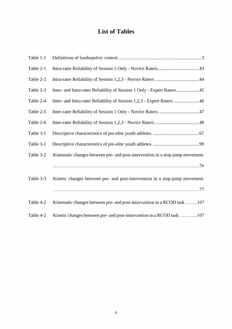
that have been adopted (Table 1-1) and the limitations of these definitions. However, based on
previous research, lumbopelvic stability can be defined as to obtain and maintain the alignment
of the body segments in a task dependant manner without substitution strategies, in both static
positions and during dynamic movements..

Chapter 1 The Problem
3
Table 1-1 Definitions of lumbopelvic control.
Reference Definition
Mills et al., (2005) The ability to control motion of the lumbar spine and
pelvis relative to an arbitrarily defined neutral
position
Panjabi (1992) The normal function of the three-component
stabilizing system is to provide sufficient stability to
the spine to match the instantaneously varying
stability demands due to changes in spinal posture,
and static and dynamic loads.
Kibler, et al., (2006) The interaction between the pelvis and lumbar and
thoracic spine, and between the pelvis and the hip
joint
The active structures of this lumbopelvic system play the primary role in maintaining
stabilisation during gross motor movements, including transverse abdominus, multifidi,
internal oblique, deep transversosponalis, erector spinae, external oblique, rectus abdominus
muscles, quadrates lumborum and the pelvic floor muscles. Spinal stability during dynamic
movements that involve the transfer of forces require the interaction of two systems (Bergmark
1989), the local and the global system, also referred to as the deep or superficial systems
(Kuszewski, Gnat et al. 2009). The local system’s role is to maintain spinal curvature and
provide lateral and sagittal stiffness to the lumbar spine to retain mechanical stability
(Bergmark 1989), and therefore responds to changes in the posture of the lumbar spine. This
local system comprises the muscles with an origin or insertion at the vertebra, with the

Chapter 1 The Problem
4
exception of the Psoas that is part of the global system. Core stabilising muscles, including the
Psoas, and intra-abdominal pressure that directly transfer load between the thoracic cage and
the pelvis are considered to be part of the global system (Bergmark 1989), and is thought to
respond to changes of the line of action of an externally applied load. It is also responsible for
changing the position of the thoracic cage in relation to the pelvis, however, both systems
respond to the magnitude of external load (Bergmark 1989).
1.1.3 Movement Screening
To implement a prevention strategy, we first must identify those athletes who are at an
increased risk of injury. As many movement screening procedures require three-dimensional
motion analysis (Hewett, Myer et al. 2005, Edwards, Steele et al. 2010, Mann, Edwards et al.
2013), there is a need for field-based movement screening that can be more easily implemented
by coaches and clinicians. To effectively predict injury risk and guide prevention strategies
within sporting communities, movement screening protocols must be injury specific, simple,
validated, reliable, cost effective and easily implemented. Despite a lack of longitudinal
evidence of the reliability and validity pertaining to the various movement screening
procedures that are available, many sporting teams have adopted these tools. Generally, these
are designed with one of two aims in mind: (1) injury risk management; and/or (2) performance
enhancement (Mottram and Comerford 2008). Specifically for injury risk management, early
identification of athletes with “poor” movement competency, yet never actually been defined
within the literature, can allow intervention strategies to be implemented to potentially prevent
the injury from occurring or re-occurring, and thus reduce the injury rates seen among the
sporting population (Parkkari, Kujala et al. 2001). Currently, movement screening tools for
injury prevention include use of dynamic movements such as jumping and landing tasks (Myer,
Ford et al. 2008, Padua, Marshall et al. 2009) or dynamic postural stability exercises (Kinzey

Chapter 1 The Problem
5
and Armstrong 1998, Cook, Burton et al. 2006). Including these functional movements in
screens of athletes movement competency provides a highly sport and skill specific movement
screen (Mottram and Comerford 2008). These functional tests identify risk factors such as
muscle flexibility and strength imbalances, impaired balance and postural stability (Kinzey and
Armstrong 1998, Cook, Burton et al. 2006), and resultant biomechanical variables (Myer, Ford
et al. 2008, Padua, Marshall et al. 2009). Yet critically, lumbopelvic stability is often over
looked when assessing lower limb injury risk (Myer, Chu et al. 2008).
One of the most popular movement screens widely employed in clinical and sporting situations,
is the Functional Movement Screen (FMS) (Cook, Burton et al. 2006, Cook, Burton et al.
2006), which has also excluded lumbopelvic stability assessment. The FMS comprises of seven
dynamic movement tasks aimed to test functional movement capacity, including the: deep
squat; hurdle step; inline lunge; shoulder mobility test; active straight leg raise; trunk stability
push-up; and rotary stability test. These movements are each scored on an ordinal scale from 0
to 3 with four categories. With a compensation defined as not complying with standard
movement expectations associated with each test (Cook, Burton et al. 2006). A score of 3
indicates the participant’s ability to perform the movement as described with no
compensations. A score of 2 denotes the participant is performing some type of compensation
while completing the movement. While a score of 1 signifies the participant is unable to
perform the movement, and a score of 0 indicated there was pain associated with performance
of the movement (Cook, Burton et al. 2006, Cook, Burton et al. 2006). The categorical scores
for each movement are summed together for a total score out of 21. Whilst previous research
has suggested that if this total composite score falls below 14, an increased injury risk is present
(Kiesel, Plisky et al. 2007, Chorba, Chorba et al. 2010), recent research has contested this

Chapter 1 The Problem
6
notion demonstrating that the FMS is unable to predict injury risk (Bardenett, Micca et al. 2015,
Bushman, Grier et al. 2016).
Inter- and inta-rater reliability is a key consideration for movement screens, given the
subjective nature of rating (Tinsley and Weiss 1975). A high level of agreement between
different raters (McHugh 2012) and high test-retest reliability of individual raters (Teyhen,
Shaffer et al. 2012) is desirable. As such, researchers have attempted to determine the reliability
in movement screening protocols that are currently used (Chorba, Chorba et al. 2010, Gribble,
Brigle et al. 2013, Gribble, Brigle et al. 2013, Smith, Chimera et al. 2013). Reliability requires
repeatability in both raters and participants, and although research has demonstrated good
reliability within some field-based movement screens (Chorba, Chorba et al. 2010, Minick,
Kiesel et al. 2010), there are limitations within both the methodology for assessing reliability
and the screening protocols themselves. Many of these studies employ the use of intra-class
correlations (ICC) to analyse and interpret inter- and intra-rater reliability (Chorba, Chorba et
al. 2010, Gribble, Brigle et al. 2013, Gribble, Brigle et al. 2013, Smith, Chimera et al. 2013).
However, this statistical method is inappropriate method, as ICC’s should only be applied to
continuous scalar data, not ordinal data in which these movement screens provide (Sim and
Wright 2005). Inter-rater reliability using Kappa scores have also been reported for the FMS
(Minick, Kiesel et al. 2010, Onate, Dewey et al. 2012, Teyhen, Shaffer et al. 2012), with authors
suggesting that there is a high reliability despite some relatively low Kappa scores (Minick,
Kiesel et al. 2010), which would indicate otherwise. While low Kappa scores have been
suggested to relate to poor scoring descriptions inherent to the screening protocols (Minick,
Kiesel et al. 2010), the FMS may not be as reliable as has been implied because of inadequate
statistical analysis and/or poor interpretation of scores.

Chapter 1 The Problem
7
Further to concerns with statistical interpretation, the presence of a learning effect may also
affect the reliability of a movement screen. The often novel movements required to be
performed during screening may be unfamiliar to athletes (e.g. a Tuck Jump (Myer, Ford et al.
2008), which might possibly lead to a familiarisation effect when first performing these tasks.
A familiarisation effect is determined by comparing repeated sessions of the same task
(Hopkins 2000), and is identified when a change in performance from session 1 to session 2
occurs due to an effect of practice or experience (Schmidt and Wrisberg 2008). The presence
of a familiarisation effect requires a familiarisation session prior to rating in order to establish
a reliable measure. It is currently unknown whether athletes require familiarisation with
movement screening procedures prior to rating their movement screens as there has been a
distinct lack of research in this area.
In addition to questions of familiarisation effect, limited research has investigated effective
rater viewing methods for the movement screen, which can also potentially affect the reliability
of the movement screening tool. Despite limited research, many sporting teams and clinicians
have adopted simple, real-time, single-viewing, field-based procedures involving manual
grading methods (Cook, Burton et al. 2006, Cook, Burton et al. 2006, Myer, Ford et al. 2008).
It has been suggested by Whiteside, Deneweth et al. (2016) that manual grading may not be
effective due to the requirement for multiple error cues relating to the movement, to be
identified simultaneously. Concern with the ability to accurately perform manual rating in real
time, relates to there being a limited attentional capacity, for example ‘bottleneck’ theories of
attention that maintain that only one of multiple high-speed tasks can be performed with
accuracy at the same time (Craik 1947). Multiple resource capacities such as Wickens et al.
(2002) accommodate the potential for a time-sharing strategy to complete multiple tasks at
once; however, the nature of the simultaneous inputs are important. The skill level of the
Chapter 1 The Problem
8
performer and the bandwidth of information processed are important determinants of
simultaneous performance success (Wickens 2002). However, it should be noted the use of
multiple visual tasks (as in the multiple error cues required to be simultaneously detected in a
real-time FMS), will likely lead to reduced performance due to interference between tasks
competing for the same processing resources (Wickens 2002). Given manual grading of
movement performance requires multiple visual cues to be detected in a short time, it is
suggested that the demands of movement assessment may be reduced by viewing a video of
the performance multiple times, focusing on different error cues each time, thereby reducing
the attentional demands (Whiteside, Deneweth et al. 2016). This would lead to an increase in
reliability of the MS tool, as raters are more accurately able to assess errors in movement.
A further limitation of the FMS and other currently available movement screens is their lack
of assessment of the stability of the lumbopelvic region, despite the role of trunk stability
during force transfer and the importance of this for both performance and injury prevention
(Myer, Chu et al. 2008). Given the lumbopelvic region serves to attach the trunk to the lower
extremities, it is a key area associated with performance enhancement through its role in force
transfer (Hibbs, Thompson et al. 2008). Also related to this force transfer function, it has been
shown to be critical in the etiology, rehabilitation and reduction of lower limb injuries (Hewett
and Myer 2011), with links to many lower limb injuries (Beckman and Buchanan 1995, Cowan,
Schache et al. 2004, Zazulak, Hewett et al. 2007, Kuszewski, Gnat et al. 2009, Edwards, Steele
et al. 2010, Hewett and Myer 2011, Mann, Edwards et al. 2013) and low back pain (Akuthota,
Ferreiro et al. 2008). It is suggested that athletes who display delayed or poor neuromuscular
control of this region during dynamic jump-landing tasks may be predisposed to lower limb
injury through poor stability, excess motion and increased forces experienced by joints (Myer,
Chu et al. 2008).

Chapter 1 The Problem
9
Although there is no validated universal definition for lumbopelvic stability, clinically
lumbopelvic stability is measured using a range of static tests such as; the straight leg lowering
test (Clark 2004), Biering-Sorensen test (Biering-Sørensen 1984), the flexor endurance test or
the side bridge test (McGill, Childs et al. 1999). These tests all provide a measure of static
lumbopelvic control, which is more appropriate to a rehabilitation setting, in comparison with
the dynamic stability that is required in a sport context. Therefore an alternative definition, and
measure of lumbopelvic stability, more suitable to athletic populations is required (Hibbs,
Thompson et al. 2008). Dynamic measures of lumbopelvic stability have been devised to meet
this need include; the lower abdominal neuromuscular assessment (Clark 2004), kneeling arm
raise and the quad arm raise test (Liemohn, Baumgartner et al. 2005). However, while including
dynamic movement, these tests only challenge lumbopelvic stability within a singular plane,
as opposed to demanding both greater magnitude and a requirement for multiple planar
stability. Given multi-planar control is required during most sport movements, these tests are
still limited in their capacity to provide a sport-specific measure of dynamic lumbopelvic
stability. Therefore, a major consideration for all movement screening is the need for a
challenging and specific lumbopelvic assessment, such as the single leg squat and/or dip test,
which have been shown to be a reliable measure of lumbopelvic control (Perrott, Pizzari et al.
2011).
To effectively identify athletes presenting an increased injury risk through movement screens
and allow intervention strategies to be implemented, it is important to determine which risk
factors should and can be identified in movement screening. Risk factors in a sporting context
are defined as any factor that may increase the potential for injury (Meeuuiisse 1991) that can
be broken down into two categories; modifiable and non-modifiable risk factors (Emery 2003).

Chapter 1 The Problem
10
Non-modifiable risk factors refer to risks that cannot be altered and modifiable risk factors to
those that can potentially be changed or altered through intervention (Emery 2003). For this
reason, injury prevention programs have focused on altering modifiable risk factors to reduce
the risk of an individual sustaining an injury. A key modifiable risk factor in lower limb injury
is an athlete’s landing technique, as the eccentric loading phase of a landing movement is highly
influential in the incidence of injury (Boden, Dean et al. 2000, Myer, Ford et al. 2008, Edwards,
Steele et al. 2010, Mann, Edwards et al. 2013). Instability or a lack of control of the trunk
segment during this phase is suggested to predispose athletes to an increased injury risk
(Edwards, Steele et al. 2010, Mann, Edwards et al. 2013). Whereby an increase in trunk motion
increases the distance between the GRF vector and joint centres, thus increasing the joint
moments and muscle activation requirements (Hewett and Myer 2011). Specifically, factors
such as an increase (Dempsey, Lloyd et al. 2009) or decrease (Sheehan, Sipprell III et al. 2012)
in trunk flexion during the weight acceptance and at initial contact respectively, increased
lateral trunk flexion (Dempsey, Lloyd et al. 2007), trunk rotation (Dempsey, Lloyd et al. 2007)
away from the direction of travel during the weight acceptance phase, increased peak hip
internal rotation and peak knee valgus angles (McLean, Huang et al. 2005) have been suggested
to play a role in increasing an individual’s risk of injury. Commonly performed in dynamic
sporting situations, both the stop-jump and unanticipated cutting task movements have been
utilised in previous research to investigated landing strategies and lumbopelvic control of
athletes at an increased risk of injury (Edwards, Steele et al. 2010, Mann, Edwards et al. 2013,
Edwards, Brooke et al. 2017). These movements are suggested to occur in approximately 70%
of playing time in a sport such as basketball (Ford, Myer et al. 2005). As these are sport specific
movement skills, alternate neuromuscular strategies that have been adopted by the athlete may
be observed through biomechanical analysis (Cowley, Ford et al. 2006).

Chapter 1 The Problem
11
Biomechanical analysis of lumbopelvic control during a COD task must be performed on an
unanticipated movement (reactive), as this will place the system under the greatest perturbation
to assess lumbopelvic stability. When an athlete is aware of the COD direction they are to
perform, as opposed to the R-COD performed in sporting situations, the preplanning of postural
and movement strategies changes both the kinematic and kinetics of the movement (Besier,
Lloyd et al. 2001). Changes such as the position or movement of the centre of gravity, altered
muscle activation timing or amplitude, and modification of reflex muscle activation are
suggested to occur when performing an non-reactive COD compared to an R-COD (Besier,
Lloyd et al. 2001). Additionally, an reactive movement should be utilised as the experimental
movement task to aid in replicating these sporting movements, as during a sporting situation
an athlete is required to suddenly respond to external stimulus such as a defender or ball making
the movement direction unanticipated (Besier, Lloyd et al. 2001).
1.1.4 Neuromuscular Landing Re-training
Traditionally, injury prevention programs have taken a local focus attempting to modify
movement at a specific joint, such as the knee joint for anterior cruciate ligament (ACL) injury
(Myer, Chu et al. 2008). However, this may not be the most effective approach as lumbopelvic
instability and lack of control has been suggested to be an underlying cause of many injury risk
variables throughout the entire lower limb complex (Hewett and Myer 2011). Altered
lumbopelvic has stability has links to groin (Cowan, Schache et al. 2004, Edwards, Brooke et
al. 2017), hamstring (Sherry and Best 2004, Chumanov, Heiderscheit et al. 2007), knee joint
(Edwards, Steele et al. 2010, Hewett and Myer 2011, Mann, Edwards et al. 2013) and ankle
joint injuries (Beckman and Buchanan 1995, Beynnon, Murphy et al. 2002). Common
plyometric jump training programs have shown conflicting results in their effectiveness to
reduce injury (Myer, Ford et al. 2006). Given balance and core stability have been associated

Chapter 1 The Problem
12
with reduced injury rates (Sherry and Best 2004, Myer, Ford et al. 2006), it may be suggested
that purely plyometric programs have neglected a critical component, that being the
development of foundational lumbopelvic stability. Previous lumbopelvic intervention
programs have elicited a training response suggested to reduce injury risk variables in athletes
during the performance of a COD following an intervention program (Bencke, Næsborg et al.
2000, Dempsey, Lloyd et al. 2009, DiStefano, Blackburn et al. 2011). These programs have
successfully altered high risk variables such as peak external knee valgus moments (Dempsey,
Lloyd et al. 2009, Cochrane, Lloyd et al. 2010) and trunk lateral flexion (Dempsey, Lloyd et
al. 2009) during COD.
The mechanisms underlying the link between biomechanical lower limb risk factors and poor
lumbopelvic or trunk control have been identified, specifically with links between increased
peak internal/external knee and hip adductor moments. Whereby an increase in lateral trunk
flexion increases the distance between the GRF vector and the knee joint centre and head of
the femur, thus increasing the knee moments and hip adduction muscle activation (Hewett and
Myer 2011). Additionally, weak hip extensors can lead to an increased activation requirement
of the hip flexors, specifically the iliopsoas, to control the lumbopelvic region and trunk region
during landing. This landing strategy is suggested to ‘stiffen’ individuals landing patterns
creating the more upright trunk landing pattern (Griffin, Agel et al. 2000), where by a reduced
degrees of freedom is suggested to increase ACL injury risk (Griffin, Agel et al. 2000,
Shimokochi, Ambegaonkar et al. 2013). In addition to associations between lumbopelvic
instability and injury risk factors, significantly increased trunk displacement following
perturbation have been identified among athletes who sustained a knee injury within a three-
year period, further increasing speculation that neuromuscular impairment of the core region
may increase an athlete’s susceptibility to a knee injury (Zazulak, Hewett et al. 2007).

Chapter 1 The Problem
13
Consequently, recent research has turned its attention to enhancing the stability of the
lumbopelvic region in relation to reducing lower limb injuries among the sporting population
(Myer, Chu et al. 2008).
Many of these intervention programs however, have been implemented within a late adolescent
or adult population (Chappell and Limpisvasti 2008, Herman, Weinhold et al. 2008, McCurdy,
Walker et al. 2012) despite research suggesting that early to mid-adolescence is a key
development phase for implementation of landing retraining injury prevention strategies
(Myer, Faigenbaum et al. 2011). During this critical phase, cognitive and motor capabilities
are more amenable to age appropriate training interventions (Myer, Faigenbaum et al. 2011),
compared to children younger than 12 years. These younger children, who despite targeted
paediatric programs, have been shown to be unable to alter lower limb biomechanics with
training (DiStefano, Blackburn et al. 2011). Dynamic neuromuscular training has been reported
to be effective for improving lower limb movement biomechanics among adolescent females
and in turn, decreasing injury risk (Myer, Ford et al. 2005). These findings suggest that this
population can be successfully re-trained to improve lower limb landing mechanics during
dynamic landing tasks. Improvements in their performance and stability during motion are
thought to occur mainly through general neuromuscular adaptations such as more
comprehensive and synchronous firing of the muscular unit (Sale 1988), rather than specific
responses to the program.
For interventions to be successful in this population, equipment must be readily available and
elements of the program suited to junior sporting organisations. With the exception of a small
number of programs such as the FIFA11+ (Bizzini, Junge et al. 2013) and FootyFirst (NoGAPS

Chapter 1 The Problem
14
2012), intervention programs that require minimal equipment, many of the other current
neuromuscular retraining programs are performed in controlled environments such as
supervised gyms with specialised equipment and/or supervision required (Hewett, Lindenfeld
et al. 1999, Myklebust, Engebretsen et al. 2003). This can create accessibility issues, especially
in rural regions where the provision of formal sporting structures and support mechanisms is
limited (Finch, Mahoney et al. 2003). Limited access to facilities may affect adherence rates
(Tappe, Duda et al. 1989), which is negatively associated with injury risk (Soligard, Nilstad et
al. 2010). Therefore, to have the greatest effect on youth sporting injury rates, it is essential
that an accessible, easily implemented field-based program is developed to aid injury
prevention among pre-elite youth athletes.
1.1.5 Lumbopelvic link to risk factors and performance
Literature has shown lumbopelvic stability to be influential in various acute and overuse lower
limb injuries. For example the overuse groin injury is commonly sustained among Australian
rules football athletes (Orchard and Seward 2002) and has been linked with altered lumbopelvic
control (Cowan, Schache et al. 2004, Edwards, Brooke et al. 2017). Athlete’s with a history of
groin pain have displayed greater variation in their lumbo-pelvic and hip joint motor control
during a R-COD task compared to those without a history (Edwards, Brooke et al. 2017). It
remains unknown if this poor lumbopelvic control during a R-COD task is the underlying
mechanism of the injury or the resultant symptom of groin injury. Furthermore, abdominal
muscle recruitment strength has also been identified as a possible risk factor for groin strains
in Australian football league athletes with chronic groin pain (Cowan, Schache et al. 2004).
This finding supports previous research that identifies the feed-forward mechanistic role of the
transverse abdominus prior to movement of the extremities (Cresswell, Oddsson et al. 1994,
Hodges and Richardson 1997). In these athletes with chronic groin pain however, this

Chapter 1 The Problem
15
activation of the transverse abdominus is delayed (Cowan, Schache et al. 2004), corresponding
with previous research that demonstrated that this muscle activation is altered with the onset
of acute experimentally induced pain (Hodges, Moseley et al. 2003).
Another injury thought to be associated with lumbopelvic control is hamstring strain injuries,
which typically occur during a forced eccentric muscle action during late swing phase in gait,
to control or decelerate a high velocity movement of the thigh segment (Whiting and Zernicke
2008). With 69% of muscle strain injuries in Australian football involving the hamstrings and
a history of previous injury being a significant risk factor for re-injury (Orchard 2001), the
identification of mechanistic risk factors is of high importance for prevention of this injury.
One possible risk factor identified for hamstring strain injuries is a lack of lumbopelvic stability
(Devlin 2000), as their anatomical attachment location of the hamstrings to the ischial
tuberosities suggests that they may play an important role in stabilisation of this region (Van
Wingerden, Vleeming et al. 1997). Chumanov et al. (1994) demonstrated this link through the
use of musculoskeletal modelling techniques, showing an increase in speed of movement
significantly increased the eccentric load placed on the hamstring muscles. This increase in
speed is said to vary with stride length based on fluctuations in the neuromuscular control.
Specifically lumbopelvic pelvic muscles, such as the iliopsoas, have a major influence on the
stretch of the hamstring muscle, suggesting that the increased lumbopelvic control is critical
for the control of the hamstring stretch during high speed movements. Stiffness of the
hamstring muscles, is measured as the ratio of change in the muscle length (passive knee
extension in supine test) to the force value causing the change in muscle length (hand held
tensometer). This stiffness is suggested to compensate for the lack of stabilisation when
lumbopelvic control is compromised, and may be a reflection of the faulty neural control of the
lumbopelvic region (Kuszewski, Gnat et al. 2009). This proposed notion provides a possible

Chapter 1 The Problem
16
explanation as to why purely mechanical stretching is not effective in reducing hamstring
muscle stiffness, as a lack of lumbopelvic control has not been addressed (Kuszewski, Gnat et
al. 2009). As a lack of hamstring flexibility is implicated as a risk factor in hamstring injuries
(Witvrouw, Danneels et al. 2003), and with a lack of lumbopelvic control implicated in muscle
stiffness (Kuszewski, Gnat et al. 2009, Schuermans, Van Tiggelen et al. 2017), this suggests
that lumbopelvic control is potentially an underlying risk factor for hamstring strain injuries.
Furthermore, this relationship between lumbopelvic control and hamstring injuries is supported
by Sherry and Best (2004) who showed a reduction in hamstring strain re-injury rates when
rehabilitation consisted of a program of progressive agility and core stability exercises.
Anterior cruciate ligament (ACL) injuries are predominantly non-contact injuries with
approximately 30% due to contact and 70% occurring due to non-contact situations (Boden,
Dean et al. 2000). With knee valgus (adduction) (McLean, Huang et al. 2004, Dempsey, Lloyd
et al. 2009), external or internal tibial rotation (Krosshaug, Nakamae et al. 2007), reduced knee
flexion (Boden, Dean et al. 2000) and lateral trunk movements (Hewett, Ford et al. 2006) as
noted as key injury risk factors for ACL injuries during COD tasks, much of the research has
focused on the effects of changing these knee joint dynamics (Hewett, Lindenfeld et al. 1999,
Myklebust, Engebretsen et al. 2003, Mandelbaum, Silvers et al. 2005). With key core
stabilising muscles such as the erector spinae and quadratus lumborum responsible for the
lateral flexion of the trunk segment (Saladin 2007), the lumbopelvic region can be suggested
to also play a vital role in the incidence of this injury. A significantly increased trunk
displacement following perturbation was seen among athletes who sustained a knee injury
within a three year period, suggesting that neuromuscular impairment of the core region may
increase an athlete’s susceptibility to a knee injury (Zazulak, Hewett et al. 2007). Hewett et al.,
(2011) proposed that this lack of neuromuscular control of the trunk region may lead to
Chapter 1 The Problem
17
increased knee loading. This increasing in knee loading was identified by Dempsey et al.
(2009) in which a lateral ground reaction force vector in respect to the femur head increases
hip adduction moments, ultimately leading to injury. Consequently recent research has turned
its attention to enhancing the stability of the lumbopelvic region in relation to reducing the
ACL injuries among the sporting population (Myer, Chu et al. 2008). This change in the forces
around the knee joint may also be influential in the development of patellofemoral pain
(Edwards, Steele et al. 2010).
Repetitive loading (Ferretti 1986) and an altered landing technique (Richards, Ajemian et al.
1996, Bisseling, Hof et al. 2007, Edwards, Steele et al. 2010) are primary risk factors for
patellar tendinopathy (PT). This overuse knee joint injury is characterised by the progressive
degeneration of the patellar tendon (Peers and Lysens 2005) that is thought to be due to
adaptation to a change in the type and direction of tendon loading sustained by the patellar
tendon during repetitive loading (Hamilton and Purdam 2004). Athletes with PT have been
shown in the literature to land with alternative landing strategies (Richards, Ajemian et al.
1996, Bisseling, Hof et al. 2007). Another key risk factor in the development of PT is the
identification of a patellar tendon abnormality on diagnostic imaging. For example,
asymptomatic junior athletes with a patellar tendon abnormality have a 4.2 times greater risk
of developing PT (Cook, Khan et al. 2000). Asymptomatic athletes with a patellar tendon
abnormality where found to, utilise an altered hip landing strategy during the horizontal landing
phase. This involved the athletes extending their hips during landing as opposed to the hip
flexion displayed by athletes with a normal patellar tendon in adult athletes (Edwards, Steele
et al. 2010) and youth basketball players (Mann, Edwards et al. 2013). This altered hip joint
strategy suggest a potential deficit in lumbopelvic control during landing.

Chapter 1 The Problem
18
A major predisposing factor in the incidence of an ankle sprain is a history of a previous ankle
sprain with 85% of ankle sprains represented a reoccurrence injury that occurs up to 12 months
following injury and in either ankle, suggesting insufficient rehabilitation (Watson 1999).
Increased postural sway has been noted in studies of athletes following an ankle injury
(Beckman and Buchanan 1995), and has also been noted as a potential risk factor for ankle
injuries in athletes (Beynnon, Murphy et al. 2002). With lumbopelvic or core musculature
responsible for the control and stabilisation of the trunk region (Brown, Vera-Garcia et al.
2006), it is plausible that a neuromuscular or strength deficiency within this region may
increase the risk of an ankle ligament injury.
Despite this, paradoxical evidence exists relating to lumbopelvic control and the implications
with injury and performance. Previous research has indicated that increased trunk lateral
flexion and rotation away from the COD direction, increases the risk of ACL injury risk
(Dempsey, Lloyd et al. 2007). Yet on the contrary, decreased trunk range of motion summed
across all three planes of motion has been associated with reduced performance in an agility
task and a countermovement jump (Edwards, Austin et al. 2017). Given this apparently
conflicting evidence, it is possible that there may exist an optimal trunk segment motion range
during dynamic jump-landing tasks that allows optimal performance without increased injury
risk. It should be noted however, that there is a lack of research surrounding the ideal
biomechanical parameters for the stop-jump and unanticipated cutting task movement and
whether a specific threshold exists for each risk factor within this population of youth athletes.
While previous research exists for adult populations, youth athletes’ research is significantly
lacking.

Chapter 1 The Problem
19
1.2 Statement of the Problem
Sporting injuries among the adolescent and early adult population are considerably higher than
that of any other age group (Medibank Private 2003). This development phase group is
susceptible to dynamic neuromuscular landing and core training strategies (Myer, Faigenbaum
et al. 2011) and display decreased lumbopelvic control (Myer, Chu et al. 2008). Conversely
these high sporting injury rates and risks among this population, identify the crucial need for
injury identification via movement screening and prevention strategies to be validated and
implemented to aid in the reduction of lower limb injuries within these athletes. While there is
some research that has identified a link between lumbopelvic control injuries (See section1.15),
this area still requires additional research.
1.3 Research Aims
The primary aim of this research is to determine the reliability of a field-based movement
competency screening, and the effect of different types of neuromuscular training intervention
programs when examining jump-landing technique and lumbopelvic stability in pre-elite youth
athletes. This will be achieved through a series of studies that aim to;
o Manuscript 1: The aim of this study was to (i) determine the inter- and intra-
rater reliability of a field-based movement screen in novice and expert raters
using different viewing methods; and (ii) evaluate the presence of a learning
and familiarisation effect for both raters and participants, respectively.
o Manuscript 2: The purpose of this study is to compare the effects of three
different 12-week intervention programs on the results of a full biomechanical
analysis during a stop-jump landing task in pre-elite youth athletes.
Chapter 1 The Problem
20
o Manuscript 3: The purpose of this study is to compare the effects of three
different 12-week intervention programs on the results of a full biomechanical
analysis during an R-COD in pre-elite youth athletes.
o Manuscript 4: This study aimed to identify which, if any, lower limb injury
risk factors are associated with increased trunk ROM during R-COD in pre-
elite youth athletes.
1.4 Research Hypotheses
It is hypothesised that lumbopelvic stability will be identified as a key factor that substantially
influence pre-elite youth athlete’s ability perform fundamental dynamic movement patterns.
The respective hypotheses are:
o Manuscript 1: It is hypothesised that the field-based movement dysfunction
screen (MDS) criteria for each movement and total score will display high
inter- and intra-rater reliability for both novice and expert raters, and that
inter- and intra-rater reliability will improve with repeated exposure to the
MDS due to a rater learning effect.
o Manuscript 2: While both core stability and landing re-training intervention
programs are suggested to improve lumbopelvic stability and lower limb
alignment, we hypothesise that the combination of both the core stability and
landing re-training programs will result in the greatest improvement (e.g. less
trunk range of motion) during the stop-jump with the strength and condition
only program not altering their stop-jump technique.
o Manuscript 3: While both core stability and landing re-training intervention
programs are suggested to improve lumbopelvic stability and lower limb
alignment, we hypothesise that a combination of both the core stability and

Chapter 1 The Problem
21
landing re-training programs will result in the greatest improvement in
lumbopelvic stability (e.g. less trunk range of motion) and lower limb
alignment during the R-COD than the other intervention programs.
o Manuscript 4: With lumbopelvic control shown to be highly influential in
numerous lower limb injuries, it is hypothesized that athletes with moderate
magnitude of trunkab-pelvis range of motion during the R-COD will employ a
strategy that is associated with lower limb injury risk compared to those with
higher and lower trunkab-pelvis range of motion.
1.5 Limitations
The following assumptions and limitations apply to this study:
1. A participant cohort of pre-elite youth athletes (current scholarship holders) within the
Western Region Academy of Sport in netball, softball, hockey, basketball or
triathletes aged between 11 to 18 years restricts results being applied to other athletic
populations and ages;
2. Field-based movement screening procedures are purely qualitative assessments; to
help reduce inter-rater error, specific criteria have been designed for each assessed
movement although this does not remove this limitation;
3. Biomechanical analysis requires athletes to perform two dynamic experimental tasks
in a laboratory environment, both commonly performed in the sports these athletes
compete in, and this study assumes the athlete will perform these tasks the same as
they would in a field-based setting;
4. Athletes will be required to adhere to the intervention program; however, while
athletes who lived in close proximity to Academy of Sport (Bathurst) completed their
sessions with the supervision of their strength and conditioning coach, those athletes

Chapter 1 The Problem
22
who lived in more remote locations were expected to compete some of their sessions
via satellite or individually with supplemented by supervised sessions only every
three weeks. Compliance is therefore difficult to control and report accurately; and
5. Although athletes are specifically told to perform their group training program and no
other program, contamination of training programs due to athletes performing
additional training cannot be entirely controlled.
1.6 Delimitations
The following delimitations apply to these studies:
1. The study population is restricted to include only pre-elite junior athletes that were
used in these investigations, therefore results are a true representation of only this
population;
2. Data collection throughout the study will be conducted using standardised protocols
within a controlled laboratory environment;
3. Intervention training programs are encouraged by the squad’s strength and
conditioning coaches to increase compliance rates in completing the training
programs; however, adherence data was not collected during this study; and
4. All movement screening and biomechanical analysis sessions will be supervised by
the primary investigator, using the same equipment to maintain consistency within the
measures.
1.7 References
Akuthota, V., Ferreiro, A., Moore, T., & Fredericson, M. (2008). Core stability exercise principles.
Current Sports Medicine Reports, 7(1), 39-44.

Chapter 1 The Problem
23
Bardenett, S. M., Micca, J. J., DeNoyelles, J. T., Miller, S. D., Jenk, D. T., & Brooks, G. S. (2015).
Functional Movement Screen normative values and validity in high school athletes: can the
FMS™ be used as a predictor of injury? International Journal of Sports Physical Therapy,
10(3), 303.
Beckman, S. M., & Buchanan, T. S. (1995). Ankle inversion injury and hypermobility: Effect on hip
and ankle muscle electromyography onset latency. Archives of Physical Medicine and
Rehabilitation, 76(12), 1138-1143.
Bencke, J., Næsborg, H., Simonsen, E. B., & Klausen, K. (2000). Motor pattern of the knee joint
muscles during side‐step cutting in European team handball. Scandinavian Journal of Medicine
& Science in Sports, 10(2), 68-77.
Bergmark, A. (1989). Stability of the lumbar spine. Acta Orthopaedica, 60(S230), 1-54.
Besier, T. F., Lloyd, D. G., Ackland, T. R., & Cochrane, J. L. (2001). Anticipatory effects on knee joint
loading during running and cutting maneuvers. Medicine & Science in Sports & Exercise, 33(7),
1176.
Beynnon, B. D., Murphy, D. F., & Alosa, D. M. (2002). Predictive factors for lateral ankle sprains: a
literature review. Journal ofAthletic Training, 37(4), 376-380.
Boden, B. P., Dean, G. S., Feagin, J., & Garrett, W. (2000). Mechanisms of anterior cruciate ligament
injury. Orthopedics, 23(6), 573-578.
Brown, S. H., Vera-Garcia, F. J., & McGill, S. M. (2006). Effects of abdominal muscle coactivation on
the externally preloaded trunk: Variations in motor control and its effect on spine stability.
Spine, 31(13), E387-E393.
Bushman, T. T., Grier, T. L., Canham-Chervak, M., Anderson, M. K., North, W. J., & Jones, B. H.
(2016). The functional movement screen and injury risk: Association and predictive value in
active men. American Journal of Sports Medicine, 44(2), 297-304.
Caine, D., Maffulli, N., & Caine, C. (2008). Epidemiology of injury in child and adolescent sports:
injury rates, risk factors, and prevention. Clinics in Sports Medicine, 27(1), 19-50.
Chappell, J. D., & Limpisvasti, O. (2008). Effect of a neuromuscular training program on the kinetics
and kinematics of jumping tasks. American Journal of Sports Medicine, 36(6), 1081-1086.

Chapter 1 The Problem
24
Chorba, R. S., Chorba, D. J., Bouillon, L. E., Overmyer, C. A., & Landis, J. A. (2010). Use of a
functional movement screening tool to determine injury risk in female collegiate athletes. North
American Journal of Sports Physical Therapy, 5(2), 47-54.
Chumanov, E. S., Heiderscheit, B. C., & Thelen, D. G. (2007). The effect of speed and influence of
individual muscles on hamstring mechanics during the swing phase of sprinting. Journal of
Biomechanics, 40(16), 3555-3562.
Clark, M. (2004). Core stabilization training in rehabiliation. In W. E. Prentice (Ed.), Rehabilitation
techniques for sports medicine and athletic training (4th ed.): McGraw-Hill Humanities/Social
Sciences/Languages.
Cochrane, J. L., Lloyd, D. G., Besier, T. F., Elliott, B. C., Doyle, T. L., & Ackland, T. R. (2010).
Training affects knee kinematics and kinetics in cutting maneuvers in sport. Medicine &
Science in Sports & Exercise, 42(8), 1535-1544.
Cook, G., Burton, L., & Hoogenboom, B. (2006a). Pre-participation screening: The use of fundamental
movements as an assessment of function–part 1. North American Journal of Sports Physical
Therapy, 1(2), 62-72.
Cook, G., Burton, L., & Hoogenboom, B. (2006b). Pre-participation screening: The use of fundamental
movements as an assessment of function–Part 2. North American Journal of Sports Physical
Therapy, 1(3), 132-139.
Cowan, S. M., Schache, A. G., Brukner, P., Bennell, K. L., Hodges, P. W., Coburn, P., & Crossley, K.
M. (2004). Delayed onset of transversus abdominus in long-standing groin pain. Medicine &
Science in Sports & Exercise, 36(12), 2040-2045.
Cowley, H. R., Ford, K. R., Myer, G. D., Kernozek, T. W., & Hewett, T. E. (2006). Differences in
neuromuscular strategies between landing and cutting tasks in female basketball and soccer
athletes. Journal of Athletic Training, 41(1), 67-73.
Craik, K. J. W. (1947). Theory of the human operator in control systems. British Journal of Psychology,
38(2), 56-61.

Chapter 1 The Problem
25
Cresswell, A., Oddsson, L., & Thorstensson, A. (1994). The influence of sudden perturbations on trunk
muscle activity and intra-abdominal pressure while standing. Experimental Brain Research,
98(2), 336-341.
Dempsey, A. R., Lloyd, D. G., Elliott, B. C., Steele, J. R., & Munro, B. J. (2009). Changing sidestep
cutting technique reduces knee valgus loading. American Journal of Sports Medicine, 37(11),
2194-2200.
Dempsey, A. R., Lloyd, D. G., Elliott, B. C., Steele, J. R., Munro, B. J., & Russo, K. A. (2007). The
effect of technique change on knee loads during sidestep cutting. Medicine & Science in Sports
& Exercise, 39(10), 1765-1773.
DiStefano, L. J., Blackburn, J. T., Marshall, S. W., Guskiewicz, K. M., Garrett, W. E., & Padua, D. A.
(2011). Effects of an age-specific anterior cruciate ligament injury prevention program on lower
extremity biomechanics in children. American Journal of Sports Medicine, 39(5), 949-957.
Edwards, S., Austin, A. P., & Bird, S. P. (2017). The role of the trunk control in athletic performance
of a reactive change-of-direction task. Journal of Strength & Conditioning Research, 31(1),
126-139.
Edwards, S., Brooke, H. C., & Cook, J. L. (2017). Distinct cut task strategy in Australian football players
with a history of groin pain. Physical Therapy in Sport, 23, 58-66.
Edwards, S., Steele, J. R., McGhee, D. E., Beattie, S., Purdam, C., & Cook, J. L. (2010). Landing
strategies of athletes with an asymptomatic patellar tendon abnormality. Medicine & Science
in Sports & Exercise, 42(11), 2072-2080.
Emery, C. A. (2003). Risk factors for injury in child and adolescent sport: a systematic review of the
literature. Clinical Journal of Sport Medicine, 13(4), 256.
Finch, C. F., Mahoney, M., Townsend, M., & Zazryn, T. (2003). Rural sports and recreational injuries
in Australia: what do we know? Australian Journal of Rural Health, 11(3), 151-158.
Finch, C. F., Valuri, G., & Ozanne-Smith, J. (1998). Sport and active recreation injuries in Australia:
evidence from emergency department presentations. British Journal of Sports Medicine, 32(3),
220-225.

Chapter 1 The Problem
26
Ford, K. R., Myer, G. D., Toms, H. E., & Hewett, T. E. (2005). Gender differences in the kinematics of
unanticipated cutting in young athletes. Medicine & Science in Sports & Exercise, 37(1), 124-
129.
Gribble, P. A., Brigle, J., Pietrosimone, B. G., Pfile, K. R., & Webster, K. A. (2013a). Intrarater
reliability of the functional movement screen. Journal of Strength & Conditioning Research,
27(4), 978-981.
Gribble, P. A., Brigle, J., Pietrosimone, B. G., Pfile, K. R., & Webster, K. A. (2013b). Intrarater
reliability of the functional movement screen. Journal of Strength & Conditioning Research,
27(4), 978-981.
Griffin, L. Y., Agel, J., Albohm, M. J., Arendt, E. A., Dick, R. W., Garrett, W. E., . . . Ireland, M. L.
(2000). Noncontact anterior cruciate ligament injuries: risk factors and prevention strategies.
Journal of the American Academy of Orthopaedic Surgeons, 8(3), 141-150.
Herman, D. C., Weinhold, P. S., Guskiewicz, K. M., Garrett, W. E., Yu, B., & Padua, D. A. (2008).
The effects of strength training on the lower extremity biomechanics of female recreational
athletes during a stop-jump task. American Journal of Sports Medicine, 36(4), 733-740.
Hewett, T. E., Lindenfeld, T. N., Riccobene, J. V., & Noyes, F. R. (1999). The effect of neuromuscular
training on the incidence of knee injury in female athletes. American Journal of Sports
Medicine, 27(6), 699-706.
Hewett, T. E., & Myer, G. D. (2011). The mechanistic connection between the trunk, hip, knee, and
anterior cruciate ligament injury. Exercise and Sport Sciences Reviews, 39(4), 161-166.
Hewett, T. E., Myer, G. D., Ford, K. R., Heidt Jr, R. S., Colosimo, A. J., McLean, S. G., . . . Succop, P.
(2005). Biomechanical measures of neuromuscular control and valgus loading of the knee
predict anterior cruciate ligament injury risk in female athletes. American Journal of Sports
Medicine, 33(4), 492-501.
Hibbs, A. E., Thompson, K. G., French, D., Wrigley, A., & Spears, I. (2008). Optimizing performance
by improving core stability and core strength. Sports Medicine, 38(12), 995-1008.
Hodges, P. W., & Richardson, C. A. (1997). Contraction of the abdominal muscles associated with
movement of the lower limb. Physical Therapy, 77(2), 132-142.

Chapter 1 The Problem
27
Hopkins, W. G. (2000). Measures of reliability in sports medicine and science. Sports Medicine, 30(1),
1-15.
Ireland, M. L. (2002). The female ACL: Why is it more prone to injury? Orthopedic Clinics, 33(4),
637-651.
Kibler, W. B., Press, J., & Sciascia, A. (2006). The role of core stability in athletic function. Sports
Medicine, 36(3), 189-198.
Kiesel, K., Plisky, P. J., & Voight, M. L. (2007). Can serious injury in professional football be predicted
by a preseason functional movement screen? North American Journal of Sports Physical
Therapy, 2(3), 147.
Kinzey, S. J., & Armstrong, C. W. (1998). The reliability of the star-excursion test in assessing dynamic
balance. Journal of Orthopaedic and Sports Physical Therapy, 27(5), 356-360.
Kreisfeld, R., Harrison, J. E., & Tovell, A. (2017). Hopsital care for Australian sports injury 2012-13.
Retrieved from Canberra: 24th January, 2018. https://www.aihw.gov.au/getmedia/080b164c-
eaa2-4f7c-b438-015797a9474e/aihw-injcat-181.pdf.aspx?inline=true
Kuszewski, M., Gnat, R., & Saulicz, E. (2009). Stability training of the lumbo‐pelvo‐hip complex
influence stiffness of the hamstrings: A preliminary study. Scandinavian Journal of Medicine
& Science in Sports, 19(2), 260-266.
Liemohn, W. P., Baumgartner, T. A., & Gagnon, L. H. (2005). Measuring core stability. Journal of
Strength & Conditioning Research, 19(3), 583.
Macintosh, J. E., Bogduk, N., & Gracovetsky, S. (1987). The biomechanics of the thoracolumbar fascia.
Clinical Biomechanics, 2(2), 78-83.
Mann, K. J., Edwards, S., Drinkwater, E. J., & Bird, S. P. (2013). A lower limb assessment tool for
athletes at risk of developing patellar tendinopathy. Medicine & Science in Sports & Exercise,
45(3), 527-533.
McCurdy, K., Walker, J., Saxe, J., & Woods, J. (2012). The effect of short-term resistance training on
hip and knee kinematics during vertical drop jumps. Journal of Strength & Conditioning
Research, 26(5), 1257-1264.

Chapter 1 The Problem
28
McGill, S. M., Childs, A., & Liebenson, C. (1999). Endurance times for low back stabilization
exercises: clinical targets for testing and training from a normal database. Archives of Physical
Medicine and Rehabilitation, 80(8), 941-944.
McHugh, M. L. (2012). Interrater reliability: The kappa statistic. Biochemia medica, 22(3), 276-282.
McLean, S. G., Huang, X., & Van Den Bogert, A. J. (2005). Association between lower extremity
posture at contact and peak knee valgus moment during sidestepping: implications for ACL
injury. Clinical Biomechanics, 20(8), 863-870.
Medibank Private. (2003). Sports Injuries Report. Melbourne, Victoria: Medibank Private Limited,
http://fulltext.ausport.gov.au/fulltext/2003/medibank/Sports_injuries_report.pdf. Retrived 24th
January, 2018.
Medibank Private. (2006). Sports Injuries Report. Melbourne, Victoria: Medibank Private Limited,
http://fulltext.ausport.gov.au/fulltext/2006/medibank/Sports_injuries_report.pdf. Retrived 24th
January, 2018.
Meeuuiisse, W. (1991). Predictability of sports injuries: what is epidemiological evidence? Sports
Medicine, 12(1), 8-15.
Mills, J. D., Taunton, J. E., & Mills, W. A. (2005). The effect of a 10-week training regimen on lumbo-
pelvic stability and athletic performance in female athletes: a randomized-controlled trial.
Physical Therapy in Sport, 6(2), 60-66.
Minick, K. I., Kiesel, K. B., Burton, L., Taylor, A., Plisky, P., & Butler, R. J. (2010). Interrater reliability
of the functional movement screen. Journal of Strength & Conditioning research, 24(2), 479-
486.
Mottram, S., & Comerford, M. (2008). A new perspective on risk assessment. Physical Therapy in
Sport, 9(1), 40-51.
Myer, G. D., Chu, D. A., Brent, J. L., & Hewett, T. E. (2008). Trunk and hip control neuromuscular
training for the prevention of knee joint injury. Clinics in Sports Medicine, 27(3), 425-448.
Myer, G. D., Faigenbaum, A. D., Ford, K. R., Best, T. M., Bergeron, M. F., & Hewett, T. E. (2011).
When to initiate integrative neuromuscular training to reduce sports-related injuries and
enhance health in youth? Current Sports Medicine Reports, 10(3), 155-166.

Chapter 1 The Problem
29
Myer, G. D., Ford, K. R., Brent, J. L., & Hewett, T. E. (2006). The effects of plyometric vs. dynamic
stabilization and balance training on power, balance, and landing force in female athletes.
Journal of Strength & ConditioningRresearch, 20(2), 345-353.
Myer, G. D., Ford, K. R., & Hewett, T. E. (2008). Tuck jump assessment for reducing anterior cruciate
ligament injury risk. Athletic Therapy Today, 13(5), 39-44.
Myer, G. D., Ford, K. R., Palumbo, J. P., & Hewett, T. E. (2005). Neuromuscular training improves
performance and lower-extremity biomechanics in female athletes. Journal of Strength and
Conditioning Research, 19(1), 51-60.
Myklebust, G., Engebretsen, L., Brækken, I. H., Skjølberg, A., Olsen, O. E., & Bahr, R. (2003).
Prevention of anterior cruciate ligament injuries in female team handball players: a prospective
intervention study over three seasons. Clinical Journal of Sport Medicine, 13(2), 71-78.
Onate, J. A., Dewey, T., Kollock, R. O., Thomas, K. S., Van Lunen, B. L., DeMaio, M., & Ringleb, S.
I. (2012). Real-time intersession and interrater reliability of the functional movement screen.
Journal of Strength & Conditioning Research, 26(2), 408-415.
Padua, D. A., Marshall, S. W., Boling, M. C., Thigpen, C. A., Garrett, W. E., & Beutler, A. I. (2009).
The landing error scoring system (LESS) is a valid and reliable clinical assessment tool of
jump-landing biomechanics. American Journal of Sports Medicine, 37(10), 1996-2002.
Palastanga, N., Field, D., & Soames, R. (2006). Anatomy and human movement: Structure and function
(Vol. 20056): Elsevier Health Sciences.
Panjabi, M. M. (1992). The stabilizing system of the spine. Part I. Function, dysfunction, adaptation,
and enhancement. Journal of Spinal Disorders, 5(4), 383-383.
Parker, R. J. (2003). Kids in gyms. Retrieved from Silverwater NSW: Retrieved on 1st February, 2018.
https://www.schn.health.nsw.gov.au/files/attachments/kids_in_gyms_guidelines.pdf
Parkkari, J., Kujala, U. M., & Kannus, P. (2001). Is it possible to prevent sports injuries? Review of
controlled clinical trials and recommendations for future work. Sports Medicine, 31(14), 985-
995.

Chapter 1 The Problem
30
Perrott, M. A., Pizzari, T., Opar, M., & Cook, J. (2011). Development of clinical rating criteria for tests
of lumbopelvic stability. Rehabilitation Research and Practice, 2012, 7.
doi:10.1155/2012/803637
Saladin, K. S. (2007). Anatomy and Physiology: the unity of form and function. (Vol. 4). New York:
McGraw Hill.
Sale, D. G. (1988). Neural adaptation to resistance training. Medicine and Science in Sports and
Exercise, 20(5 Suppl), S135-145.
Schmidt, R. A., & Wrisberg, C. A. (2008). Motor learning and performance: A situation-based learning
approach. Champaign: Human kinetics.
Sheehan, F. T., Sipprell III, W. H., & Boden, B. P. (2012). Dynamic sagittal plane trunk control during
anterior cruciate ligament injury. American Journal of Sports Medicine, 40(5), 1068-1074.
Sherry, M. A., & Best, T. M. (2004). A comparison of 2 rehabilitation programs in the treatment of
acute hamstring strains. Journal of Orthopaedic and Sports Physical Therapy, 34(3), 116-125.
Shimokochi, Y., Ambegaonkar, J. P., Meyer, E. G., Lee, S. Y., & Shultz, S. J. (2013). Changing sagittal
plane body position during single-leg landings influences the risk of non-contact anterior
cruciate ligament injury. Knee Surgery, Sports Traumatology, Arthroscopy, 21(4), 888-897.
Sim, J., & Wright, C. C. (2005). The kappa statistic in reliability studies: use, interpretation, and sample
size requirements. Physical Therapy, 85(3), 257-268.
Smith, C. A., Chimera, N. J., Wright, N. J., & Warren, M. (2013). Interrater and intrarater reliability of
the functional movement screen. Journal of Strength & Conditioning Research, 27(4), 982-
987.
Soligard, T., Nilstad, A., Steffen, K., Myklebust, G., Holme, I., Dvorak, J., . . . Andersen, T. E. (2010).
Compliance with a comprehensive warm-up programme to prevent injuries in youth football.
British Journal of Sports Medicine, 44(11), 787-793.
Statistics, A. B. o. (2011). Perspectives on sport. Retrieved onJanurary 10, 2018, from
AusStatsDatabase:
http://www.abs.gov.au/ausstats/[email protected]/ProductsbyReleaseDate/BE7F3875EAFF2652CA25
7A390017373E?OpenDocument

Chapter 1 The Problem
31
Tappe, M. K., Duda, J. L., & Ehrnwald, P. M. (1989). Perceived barriers to exercise among adolescents.
Journal of School Health, 59(4), 153-155.
Teyhen, D. S., Shaffer, S. W., Lorenson, C. L., Halfpap, J. P., Donofry, D. F., Walker, M. J., . . . Childs,
J. D. (2012). The functional movement screen: A reliability study. Journal of Orthopaedic &
Sports Physical Therapy, 42(6), 530-540.
Tinsley, H. E., & Weiss, D. J. (1975). Interrater reliability and agreement of subjective judgments.
Journal of Counseling Psychology, 22(4), 358.
Whiteside, D., Deneweth, J. M., Pohorence, M. A., Sandoval, B., Russell, J. R., McLean, S. G., . . .
Goulet, G. C. (2016). Grading the functional movement screen: A comparison of manual (real-
time) and objective methods. Journal of Strength & Conditioning Research, 30(4), 924-933.
Wickens, C. D. (2002). Multiple resources and performance prediction. Theoretical Issues in
Ergonomics Science, 3(2), 159-177.
Willson, J. D., Dougherty, C. P., Ireland, M. L., & Davis, I. M. C. (2005). Core stability and its
relationship to lower extremity function and injury. Journal of the American Academy of
Orthopaedic Surgeons, 13(5), 316-325.
Zazulak, B. T., Hewett, T. E., Reeves, N. P., Goldberg, B., & Cholewicki, J. (2007). Deficits in
neuromuscular control of the trunk predict knee injury risk. American Journal of Sports
Medicine, 35(7), 1123-1130.

30
Context of Manuscript in Relation to the Thesis
The manuscript below is an investigation into the first step of injury prevention, injury
risk identification through movement screening. This manuscript will provide an analysis
of the inter- and intra-rater reliability of a field-based movement screen and the learning
and familiarisation effect of raters and atheltes. Results and issues identified within this
study may offer valuable insight into movement screening procedure requirements and
current limitations of these procedures.

Chapter 2: Reliability of Movement Screen
31
Chapter 2. The inter- and intra-rater reliability and
learning effect of a movement dysfunction screen for
pre-elite youth athletes in novice and expert raters
This chapter is an amended version of the manuscript: Mann K, O’ Dwyer N, Bruton M,
Edwards S. The inter- and intra-rater reliability and learning effect of a movement
dysfunction screen for pre-elite youth athletes in novice and expert raters. To be submitted
to the Sports Medicine.
Abstract
Purpose: Field-based movement screens (MSs) are frequently used by coaches and
clinicians to assess risk of lower limb injury, but there is conflicting evidence for their
reliability. These MSs are also limited in that often they do not assess lumbopelvic
stability, despite its strong association with lower limb injuries. This study aimed to
determine the reliability of a field-based movement dysfunction screen (MDS) in novice
and expert raters, and to ascertain whether there was a familiarisation effect of the athletes
performing or the assessors rating the MDS.
Methods: Fifty-five pre-elite youth athletes performed the MDS on three separate
occasions and videos of their performance were rated three times in randomised order by
18 raters. Kappa score, percentage agreement and intra-class correlation (ICC) were
calculated both for each movement and for the MDS composite score. Reliability was
established using inter- and intra-rater reliability of novice and expert raters, with learning
and familiarisation effects measured across repeated exposure of raters and athletes,
respectively.

Chapter 2: Reliability of Movement Screen
32
Results: For individual movements, inter- and intra-rater reliability varied across novice
and expert raters based on Kappa score and percentage agreement, but reliability
increased between sessions in both novices and experts, from moderate to excellent based
on the MDS composite score. The ICC exhibited excellent reliability in both expert and
novice raters.
Conclusions: Both novice and expert raters demonstrated high variability to rate any
single movement reliably. Yet when the individual movements were summed (MDS total
score), excellent reliability was observed, providing preliminary evidence that the MDS
can be reliably used to assess overall movement competency. This study also highlights
that the specific movement tasks within a MDC are a learned skill and require participant
familiarisation before data are collected.
Key points
A familiarisation session is required prior to recording MDS performance because
a ‘familiarisation effect’ is present in pre-elite youth athletes performing these novel
tasks.
Individual movements of the MDS are not reliable with the current three-point
grading system. Future research should examine grading systems containing seven
or more categories.
Total MDS composite score rather than individual movement scores should be
utilised to reliably determine an athlete’s overall movement competency.
2.1 Introduction
Due to the substantial financial and social benefits of reducing injury prevalence, simple
and accessible movement screens (MSs) have been popular tools across all levels of the
sporting community (Mottram and Comerford 2008). With injury risk management the

Chapter 2: Reliability of Movement Screen
33
primary aim of most MSs, screening aims to quantify movement dysfunction in an athlete
using procedures that may include identification of biomechanical symptoms in dynamic
landing tasks (Myer, Ford et al. 2008, Padua, Marshall et al. 2009), dynamic postural
stability exercises (Cook, Burton et al. 2006, Cook, Burton et al. 2006), range of motion
(Bradley and Portas 2007) or joint proprioception deficiencies (Ebenbichler, Oddsson et
al. 2001). Early identification of movement dysfunction via screening (Mann, Edwards et
al. 2013), may in turn allow implementation of training programs for injury reduction and
therefore lead to lower injury rates among youth sporting populations (Parkkari, Kujala
et al. 2001).
For the implementation of MS within the sporting communities, MS protocols must
attempt to meet a number of requirements, such as being simple, validated, reliable, cost
effective, easily implemented and relevant to sport-specific injuries. Unfortunately, many
of the current reliable injury risk identification protocols (Hewett, Myer et al. 2005,
Edwards, Steele et al. 2010, Mann, Edwards et al. 2013) are not cost effective or easily
implemented because they require gold-standard three-dimensional biomechanical
motion analysis. One screening tool that has been widely adopted by coaches and
clinicians is the Functional Movement Screen (FMS) (Cook, Burton et al. 2006, Cook,
Burton et al. 2006). The FMS comprises seven dynamic movement tasks: the deep squat,
hurdle step, inline lunge, shoulder mobility test, active straight leg raise, trunk stability
push-up and rotary stability test. These movements are each scored on an ordinal scale
from one to three, with four categories, and these categorical scores are then summed for
a total score out of 21 (Cook, Burton et al. 2006, Cook, Burton et al. 2006). Whilst
research has suggested if this total composite score falls below 14, increased injury risk
is present (Kiesel, Plisky et al. 2007, Chorba, Chorba et al. 2010), yet recent research has
Chapter 2: Reliability of Movement Screen
34
refuted this demonstrating that the FMS is unable to predict injury risk (Bardenett, Micca
et al. 2015, Bushman, Grier et al. 2016).
A substantial limitation of the FMS and other currently available movement screens is
their lack of assessment of the lumbopelvic region (Myer, Chu et al. 2008), despite the
importance of this region for trunk stability during force transfer during motion. Since the
lumbopelvic region attaches the trunk to the lower extremities, it is a key area associated
with performance enhancement (Hibbs, Thompson et al. 2008) and is thought to be
critical in the etiology, rehabilitation and reduction of lower limb injuries (Hewett and
Myer 2011). Poor lumbopelvic stability has been linked to many lower limb injuries
(Beckman and Buchanan 1995, Cowan, Schache et al. 2004, Zazulak, Hewett et al. 2007,
Kuszewski, Gnat et al. 2009, Edwards, Steele et al. 2010, Hewett and Myer 2011, Mann,
Edwards et al. 2013) and low back pain (Akuthota, Ferreiro et al. 2008). It is thought that
athletes who display delayed or poor neuromuscular control of this region during dynamic
jump-landing tasks may be predisposed to lower limb injury through poor stability, excess
motion and increased forces experienced by joints (Myer, Chu et al. 2008). Therefore, a
major consideration for all movement screening is the inclusion of lumbopelvic
assessment, with the single leg squat and/or dip test shown to be a reliable measure of
lumbopelvic control (Perrott, Pizzari et al. 2011).
A key requirement of any MS is that it must exhibit high inter- and intra-rater reliability;
that is to say, a high level of agreement between different raters (McHugh 2012) and high
test-retest reliability of individual raters (Teyhen, Shaffer et al. 2012). These factors are
critical for movement screening because of the observational variance that can occur
within subjective rating (Tinsley and Weiss 1975). Since movement screening requires
the ability of expert raters to observe and identify movement errors (Cook, Burton et al.

Chapter 2: Reliability of Movement Screen
35
2006, Cook, Burton et al. 2006, Myer, Ford et al. 2008, Padua, Boling et al. 2011), the
test protocol must be specific enough to provide a reproducible result between raters and
across sessions. Although studies have demonstrated good reliability within some field-
based movement screens (Chorba, Chorba et al. 2010, Minick, Kiesel et al. 2010), there
are methodological limitations. Many studies (Chorba, Chorba et al. 2010, Gribble, Brigle
et al. 2013, Gribble, Brigle et al. 2013, Smith, Chimera et al. 2013) have employed the
intra-class correlation coefficient (ICC) to assess reliability. This statistical procedure
however, should only be applied to continuous scalar data, not ordinal data (Sim and
Wright 2005) such as used in the FMS. More appropriate statistical methodology for
reliability in ordinal data is the Kappa score (Sim and Wright 2005) or weighted Kappa,
which places more importance on large rather than small differences in ratings between
conditions (Sim and Wright 2005). Some studies (Minick, Kiesel et al. 2010, Onate,
Dewey et al. 2012, Teyhen, Shaffer et al. 2012) have utilised Kappa to determine inter-
and intra-rater reliability, but high reliability has been reported despite low Kappa scores
for some movements (i.e. rotary final score k=0.43 (Minick, Kiesel et al. 2010), inline
lunge k=0.45 (Teyhen, Shaffer et al. 2012), hurdle step total score k=0.31 (Onate, Dewey
et al. 2012)). While these low Kappa scores have been suggested to result from poor
scoring descriptions (Minick, Kiesel et al. 2010), the FMS may not be as reliable as
previously implied because of incorrect statistical analysis and poor interpretation of
previous findings.
Often when an athlete performs a movement screen, they have never performed some of
the movements within the screening tool previously (e.g. a tuck jump (Myer, Ford et al.
2008)) and so it is likely that familarisation occurs when first performing these tasks. A
familiarisation effect can be identified via a comparison of repeated sessions of the same
task (Hopkins 2000). If a change in performance occurs between sessions because of

Chapter 2: Reliability of Movement Screen
36
practice or experience (Schmidt and Wrisberg 2008), then a familiarisation session(s),
must be provided first in order to establish a reliable measure. It is currently unknown
whether athletes require familiarisation with procedures prior to rating their movement
competency for novel screening instruments.
The viewing methods are an additional component of the MS protocol, which may
influence the reliability of the tool, and to the authors’ knowledge, this has not been
previously investigated. Many sporting teams and clinicians have adopted simple, real-
time, single-viewing, field-based procedures utilising manual grading methods (Cook,
Burton et al. 2006, Cook, Burton et al. 2006, Myer, Ford et al. 2008). Whiteside,
Deneweth et al. (2016) have highlighted that there may be an inability of the human brain
to perform manual rating of multiple error cues simultaneously in a real-time setting. The
limit on the capacity of the brain to perform dual tasks was originally described as a
‘bottleneck’, whereby only one of multiple high-speed tasks can be performed at the one
time (Craik 1947). Yet more recent multiple resources theory suggests that the human
brain can utilise a time-sharing strategy to complete multiple tasks at once (Wickens
2002), although the level of skill of the performer and the bandwidth of information
processed are important determinants of performance. It is however, suggested that when
multiple visual tasks are required, performance is reduced due to interference between
tasks (Wickens 2002). Since manual grading of movement performance requires multiple
visual cues to be detected over a short time, the cognitive demands of movement
assessment may be reduced by viewing a video of the performance multiple times,
focusing on different error cues each time, thereby reducing the attentional demands
(Whiteside, Deneweth et al. 2016) and leading to an increase in reliability of the MS tool.

Chapter 2: Reliability of Movement Screen
37
Therefore, the aim of this study was to (i) determine the inter- and intra-rater reliability
of a field-based movement screen in novice and expert raters using different viewing
methods; and (ii) evaluate the presence of a learning and familiarisation effect for both
raters and participants, respectively. It is hypothesised that the field-based MDS criteria
for each movement and total score will display high inter- and intra-rater reliability for
both novice and expert raters, and that inter- and intra-rater reliability will improve with
repeated exposure to the MDS due to a learning effect.
2.2 Methods
2.2.1 Participants
Fifty-five pre-elite youth athletes (15.0±1.6 years; n=33 athletics, n=10 BMX and n=8
surfing) who had never previously performed movement screening were recruited from
the Hunter Academy of Sport (New South Wales, Australia). The Hunter Academy of
Sport is one of eleven regional academies across New South Wales, Australia who aim
to assist regional pre-elite youth athletes in athletic development. Informed consent was
obtained from all participants prior to data collection and all methods were approved by
the local Human Research Ethics Committee (2012/157, Appendix 1).
Each athlete performed the MDS that comprised six dynamic movements on three
separate occasions (data collection session 1, 2, 3), with a minimum four-week washout
period between each data collection session. The dynamic movements were: Tuck Jump,
Overhead Squat, Single Leg Squat, Dip Test, Forward Lunge and Prone Hold (details of
the task requirements are outlined in Appendix 2). The performance of each movement
was videoed in the sagittal and lateral planes at 240 Hz (ZR-200, Casio Computer Co.,
Ltd, Tokyo, Japan).

Chapter 2: Reliability of Movement Screen
38
2.2.2 Video data analysis
Eighteen individuals (n = 6 expert; n = 12 novice) rated the performance of the 55 athletes.
The raters were divided into six groups based on three variables (Figure 2-1). First, there
were novice and expert raters. An expert (E) rater was defined as an exercise science
professional with a minimum of one year of experience completing greater than 150
movement screens, while a novice (N) rater was defined as an individual with less than
one year of movement screening experience. Second, the viewing method was either
single or multiple views of each movement. A single (Single) video viewing of a
movement involved the rater watching the sagittal and lateral plane videos of a movement
task once only, and assessing all the movement criteria at once. A multiple (Multiple)
video viewing of a movement involved the rater watching the sagittal and lateral plane
videos of a movement and assessing only two criteria, then re-watching the videos and
assessing two different criteria, and repeating this procedure until all criteria were
assessed for that movement. Third, the video data that was viewed was either data
collection from session 1 viewed three times, in separate rating sessions (Session 1), or
data collection from sessions 1, 2 and 3, each viewed once in separate rating sessions
(Session 1,2,3).

Chapter 2: Reliability of Movement Screen
39
Figure 2-1 Raters divided into groups based on MDS viewing method.
The six rating groups that were formed based on these variables were as follows (Figure
1). Expert (E-1-Single; n = 3) and novice raters (N-1-Single; n = 3) undertook single video
viewings for each movement from data collection session 1 only, on three separate
occasions. Different expert (E-123-Single; n = 3) and novice (N-123-Single; n = 3) raters
undertook single video viewings, on separate occasions, for each movement from data
collection sessions 1, 2 and 3. Novices (N-1-Multiple; n = 3) rated each movements, with
multiple video viewings, from data collection session 1 only, on three separate occasions.
Different novices (N-123-Multiple; n = 3) rated movements, with multiple viewings, from
data collection sessions 1, 2 and 3 on separate occasions.
The purpose of these six groups was as follows. Ratings of data collection sessions 1, 2
and 3 were compared in order to determine the effect of familiarisation by the athletes
performing the movements. All raters, regardless of rating condition, carried out ratings
on three separate occasions in order to evaluate any effect of learning by the raters.
Experts and novices were compared in order to determine the effect of rating experience
on the reliability of the ratings. Single and multiple viewings of the movement videos
Raters
(n=18)
Novice
(n=12)
Single (n=6)
Session 1 only (n=3)
Session 1,2,3 (n=3)
Multiple (n=6)
Session 1 only (n=3)
Session 1,2,3 (n=3)
Expert
(n=6)
Single (n=6)
Session 1 (n=3)
Session 1,2,3 (n=3)

Chapter 2: Reliability of Movement Screen
40
were compared in order to determine the effect of simplifying the task for the rater by
reducing the number of criteria that were assessed on each viewing.
Each rater scored each movement based on the presence or absence of errors (see
Appendix 1) and counted the errors to yield a score (1, 2 or 3) for that movement (a zero
for pain was not applicable in this study). Then, they summed the scores for all six
movements to determine a composite score out of 27 (total score).
2.2.3 Statistical analysis
Kappa, intra-class correlation (ICC; 2,1) and percentage agreement for each of the six
movements were calculated to determine intra- and inter-rater reliability as a pairwise
comparison between each rater and analysis method. Kappa was defined as slight (0.00-
0.20), fair (0.21-0.40), moderate (0.41-0.60), substantial (0.61-0.80) and almost perfect
(0.81-1.00) (Landis and Koch 1977). Intra-class correlations are used to indicate the
relationship between scalar data (Baumgartner, Strong et al. 2006) and were defined as
poor (<0.40), fair/good (0.40-0.75) and excellent (>0.75) (Fleiss 1986). Percentage
agreement was calculated to determine the ratio of the number of occasions that both
observers agree the behaviour occurred to the number of all occasions (i.e. the sum of the
occasions that the observers agree plus the occasions on which they disagree). This ratio
was then converted to a percentage (Birkimer and Brown 1979). To define percentage
agreement, the following categories were used, poor (<50%), moderate (51-79%) and
excellent (>80%) (Landis and Koch 1977). Statistical analyses were performed using
SPSS statistical package (Version 17.0.1, SPSS Inc, Chicago, IL).

Chapter 2: Reliability of Movement Screen
41
2.3 Results
2.3.1 Movement Reliability
2.3.1.1 Intra-rater reliability
As seen in Table 2-1 for the novice single rater reliability when comparing session 1v2
in the single view of the performance of the movement, was shown to have slight to fair
Kappa scores across all movements, except for the moderate score in the right lunge
(0.43). Whereas the moderate percentage agreement was observed for all movements with
the right single leg squat scoring excellent agreement (80%). Inter-class correlations for
the total composite score indicated excellent reliability (0.85) with a 95% confidence
interval of 0.81.
2.3.1.2 Inter-rater reliability
Inter-rater reliability of multiple novice raters is depicted in Table 2-5 (Session 1, Session
2 and Session 3, Single View Session). When comparing sessions 1, 2 & 3 Kappa
reliability was found to be slight to fair across all movements. Percentage agreement
displayed moderate reliability in all movements, yet both the right and left dip tests and
the prone hold indicated poor reliability, and the tuck jump agreement decreased from
moderate to poor from session 1 to session 2 and 3 respectively. Total composite score
for the inter-class correlations obtained a fair/good reliability in session 1 that increased
to excellent in session 2 and 3 with a 95% confidence interval of 0.68, 0.64 and 0.55
across session 1, 2 and 3, respectively.
2.3.2 Athlete Repeatability
Athlete repeatability, as seen in Table 2-6 (Session 1, Session 2 and Session 3, Single
View Session). Kappa scores fluctuated between slight to fair across the 3 sessions across

Chapter 2: Reliability of Movement Screen
42
most movements, apart from the prone hold and the right dip test that displayed moderate
reliability in session 1 and substantial reliability in session 2, respectively. Percentage
agreement saw an increase in the reliability in all movements from session 1 to session 3,
with the exception of the overhead squat and the prone hold that maintained moderate
percentage agreement reliability across all sessions. Excellent reliability of the total
composite score that steadily increased from 0.85 to 0.92 and 0.95 from session 1 to
session 3.
2.3.3 Rater Learning Effect
Rater repeatability, as seen in Table 2-2 (Session 1v2, Session 2v3, Single View Session),
identified an increase in Kappa across most movements excluding the overhead and single
leg squats. Percentage agreement however increased from session 1v2 to session 2v3 in
most movements, except for the right and left single leg squat, and the prone hold that
displayed a negligible decrease or remained unchanged. Intra-class correlations identified
a substantial increase from fair/good (0.51) to excellent (0.82) reliability in the total
composite scores, with a 95% confidence interval of 0.44 and 0.77, respectively.
2.3.4 Single View Screening versus Multiple Views Screening
Tables 2-1, 2-2, 2-5 and 2-6 identify the results of a rater performing a single view
screening versus multiple views screening of the movement. Kappa, percentage
agreement and inter-class correlations identify minimal variation in the reliability across
all conditions when comparing the Single View and Multiple Views.
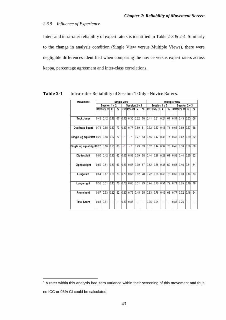
Chapter 2: Reliability of Movement Screen
43
2.3.5 Influence of Experience
Inter- and intra-rater reliability of expert raters is identified in Table 2-3 & 2-4. Similarly
to the change in analysis condition (Single View versus Multiple Views), there were
negligible differences identified when comparing the novice versus expert raters across
kappa, percentage agreement and inter-class correlations.
Table 2-1 Intra-rater Reliability of Session 1 Only - Novice Raters.
Movement Single View Multiple View
Session 1 v 2 Session 2 v 3 Session 1 v 2 Session 2 v 3
ICC 95% CI k % ICC 95% CI k % ICC 95% CI k % ICC 95% CI k %
Tuck Jump 0.48 0.42 0.18 67 0.40 0.30 0.22 78 0.41 0.31 0.24 61 0.51 0.43 0.33 66
Overhead Squat 0.71 0.65 0.33 73 0.80 0.77 0.59 81 0.72 0.67 0.45 71 0.66 0.59 0.37 68
Single leg squat left 0.29 0.18 0.22 77 - 1 - 1 0.27 83 0.55 0.47 0.38 77 0.48 0.42 0.39 82
Single leg squat right 0.27 0.16 0.25 80 - 1 - 1 0.29 83 0.52 0.44 0.37 78 0.46 0.38 0.36 80
Dip test left 0.50 0.42 0.30 62 0.65 0.59 0.39 68 0.44 0.36 0.23 64 0.52 0.44 0.25 62
Dip test right 0.59 0.51 0.33 63 0.63 0.57 0.39 67 0.62 0.56 0.36 69 0.53 0.46 0.31 64
Lunge left 0.54 0.47 0.26 73 0.73 0.68 0.52 78 0.72 0.68 0.48 76 0.65 0.60 0.44 73
Lunge right 0.58 0.51 0.43 76 0.70 0.65 0.51 79 0.74 0.70 0.51 79 0.71 0.65 0.49 76
Prone hold 0.57 0.53 0.32 52 0.80 0.75 0.45 65 0.83 0.78 0.45 63 0.77 0.72 0.48 64
Total Score 0.85 0.81 - - 0.89 0.87 - - 0.95 0.94 - - 0.88 0.76 - -
1 A rater within this analysis had zero variance within their screening of this movement and thus
no ICC or 95% CI could be calculated.

Chapter 2: Reliability of Movement Screen
44
Table 2-2 Intra-rater Reliability of Session 1,2,3 - Novice Raters.
Movement Single View Multiple View
Session 1 v 2 Session 2 v 3 Session 1 v 2 Session 2 v 3
ICC 95% CI k % ICC 95% CI k % ICC 95% CI k % ICC 95% CI k %
Tuck Jump 0.12 -0.05 -0.02 45 0.34 0.16 0.05 49 -0.02 -0.21 0.09 72 -0.04 -0.30 0.00 69
Overhead Squat 0.59 0.52 0.31 62 0.49 0.36 0.29 70 0.48 0.36 0.28 66 0.57 0.48 0.27 66
Single leg squat left 0.46 0.36 0.16 62 0.34 0.20 0.07 59 0.14 -0.04 0.03 62 0.61 0.52 0.39 73
Single leg squat right 0.47 0.37 0.20 61 0.47 0.36 0.19 61 0.46 0.37 0.20 66 0.60 0.50 0.48 75
Dip test left 0.35 0.24 0.11 57 0.54 0.47 0.25 61 0.14 -0.01 0.04 58 0.43 0.30 0.22 66
Dip test right 0.01 -0.19 -0.07 50 0.41 0.27 0.09 54 0.32 0.19 0.14 62 0.55 0.44 0.30 68
Lunge left 0.30 0.18 0.23 63 0.63 0.54 0.39 72 0.22 0.07 0.21 71 0.35 0.18 0.36 73
Lunge right 0.22 0.07 0.06 59 0.59 0.50 0.42 72 0.35 0.24 0.19 68 0.54 0.42 0.36 73
Prone hold 0.44 0.35 0.21 68 0.60 0.51 0.23 59 0.55 0.48 0.20 49 0.56 0.46 0.18 52
Total score 0.51 0.44 - - 0.82 0.77 - - 0.55 0.47 - - 0.83 0.81 - -

Chapter 2: Reliability of Movement Screen
45
Table 2-3 Inter- and Intra-rater Reliability of Session 1 Only - Expert Raters.
Movement Session 1 Only
Session 1 v 2 Session 2 v 3 Session 1 Session 2 Session 3
ICC 95% CI k % ICC 95% CI k % ICC 95% CI k % ICC 95% CI k % ICC 95% CI k %
Tuck Jump 0.43 -0.05 0.41 77 0.34 0.23 0.22 77 0.26 0.20 0.18 63 0.48 0.42 0.36 72 0.28 0.19 0.20 71
Overhead Squat 0.70 0.52 0.58 78 0.61 0.55 0.45 82 0.65 0.56 0.32 67 0.58 0.50 .040 73 0.69 0.60 0.43 73
Single leg squat left 0.15 0.36 0.49 85 0.03 -0.11 0.06 83 0.42 0.35 0.24 81 0.29 0.18 0.20 89 0.57 0.51 0.29 87
Single leg squat right 0.17 0.37 0.46 85 0.09 -0.09 0.03 87 0.34 0.25 0.17 80 0.15 0.03 0.30 93 0.59 0.53 0.31 87
Dip test left 0.32 0.24 0.04 53 0.43 0.34 0.22 55 0.33 0.26 0.07 39 0.51 0.43 0.39 70 0.37 0.26 0.24 55
Dip test right 0.30 -0.19 0.25 43 0.49 0.41 0.17 58 0.26 0.20 0.06 39 0.39 0.30 0.26 66 0.61 0.54 0.35 62
Lunge left 0.23 0.18 0.07 55 0.57 0.50 0.35 63 0.10 0.06 0.07 36 0.56 0.49 0.35 65 0.54 0.45 0.33 64
Lunge right 0.34 0.07 -0.10 58 0.53 0.46 0.28 68 0.19 0.15 0.05 35 0.39 0.32 0.25 62 0.52 0.44 0.31 63
Prone hold 0.46 0.35 0.24 45 0.50 0.43 0.23 57 0.25 0.15 -0.01 24 0.03 -0.05 0.10 31 0.39 0.34 0.17 30
Total Score 0.81 0.44 - - 0.85 0.83 - - 0.71 0.49 - - 0.88 0.86 - - 0.85 0.81 - -

Chapter 2: Reliability of Movement Screen
46
Table 2-4 Inter- and Intra-rater Reliability of Session 1,2,3 - Expert Raters.
Movement Session 1,2,3
Session 1 v 2 Session 2 v 3 Session 1 Session 2 Session 3
ICC 95% CI k % ICC 95% CI k % ICC 95% CI k % ICC 95% CI k % ICC 95% CI k %
Tuck Jump 0.14
-0.04 0.2 64 0.29 0.25 -0.01 62 0.16 0.13 0.08 46 0.37 0.27 0.12 52 0.5 0.28 0.24 52
Overhead Squat 0.61 0.52 0.37 66 0.58 0.48 0.38 73 0.56 0.48 0.34 61 0.4 0.28 0.17 48 0.59 0.48 0.32 64
Single leg squat left 0.38 0.25 0.28 73 0.61 0.52 0.31 77 0.4 0.31 0.19 61 0.1 0.1 -0.01 31 0.22 0.17 0.08 39
Single leg squat right 0.27 0.14 0.32 77 0.26 0.12 0.23 73 0.41 0.36 0.14 65 0 0.01 - 2 36 0.18 0.14 0.1 42
Dip test left 0.54 0.43 0.28 57 0.72 0.65 0.5 73 0.49 0.38 0.11 44 0.26 0.2 -0.21 20 0.25 0.19 0.01 23
Dip test right 0.49 0.37 0.22 58 0.57 0.48 0.38 67 0.53 0.4 0.14 50 0.26 0.21 -0.07 14 0.24 0.18 0.07 33
Lunge left 0.32 0.27 0.15 55 0.66 0.58 0.43 72 0.47 0.37 0.01 63 0.42 0.26 -0.07 29 0.41 0.26 -0.03 28
Lunge right 0.42 0.37 0.18 58 0.51 0.4 0.33 69 0.4 0.3 0.09 63 0.33 0.23 -0.1 31 0.41 0.29 -0.15 19
Prone hold 0.58 0.49 0.18 50 0.73 0.66 0.32 65 0.12 0.1 0.12 28 0.19 0.17 0.03 17 0.12 0.13 0.02 11
Total Score 0.53 0.42 - - 0.82 0.8 - - 0.72 0.44 - - 0.76 0.39 - - 0.8 0.53 - -
2 A rater within this analysis had zero variance within their screening of this movement and thus no Kappa value could be calculated.

Chapter 2: Reliability of Movement Screen
47
Table 2-5 Inter-rater Reliability of Session 1 Only - Novice Raters.
Movement Single View Multiple View
Session 1 Session 2 Session 3 Session 1 Session 2 Session 3
ICC 95% CI k % ICC 95% CI k % ICC 95% CI k % ICC 95% CI k % ICC 95% CI k % ICC 95% CI k %
Tuck Jump 0.50 0.43 0.28 56 0.29 0.27 0.16 45 0.39 0.34 0.12 49 0.30 0.22 0.11 49 0.27 0.17 0.22 59 0.19 0.13 0.09 47
Overhead Squat 0.65 0.57 0.38 64 0.56 0.47 0.34 59 0.60 0.49 0.31 61 0.67 0.59 0.38 65 0.61 0.53 0.29 59 0.42 0.32 0.10 45
Single leg squat left 0.30 0.25 0.13 60 0.06 0.00 0.08 63 0.12 0.04 0.06 72 0.37 0.30 0.18 62 0.60 0.54 0.43 76 0.07 -0.01 0.05 71
Single leg squat right 0.32 0.22 0.09 61 0.08 0.02 0.14 65 0.17 0.10 0.32 71 0.37 0.33 0.29 63 0.52 0.43 0.25 70 0.19 0.12 0.13 68
Dip test left 0.33 0.25 0.03 40 0.34 0.26 0.05 33 0.32 0.25 0.08 40 0.43 0.33 0.08 50 0.44 0.36 0.14 61 0.38 0.27 -0.04 36
Dip test right 0.35 0.26 0.07 44 0.50 0.36 0.11 41 0.44 0.36 0.16 49 0.45 0.35 0.09 49 0.59 0.49 0.24 62 0.28 0.21 -0.03 45
Lunge left 0.16 0.04 0.08 62 0.30 0.20 0.03 58 0.28 0.18 0.11 52 0.47 0.38 0.16 53 0.55 0.46 0.21 63 0.09 0.04 0.04 45
Lunge right 0.30 0.19 0.20 65 0.51 0.42 0.22 68 0.25 0.17 0.07 51 0.46 0.38 0.16 64 0.48 0.40 0.14 63 0.19 0.13 0.06 48
Prone hold 0.12 0.09 0.06 17 0.39 0.28 0.08 21 0.50 0.40 0.19 37 0.70 0.60 0.19 40 0.60 0.48 0.25 45 0.53 0.41 0.26 47
Total Score 0.75 0.68 - - 0.85 0.64 - - 0.73 0.55 - - 0.84 0.57 - - 0.91 0.77 - - 0.70 0.48 - -

Chapter 2: Reliability of Movement Screen
48
Table 2-6 Inter-rater Reliability of Session 1,2,3 - Novice Raters.
Movement Single View Multiple View
Session 1 Session 2 Session 3 Session 1 Session 2 Session 3
ICC 95% CI k % ICC 95% CI k % ICC 95% CI k % ICC 95% CI k % ICC 95% CI k % ICC 95% CI k %
Tuck Jump 0.35 0.30 0.09 44 0.53 0.43 0.25 54 0.49 0.38 0.14 48 0.23 0.19 0.08 43 0.11 0.09 0.06 40 -0.02 -0.13 0.06 39
Overhead Squat 0.53 0.43 0.24 57 0.49 0.36 0.16 45 0.56 0.44 0.30 56 0.63 0.57 0.36 65 0.35 0.25 0.13 51 0.61 0.49 0.31 61
Single leg squat left 0.31 0.23 0.12 46 0.24 0.18 0.02 36 0.33 0.27 0.17 49 0.17 0.13 0.08 45 0.37 0.28 0.16 50 0.49 0.39 0.23 56
Single leg squat right 0.36 0.28 0.09 40 0.25 0.19 0.00 41 0.40 0.31 0.17 47 0.45 0.34 0.19 50 0.39 0.30 0.16 53 0.49 0.36 0.18 55
Dip test left 0.41 0.33 0.18 55 0.36 0.28 0.20 53 0.40 0.33 0.11 53 0.26 0.19 0.00 45 0.30 0.22 0.02 48 0.29 0.15 0.00 51
Dip test right 0.24 0.19 0.08 49 0.49 0.40 0.66 54 0.38 0.33 0.14 39 0.26 0.21 -0.03 35 0.16 0.10 0.03 42 0.34 0.25 0.08 59
Lunge left 0.27 0.18 0.10 50 0.48 0.34 0.17 53 0.44 0.34 0.23 57 0.27 0.23 0.20 61 0.09 -0.01 -0.07 54 0.40 0.30 0.11 58
Lunge right 0.19 0.13 0.07 46 0.28 0.15 0.07 48 0.38 0.28 0.15 52 0.42 0.35 0.17 57 0.15 0.11 0.08 54 0.19 0.10 0.15 61
Prone hold 0.28 0.21 0.45 69 0.19 0.08 0.07 52 0.69 0.58 0.29 55 0.33 0.19 0.05 27 0.42 0.29 0.11 38 0.26 0.19 0.09 32
Total score 0.85 0.64 - - 0.92 0.74 - - 0.95 0.82 - - 0.78 0.53 - - 0.86 0.61 - - 0.93 0.82 - -

Chapter 2: Reliability of Movement Screen
49
2.4 Discussion
The ability to determine movement dysfunction in order to intervene to an individual to
potentially reduce their prevalence of musculoskeletal injuries has enormous appeal due
to the detrimental social and economic effects of sporting injuries on athletes (Caine,
Maffulli et al. 2008). A more feasible solution to identify altered movement strategies
than the costly and time-consuming process of three-dimensional biomechanical motion
analysis (McLean, Walker et al. 2005) is to develop a reliable field-based movement
screen that can be easily implemented by coaches and clinicians alike. For any movement
screen to be implemented it must be reliable (Padua, Boling et al. 2011). This study is the
first to demonstrate that pre-elite youth athletes display a familiarisation effect when
performing novel movement screen tasks, which was not observed on further exposure to
performing the tasks. The results also demonstrate a lack of reliability in individual screen
tasks, however excellent reliability was observed when using the total composite score.
Previous research has inappropriately employed ICCs to determine the reliability of
screening tools for ordinal data (Chorba, Chorba et al. 2010, Gribble, Brigle et al. 2013),
which is an incorrect statistical procedure for categorical data, as ICCs are only
appropriate for scalar data (Sim and Wright 2005). Within this study, ICCs were
employed only for purposes of comparison with previous research of individual
movement tasks (ordinal data), and to assess reliability for the total composite score
(scalar data). This study employed the kappa score as it is an appropriate statistical
measure of reliability of categorical data to assess “true” agreement (Sim and Wright
2005), in addition to the percentage agreement (Mitchell 1979) for individual movements
of the MDS.
Novice and expert raters within this study were deemed too variable to reliably rate any
of the individual movements within the MDS between rating sessions. This variability
has been suggested to be due to the complex nature of viewing multiple identifiable

Chapter 2: Reliability of Movement Screen
50
features at once, thought to interfere with information processing (Whiteside, Deneweth
et al. 2016). The results of this study however, indicate that repeated viewing of the
movement screen (N-Multiple-1 and N-Multiple-123), to allow reduced number of
identifiable features at once, does not result in a learning effect or improve reliability of
the rater. The reduced rater reliability observed in the individual movements may be
attributed to the small scale on which the movement is scored (Preston and Colman 2000).
The current individual movement scoring system requires each of the movement errors
observed to counted up and placed into a category (1, 2 or 3). Research has suggested that
scoring criterion of only three categories results in reduced reliability, validity and
discrimination, whereas scales between 7-10 categories are most reliable (Preston and
Colman 2000). Furthermore, research has shown increased sensitivity when utilising error
count as opposed to the three point category method (Perrott, Pizzari et al. 2017). It is
therefore suggested that the criterion composing of a summation of the number of errors
for each movement (total composite score) be utilised for scoring the individual
movements of the MDS instead of the typical three categories used within this study and
the FMS.
The categorical limits applied to movement screening are often arbitrary with data lost
due to their categorical nature (Sim and Wright 2005). This choice of categorical limit
and loss of data can often result in a reduction of statistical power, leaving the kappa score
meaningless (Sim and Wright 2005). This reduction in statistical power was likely to be
confounded by skewed data distribution of the data for each individual movement task
(Sim and Wright 2005) (Table 2-7). That is, most athletes displayed numerous movement
errors, leading to majority of participants being rated as a one or two for individual
movements (Percentage of participants in category 1 and 2: tuck jump 96%; overhead
squat 86%; single leg squat left 95% and right 95%; dip test left 84% and right 83%;
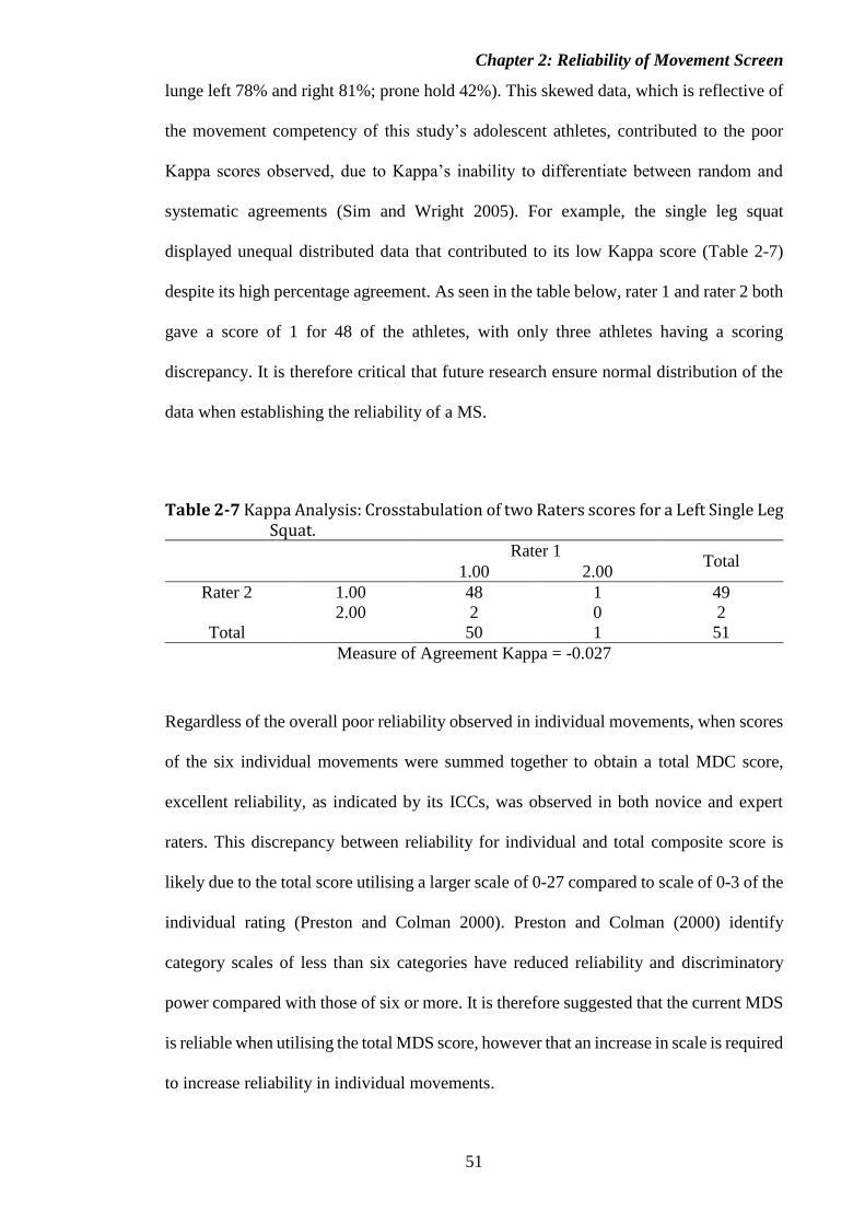
Chapter 2: Reliability of Movement Screen
51
lunge left 78% and right 81%; prone hold 42%). This skewed data, which is reflective of
the movement competency of this study’s adolescent athletes, contributed to the poor
Kappa scores observed, due to Kappa’s inability to differentiate between random and
systematic agreements (Sim and Wright 2005). For example, the single leg squat
displayed unequal distributed data that contributed to its low Kappa score (Table 2-7)
despite its high percentage agreement. As seen in the table below, rater 1 and rater 2 both
gave a score of 1 for 48 of the athletes, with only three athletes having a scoring
discrepancy. It is therefore critical that future research ensure normal distribution of the
data when establishing the reliability of a MS.
Table 2-7 Kappa Analysis: Crosstabulation of two Raters scores for a Left Single Leg Squat.
Rater 1
Total 1.00 2.00
Rater 2 1.00 48 1 49
2.00 2 0 2
Total 50 1 51
Measure of Agreement Kappa = -0.027
Regardless of the overall poor reliability observed in individual movements, when scores
of the six individual movements were summed together to obtain a total MDC score,
excellent reliability, as indicated by its ICCs, was observed in both novice and expert
raters. This discrepancy between reliability for individual and total composite score is
likely due to the total score utilising a larger scale of 0-27 compared to scale of 0-3 of the
individual rating (Preston and Colman 2000). Preston and Colman (2000) identify
category scales of less than six categories have reduced reliability and discriminatory
power compared with those of six or more. It is therefore suggested that the current MDS
is reliable when utilising the total MDS score, however that an increase in scale is required
to increase reliability in individual movements.

Chapter 2: Reliability of Movement Screen
52
Despite a learning effect previously demonstrated in novel dynamic movement tasks
(Hansen, Dieckmann et al. 2000), the familiarisation effect within movement screening
has not previously been determined. This study identifies a familiarisation effect in
athletes during initial performance of the MDS, as seen with the total composite score
during ‘session 123’ video viewing (N-123-Single, N-123-Multiple and E-123-Single)
increasing from moderate to excellent reliability from session1v2 to 2v3. This highlights
that this MDS requires at least one familiarisation session in which the athlete performs
the MDS prior to rating. It is plausible that more than one familiarisation sessions may be
required but the exact number cannot be ascertain within this current study. Without
familiarisation of the MDS, the recorded scores of the individual MDS movements cannot
be considered a true representation of the athlete’s movement competency. Conversely,
no learning effect was present among either novice nor expert raters for the total MDS
score during ‘single’ video viewing (N-1-Single and E-1-Single) of the movement. The
excellent reliability of the total composite score as a single viewing highlights that this
MDS can be reliably replicated in real-time field-based environments which the MDS
would be typically employed.
The authors acknowledge potential limitations within this study. Raters within this study
were volunteers and had extensive volumes of movement screens to rate. This could have
led to reduced attention span during some screening due to the tedious nature of the
sessions. The movement screening was recorded on two video cameras (lateral and
sagittal views). When watching these videos, it was only possible to watch one view at a
time, which therefore increased the time required to view each MDS.

Chapter 2: Reliability of Movement Screen
53
2.5 Conclusions
The results of this study indicate that a rater’s movement screening experience does not
affect their reliability and for raters scoring the MDS. Results also indicate that the total
composite score can be reliably used to determine movement competency; however, the
individual movements scores should not be relied on. The requirement of athlete
familiarisation prior to MDS rating has also been identified. Therefore, it is suggested
that MDS classification be based on the total composite score, following a familiarisation
session, and that future research increase the scale for individual movement screening
scores to obtain more reliable individual movement scores.
2.6 Acknowledgements
The authors gratefully acknowledge the Hunter Academy of Sport for providing the
participants for this study and all the contribution of the raters who generously gave their
time to complete the movement screening for this study.
2.7 References
1. Mottram S, Comerford M. A new perspective on risk assessment. Phys Ther in Sport.
2008;9(1):40-51.
2. Padua DA, Marshall SW, Boling MC, Thigpen CA, Garrett WE, Beutler AI. The landing error
scoring system (LESS) is a valid and reliable clinical assessment tool of jump-landing
biomechanics. Am J Sport Med. 2009;37(10):1996-2002.
3. Myer GD, Ford KR, Hewett TE. Tuck jump assessment for reducing anterior cruciate ligament
injury risk. Athlet Ther Today. 2008;13(5):39-44.
4. Cook G, Burton L, Hoogenboom B. Pre-participation screening: The use of fundamental
movements as an assessment of function–Part 1. N Am J Sports Phys Ther. 2006;1(2):62-72.

Chapter 2: Reliability of Movement Screen
54
5. Cook G, Burton L, Hoogenboom B. Pre-participation screening: The use of fundamental
movements as an assessment of function–Part 2. N Am J Sports Phys Ther. 2006;1(3):132-9.
6. Bradley PS, Portas MD. The relationship between preseason range of motion and muscle strain
injury in elite soccer players. J Strength Cond Res. 2007;21(4):1155-9.
7. Ebenbichler GR, Oddsson LI, Kollmitzer J, Erim Z. Sensory-motor control of the lower back:
Implications for rehabilitation. Med Sci Sports Exerc. 2001;33(11):1889-98.
8. Mann KJ, Edwards S, Drinkwater EJ, Bird SP. A lower limb assessment tool for athletes at risk
of developing patellar tendinopathy. Med Sci Sports Exerc. 2013;45(3):527-33.
9. Parkkari J, Kujala UM, Kannus P. Is it possible to prevent sports injuries? Review of controlled
clinical trials and recommendations for future work. Sports Med. 2001;31(14):985-95.
10. Hewett TE, Myer GD, Ford KR, Heidt Jr RS, Colosimo AJ, McLean SG et al. Biomechanical
measures of neuromuscular control and valgus loading of the knee predict anterior cruciate
ligament injury risk in female athletes. Am J Sport Med. 2005;33(4):492-501.
11. Edwards S, Steele JR, McGhee DE, Beattie S, Purdam C, Cook JL. Landing strategies of
athletes with an asymptomatic patellar tendon abnormality. Med Sci Sport Exer.
2010;42(11):2072-80.
12. Kiesel K, Plisky PJ, Voight ML. Can serious injury in professional football be predicted by a
preseason functional movement screen? N Am J Sports Phys Ther. 2007;2(3):147.
13. Chorba RS, Chorba DJ, Bouillon LE, Overmyer CA, Landis JA. Use of a functional movement
screening tool to determine injury risk in female collegiate athletes. N Am J Sports Phys Ther.
2010;5(2):47-54.
14. Bardenett SM, Micca JJ, DeNoyelles JT, Miller SD, Jenk DT, Brooks GS. Functional
Movement Screen normative values and validity in high school athletes: can the FMS™ be
used as a predictor of injury? Int J Sports Phys Ther. 2015;10(3):303.
15. Bushman TT, Grier TL, Canham-Chervak M, Anderson MK, North WJ, Jones BH. The
functional movement screen and injury risk: Association and predictive value in active men.
Am J Sport Med. 2016;44(2):297-304.
16. Myer GD, Chu DA, Brent JL, Hewett TE. Trunk and hip control neuromuscular training for
the prevention of knee joint injury. Clin Sport Med. 2008;27(3):425-48.

Chapter 2: Reliability of Movement Screen
55
17. Hibbs AE, Thompson KG, French D, Wrigley A, Spears I. Optimizing performance by
improving core stability and core strength. Sports Med. 2008;38(12):995-1008.
18. Hewett TE, Myer GD. The mechanistic connection between the trunk, hip, knee, and anterior
cruciate ligament injury. Exerc Sport Sci Rev. 2011;39(4):161-6.
19. Cowan SM, Schache AG, Brukner P, Bennell KL, Hodges PW, Coburn P et al. Delayed onset
of transversus abdominus in long-standing groin pain. Med Sci Sports Exerc.
2004;36(12):2040-5.
20. Kuszewski M, Gnat R, Saulicz E. Stability training of the lumbo‐pelvo‐hip complex influence
stiffness of the hamstrings: A preliminary study. Scand J Med Spor. 2009;19(2):260-6.
21. Beckman SM, Buchanan TS. Ankle inversion injury and hypermobility: Effect on hip and
ankle muscle electromyography onset latency. Arch Phys Med Rehab. 1995;76(12):1138-43.
22. Zazulak BT, Hewett TE, Reeves NP, Goldberg B, Cholewicki J. Deficits in neuromuscular
control of the trunk predict knee injury risk. Am J Sports Med. 2007;35(7):1123-30.
23. Akuthota V, Ferreiro A, Moore T, Fredericson M. Core stability exercise principles. Curr
Sport Med Rep. 2008;7(1):39-44.
24. Perrott MA, Pizzari T, Opar M, Cook J. Development of clinical rating criteria for tests of
lumbopelvic stability. Rehabil Res Pract. 2011;2012:7.
25. McHugh ML. Interrater reliability: The kappa statistic. Biochem Medica. 2012;22(3):276-82.
26. Teyhen DS, Shaffer SW, Lorenson CL, Halfpap JP, Donofry DF, Walker MJ et al. The
functional movement screen: A reliability study. J Orthop Sports Phys Ther. 2012;42(6):530-
40.
27. Tinsley HE, Weiss DJ. Interrater reliability and agreement of subjective judgments. J Couns
Psychol. 1975;22(4):358.
28. Padua DA, Boling MC, DiStefano LJ, Onate JA, Beutler AI, Marshall SW. Reliability of the
landing error scoring system-real time, a clinical assessment tool of jump-landing
biomechanics. J Sport Rehabil. 2011;20:145-56.
29. Minick KI, Kiesel KB, Burton L, Taylor A, Plisky P, Butler RJ. Interrater reliability of the
functional movement screen. J Strength Cond Res. 2010;24(2):479-86.
Chapter 2: Reliability of Movement Screen
56
30. Gribble PA, Brigle J, Pietrosimone BG, Pfile KR, Webster KA. Intrarater reliability of the
functional movement screen. J Strength Cond Res. 2013;27(4):978-81.
31. Smith CA, Chimera NJ, Wright NJ, Warren M. Interrater and intrarater reliability of the
functional movement screen. J Strength Cond Res. 2013;27(4):982-7.
32. Gribble PA, Brigle J, Pietrosimone BG, Pfile KR, Webster KA. Intrarater reliability of the
functional movement screen. J Strength Cond Res. 2013;27(4):978-81.
33. Sim J, Wright CC. The kappa statistic in reliability studies: use, interpretation, and sample
size requirements. Phys Ther. 2005;85(3):257-68.
34. Onate JA, Dewey T, Kollock RO, Thomas KS, Van Lunen BL, DeMaio M et al. Real-time
intersession and interrater reliability of the functional movement screen. J Strength Cond Res.
2012;26(2):408-15.
35. Hopkins WG. Measures of reliability in sports medicine and science. Sports Med.
2000;30(1):1-15.
36. Schmidt RA, Wrisberg CA. Motor learning and performance: A situation-based learning
approach. Champaign: Human kinetics; 2008.
37. Whiteside D, Deneweth JM, Pohorence MA, Sandoval B, Russell JR, McLean SG et al.
Grading the functional movement screen: A comparison of manual (real-time) and objective
methods. J Strength Cond Res. 2016;30(4):924-33.
38. Craik KJW. Theory of the human operator in control systems. Brit J Psychol. 1947;38(2):56-
61.
39. Wickens CD. Multiple resources and performance prediction. Theor Issues in Ergon Sci.
2002;3(2):159-77.
40. Landis JR, Koch GG. The measurement of observer agreement for categorical data.
Biometrics. 1977;33:159-74.
41. Baumgartner TA, Strong CH, Hensley LD. Conducting and reading research in health and
human performance (3rd Ed.). New York: McGraw-Hill; 2006.
42. Fleiss JL. Reliability of measurement. The Design and Analysis of Clinical Experiments. New
York: John Wiley and Sons, Inc; 1986. p. 2-32.

Chapter 2: Reliability of Movement Screen
57
43. Birkimer JC, Brown JH. Back to basics: Percentage agreement measures are adequate, but
there are easier ways. J Appl Behav Anal. 1979;12(4):535-43.
44. Caine D, Maffulli N, Caine C. Epidemiology of injury in child and adolescent sports: injury
rates, risk factors, and prevention. Clin Sport Med. 2008;27(1):19-50.
45. McLean S, Walker K, Ford K, Myer G, Hewett T, Van Den Bogert A. Evaluation of a two
dimensional analysis method as a screening and evaluation tool for anterior cruciate ligament
injury. Brit J Sport Med. 2005;39(6):355-62.
46. Mitchell SK. Interobserver agreement, reliability, and generalizability of data collected in
observational studies. Psychol Bull. 1979;86(2):376.
47. Preston CC, Colman AM. Optimal number of response categories in rating scales: reliability,
validity, discriminating power, and respondent preferences. Acta Psychol. 2000;104(1):1-15.
48. Perrott M, Pizzari T, Cook J. Assessment of lumbopelvic stability: Beyond a three-point rating
scale. J Sci Med Sport. 2017;20:25-6.
49. Hansen M, Dieckmann B, Jensen K, Jakobsen B. The reliability of balance tests performed
on the kinesthetic ability trainer (KAT 2000). Knee Surg Sport Tr A. 2000;8(3):180-5.
60
Context of Manuscript in Relation to the Thesis
This manuscript is an investigation into the effectiveness of whether a 12-week
intervention program can modify jump-landing strategies in pre-elite youth athletes. The
stop-jump task was chosen as the experimental task as many lower limb injuries and
intervention programs are associate with landing mechanics during a stop-jump
movement. However, many of these investigations have been conducted within an adult
population despite the adolescents population being identified as a key population at risk
of lower limb injury. With specialised equipment required for many of these intervention
programs, it creates accessibility issues in rural and remote communities that often do not
have access to these resources. This manuscript addresses these limitations in previous
research by providing a 3D biomechanical motion analysis of a stop-jump task and
evaluates the effectiveness of four intervention programs (core, landing, core and landing
and control) to modify jump-landing strategies and variables associated with lower limb
injury risk during landing. Results of this manuscript will provide coaches and clinicians
valuable information on the pre-elite youth athlete population and their understanding of
training program adaptations of these athletes with limited access to facilities and
practical recommendations for injury prevention training

Chapter 3: Intervention programs alters stop-jump technique
61
Chapter 3. Stop-jump landing technique is modified by
neuromuscular training programs in pre-elite youth
athletes
This chapter is an amended version of the manuscript: Mann K, O’ Dwyer N, Bruton M,
Edwards S. Stop-jump landing technique is modified by neuromuscular training programs
in pre-elite youth athletes. To be submitted to the Medicine and Science in Sports and
Exercise.
ABSTRACT
Purpose: Neuromuscular training interventions for the reduction of lower limb injuries
have been widespread; however, the current local focus on injury prevention at specific
joints, when compared to a focus on the global kinetic chain, is less effective, as
evidenced by high rates of re-injury. Research suggests that lumbopelvic stability may be
critical in the prevention of lower limb injuries in athletes as a global focus in injury
prevention. This study’s purpose was to compare the effects of three different 12-week
intervention programs on the results of a full biomechanical analysis during a stop-jump
landing tasks in pre-elite youth athletes.
Methods: Eighty-Nine junior pre-elite athletes with no current signs or symptoms of
injury were recruited from the Western Region Academy of Sport. Five successful stop-
jump tasks were completed both before and after exposure to one of three different 12-
week training programs, or the control program, in conjunction with a strength and
conditioning program for each participant. Mixed design factorial analysis of variance
(ANOVA) was used to determine statistically significant between-group differences
(P≤0.05).

Chapter 3: Intervention programs alters stop-jump technique
62
Results: There was no main effect of intervention type for any kinematic or kinetic
variables; however, athletes used a straight trunk posture as shown via increased L5-S1
extension, thoracic-abdominal flexion identified across the landing phase (pre = 7.2 ±
1.0°; post=4.2±0.9°) in post- than pre-intervention testing. Post-intervention testing
showed significantly reduced hip flexion; T12-L1 rotation; hip external rotation, and
increased L5-S1 flexion with a decrease in L5-S1 rotation range of motion (ROM), an
increase in peak L5-S1 angles and a decrease in L5-S1 peak extension angles within the
landing phase than pre-intervention testing. Significantly decreased medial ground
reaction force (GRF)s and increased lateral GRFs accompanied by a significant decrease
in duration from initial contact (IC) to peak anterior force and a decrease in IC-FV1
loading rate was also identified in post- compared to pre-intervention testing.
Conclusions: Results of this study demonstrate that regardless of intervention program,
all athletes displayed similar changes pre- vs post-intervention results. It is therefore
recommended that a single strength and conditioning program can be implemented in pre-
elite youth athletes with poor movement competency as an injury reduction strategy.
Interventions with alternate feedback or implemented within athletes with increased
movement competency may provide more effective results than the current programs
investigated.
Keywords: Injury, biomechanics, movement screening, prevention through prediction,
dynamic landing
3.1 Introduction
The cost of sports injuries in Australia is increasing with an estimated cost of AUS$1.5
billion dollars in 2003 (Medibank Private 2003), rising to AUS$2 billion dollars in 2006
(Medibank Private 2006); however, a lack of population-wide data has limited more
Chapter 3: Intervention programs alters stop-jump technique
63
recent figures of the exact extent of injury cost in Australia (Finch 2011). Previous
research has identified a relationship between growth, maturation and age that may
predispose young athletes to injury (Emery 2003, Myer, Chu et al. 2008), specifically
lower limb injuries that are more prevalent following adolescence (Finch, Valuri et al.
1998). The adolescent population has a higher incident of sporting injury (Medibank
Private 2003), increased adaptability of the tendon to load and stress (Tuite, Renström et
al. 1997), and following the pre-pubertal stage of maturation, high risk landing techniques
begin to emerge (Hewett, Myer et al. 2004, Sigward, Pollard et al. 2012, Sigward, Pollard
et al. 2012). Due to the substantial cost and potentially devastating effect of sporting
injuries, injury reduction programs are becoming increasingly popular with the aim of
reducing injury risk factors.
Landing technique is considered a key modifiable risk factor in lower limb injury,
specifically the eccentric loading phase of a landing movement that is highly influential
in the incidence of lower limb injuries (Boden, Dean et al. 2000, Myer, Ford et al. 2008,
Edwards, Steele et al. 2010, Mann, Edwards et al. 2013). Landing risk factors such as
increased (Richards, Ajemian et al. 1996, Edwards, Steele et al. 2010, Mann, Edwards et
al. 2013) or decreased (Boden, Dean et al. 2000) knee joint flexion, incorrect hip
movement strategies (Edwards, Steele et al. 2010, Mann, Edwards et al. 2013), increased
knee internal rotation (Hewett, Myer et al. 2005) and increase hip adduction angles
(Edwards, Steele et al. 2010) have been identified as biomechanical injury risk factors
during a jump landing task. Dynamic neuromuscular landing re-training programs in
adolescent female volleyball athletes has been shown to decrease forces and modify
critical biomechanical injury risk factors during landing. For risk factors such as peak
GRF’s and peak knee moments within the eccentric loading phase (Hewett, Stroupe et al.
1996), landing retraining is a commonly used intervention for the reduction of lower limb
Chapter 3: Intervention programs alters stop-jump technique
64
injuries (Hewett, Lindenfeld et al. 1999, Myklebust, Engebretsen et al. 2003,
Mandelbaum, Silvers et al. 2005). Many of these programs however, have been
implemented within a late adolescent or adult population (Chappell and Limpisvasti 2008,
Herman, Weinhold et al. 2008, McCurdy, Walker et al. 2012). This is despite research
suggesting that early adolescence population are at a key development phase for
implementation of landing retraining injury prevention strategies (Myer, Faigenbaum et
al. 2011). During the critical early adolescence phase, cognitive and motor capabilities
are highly susceptible to age appropriate training interventions (Myer, Faigenbaum et al.
2011). Before early adolescence (children younger than 12 years) is not the most
appropriate time to implement interventions as they are unable to alter lower limb
biomechanics with training (DiStefano, Blackburn et al. 2011). Dynamic neuromuscular
training programs in adolescent females has been reported to be effective for improving
their lower limb movement mechanics during landing and in turn, decreasing injury risk
(Myer, Ford et al. 2005). Large gains in performance and stability within the novice
populations are thought to occur mainly through neuromuscular adaptations such as more
comprehensive and synchronous firing of the muscular unit. These neuromuscular
adaptations can result in improved skill and coordination (Rutherford and Jones 1986),
which supports the notion that the novice adolescent population as at a key time for
intervention strategies to be implemented aimed at reducing their injury risk (Myer,
Faigenbaum et al. 2011).
For interventions to be successful within this population, equipment must be readily
available and strategies suited to junior sporting organisations. Many of the current
neuromuscular retraining programs are performed in controlled environments such as
supervised gyms and require specialised equipment (Hewett, Lindenfeld et al. 1999,
Myklebust, Engebretsen et al. 2003). This can create access issues, especially in rural
Chapter 3: Intervention programs alters stop-jump technique
65
regions where access to formal sporting structures and support mechanisms is limited
(Finch, Mahoney et al. 2003). Reduced access may affect adherence rates (Tappe, Duda
et al. 1989), which is negatively associated with injury risk (Soligard, Nilstad et al. 2010).
Therefore, to have the greatest effect on adolescent sporting injury rates, it is essential
that an accessible, easily implemented field based program is developed to aid injury
prevention among pre-elite youth athletes.
A key focus of neuromuscular prevention strategies has been to improve an individual’s
landing technique (Hewett, Stroupe et al. 1996, Myer, Ford et al. 2005). Yet focusing on
a specific joint (e.g. the knee joint for ACL injuries) and its symptoms (e.g. increased
knee abduction in ACL injuries) to prevent lower limb injuries is ineffective, as high rates
of injury and re-injury remain (Mottram and Comerford 2008) despite advanced in sports
medicine and rehabilitation (Hewett, Ford et al. 2006). A broader focus on the entire
kinetic chain might therefore be warranted that incorporates the lumbopelvic region. An
athlete who displays delayed or poor neuromuscular control in the lumbopelvic region
during dynamic landing tasks may be predisposed to a lower limb injury through poor
stability, motion and increased forces experienced by the lower limb joints (Myer, Chu et
al. 2008). Not only is the lumbopelvic region considered to play a critical role in the
aetiology, rehabilitation and prevention of lower limb injuries (Beckman and Buchanan
1995, Beynnon, Murphy et al. 2002, Cowan, Schache et al. 2004, Sherry and Best 2004,
Willson, Dougherty et al. 2005, Chumanov, Heiderscheit et al. 2007, Edwards, Steele et
al. 2010, Hewett and Myer 2011, Mann, Edwards et al. 2013, Edwards, Brooke et al.
2017) such as lower back (Akuthota, Ferreiro et al. 2008) and groin pain (Edwards,
Brooke et al. 2017), it has also been associated with performance enhancement during R-
COD (Edwards, Austin et al. 2017). Furthermore, trunk stabilisation during dynamic
tasks is of particular concern during the adolescent phase, when athletes undergo rapid

Chapter 3: Intervention programs alters stop-jump technique
66
increases in height and body mass due to puberty (Myer, Chu et al. 2008). This is largely
a result of an increase in the length of the long bones, which will increase the height of
centre of gravity (Myer, Chu et al. 2008) and challenge trunk stabilisation (Myer, Chu et
al. 2008) of the adolescent athlete. Therefore, the purpose of this study is to compare the
effects of three different 12-week intervention programs on the results of a full
biomechanical analysis during a stop-jump landing tasks in pre-elite youth athletes.
3.2 Methods
3.2.1.1 Participants
Eighty-nine junior pre-elite athletes (age = 15.2 ± 2.2 years, height = 171.1 ± 9.4 cm;
mass = 61.9 ± 12.3 kg) with no current signs or symptoms of injury were recruited from
the Western Region Academy of Sports Basketball, Hockey, Netball, Triathlon and
Softball teams (Table 3-1). A total of 67 athletes completed both pre- and post-
intervention testing, and were included within the analysis. Written informed consent was
obtained from each participant prior to data collection, with parental/guardian consent
obtained for minors. All methods were approved by the institution’s Human Research
Ethics Committee (2012/157, Appendix 2).

Chapter 3: Intervention programs alters stop-jump technique
67
Table 3-1 Descriptive characteristics of pre-elite youth athletes (n- no additional training; C–Core; L-Landing; C&L-Core & Landing).
Variable Pre-Intervention Post-Intervention n C L C&L TOTAL n C L C&L TOTAL Sport
Netball 3 5 4 8 20 1 4 4 6 15 Basketball 8 2 4 1 15 7 2 3 1 13 Hockey 7 10 11 3 32 6 7 10 3 26 Softball 1 2 1 3 7 1 1 1 3 6 Triathlon 0 2 1 4 7 0 1 1 1 3 Other 3 2 0 2 7 1 1 0 2 4 TOTAL 22 23 21 22 88 16 16 19 16 67
Sex Male 10 6 10 2 28 7 3 8 1 19 Female 12 17 11 20 61 9 13 11 15 48 TOTAL 22 23 21 22 88 16 16 19 16 67
Movement Screening Total Score
16
Very Low 5 2 6 2 15 3 1 4 4 12 Low 16 20 15 19 70 13 15 15 12 55 Moderate 1 1 0 1 3 0 0 0 0 0 High 0 0 0 0 0 0 0 0 0 0 TOTAL 22 23 21 22 88 16 16 19 16 67
Tanner Scale Genital
1 1 0 0 0 1 0 0 0 0 0 2 0 1 1 0 2 2 0 0 0 2 3 3 8 4 5 20 3 3 4 7 17 4 7 7 9 9 32 5 11 7 7 30 5 7 3 5 4 19 5 2 7 2 16 Did not
answer 4 4 2 4 14 1 0 1 0 2
TOTAL 22 23 21 22 88 16 16 19 16 67 Tanner Scale Pubic Hair
1
1
1 0 1 0 2 0 0 0 0 0 2 1 2 0 0 3 3 1 1 0 5 3 3 5 3 4 15 2 4 3 7 16 4 5 9 11 9 34 5 9 8 7 29 5 8 3 4 5 20 5 2 6 2 15 Did not
answer 4 4 2 4 14 1 0 1 0 2
TOTAL 22 23 21 22 88 16 16 19 16 67 POMS Scale
Prepuberty 0 0 0 0 0 0 0 1 0 1 2 2 2 0 6 1 0 1 0 2 Midpubertal 2 3 1 3 9 3 2 0 1 6 Postpubertal 15 14 17 17 63 12 14 16 15 57 Did not
answer 3 4 1 2 10 0 0 1 0 1
TOTAL 22 23 21 22 88 16 16 19 16 67

Chapter 3: Intervention programs alters stop-jump technique
68
3.2.2 Experimental Task
Since eccentric control during a landing task is commonly implicated in altered landing
patterns during a stop-jump task (Edwards, Steele et al. 2010, Mann, Edwards et al. 2013),
each participant performed five successful stop-jump tasks before and after the
completion of a 12-week intervention program. A successful stop–jump was defined as a
participant: obtaining an adequate approach speed of between 3.5 and 4.5 m∙s-1, measured
using infrared timing lights (Smart Speed, Fusion Sport, Queensland, Australia); placing
a foot wholly on the force platform during the horizontal landing phase; and contacting
the ball suspended from the ceiling with both hands. The approach speed was based on
10 m sprint times from the Western Region Academy of Sports previous years (4.0 ± 0.1
m·s-1), as these are considered typical values within this cohort. During task
familiarisation, the jump height effort was standardized for each participant by
positioning the ball at the maximum height each participant could touch the ball with both
hands after performing the horizontal landing phase of the stop-jump task.
3.2.3 Experimental Procedure
Preceding biomechanical testing, a familiarisation session of the movement screening
protocol was conducted. During biomechanical testing, a static trial was performed prior
to completing a 5-minute self-paced warm-up on a cycle ergometer (Monark 828E,
Varburg, Sweden) and a movement dysfunction screen (Appendix 2). Each movement
within this screening tool (tuck jump, overhead squat, single leg squat, dip test, forward
lunge, prone hold) was scored based on the presence or absence of errors observed by a
single rater in single view of the sagittal and frontal plane videos (see Appendix 2). After
viewing the videos, the rater and counted the errors for the individual movement to
allocate a categorical score (1, 2 or 3) for that movement (a zero for pain was not

Chapter 3: Intervention programs alters stop-jump technique
69
applicable in this study). The categorical scores of each of the six individual movements
were summed to determine a total composite score out of 27 (total score).
The participant performed five successful dynamic stop-jump trials after being
familiarised with the movement. During each trial, the three-dimensional ground reaction
forces generated at landing were recorded (2,500 Hz) using two multichannel force
platforms with built-in charge amplifiers (Type 9281CA, Kistler, Winterthur,
Switzerland; Type9821EA, Kistler, Winterthur, Switzerland) embedded in the floor and
each connected to control units (Type 5233A, Kistler, Winterthur, Switzerland). The
participant’s three-dimensional lower limb and trunk motion was recorded (250 Hz) using
12 Qualisys Oqus 300+ camera system (Qualisys AB, Göteborg, Sweden). Passive
reflective markers were placed on each participant’s lower limbs, pelvis and torso, on the
shoe at the first and fifth metatarsal head, mid anterior foot and calcaneus, lateral and
medial malleolus, lateral and medial femoral epicondyle, four-marker cluster placed on
the leg and thigh, greater trochanter, anterior superior iliac spine, posterior superior iliac
spine, iliac crest, sternal notch, xiphoid process, acromion, lumbo-sacral (L5-S1)
intervertebral joint space, thoraco-lumbar (T12-L1) intervertebral joint space, bilaterally
on the ribcage at the level of the T12-L1 intervertebral joint space and immediately
superior to the iliac crest marker, and five tracking markers placed on the lumbar region.
To avoid losing view of the passive reflective markers, the participants wore minimal
clothing (shorts), and their athletic running shoes and socks.
3.2.4 Intervention Program
Each participant was randomised into one of four intervention programs: core; landing
retraining, core and landing retraining; or control. Appendix 3 details the four specific
exercises within each intervention program and their associated competency criteria that

Chapter 3: Intervention programs alters stop-jump technique
70
had to be met by the participant at each level before moving on to the next stage of the
specific intervention program. Each athlete was provided with a compact disk (CD) that
included videos of all the exercises for each phase of their program. In addition to their
program, all athletes also performed a standard strength and conditioning program
provided by the Western Region Academy of Sport (Appendix 5), consisting of four key
exercises (push, pull, squat and lunge). Strength and Conditioning interns (undergraduate
exercise and sports science students) monitored the athletes progress throughout the
program and performed movement competency testing of these athletes every three
weeks. The protocol for each intervention was as follows:
Common strength and conditioning program: This program was designed and
implemented by the Western Region Academy of Sport and encompassed a dynamic
warm up, a push, pull, squat and lunge resistance exercise and cool down stretches,
performed by all athletes (Appendix 5).
Program 1 – Core program: This program implemented a range of functional balance and
postural stability core exercises designed to improve lumbopelvic stability. Athlete’s
undertaking this program progressed through the four levels of core stabilisation training;
stabilisation, stabilisation and strength, integrated stabilisation strength and explosive
stabilisation using the core stabilisation guidelines outlined by Clarke (2004).
Program 2 – Landing retraining program: The landing re-training program included basic
strength, stability and plyometric exercises in attempt to re-train the landing technique of
the athletes included within this program. Athletes progressed from basic strength and
stability to the six stages of plyometric training outlined by Chu (1999).
Program 3 – Core program and landing retraining program: This program combined both
the core stability and landing re-training programs together by incorporating a half load
of each training program. The design of this program was to provide a core stability and

Chapter 3: Intervention programs alters stop-jump technique
71
landing re-training exercises that also followed the guidelines outlined by Clark (2004)
and Chu (1999).
Program 4 – Control program: This program did not perform any additional intervention
training on top of their strength and conditioning program provided by the Western
Region Academy of Sport.
3.2.5 Data Reduction and Analysis
Analysis of the three-dimensional kinematic and kinetic data was performed using Visual
3D software (Version 5, C-Motion, Germantown, MD), and GRF data analysed in a
custom built LabView program (Lab-View 2014, National Instruments, Austin, TX,
USA). Fourth-order zero-phase Butterworth low-pass digital filters were used to filter all
raw kinematic coordinates; GRF’s; free moments and centre of pressure data prior to
calculating individual joint kinematics and net internal joint moments (fc = 18 Hz),
whereas the raw GRFs were filtered separately to calculate GRF variables (fc = 50 Hz).
Segment masses were defined from Zatsiorsky, Seluyanov et al. (1990) for the foot, shank
and thigh segments, and Pearsall, Reid et al. (1996) for the pelvis, lumbar, thorax and
trunk segments. Segmental inertial properties of each segment were modelled using
geometric primitives (Hanavan 1964) with the foot, shank and thigh defined as a frusta
of a right cone, and the pelvis, lumbar, thorax and trunk defined as elliptical cylinders
(Seay, Selbie et al. 2008). To support Cartesian local coordinate system sign conventions,
all segments were defined as follows: x-axis as medio-lateral, y-axis as anterior-posterior
and z-axis as the vertical direction. Loads applied to each joint were defined as net internal
joint moments that were normalised to each individual participant’s body mass and
height, and ground reaction forces were normalised to body weight to account for
variations between participants. Using the 18 Hz filtered kinetic data, the horizontal
Chapter 3: Intervention programs alters stop-jump technique
72
landing phase was defined from initial contact (IC) when the vertical GRF exceeded 10
N to the first peak (FV1) and local minimum (FV2), to the second vertical GRF peak (FV3)
and then to take-off (TO) when the vertical GRF fell below 10 N. For each trial,
intersegment joint angles of the ankle, knee, hip, L5-S1 (lumbar segment relative to the
pelvis), T12-L1 (lumbar segment relative to the thoracic segment), Thoracic_Ab-Pelvis
(trunk segment relative to the pelvis segment) at the time of IC, FV1, FV2, FV3, and TO
were calculated. Peak ankle, knee, hip, L5-S1, and T12-L1 joint angles and net internal
moments, and peak GRF and impulses, were identified during the phase from IC to TO.
While both the left and right lower limb data were analysed, only the left lower limb was
included in statistical analysis, as lower limb were show to be symmetrical (Appendix 4).
3.2.6 Statistical Analysis
A G*Power calculation of the required sample size was performed for an actual power of
0.96. This calculation showed a total sample size of 56 athletes was required, suggesting
that this study is adequately powered. A series of mixed-design factorial analyses of
variance (ANOVA) were performed on the outcome variables that consisted of joint
angles at specific time events, peak joint angles, ranges of motions (ROMs), ground
reaction forces (GRFs) and joint moments during the stop-jump task. The primary effect
of interest was the independent factor of intervention (core, landing, core-landing and
control), and the secondary repeated measures factor was that of time (pre- and post-
intervention). There were four factors for analyses of joint angles
(intervention*pre/post*events*angles). Time events comprised five levels; the discrete
GRF time-points within the stance phase of the R-COD (IC, FV1, FV2, FV3, TO), while
angles comprised of 18 levels; six joints (ankle, knee hip, L5-S1, T12-L1,
TrunkAb_Pelvis ) in three respective planes (x,y,z). There were three factors for the other
kinematic analyses of ROM (intervention*pre/post*ROM) and peak angles

Chapter 3: Intervention programs alters stop-jump technique
73
(intervention*pre/post*peak angles). Three factors were also used for each of the kinetic
variable analyses of joint moments (intervention*pre/post*moments), GRF peaks
(pre/post*GRF peaks), GRF impulse (pre/post*GRF impulse) and GRF timing
(intervention*pre/post*GRF timing). It was to be expected that the outcome variables
(joint angles, segment alignments, joint moments, ROMs and GRFs) would vary
significantly across the stages of the stop-jump landing phase. Similarly, significant
differences were to be expected between the variables within each category of outcome
variable. For example, differences would be expected between joint angles and between
segment alignments. Such differences, however, were not relevant to the primary
hypotheses concerning the effects of the interventions or pre/post testing. Therefore, only
main effects or interactions that involved the factors of intervention and/or pre/post
testing were reported here.
All tests were conducted using Statistica (v.13, StatSoft Inc., Tulsa, OK, USA), with
significant results reported where p<0.05; Tukey Post hoc tests were used to identify
where the significant effect occurred. Assumptions of constant variance and normality
were examined for all data through their residuals and distribution Kolmogorov Smirnov
test with Lilliefors correction, respectively, and both were satisfied and accepted. If Tukey
post hoc did not reveal the significant effect where a significant interaction was indicated
in the ANOVA results, univariate and independent t-tests were employed to assess which
variable was responsible for the interaction and effect size analyses, respectively. Effects
sizes (p) were defined as trivial (<0.0099), small (0.0099-0.0588), moderate (0.0588-
0.1379), and large (>0.1379) sizes (Richardson 2011).

Chapter 3: Intervention programs alters stop-jump technique
74
3.3 Results
3.3.1 Kinematics
Joint angles showed no significant main effect of intervention (F3,62=0.23, p=0.88,
=0.0109) or pre/post (F1,62=0.18, p=0.67, =0.0029); however, significant interactions
between pre/post*events (F4,248=4.10, p=0.003, =0.0620), pre/post*angles
(F17,1054=3.71, p<0.001, =0.0565) and pre/post*events*angles (F68,4216=2.04, p<0.001,
=0.0318; Table 3-2) were present. Post hoc tests failed to identify the source of the
significance in pre/post*events yet revealed that from pre- to post-intervention, the grand
mean across all events was greater for L5-S1 extension (pre= 11.8±1.1°; post= 1.3±0.9°;
p<0.01) and thoracic-abdominal (pre= 7.2±1.0°; post= 4.2±0.9°; p<0.05) joint angles pre-
compared to post-intervention. Comparing pre- to post-intervention, a significant
reduction in hip flexion at IC (p<0.001), FV1 (p<0.001); thoracic abdominal flexion at all
events (p<0.001); T12-L1 rotation at FV1 (p<0.05); hip external rotation at FV3 (p<0.01)
and increased L5-S1L5-S1 flexion at all events (p<0.001) was identified. Whilst joint
ROM showed no main effect of intervention (F3,62=0.51, p=0.68, =0.0242) or pre/post
(F1,62=2.82, p=0.10, =0.0435), there was a significant pre/post*ROM interaction
(F413,531=-1403.51, p=0.57, =0.2189, Table 3-2). Post hoc analysis identified a decrease
in L5-S1 rotation ROM (p<0.01) when comparing pre- to post-intervention. Again, there
were no main effects of intervention (F3,62=0.14, p=0.93, =0.0068) or pre/post
(F1,62=3.81, p=0.06, =0.0578) for peak joint angles, but a significant pre/post*peak
angle interaction (F29,1798=2.77, p<0.001, =0.0428) were observed. Post hoc analysis
identified an increase in peak L5-S1 flexion angles (p<0.05), and a decrease in L5-S1
peak extension angles (p<0.05) from pre- to post-intervention. No main effects of
intervention (F3,62=0.11, p=0.95, =0.0053) or pre/post (F1,62=0.86, p=0.35, =0.0138)
for segment angles were observed. There was a significant pre/post*events*segment
interaction (F80,4960=2.00, p<0.001, =0.0312; Table 3-2) which post hoc revealed the

Chapter 3: Intervention programs alters stop-jump technique
75
stance foot was in a more dorsiflexed position at IC, plantarflexion position at FV2 and
FV3 and TO, reduced lumbar rotation at FV1, and the thigh segment to be less internally
rotated at FV3 and TO post- versus pre-intervention (p<0.001).

Chapter 3: Intervention programs alters stop-jump technique
76
Table 3-2 Kinematic changes between pre- and post-intervention in a stop-jump movement.
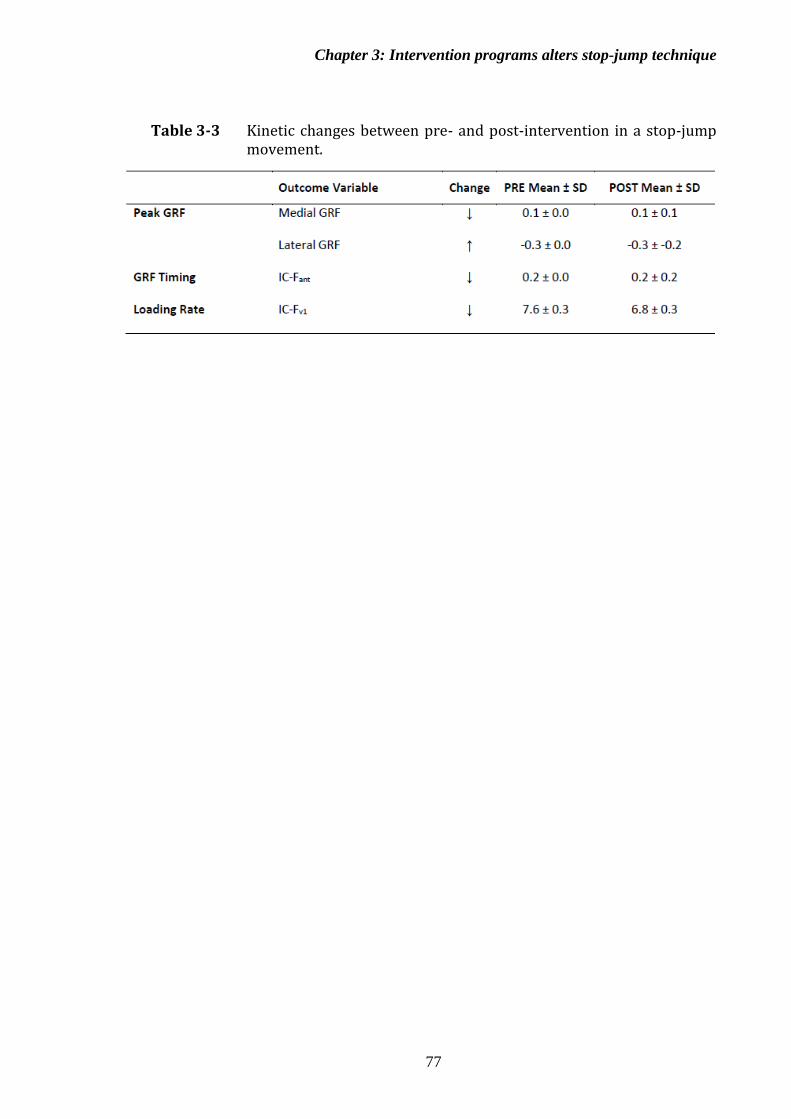
Chapter 3: Intervention programs alters stop-jump technique
77
Table 3-3 Kinetic changes between pre- and post-intervention in a stop-jump movement.

Chapter 3: Intervention programs alters stop-jump technique
78
3.3.2 Kinetics
No significant main effects of intervention for joint moments (F3,62=0.98, p=0.41,
=0.0452) or any interactions were observed. Peak GRF showed a significant main
effect for pre/post (F1,84=51.72, p=0.00, =0.9368) yet not for intervention (F3,244=0.42,
p=0.79, =0.7985), and a significant pre/post*peak GRF interaction was observed
(F4,336=11.53, p<0.001, =0.0066; Table 3-3). When comparing pre- to post-
intervention, post hoc analysis identified a significant decrease in medial GRFs (p<0.001)
but an increase in lateral GRFs (p<0.001). Although no main effect of intervention was
identified in GRF timing (F3,84=0.82, p=0.49, =0.0334), a pre/post*GRF timing
interaction was also evident (F7,588=3.74, p<0.001, =0.2067; Table 3-3). Post hoc
analysis identified a significant pre/post-intervention difference, with a significant
decrease in the duration from IC to FANT displayed (p<0.05). There was no main effect of
intervention identified in LR FV1 (F1,62=7.50, p=0.01, =0.1079); however, a significant
interaction of pre/post revealed it decrease from pre- to post-intervention identified by
post hoc analysis.
3.4 Discussion
With injury prevalence rates high and the cost of sports injuries increasing (Medibank
Private 2003, Medibank Private 2006), injury reduction programs are an appealing option
for coaches and clinicians to reduce injury risk in their athletes. The current study is the
first to compare the effects of three different field-based injury reduction programs on the
stop-jump landing mechanics among pre-elite youth athletes. Results revealed no
significant differences between the interventions; however, a significant change in
landing mechanics were identified between pre/post 12-week intervention. Specifically,

Chapter 3: Intervention programs alters stop-jump technique
79
all youth athletes, regardless of training intervention group, landed in a more upright trunk
relative to pelvis position in post-intervention than pre-intervention testing. Youth
athletes generally lack previous exposure to age appropriate strength and conditioning
training, due to policies outlining the requirement for supervision by qualified
professionals prior to the age of 16 (Parker 2003). Within this study, screening revealed
that the athletes displayed poor movement competency total scores, which may be
indicative of a lack of training. Neuromuscular training often elicits a training effect that
is commonly identified in strength interventions programs, whereby the neuromuscular
activation patterns become more succinct leading to an improved performance (Sale
1988). Thus, the altered movement pattern between pre- and post-intervention is likely
related to an increase in neuromuscular control as a result of general training rather than
any specific intervention program.
Research has identified adolescent athletes as an ideal target population to retrain
(DiStefano, Padua et al. 2009). Older populations have a reduced ability for tissues to
adapt in response to internal and external loads (Tuite, Renström et al. 1997); highlighted
in an older cohort of 18-30 years old who failed to alter their stop-jump mechanics after
a 9-week strength program (Herman, Weinhold et al. 2008). Younger athletes (<13years)
have also been identified as displaying no training benefits, suggested to be due to
alternate requirement for early adolescent athletes. This early adolescent population are
suggested to require internal feedback, such as bend your knees, in contrast to the external
feedback required for older populations (DiStefano, Padua et al. 2009). The middle period
of adolescence phase appears to be the optimal period for neuromuscular intervention to
elicit a positive effect. The present study supports this notion as strength training for youth
athletes (15.2±2.2 years) was able to alter jump-landing mechanics after a 12-week
intervention program. It is likely that differences in training age for strength training may

Chapter 3: Intervention programs alters stop-jump technique
80
contribute to the different findings between this and other studies. Therefore, strength
training can successfully alter jump-landing mechanics in those adolescent athletes who
self-report no or little strength training experience.
Irrespective of the type of intervention program, this study identified significant pre/post
program differences in landing mechanics , with all athletes landing in a more upright
trunk relative to pelvis position. While this upright trunk landing posture in itself has been
suggested to be an ACL injury risk factor (Griffin, Agel et al. 2000, Shimokochi,
Ambegaonkar et al. 2013), the present study did not observe any ACL landing risk factors
associated with this landing strategy. That is, no indications of higher posterior GRF,
lower knee flexion at IC or greater peak knee abduction during landing were observed,
all of which have been defined as key injury factors (Shimokochi, Ambegaonkar et al.
2013). It is possible that the youth athletes here, most of whom commenced the program
with poor movement competency, were attempting to modify their landing strategy by
adopting a ‘guarding’ (van der Hulst, Vollenbroek-Hutten et al. 2010) or ‘splinting’
(Arendt-Nielsen, Graven-Nielsen et al. 1996) technique. While a universal definition of
this phenomenon does not exist in the literature (van der Hulst, Vollenbroek-Hutten et al.
2010), it has been characterised by a general increase in muscular tension, specifically
increased activation of the superficial core musculature, and has also been related to low
back pain (Ahern, Follick et al. 1988, Arendt-Nielsen, Graven-Nielsen et al. 1996).
Strength and Conditioning training often enforces a similar protective strategy with
control, good posture and bracing as correct technique (Baechle and Earle 2004) as a
strategy to protect athletes from injuries during training (Faigenbaum, Kraemer et al.
2009). We postulate that the splinting strategy for maintaining a upright trunk posture
may have been a transfer training effect from the strength and conditioning training that
all athletes undertook, which influenced this modification of the dynamic stop-jump
movement performance. It is unknown however, whether this altered movement strategy

Chapter 3: Intervention programs alters stop-jump technique
81
has been adopted by these athletes due to their novice status of training, whether it is
attributed to the early learning stage in which athletes typically display stiff looking
movements (Schmidt and Wrisberg 2008), and/or if these altered patterns are beneficial
or detrimental to lower limb injury risk.
Adopting a ‘guarding’ technique, landing in a more upright position by reducing trunk
relative to pelvis flexion and rotation, may have been an attempt by the participants to
control their body segment stability during the task in a strategy to reduced their degrees
of freedom, i.e the complexity of the task (Wang, O’Dwyer et al. 2013). Increased trunk
movement has been shown to be indicative of increased risk of knee injury due to the
influence on dynamic stability of the knee joint and low back injury (Zazulak, Hewett et
al. 2007). With both lower trunk motion (Griffin, Agel et al. 2000, Shimokochi,
Ambegaonkar et al. 2013) and greater trunk motion (Zazulak, Hewett et al. 2007) both
associated with knee injuries, it is plausible that there is an optimal middle range of trunk
motion that is ideal for reducing injury risk. This would be similar to findings at the knee,
where decreased and increased knee flexion has been associated with ACL injuries
(Boden, Dean et al. 2000) and patellar tendinopathy (Edwards, Steele et al. 2010),
respectively. However, further research is required to determine if this this notion is so.
McGill (2010) suggested a necessary balance between increased stability and mobility
for injury prevention and optimal performance. This notion supports recent research that
highlighted the need for athletes to possess some trunk motion when performing a R-
COD movement, to effectively utilise the stretch shortening cycle for optimal force
production (Edwards, Austin et al. 2017). This is supported by additional research that
found that those with chronic low back pain who employed a ‘splinted’ movement
strategy displayed decreased movement speed and power (Arendt-Nielsen, Graven-
Nielsen et al. 1996). It is therefore suggested that employing the splinting or guarding

Chapter 3: Intervention programs alters stop-jump technique
82
mechanism could potentially reduce the reactive athletic capability of these pre-elite
youth athletes, yet the effect on injury risk utilising this strategy remains unclear.
Results of this study did not identify any differences in ankle or knee mechanics between
pre/post programs, which could be a result of the feedback strategies utilised during the
intervention programs. Research has determined that externally focused feedback
(implicit), focusing on the result of the movement such as a ‘landing softly’, is
increasingly beneficial for learning. This is as opposed to internally focused feedback
(explicit) such as ‘increase your knee flexion’ during landing, focusing on the
performance of the movement, as it develops automatic motor control resulting in more
effective and faster learning (Wulf, Shea et al. 2010). With females suggested to be less
responsive to explicit feedback (Benjaminse, Otten et al. 2017), the proportionately
greater numbers of female subjects in the present study may have had bearing on this
study’s results. It is unknown whether increased time in the intervention program can
elicit a response in female athletes. The use of explicit instructions for the desired landing
and the provision of internal feedback, has previously been common in landing retraining
programs (Donnelly, Elliott et al. 2012). The results of this study suggests explicit
learning intervention programs in novice skill adolescent athletes was ineffective in
altering the ankle and knee mechanics, and thus altering landing technique.
A significant decrease in hip flexion at IC during the stop-jump post- versus pre-
intervention in this current study concurs with the findings of Chappell et al., (2008)
neuromuscular intervention program in late adolescent female basketball and soccer
players. It is likely that this decrease in hip flexion along with less trunk relative to their
pelvis flexion (upright trunk position) enable the participants to return their centre of mass

Chapter 3: Intervention programs alters stop-jump technique
83
over their base of support to maintain their equilibrium control of their landing technique.
This more upright trunk position is further explained by the decrease in T12-L1 flexion
moment identified within this study, which is suggested to be due to a reduction in the
moment arm of the trunk segment moving closer to the location of the centre of mass.
Landing in a more upright trunk position, is postulated to be due to an increase in core
musculature control reducing the requirement of the hip flexors to assist in stabilising the
lumbopelvic region, though this notion requires verification.
A decrease in hip external rotation to a more anatomical hip position at FV3, was also
identified post-intervention compared to pre-intervention in this current study and in
previous research (Chappell and Limpisvasti 2008). The splinting or guarding hypothesis
(van der Hulst, Vollenbroek-Hutten et al. 2010) is postulated to also explain this decrease,
with athletes suggested to reduce the degrees of freedom in their movements order to
increase hip joint stability. Hip internal rotation position has been associated with a more
anatomical position in athlete with a history of groin injury (Edwards, Brooke et al. 2017)
but also greater rotation in ACL injuries during the weight acceptance phase (Koga,
Nakamae et al. 2018). Yet here in this study this decrease in internal hip rotation occurred
within the propulsion phase as opposed to the weight acceptance phase reported in either
groin (Edwards, Brooke et al. 2017) or ACL injuries (Koga, Nakamae et al. 2018) .
Therefore, this decrease in hip internal rotation was not deemed as an increase in injury
risk.
An increased posterior GRF is indicative of ACL injury risk (Dai, Herman et al. 2012).
This study identified an increase in posterior impulse and a decrease in GRF loading rate
when comparing post- versus pre-intervention. It is suggested that these kinetics variables

Chapter 3: Intervention programs alters stop-jump technique
84
in conjunction with hip and knee kinematics during a stop-jump movement, play a vital
role in ACL injury mechanics. Without a significant increase in peak posterior GRF, the
increase in loading rate (IC-FV1), and increase in time from IC to anterior and lateral peak
GRF’s present within the current study was not deemed as an increased risk for ACL
injury.
The authors acknowledge the potential limitations of the current study. The 12-week
intervention was designed for implementation in a non-gymnasium setting to alleviate the
common problem of access and supervision for the rural and remote athletes. While this
intervention allowed a real-world situational approach, a more controlled environment
may have produced altered adherence that may have affected this study’s findings. It
should be noted that compliance rates for injury prevention programs are closely related
to success (Benjaminse, Otten et al. 2017). This current study did not record adherence
data as athletes who lived in close proximity to Bathurst completed their sessions with
the supervision of their strength and conditioning coach. Athletes who lived in more
remote locations were expected to compete some of their sessions via satellite or
individually. The precise adherence rate is therefore unknown. A major unavoidable
limitation within this study is the lack of a genuine control group as it is virtually
impossible to obtain a sporting group that receives no training at all, due to the nature of
this study’s population.
3.5 Conclusions
Results indicated that regardless of the intervention, all athletes displayed similar post-
intervention changes. This suggests that a single strength and conditioning program, as
opposed to a more complex intervention program, may be implemented in pre-elite youth

Chapter 3: Intervention programs alters stop-jump technique
85
athletes with poor movement competency to provide changes in landing patterns,
however a learning effect cannot be eliminated. While it is unknown whether the splinting
or guarding strategy implemented by athletes is beneficial or detrimental to performance
or alter injury risk, longitudinal research is required to determine whether this landing
strategy is maintained after this study or is just the first step in learning to land in a more
controlled manner.
3.6 Acknowledgements
The authors gratefully acknowledge the Western Region Academy of Sport for providing
the participant pool from which participants were recruited for this study.
3.7 References
Ahern, D. K., Follick, M. J., Council, J. R., Laser-Wolston, N., & Litchman, H. (1988).
Comparison of lumbar paravertebral EMG patterns in chronic low back pain patients
and non-patient controls. Pain, 34(2), 153-160.
Akuthota, V., Ferreiro, A., Moore, T., & Fredericson, M. (2008). Core stability exercise
principles. Current Sports Medicine Reports, 7(1), 39-44.
Arendt-Nielsen, L., Graven-Nielsen, T., Svarrer, H., & Svensson, P. (1996). The influence of low
back pain on muscle activity and coordination during gait: a clinical and experimental
study. Pain, 64(2), 231-240.
Baechle, T., & Earle, R. (2004). Resistance training exercise techniques. In R. Earle & T. R.
Baechle (Eds.), NSCA's Essentials of Personal Training. Champaign, IL: Human
Kinetics.
Beckman, S. M., & Buchanan, T. S. (1995). Ankle inversion injury and hypermobility: Effect on
hip and ankle muscle electromyography onset latency. Archives of Physical Medicine
and Rehabilitation, 76(12), 1138-1143.

Chapter 3: Intervention programs alters stop-jump technique
86
Benjaminse, A., Otten, B., Gokeler, A., Diercks, R. L., & Lemmink, K. A. (2017). Motor learning
strategies in basketball players and its implications for ACL injury prevention: A
randomized controlled trial. Knee Surgery, Sports Traumatology, Arthroscopy, 25(8),
2365-2376.
Beynnon, B. D., Murphy, D. F., & Alosa, D. M. (2002). Predictive factors for lateral ankle sprains:
A literature review. Journal of Athletic Training, 37(4), 376-380.
Boden, B. P., Dean, G. S., Feagin, J., & Garrett, W. (2000). Mechanisms of anterior cruciate
ligament injury. Orthopedics, 23(6), 573-578.
Chappell, J. D., & Limpisvasti, O. (2008). Effect of a neuromuscular training program on the
kinetics and kinematics of jumping tasks. American Journal of Sports Medicine, 36(6),
1081-1086.
Chu, D. A. (1999). Plyometrics in sports injury rehabilitation and training. Athletic Therapy
Today, 4(3), 7-11.
Chumanov, E. S., Heiderscheit, B. C., & Thelen, D. G. (2007). The effect of speed and influence
of individual muscles on hamstring mechanics during the swing phase of sprinting.
Journal of Biomechanics, 40(16), 3555-3562.
Clark, M. (2004). Core stabilization training in rehabiliation. In W. E. Prentice (Ed.),
Rehabilitation Techniques for Sports Medicine and Athletic Training (4th ed.):
McGraw-Hill Humanities/Social Sciences/Languages.
Cowan, S. M., Schache, A. G., Brukner, P., Bennell, K. L., Hodges, P. W., Coburn, P., &
Crossley, K. M. (2004). Delayed onset of transversus abdominus in long-standing groin
pain. Medicine & Science in Sports & Exercise, 36(12), 2040-2045.
Dai, B., Herman, D., Liu, H., Garrett, W. E., & Yu, B. (2012). Prevention of ACL injury, part I:
injury characteristics, risk factors, and loading mechanism. Research in Sports
Medicine, 20(3-4), 180-197.
DiStefano, L. J., Blackburn, J. T., Marshall, S. W., Guskiewicz, K. M., Garrett, W. E., & Padua,
D. A. (2011). Effects of an age-specific anterior cruciate ligament injury prevention

Chapter 3: Intervention programs alters stop-jump technique
87
program on lower extremity biomechanics in children. American Journal of Sports
Medicine, 39(5), 949-957.
DiStefano, L. J., Padua, D. A., DiStefano, M. J., & Marshall, S. W. (2009). Influence of age, sex,
technique, and exercise program on movement patterns after an anterior cruciate
ligament injury prevention program in youth soccer players. American Journal of Sports
Medicine, 37(3), 495-505.
Donnelly, C. J., Elliott, B. C., Doyle, T. L., Finch, C. F., Dempsey, A. R., & Lloyd, D. G. (2012).
Changes in knee joint biomechanics following balance and technique training and a
season of Australian football. British Journal of Sports Medicine, 917-922.
Edwards, S., Austin, A. P., & Bird, S. P. (2017). The role of the trunk control in athletic
performance of a reactive change-of-direction task. Journal of Strength & Conditioning
Research, 31(1), 126-139.
Edwards, S., Brooke, H. C., & Cook, J. L. (2017). Distinct cut task strategy in Australian football
players with a history of groin pain. Physical Therapy in Sport, 23, 58-66.
Edwards, S., Steele, J. R., Cook, J. L., Purdam, C. R., & McGhee, D. E. (2012). Lower limb
movement symmetry cannot be assumed when investigating the stop-jump landing.
Medicine & Science in Sports & Exercise, 44(6), 1123-1130.
Edwards, S., Steele, J. R., McGhee, D. E., Beattie, S., Purdam, C., & Cook, J. L. (2010). Landing
strategies of athletes with an asymptomatic patellar tendon abnormality. Medicine &
Science in Sports & Exercise, 42(11), 2072-2080.
Emery, C. A. (2003). Risk factors for injury in child and adolescent sport: a systematic review of
the literature. Clinical Journal of Sport Medicine, 13(4), 256.
Faigenbaum, A. D., Kraemer, W. J., Blimkie, C. J., Jeffreys, I., Micheli, L. J., Nitka, M., &
Rowland, T. W. (2009). Youth resistance training: updated position statement paper
from the national strength and conditioning association. Journal of Strength &
Conditioning Research, 23, S60-S79.
Chapter 3: Intervention programs alters stop-jump technique
88
Finch, C. F. (2011). Getting sports injury prevention on to public health agendas–addressing the
shortfalls in current information sources. British Journal of Sports Medicine, 46(1), 70-
74.
Finch, C. F., Mahoney, M., Townsend, M., & Zazryn, T. (2003). Rural sports and recreational
injuries in Australia: What do we know? Australian Journal of Rural Health, 11(3),
151-158.
Finch, C. F., Valuri, G., & Ozanne-Smith, J. (1998). Sport and active recreation injuries in
Australia: evidence from emergency department presentations. British Journal of Sports
Medicine, 32(3), 220-225.
Griffin, L. Y., Agel, J., Albohm, M. J., Arendt, E. A., Dick, R. W., Garrett, W. E., . . . Ireland, M.
L. (2000). Noncontact anterior cruciate ligament injuries: risk factors and prevention
strategies. Journal of the American Academy of Orthopaedic Surgeons, 8(3), 141-150.
Herman, D. C., Weinhold, P. S., Guskiewicz, K. M., Garrett, W. E., Yu, B., & Padua, D. A.
(2008). The effects of strength training on the lower extremity biomechanics of female
recreational athletes during a stop-jump task. American Journal of Sports Medicine,
36(4), 733-740.
Hewett, T. E., Ford, K. R., & Myer, G. D. (2006). Anterior cruciate ligament injuries in female
athletes: Part 2, a meta-analysis of neuromuscular interventions aimed at injury
prevention. American Journal of Sports Medicine, 34(3), 490.
Hewett, T. E., Lindenfeld, T. N., Riccobene, J. V., & Noyes, F. R. (1999). The effect of
neuromuscular training on the incidence of knee injury in female athletes. American
Journal of Sports Medicine, 27(6), 699-706.
Hewett, T. E., & Myer, G. D. (2011). The mechanistic connection between the trunk, hip, knee,
and anterior cruciate ligament injury. Exercise and Sport Sciences Reviews, 39(4), 161-
166.
Chapter 3: Intervention programs alters stop-jump technique
89
Hewett, T. E., Myer, G. D., & Ford, K. R. (2004). Decrease in neuromuscular control about the
knee with maturation in female athletes. Journal of Bone and Joint Surgery, 86(8),
1601-1608.
Hewett, T. E., Stroupe, A. L., Nance, T. A., & Noyes, F. R. (1996). Plyometric training in female
athletes. American Journal of Sports Medicine, 24(6), 765-773.
Mandelbaum, B. R., Silvers, H. J., Watanabe, D. S., Knarr, J. F., Thomas, S. D., Griffin, L. Y., .
. . Garrett Jr, W. (2005). Effectiveness of a neuromuscular and proprioceptive training
program in preventing anterior cruciate ligament injuries in female athletes. American
Journal of Sports Medicine, 33(7), 1003-1010.
Mann, K. J., Edwards, S., Drinkwater, E. J., & Bird, S. P. (2013). A lower limb assessment tool
for athletes at risk of developing patellar tendinopathy. Medicine & Science in Sports &
Exercise, 45(3), 527-533.
McCurdy, K., Walker, J., Saxe, J., & Woods, J. (2012). The effect of short-term resistance training
on hip and knee kinematics during vertical drop jumps. Journal of Strength and
Conditioning Research, 26(5), 1257-1264.
McGill, S. M. (2010). Core training: Evidence translating to better performance and injury
prevention. Strength & Conditioning Journal, 32(3), 33.
Medibank Private. (2003). Sports Injuries Report. Melbourne, Victoria: Medibank Private
Limited,http://fulltext.ausport.gov.au/fulltext/2003/medibank/Sports_injuries_report.p
df. Retrieved 24th January, 2018.
Medibank Private. (2006). Sports Injuries Report. Melbourne, Victoria: Medibank Private
Limited,
http://fulltext.ausport.gov.au/fulltext/2006/medibank/Sports_injuries_report.pdf.
Retrieved 24th January, 2018.
Mottram, S., & Comerford, M. (2008). A new perspective on risk assessment. Physical Therapy
in Sport, 9(1), 40-51.

Chapter 3: Intervention programs alters stop-jump technique
90
Myer, G. D., Chu, D. A., Brent, J. L., & Hewett, T. E. (2008). Trunk and hip control
neuromuscular training for the prevention of knee joint injury. Clinics in Sports
Medicine, 27(3), 425-448.
Myer, G. D., Faigenbaum, A. D., Ford, K. R., Best, T. M., Bergeron, M. F., & Hewett, T. E.
(2011). When to initiate integrative neuromuscular training to reduce sports-related
injuries and enhance health in youth? Current Sports Medicine Reports, 10(3), 155-166.
Myer, G. D., Ford, K. R., & Hewett, T. E. (2008). Tuck jump assessment for reducing anterior
cruciate ligament injury risk. Athletic Therapy Today, 13(5), 39-44.
Myer, G. D., Ford, K. R., Palumbo, J. P., & Hewett, T. E. (2005). Neuromuscular training
improves performance and lower-extremity biomechanics in female athletes. Journal of
Strength & Conditioning Research, 19(1), 51-60.
Myklebust, G., Engebretsen, L., Brækken, I. H., Skjølberg, A., Olsen, O. E., & Bahr, R. (2003).
Prevention of anterior cruciate ligament injuries in female team handball players: a
prospective intervention study over three seasons. Clinical Journal of Sport Medicine,
13(2), 71-78.
Parker, R. J. (2003). Kids in gyms. Retrieved from Silverwater NSW: 1st February, 2018.
https://www.schn.health.nsw.gov.au/files/attachments/kids_in_gyms_guidelines.pdf
Rutherford, O., & Jones, D. (1986). The role of learning and coordination in strength training.
European Journal of Applied Physiology & Occupational Physiology, 55(1), 100-105.
Sale, D. G. (1988). Neural adaptation to resistance training. Medicine & Science in Sports &
Exercise, 20(5), 135-145.
Schmidt, R. A., & Wrisberg, C. A. (2008). Motor learning and performance: A situation-based
learning approach. Champaign: Human kinetics.
Sherry, M. A., & Best, T. M. (2004). A comparison of 2 rehabilitation programs in the treatment
of acute hamstring strains. Journal of Orthopaedic & Sports Physical Therapy, 34(3),
116-125.

Chapter 3: Intervention programs alters stop-jump technique
91
Shimokochi, Y., Ambegaonkar, J. P., Meyer, E. G., Lee, S. Y., & Shultz, S. J. (2013). Changing
sagittal plane body position during single-leg landings influences the risk of non-contact
anterior cruciate ligament injury. Knee Surgery, Sports Traumatology, Arthroscopy,
21(4), 888-897.
Sigward, S. M., Pollard, C. D., Havens, K. L., & Powers, C. M. (2012). Influence of sex and
maturation on knee mechanics during side-step cutting. Medicine & Science in Sports
& Exercise, 44(8), 1497-1503.
Sigward, S. M., Pollard, C. D., & Powers, C. M. (2012). The influence of sex and maturation on
landing biomechanics: implications for anterior cruciate ligament injury. Scandinavian
Journal of Medicine & Science in Sports, 22(4), 502-509.
Soligard, T., Nilstad, A., Steffen, K., Myklebust, G., Holme, I., Dvorak, J., . . . Andersen, T. E.
(2010). Compliance with a comprehensive warm-up programme to prevent injuries in
youth football. British Journal of Sports Medicine, 44(11), 787-793.
Tappe, M. K., Duda, J. L., & Ehrnwald, P. M. (1989). Perceived barriers to exercise among
adolescents. Journal of School Health, 59(4), 153-155.
Tuite, D. J., Renström, P., & O'brien, M. (1997). The aging tendon. Scandinavian Journal of
Medicine & Science in Sports, 7(2), 72-77.
van der Hulst, M., Vollenbroek-Hutten, M. M., Rietman, J. S., & Hermens, H. J. (2010). Lumbar
and abdominal muscle activity during walking in subjects with chronic low back pain:
Support of the “guarding” hypothesis? Journal of Electromyography & Kinesiology,
20(1), 31-38.
Wang, X., O’Dwyer, N., & Halaki, M. (2013). A review on the coordinative structure of human
walking and the application of principal component analysis. Neural Regeneration
Research, 8(7), 662.
Willson, J. D., Dougherty, C. P., Ireland, M. L., & Davis, I. M. C. (2005). Core stability and its
relationship to lower extremity function and injury. Journal of the American Academy
of Orthopaedic Surgeons, 13(5), 316-325.

Chapter 3: Intervention programs alters stop-jump technique
92
Wulf, G., Shea, C., & Lewthwaite, R. (2010). Motor skill learning and performance: a review of
influential factors. Medical Education, 44(1), 75-84.
Zazulak, B. T., Hewett, T. E., Reeves, N. P., Goldberg, B., & Cholewicki, J. (2007). Deficits in
neuromuscular control of the trunk predict knee injury risk. American Journal of Sports
Medicine, 35(7), 1123-1130.

93
Context of Manuscript in Relation to the Thesis
This manuscript builds on this thesis investigation into the effectiveness of whether a 12-
week intervention program can modify reactive change-of-direction (R-COD) strategies
in pre-elite youth athletes, specifically this manuscript focuses on a R-COD movement
strategy. The R-COD was chosen as the experimental task as this is task used to
investigate key lower limb injuries, such as the anterior cruciate ligament injury and groin
pain, and are repetitively performed within a game and/or training environment. Much of
the previous literature on COD and injury prevention studies have been completed within
an adult population; however, it is unknown whether we can modify the landing strategies
within a youth population. This manuscript addresses these limitations in previous
research by providing a 3D biomechanical analysis of a R-COD task and examines the
effectiveness of four intervention programs (core, landing, core and landing and control)
to modify R-COD strategy and variables associated with lower limb injury risk within a
youth population. Results of this manuscript will provide coaches and clinicians valuable
information on how pre-elite youth athletes move, improve their understanding of how
an intervention leads to training adaptations and provide practical recommendations for
coaches and clinicians.

Chapter 4: Intervention programs alters R-COD technique
94
Chapter 4. Any type of neuromuscular training program can
modify pre-elite youth athletes reactive change-of-direction
technique
This chapter is an amended version of the manuscript: Mann K, O’ Dwyer N, Bruton M, Edwards S.
Any type of neuromuscular training program can modify pre-elite youth athletes reactive change-of-
direction technique. To be submitted to the American Journal of Sports Medicine.
Background: Neuromuscular training is commonly used in anterior cruciate ligament (ACL) injury
prevention programs, yet current intervention strategies are not delivering the desired reduction in
injury rates. It is likely these interventions programs are limited in their effectiveness as they often
target specific joints, not including the entire kinetic chain such as the lumbopelvic region, and are
very unlikely to be implemented in rural regions with limited access to facilities.
Purpose: This study’s purpose was to determine the most effective 12-week intervention program to
reduce lower limb injury risk during a reactive change-of-direction (R-COD) maneuver in pre-elite
youth athletes.
Study Design: Controlled laboratory study.
Methods: Eighty-nine pre-elite youth athletes with no current signs or symptoms of injury were
recruited from a regional Academy of Sport. Five successful R-COD on each leg were performed by
four groups, both before and after completing 12-week training program. Every group completed a
standard strength and conditioning program, with three of four groups completing either an additional
core, landing or core-landing component. Mixed model factorial analysis of variance (ANOVA) was
used to determine statistically significant between-group differences (P≤0.05).
Results: While no main effect of intervention type for any kinematic or kinetic variables were
identified, all athletes displayed a guarding or splinting trunk posture during their R-COD,

Chapter 4: Intervention programs alters R-COD technique
95
exemplified by reduced L5-S1 flexion, and trunkab-pelvis extension post- compared to pre-
intervnetion. This strategy contributed to a significant decrease in T12-L1 flexion moment and
increased posterior impulse was also identified between pre- and post-intervention testing.
Conclusions: Results of this study showed no differences between intervention programs to modify
R-COD, suggesting that a single strength and conditioning program can be utilised to modify landing
patterns within pre-elite youth athletes. This general strength and conditioning program is postulated
to have led to all groups increasing their trunk control via utilising a trunk splinting strategy post-
compared to pre-intervention
Clinical Relevance: Athletes R-COD technique can be altered using a simple strength and
conditioning program in youth athletes as opposed to a specialised program.
Key Terms: Injury, biomechanics, movement screening, prevention through prediction, dynamic
landing
4.1 INTRODUCTION
The knee is the most commonly injured joint in Australia, (Medibank Private 2006) with a marked
increase in knee injuries between ages 10-14 and 15-17 years (Kreisfeld, Harrison et al. 2017). The
anterior cruciate ligament (ACL) resides within the internal capsule of the knee joint and acts to
prevent anterior translation of the tibia with respect to the femur (Palastanga, Field et al. 2006), and
was injured in approximately 3,125 Australian individuals in 2012-13 (Kreisfeld, Harrison et al.
2017). The cost of knee injuries however, is substantial with major injuries that require surgical
intervention costing on average between AUS $11,000 and $16,500 (Medibank Private 2006). While
ACL injury occurs in both contact and non-contact situations, it is estimated that approximately 70%
of injuries are a result of a non-contact scenario (Boden, Dean et al. 2000). The detrimental effects
of ACL injury persist long term after the injury, with an increased risk of early-onset osteoarthritis
(Lohmander, Östenberg et al. 2004).

Chapter 4: Intervention programs alters R-COD technique
96
The high prevalence (Kreisfeld, Harrison et al. 2017), substantial costs (Medibank Private 2006) and
detrimental long term effects (Lohmander, Östenberg et al. 2004) of ACL injury make injury
reduction a worthwhile pursuit. Injury reduction programs have been implemented across a range of
age groups (Hewett, Lindenfeld et al. 1999, Wedderkopp, Kaltoft et al. 1999, Myklebust,
Engebretsen et al. 2003, Mandelbaum, Silvers et al. 2005). Targeting an adolescent population with
injury reduction programs is critical as early adolescence injury rates begin to increase (Kreisfeld,
Harrison et al. 2017) and the rapid increases in height and body mass increase the height of the
location of centre of mass (Myer, Chu et al. 2008) that can cause the trunk segment to become harder
to stabilise during motion. This difficulty in stabilising the trunk during motion can be further
compounded by an absence of sufficient neuromuscular control and strength (Myer, Chu et al. 2008)
and is suggested to cause increased knee joint moments (Hewett and Myer 2011), making the
adolescence population a key population for intervention training strategies.
ACL injuries are multifactorial, having a combination of both modifiable and non-modifiable risk
factors (van Mechelen, Hlobil et al. 1992). Modifiable risk factors such as landing technique are often
the focus of injury prevention programs; the eccentric loading phase is critical phase associated with
many lower limb injuries (Boden, Dean et al. 2000, Myer, Ford et al. 2008, Edwards, Steele et al.
2010, Mann, Edwards et al. 2012). Within the eccentric loading phase, biomechanical variables such
as knee abduction angles (McLean, Huang et al. 2005), net external knee abduction moment
(Dempsey, Lloyd et al. 2009), external or internal tibial rotation (Krosshaug, Nakamae et al. 2007),
reduced knee flexion (Boden, Dean et al. 2000) and lateral trunk movements away from the direction
of travel (Dempsey, Lloyd et al. 2007) have been associated with ACL injury during a COD task.
Dynamic neuromuscular landing re-training in adolescent female volleyball athletes has been shown
to successfully decrease joint forces and modify critical ACL biomechanical risk factors within this
deceleration phase (Hewett, Stroupe et al. 1996). As such, landing retraining programs is a popular
intervention for preventing ACL injury (Hewett, Lindenfeld et al. 1999, Myklebust, Engebretsen et

Chapter 4: Intervention programs alters R-COD technique
97
al. 2003, Mandelbaum, Silvers et al. 2005). However, the need for controlled environments or
specialised equipment in the aforementioned programs may affect adherence rates, due to the
challenges in accessing appropriate facilities (Tappe, Duda et al. 1989), especially among rural and
remote athletes (Finch, Mahoney et al. 2003). As greater adherence rates are associated with a
decrease in injury risk (Soligard, Nilstad et al. 2010), providing a field based injury reduction
program that is easy to implement and access for athletes is essential for any injury reduction
program.
Injury reduction strategies that aim to correct or modify lower limb patterns of movement during
landing, are often the focus of ACL injury prevention programs (Hewett, Stroupe et al. 1996, Myer,
Ford et al. 2005). However, high rates of injury and re-injury (Mottram and Comerford 2008) suggest
that this joint specific approach (e.g. the knee joint for ACL injuries), targeting underlying mechanics
associated with the specific joint are not effective as opposed to a global kinetic chain focus that also
incorporate the lumbopelvic region. Delayed or poor neuromuscular control of the lumbopelvic
region during dynamic landing tasks may predispose athletes to a lower limb injury through poor
stability, motion and increased forces experienced by the lower limb joints (Myer, Chu et al. 2008).
As lumbopelvic instability has been linked to groin (Cowan, Schache et al. 2004, Edwards, Brooke
et al. 2017), hamstring strain (Sherry and Best 2004, Chumanov, Heiderscheit et al. 2007), knee joint
(Edwards, Steele et al. 2010, Hewett and Myer 2011, Mann, Edwards et al. 2012) and ankle joint
injuries (Beckman and Buchanan 1995, Beynnon, Murphy et al. 2002), lumbopelvic instability may
be a key area in relation to the prevention of lower limb injuries in athletes.
Dynamic neuromuscular training has been shown to be an effective intervention for improving
biomechanical injury risk factors among adolescents during drop jump landing (DiStefano, Padua et
al. 2009) and in turn, decreasing injury risk (Myer, Ford et al. 2005). This suggests that this
population can be successfully re-trained, in a more complex sport specific movement such as the R-
COD task. These changes in landing technique within this population are thought to occur mainly

Chapter 4: Intervention programs alters R-COD technique
98
through increased neuromuscular adaptations, such as more comprehensive and synchronous firing
of the muscular unit (Sale 1988). Therefore, the purpose of this study is to compare the effectiveness
of three different 12-week intervention programs to modify the R-COD technique in pre-elite youth
athletes.
4.2 MATERIAL AND METHODS
4.2.1 Participants
Eighty-nine junior pre-elite athletes (age = 15.2 ± 2.2 years, height = 171.1 ± 9.4 cm; mass = 61.9 ±
12.3 kg) with no current signs or symptoms of injury were recruited from the Western Region
Academy of Sports Basketball, Hockey, Netball, Triathlon and Softball teams (Error! Reference
source not found.). A total of 67 athletes completed both baseline (pre) and post-intervention testing,
and were included in the analysis. Written informed consent was obtained from each participant prior
to data collection, with parental/guardian consent obtained for minors. All methods were approved
by the institution’s Human Research Ethics Committee (2012/157, Appendix 1).
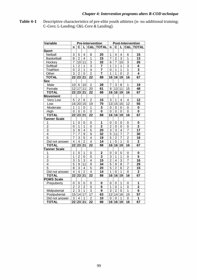
Chapter 4: Intervention programs alters R-COD technique
99
Table 4-1 Descriptive characteristics of pre-elite youth athletes (n- no additional training; C–Core; L-Landing; C&L-Core & Landing).
Variable Pre-Intervention Post-Intervention n C L C&L TOTAL n C L C&L TOTAL Sport
Netball 3 5 4 8 20 1 4 4 6 15 Basketball 8 2 4 1 15 7 2 3 1 13 Hockey 7 10 11 3 32 6 7 10 3 26 Softball 1 2 1 3 7 1 1 1 3 6 Triathlon 0 2 1 4 7 0 1 1 1 3 Other 3 2 0 2 7 1 1 0 2 4 TOTAL 22 23 21 22 88 16 16 19 16 67
Sex Male 10 6 10 2 28 7 3 8 1 19 Female 12 17 11 20 61 9 13 11 15 48 TOTAL 22 23 21 22 88 16 16 19 16 67
Movement Screening Total Score
16
Very Low 5 2 6 2 15 3 1 4 4 12 Low 16 20 15 19 70 13 15 15 12 55 Moderate 1 1 0 1 3 0 0 0 0 0 High 0 0 0 0 0 0 0 0 0 0 TOTAL 22 23 21 22 88 16 16 19 16 67
Tanner Scale Genital
1 1 0 0 0 1 0 0 0 0 0 2 0 1 1 0 2 2 0 0 0 2 3 3 8 4 5 20 3 3 4 7 17 4 7 7 9 9 32 5 11 7 7 30 5 7 3 5 4 19 5 2 7 2 16 Did not answer 4 4 2 4 14 1 0 1 0 2 TOTAL 22 23 21 22 88 16 16 19 16 67
Tanner Scale Pubic Hair
1
1
1 0 1 0 2 0 0 0 0 0 2 1 2 0 0 3 3 1 1 0 5 3 3 5 3 4 15 2 4 3 7 16 4 5 9 11 9 34 5 9 8 7 29 5 8 3 4 5 20 5 2 6 2 15 Did not answer 4 4 2 4 14 1 0 1 0 2 TOTAL 22 23 21 22 88 16 16 19 16 67
POMS Scale Prepuberty 0 0 0 0 0 0 0 1 0 1 2 2 2 0 6 1 0 1 0 2 Midpubertal 2 3 1 3 9 3 2 0 1 6 Postpubertal 15 14 17 17 63 12 14 16 15 57 Did not answer 3 4 1 2 10 0 0 1 0 1 TOTAL 22 23 21 22 88 16 16 19 16 67

Chapter 4: Intervention programs alters R-COD technique
100
4.2.2 Experimental Task
A movement screening familiarisation was performed followed by a data collection session before
and after completion of a 12-week intervention program. Each data collection session involved
participants to perform five R-COD on each leg and the movement screening protocol.
The movement screening protocol, outlined in detail in Appendix 2, was videoed and involved each
participant performing six movements (tuck jump, overhead squat, single leg squat, dip test, forward
lunge and prone hold). Each movement was scored based on the presence or absence of errors by a
single rater (KM) who counted the errors to yield a score of either 1,2 or 3 for that movement. Then,
the rater summed the scores for all six movements to determine a total composite score out of 27.
The R-COD involved the participant accelerating forward (mean approach speed 4.00.1 ms-1) for
7 metres in a straight line towards two force platforms. It was followed by a plant-and-cut manoeuvre
at an angle between 30° to 60° originating at the centre of the force plates (mean exit speed 4.30.1
ms-1). Line markings were made on the floor using duct tape outlining the angle of cut. A skeleton
placed 2.8 m from the base of the force platforms to simulate a defensive opponent in a game
environment (Besier, Lloyd et al. 2001). An automatic light-emitting timing gates protocol (4-gate-
cut protocol; Smart Speed, Fusion Sport, Queensland, Australia) signalled the participant a right or
left COD when they reached a two-metre distance from the force platforms. A successful trial was
defined as the: participants’ whole foot being in contact with the force platforms during the plant-
and-cut (opposite foot to the direction of the cut task (i.e. right foot for left R-COD); the participants
cut direction matching the light-emitting diode; and the approach speed was between 3.5 and 4.5 ms-
1. To ensure sufficient recovery between trials and to minimise effects of fatigue, participants were
given a 30 second rest between each trial, and a minimum of 5-minute following every 20 trials.

Chapter 4: Intervention programs alters R-COD technique
101
4.2.3 Experimental Procedure
A static trial and movement competency screen (Appendix 2) was performing prior to completing a
5-minute self-paced warm-up on a cycle ergometer (Monark 828E, Varburg, Sweden). The
participant was then familiarised with the COD before performing the experimental task. Ground
reaction forces generated at landing were recorded (2400 Hz) using two multichannel force platforms
with built-in charge amplifiers (Type 9281CA, Kistler, Winterthur, Switzerland; Type9821EA,
Kistler, Winterthur, Switzerland) embedded in the floor and connected to control units (Type 5233A,
Kistler, Winterthur, Switzerland) for each trial. Using a 12 Qualisys Oqus 300+ camera system
(Qualisys AB, Göteborg, Sweden), three-dimensional lower limb and trunk motion was recorded
(250 Hz) for each participant. Passive reflective markers were placed on each participant’s lower
limbs, pelvis and torso, on the shoe at the first and fifth metatarsal head, mid anterior foot and
calcaneus, lateral and medial malleolus, lateral and medial femoral epicondyle, a four-marker cluster
placed on the leg and thigh, greater trochanter, anterior superior iliac spine, posterior superior iliac
spine, iliac crest, sternal notch, xiphoid process, acromion, lumbo-sacral (L5-S1) intervertebral joint
space, thoraco-lumbar (T12-L1) intervertebral joint space, bilaterally on the ribcage at the level of
the T12-L1 intervertebral joint space and immediately superior to the iliac crest marker, and five
tracking markers placed on the lumbar region. The participants wore minimal clothing (shorts) to
avoid interference with the passive reflective markers, and their athletic running shoes and socks.
4.2.4 Intervention Program
Participants were randomly allocated into one of four intervention programs: core plus standard
strength and conditioning; landing retraining plus standard strength and conditioning, core and
landing retraining plus standard strength and conditioning; or only standard strength and conditioning
(control; Appendix 3). Every participant performed a standard strength and conditioning program
consisting of four key exercises (push, pull, squat and lunge). To aid participants in adhering to the

Chapter 4: Intervention programs alters R-COD technique
102
program, they were provided with a compact-disk (CD) that had videos of all exercises for each phase
of their program. At the beginning and every three weeks after the commencement of the program,
Strength and Conditioning interns completed the movement competency testing to record each
athlete’s progression in the intervention program and assessed their execution of their selected
exercises. Each intervention program required participants to meet a competency criterion before
advancing to the next level of the program (Appendix 3). Below outlines the rationale for each
intervention.
Strength and conditioning program: Designed and implemented by the Academy of Sport, this
program incorporated a dynamic warm up, a push, pull, squat and lunge resistance exercise and cool-
down stretches (Appendix 5).
Program 1 – Core program: This program was designed to include a range of functional balance and
postural stability core exercises to improve lumbopelvic stability. Athlete’s in this program
progressed through the four levels of core stabilisation training; stabilisation, stabilisation and
strength, integrated stabilisation strength and explosive stabilisation using the core stabilisation
guidelines outlined by Clarke (2004).
Program 2 – Landing retraining program: This landing re-training program included basic strength,
stability and plyometric concepts to re-train landing technique. Athletes progressed from basic
strength and stability to the six stages of plyometric training outlined by Chu (1999).
Program 3 – Core program and landing retraining program: This program was designed to combine
both the core stability and landing re-training programs together by incorporating a half load of each
training program to provide a core stability and landing re-training exercises that also followed the
guidelines outlined by Clark (2004) and Chu (1999).
Program 4 – Control program: This program did not perform any additional intervention training in
conjunction with their strength and conditioning program provided by the Academy of Sport.

Chapter 4: Intervention programs alters R-COD technique
103
4.2.5 Data Reduction and Analysis
Visual 3D software (Version 5, C-Motion, Germantown, MD) was used for analysis of the three-
dimensional kinematic and kinetic data for angles and joint moments, with a custom built LabView
program (Lab-View 2010, National Instruments, Austin, TX, USA) for GRF data analysis. All raw
kinematic coordinates, GRF’s, free moments and centre of pressure data were filtered through a
fourth-order zero-phase Butterworth low-pass digital filter prior to calculating individual joint
kinematics and net internal joint moments (fc = 18 Hz), and raw GRFs to calculate GRF variables (fc
= 50 Hz).
Segment masses were defined from Zatsiorsky et al. (1990) for the foot, shank and thigh segments,
and from Pearsall et al. (1996) for the pelvis, lumbar, thorax, and trunk segments. Segmental inertial
properties of each segment were modelled using geometric primitives (Hanavan 1964) with the foot,
shank, and thigh defined as a frusta of a right cone, and the pelvis, lumbar, thorax, and trunk defined
as elliptical cylinders (Seay, Selbie et al. 2008). To support Cartesian local coordinate system sign
conventions, all segments were defined as follows: x-axis as medio-lateral, y-axis as anterior-
posterior and z-axis as the vertical direction. Loads applied to each joint were defined as internal net
joint moments that were normalised to each individual participant’s body mass multiplied by height,
and ground reaction forces were normalised to body weight to account for variations between
participants. Using the 18 Hz filtered kinetic data, the horizontal landing phase was defined from
initial contact (IC) when the vertical ground reaction force exceeded 10 N to the first peak (FV1) and
local minimum (FV2), to the second GRFV peak (FV3) and then to take-off (TO) when the vertical
GRF fell below 10 N. For each trial, intersegment joint angles of the ankle, knee, hip, L5-S1 (lumbar
segment relative to the pelvis), T12-L1 (lumbar segment relative to the thoracic segment),
Thoracic_Ab-Pelvis (trunk segment relative to the pelvis segment) at the time of IC, FV1, FV2, FV3,
and TO were calculated. Peak net internal ankle, knee, hip, L5-S1, and T12-L1 joint moments, and
peak anteroposterior, mediolateral and vertical GRF were identified during the ground contact period

Chapter 4: Intervention programs alters R-COD technique
104
from IC to TO. While both the left and right lower limb data were analysed, only the left lower limb
was included in statistical analysis, as lower limb were show to be symmetrical (Appendix 4).
4.2.6 Statistical Analysis
A G*Power calculation of the required sample size was performed for an actual power of 0.96. This
calculation showed a total sample size of 56 athletes was required, suggesting that this study is
adequately powered A series of mixed-design factorial analyses of variance (ANOVA) were
performed on the outcome variables that consisted of joint angles, peak joint angles, ranges of
motions (ROMs), GRFs and joint moments during performance of the R-COD. The primary effect
of interest was the independent factor of intervention (core, landing, core-landing and control), and
the secondary repeated measures factor was that of time (pre- and post-intervention). There were four
factors for analyses of joint angles (intervention*pre/post*events*angles). Events comprised of five
levels; the discrete GRF time-points within the stance phase of the COD (IC, FV1, FV2, FV3, TO),
while angles comprised of 18 levels; six joints (ankle, knee hip, L5-S1, T12-L1, thoracic abdominal
pelvis) in three respective planes (x,y,z). There were three factors for the other kinematic analyses of
ROM (intervention*pre/post*ROM) and peak angles (intervention*pre/post*peak angles). There
were also three factors for each of the kinetic variable analyses of joint moments
(intervention*pre/post*moments), GRF peaks (pre/post*GRF peaks), GRF impulse (pre/post*GRF
impulse) and GRF timing (intervention*pre/post*GRF timing. It was to be expected that the
outcomes variables (joint angles, segment alignments, joint moments, ROMs and GRFs) would vary
significantly across the stages of the horizontal landing phase. Similarly, significant differences were
to be expected between the variables within each category of outcome variable. For example,
differences would be expected between joint angles and between segment alignments. Such
differences, however, were not relevant to the primary hypotheses concerning the effects of the
interventions or pre/post testing. Therefore, only main effects or interactions that involved the factors
of intervention and/or pre/post testing were reported here.

Chapter 4: Intervention programs alters R-COD technique
105
All tests were conducted using Statistica (v.13, StatSoft Inc., Tulsa, OK, USA), with significant
results reported for p<0.05 and Tukey Post hoc tests used to identify where the significant effect
occurred. All data were tested for the assumptions of normality of distribution and sphericity, with
multivariate ANOVA considered when sphericity was not satisfied. Assumptions of constant
variance and normality were examined through residuals and were both satisfied and accepted.
Where the Tukey post hoc did not reveal the significant effect when a significant interaction was
indicated in the ANOVA results, univariate and independent t-tests were employed to assess which
variable was responsible for the interaction and effect size analyses, respectively. Effects sizes (p)
were defined as trivial (<0.0099), small (0.0099-0.0588), moderate (0.0588-0.1379), and large
(>0.1379) sizes (Richardson 2011).
4.3 RESULTS
4.3.1 Kinematics
Joint angles showed no significant main effect of intervention group (F3,62=0.46, p=0.71, =0.0219),
or pre/post time point (F1,62=0.006, p=0.94, =0.0001); however, significant interactions between
pre/post*angles (F17,1054=295.4, p<0.001, =0.0383) and pre/post*events*angles (F68,4216=1.42,
p=0.02, =0.0225, Table 4-2) were present. Post hoc tests failed to determine the source of the
effect in the pre/post*angles interaction. It was revealed that pre- compared to post-intervention, in
the pre/post*events*angles interaction, that there was significantly reduced L5-S1 flexion at IC
(p<0.001), FV1 (p<0.001), FV2 (p<0.001), FV3 (p<0.001), TO (p<0.041), thoracic abdominal
extension at IC (p<0.05), T12-L1 extension at TO (p<0.001), increased forefoot abduction at FV1
(p<0.05) and hip external rotation at FV3 (p<0.05). There were no main effects of intervention
(F3,62=2.59, p=0.06, =0.1112) or pre/post (F1,62=1.05, p=0.31, =0.0167), and no significant
interactions for joint range of motion were observed. Nor were there any main effects of intervention

Chapter 4: Intervention programs alters R-COD technique
106
(F3,62=0.49, p=0.69, =0.0230), or pre/post (F1,62=0.02 p=0.89, =0.0003), nor significant
interactions for peak joint angles or joint segments displayed.
4.3.2 Kinetics
For joint moments, there were no main effects of intervention (F3,62=1.64, p=0.19, =0.0734), or
pre/post (F1,62=1.94 p=0.17, =0.0303; Table 4-3) observed. However, a significant interaction was
identified between pre/post*moments (F29,1798=1.98 p=0.001, =0.0309; Table 4-3). Post hoc
analysis identified a significant decrease in T12-L1 flexion moment (p<0.05) between pre- and post-
intervention were observed. A significant main effect of pre- post-intervention was reported for peak
GRF (F4,248=2.61, p=0.04, =0.0404); however, post hoc analysis failed to identify the source of the
significant interaction. No main effect of intervention group (F3,62=0.47, p=0.70, =0.0223), or
pre/post (F1,62=4.57, p=0.37, =0.0686), or any interactions were identified in GRF timing.

Chapter 4: Intervention programs alters R-COD technique
107
Table 4-2 Kinematic changes between pre- and post-intervention in a RCOD task.
Table 4-3 Kinematic changes between pre- and post-intervention in RCOD task.
Chapter 4: Intervention programs alters R-COD technique
108
4.4 DISCUSSION
Prevention programs for ACL injury are increasingly popular, with programs successfully
shown to alter landing technique (Hewett, Lindenfeld et al. 1999, Wedderkopp, Kaltoft
et al. 1999, Mandelbaum, Silvers et al. 2005). Yet the unchanged rate of injury observed
(Medibank Private 2003, Medibank Private 2006) may be due to these programs not being
adopted by the general population. A barrier to implementation of and adherence to these
programs, that can directly impact the effectiveness of prevention program (Benjaminse,
Otten et al. 2017) is that many programs require specific equipment or specialised trainers
(Söderman, Werner et al. 2000, Myklebust, Engebretsen et al. 2003, Olsen, Myklebust et
al. 2005, Petersen, Braun et al. 2005). This present study’s field-based neuromuscular
training intervention programs were easily implemented without the need for specialised
equipment or supervision and were shown they could alter youth athletes R-COD
technique regardless of the type of intervention program. Irrespective of the type of
intervention, athletes displayed a less flexed trunk-pelvis position during the R-COD
post- compared to pre-intervention. Minimal differences displayed between the
intervention programs is postulated to be attributed to the overall poor movement
competency of all participants at commencement of the program along with their
lack/minimum prior strength training exposure. A similar training effect has been
identified in strength training of novice individuals, where an increase in performance is
related to more succinct neuromuscular patterns (Sale 1988). Important to note is that
previous research has identified that athletes displaying poor movement screening prior
to intervention obtain a greater effect of training (DiStefano, Padua et al. 2009), an effect
likely to contribute to this study’s results.
Early adolescent athletes are a key population for implementation of injury reduction
programs. This population are key given the reduced resistance to internal and external

Chapter 4: Intervention programs alters R-COD technique
109
loads and reduction in adaptability seen in the developing tendon (Tuite, Renström et al.
1997), along with the increase in injuries identified within late adolescents and yearly
adulthood (Kreisfeld, Harrison et al. 2017). This study strengthens previous research
suggesting that successful training programs can be implemented within this adolescent
population (DiStefano, Padua et al. 2009). However without a ‘real’ control group (i.e.
no training), it remains questionable whether this was in fact a training or learning effect.
It should be noted that the retention of the training adaption three months post-
intervention may be poor (Prapavessis, McNair et al. 2003), and it remains unknown
whether the participants in this current study maintained their altered R-COD strategy.
The minimal pre/post-interventions differences identified in the lower limb mechanics in
the current study may be associated with the participant age range, from 12 to 16 years,
utilised in this study. Age has been shown to be a factor in which feedback strategies and
phrasing of directions need to be utilised to produce a significant effect on motor learning
(Benjaminse, Gokeler et al. 2015). It is possible that this lack of difference between the
interventions may be due to the more traditional explicit feedback incorporated within the
program (e.g. keep your trunk stable, don’t let your knee medially collapse), as opposed
to implicit feedback that is reported to ehnance autonomous learning and retention
(Benjaminse, Otten et al. 2017). Children younger than 13 years of age require more
simplistic feedback strategies to elicit a change in movement patterns, as proposed by
previous research that suggests implicit feedback mechanisms are beneficial in speeding
up the progression through the learning phases to enable automaticity (Benjaminse,
Gokeler et al. 2015). However, with children younger than 13 responding to internal
feedback, different age groups may respond to different feedback strategies depending on
their learning phase. The use of internal feedback is common in prevention programs
(Donnelly, Elliott et al. 2012); however, it may explain the limited success in changing

Chapter 4: Intervention programs alters R-COD technique
110
the ankle and knee landing biomechanics in the current study and previous research
(Donnelly, Elliott et al. 2012).
Successfully altering high risk ACL movement patterns, such as knee flexion at IC, has
been shown in dynamic singular plane movements such as the stop-jump (Chappell and
Limpisvasti 2008) but not multi-planar movement such as the COD (Donnelly, Elliott et
al. 2012). A COD task requires sagittal plane running followed by rapid deceleration,
with an abrupt change direction through rotation of the body and subsequent re-
acceleration (Kristianslund and Krosshaug 2013). In contrast, a stop-jump requires
acceleration, deceleration and a vertical jump that are all primarily performed within the
sagittal plane. This lack of training modification seen in R-COD, may be due to the high
intensity, rapid and single leg supported nature of the R-COD that causes significantly
higher knee joint moments that increases the risk of ACL injury (Kristianslund and
Krosshaug 2013).
No significant pre-/post-intervention differences in the R-COD mechanics were found
between interventions, suggesting a general, rather than a specific effect of the training is
effective in adolescent athletes with no/minimal strength training experience. This study’s
finding is in agreement with previous research that athletes with poor movement
competency, such as observed in this study, receive the most benefit from training
interventions (DiStefano, Padua et al. 2009). It is suggested that this may be due to a lack
of previous structured training, initiated a training effect similar to that seen in strength
training (Sale 1988), whereby an increase in the function of the neuromuscular pathways
occurs to improve performance. It is plausible that this non-specific effect of core, landing
and core and landing type of intervention program may not be observed in youth athletes

Chapter 4: Intervention programs alters R-COD technique
111
with greater strength training experiences as previous research that failed to detect
changes in landing technique adults with previous strength training experience following
a 9-week strength program (Herman, Weinhold et al. 2008).
One significant finding of this current study was the significant reduction in trunkab-
pelvis flexion in post- compared to pre-intervention, a key aim in neuromuscular
intervention program in adults (King, Franklyn-Miller et al. 2018). An upright trunk
posture during landing has been associated with increased ACL injury risk (Griffin, Agel
et al. 2000, Shimokochi, Ambegaonkar et al. 2013) and decrease performance (Edwards,
Austin et al. 2017) during a R_COD. It is possible that this change in trunkab-pelvis
flexion observed in this current study represents an increased ACL injury risk in these
athletes and poorer athletic performance pre- versus post-intervention. The ‘guarded’
posture is suggested here to be related to the focus of the strength and conditioning
programs on maintaining trunk control and posture throughout movements. A similar
‘guarding’ technique has been previously identified in chronic low back pain (Arendt-
Nielsen, Graven-Nielsen et al. 1996); however, it is unknown whether this movement
strategy is beneficial or detrimental to an athlete, or whether this is a permanent change
or an early stage learning strategy to simplify the movement. Athletes employed this
‘guarding’ technique to effectively reduce their degrees of freedom and thus their
movement complexity in a strategy to react to stimuli and increase their ability to control
their movement. Given there was no concomitant increase in FPOST, decrease in knee
flexion at IC, peak knee abduction nor peak knee adduction joint moment, we suggest
that overall less flexed trunkab-pelvis posture did not result in an increased ACL injury
risk. This is further confirmed by the reduced T12-L1 flexion moment identified within
the landing phase, likely to be due to the reduction in T12-L1 moment arm, with the
reduced flexion of the trunkab-pelvis.

Chapter 4: Intervention programs alters R-COD technique
112
Finally, a decrease in FV loading rate post-intervention was that only kinetic variable
identified compared to pre-intervention, a finding considered to be a protective effect
against lower limb injury. No other GRF variables indicative of ACL injury risk such as
FPOST (Dai, Herman et al. 2012) or peak net internal knee adduction joint moment
(Hewett, Myer et al. 2005) (note different methodology: peak net external knee valgus
moment is equivalent to peak net internal knee adduction moment) were observed in this
study.
The authors acknowledge the potential limitations of the current study. To aid with
adherence for the rural and remote athletes, the intervention program was designed as a
field-based prevention program for a non-gymnasium setting. While a non-gymnasium
setting permitted a more realistic approach, a more controlled setting may have presented
different findings. Research has shown that adherence to injury prevention programs is
closely associated to the success of the program (Benjaminse, Otten et al. 2017); however,
adherence data was not collected as many athletes live up to 266 km away from the
Academy of Sport. Additionally, supervision by the strength and conditioning coaches
was provided to athletes who lived near the Academy of Sport, while athletes who lived
more remotely were provided with satellite sessions or individual completed the sessions
by themselves.
Results of this study showed minimal changes between the intervention programs,
suggesting that a single strength and conditioning program may be utilised to modify
landing patterns within pre-elite youth athletes, however a learning effect cannot be
eliminated. Increased trunk control, as seen by a reduction in trunkab-pelvis flexion in

Chapter 4: Intervention programs alters R-COD technique
113
post- compared to pre-intervention is suggested to be a result of the general strength and
conditioning program completed by all athletes within the study.
4.5 AKNOWLEDGEMENTS
The authors gratefully acknowledge the Western Region Academy of Sport for providing
the participant pool from which participants were recruited for this study.
4.6 REFERENCES
Arendt-Nielsen, L., Graven-Nielsen, T., Svarrer, H., & Svensson, P. (1996). The influence of low
back pain on muscle activity and coordination during gait: a clinical and experimental
study. Pain, 64(2), 231-240.
Beckman, S. M., & Buchanan, T. S. (1995). Ankle inversion injury and hypermobility: Effect on
hip and ankle muscle electromyography onset latency. Archives of Physical Medicine and
Rehabilitation, 76(12), 1138-1143.
Benjaminse, A., Gokeler, A., Dowling, A. V., Faigenbaum, A., Ford, K. R., Hewett, T. E., . . .
Myer, G. D. (2015). Optimization of the Anterior Cruciate Ligament injury prevention
paradigm: Novel feedback techniques to enhance motor learning and reduce injury risk.
Journal of Orthopaedic & Sports Physical Therapy, 45(3), 170-182.
Benjaminse, A., Otten, B., Gokeler, A., Diercks, R. L., & Lemmink, K. A. (2017). Motor learning
strategies in basketball players and its implications for ACL injury prevention: A
randomized controlled trial. Knee Surgery, Sports Traumatology, Arthroscopy, 25(8),
2365-2376.
Besier, T. F., Lloyd, D. G., Ackland, T. R., & Cochrane, J. L. (2001). Anticipatory effects on knee
joint loading during running and cutting maneuvers. Medicine & Science in Sports &
Exercise, 33(7), 1176.

Chapter 4: Intervention programs alters R-COD technique
114
Beynnon, B. D., Murphy, D. F., & Alosa, D. M. (2002). Predictive factors for lateral ankle sprains:
A literature review. Journal of Athletic Training, 37(4), 376-380.
Boden, B. P., Dean, G. S., Feagin, J., & Garrett, W. (2000). Mechanisms of anterior cruciate
ligament injury. Orthopedics, 23(6), 573-578.
Chappell, J. D., & Limpisvasti, O. (2008). Effect of a neuromuscular training program on the
kinetics and kinematics of jumping tasks. American Journal of Sports Medicine, 36(6),
1081-1086.
Chu, D. A. (1999). Plyometrics in sports injury rehabilitation and training. Athletic Therapy
Today, 4(3), 7-11.
Chumanov, E. S., Heiderscheit, B. C., & Thelen, D. G. (2007). The effect of speed and influence
of individual muscles on hamstring mechanics during the swing phase of sprinting.
Journal of Biomechanics, 40(16), 3555-3562.
Clark, M. (2004). Core stabilization training in rehabiliation. In W. E. Prentice (Ed.),
Rehabilitation Techniques for Sports Medicine and Athletic Training (4th ed.): McGraw-
Hill Humanities/Social Sciences/Languages.
Cowan, S. M., Schache, A. G., Brukner, P., Bennell, K. L., Hodges, P. W., Coburn, P., &
Crossley, K. M. (2004). Delayed onset of transversus abdominus in long-standing groin
pain. Medicine & Science in Sports & Exercise, 36(12), 2040-2045.
Dai, B., Herman, D., Liu, H., Garrett, W. E., & Yu, B. (2012). Prevention of ACL injury, part I:
injury characteristics, risk factors, and loading mechanism. Research in Sports Medicine,
20(3-4), 180-197.
Dempsey, A. R., Lloyd, D. G., Elliott, B. C., Steele, J. R., & Munro, B. J. (2009). Changing
sidestep cutting technique reduces knee valgus loading. American Journal of Sports
Medicine, 37(11), 2194-2200.
DiStefano, L. J., Padua, D. A., DiStefano, M. J., & Marshall, S. W. (2009). Influence of age, sex,
technique, and exercise program on movement patterns after an anterior cruciate ligament
injury prevention program in youth soccer players. American Journal of Sports Medicine,
37(3), 495-505.

Chapter 4: Intervention programs alters R-COD technique
115
Donnelly, C. J., Elliott, B. C., Doyle, T. L., Finch, C. F., Dempsey, A. R., & Lloyd, D. G. (2012).
Changes in knee joint biomechanics following balance and technique training and a
season of Australian football. British Journal of Sports Medicine, 917-922.
Edwards, S., Brooke, H. C., & Cook, J. L. (2017). Distinct cut task strategy in Australian football
players with a history of groin pain. Physical Therapy in Sport, 23, 58-66.
Edwards, S., Steele, J. R., McGhee, D. E., Beattie, S., Purdam, C., & Cook, J. L. (2010). Landing
strategies of athletes with an asymptomatic patellar tendon abnormality. Medicine &
Science in Sports & Exercise, 42(11), 2072-2080.
Finch, C. F., Mahoney, M., Townsend, M., & Zazryn, T. (2003). Rural sports and recreational
injuries in Australia: What do we know? Australian Journal of Rural Health, 11(3), 151-
158.
Griffin, L. Y., Agel, J., Albohm, M. J., Arendt, E. A., Dick, R. W., Garrett, W. E., . . . Ireland, M.
L. (2000). Noncontact anterior cruciate ligament injuries: risk factors and prevention
strategies. Journal of the American Academy of Orthopaedic Surgeons, 8(3), 141-150.
Herman, D. C., Weinhold, P. S., Guskiewicz, K. M., Garrett, W. E., Yu, B., & Padua, D. A.
(2008). The effects of strength training on the lower extremity biomechanics of female
recreational athletes during a stop-jump task. American Journal of Sports Medicine,
36(4), 733-740.
Hewett, T. E., Ford, K. R., Myer, G. D., Wanstrath, K., & Scheper, M. (2006). Gender differences
in hip adduction motion and torque during a single‐leg agility maneuver. Journal of
Orthopaedic Research, 24(3), 416-421.
Hewett, T. E., Lindenfeld, T. N., Riccobene, J. V., & Noyes, F. R. (1999). The effect of
neuromuscular training on the incidence of knee injury in female athletes. American
Journal of Sports Medicine, 27(6), 699-706.
Hewett, T. E., & Myer, G. D. (2011). The mechanistic connection between the trunk, hip, knee,
and anterior cruciate ligament injury. Exercise and Sport Sciences Reviews, 39(4), 161-
166.
Hewett, T. E., Stroupe, A. L., Nance, T. A., & Noyes, F. R. (1996). Plyometric training in female
athletes. American Journal of Sports Medicine, 24(6), 765-773.

Chapter 4: Intervention programs alters R-COD technique
116
Kreisfeld, R., Harrison, J. E., & Tovell, A. (2017). Hopsital care for Australian sports injury
2012-13. Retrieved from Australian Institute of Health and Welfare, Canberra.24th
January, 2018. https://www.aihw.gov.au/getmedia/080b164c-eaa2-4f7c-b438-
015797a9474e/aihw-injcat-181.pdf.aspx?inline=true
Kristianslund, E., & Krosshaug, T. (2013). Comparison of drop jumps and sport-specific sidestep
cutting: Implications for Anterior Cuciate Ligament injury risk screening. American
Journal of Sports Medicine, 41(3), 684-688.
Krosshaug, T., Nakamae, A., Boden, B. P., Engebretsen, L., Smith, G., Slauterbeck, J. R., . . .
Bahr, R. (2007). Mechanisms of anterior cruciate ligament injury in basketball. American
Journal of Sports Medicine, 35(3), 359-367.
Lohmander, L. S., Östenberg, A., Englund, M., & Roos, H. (2004). High prevalence of knee
osteoarthritis, pain, and functional limitations in female soccer players twelve years after
anterior cruciate ligament injury. Arthritis & Rheumatology, 50(10), 3145-3152.
Mandelbaum, B. R., Silvers, H. J., Watanabe, D. S., Knarr, J. F., Thomas, S. D., Griffin, L. Y., .
. . Garrett Jr, W. (2005). Effectiveness of a neuromuscular and proprioceptive training
program in preventing anterior cruciate ligament injuries in female athletes. American
Journal of Sports Medicine, 33(7), 1003-1010.
Mann, K. J., Edwards, S., Drinkwater, E. J., & Bird, S. P. (2013). A lower limb assessment tool
for athletes at risk of developing patellar tendinopathy. Medicine & Science in Sports &
Exercise, 45(3), 527-533.
McLean, S. G., Huang, X., Su, A., & van den Bogert, A. J. (2004). Sagittal plane biomechanics
cannot injure the ACL during sidestep cutting. Clinical Biomechanics (Bristol, Avon),
19(8), 828-838.
Medibank Private. (2006). Sports Injuries Report. Melbourne, Victoria: Medibank Private
Limited,
http://fulltext.ausport.gov.au/fulltext/2006/medibank/Sports_injuries_report.pdf.
Retrieved 24th January, 2018.
Mottram, S., & Comerford, M. (2008). A new perspective on risk assessment. Physical Therapy
in Sport, 9(1), 40-51.
Chapter 4: Intervention programs alters R-COD technique
117
Myer, G. D., Chu, D. A., Brent, J. L., & Hewett, T. E. (2008). Trunk and hip control
neuromuscular training for the prevention of knee joint injury. Clinics in Sports Medicine,
27(3), 425-448.
Myer, G. D., Ford, K. R., & Hewett, T. E. (2008). Tuck jump assessment for reducing anterior
cruciate ligament injury risk. Athletic Therapy Today, 13(5), 39-44.
Myer, G. D., Ford, K. R., Palumbo, J. P., & Hewett, T. E. (2005). Neuromuscular training
improves performance and lower-extremity biomechanics in female athletes. Journal of
Strength and Conditioning Research, 19(1), 51-60.
Myklebust, G., Engebretsen, L., Brækken, I. H., Skjølberg, A., Olsen, O. E., & Bahr, R. (2003).
Prevention of anterior cruciate ligament injuries in female team handball players: a
prospective intervention study over three seasons. Clinical Journal of Sport Medicine,
13(2), 71-78.
Olsen, O.-E., Myklebust, G., Engebretsen, L., Holme, I., & Bahr, R. (2005). Exercises to prevent
lower limb injuries in youth sports: cluster randomised controlled trial. The BMJ,
330(7489), 449.
Palastanga, N., Field, D., & Soames, R. (2006). Anatomy and human movement: Structure and
function 5th Edition, Elsevier Health Sciences.
Petersen, W., Braun, C., Bock, W., Schmidt, K., Weimann, A., Drescher, W., . . . Hedderich, J.
(2005). A controlled prospective case control study of a prevention training program in
female team handball players: the German experience. Archives of Orthopaedic &
Trauma Surgery, 125(9), 614.
Prapavessis, H., McNair, P. J., Anderson, K., & Hohepa, M. (2003). Decreasing landing forces in
children: the effect of instructions. Journal of Orthopaedic & Sports Physical Therapy,
33(4), 204-207.
Sale, D. G. (1988). Neural adaptation to resistance training. Medicine and Science in Sports and
Exercise, 20(5), 135-145.
Sherry, M. A., & Best, T. M. (2004). A comparison of 2 rehabilitation programs in the treatment
of acute hamstring strains. Journal of Orthopaedic & Sports Physical Therapy, 34(3),
116-125.

Chapter 4: Intervention programs alters R-COD technique
118
Shimokochi, Y., Ambegaonkar, J. P., Meyer, E. G., Lee, S. Y., & Shultz, S. J. (2013). Changing
sagittal plane body position during single-leg landings influences the risk of non-contact
anterior cruciate ligament injury. Knee Surgery, Sports Traumatology, Arthroscopy,
21(4), 888-897.
Söderman, K., Werner, S., Pietilä, T., Engström, B., & Alfredson, H. (2000). Balance board
training: prevention of traumatic injuries of the lower extremities in female soccer
players? Knee Surgery, Sports Traumatology, Arthroscopy, 8(6), 356-363.
Soligard, T., Nilstad, A., Steffen, K., Myklebust, G., Holme, I., Dvorak, J., . . . Andersen, T. E.
(2010). Compliance with a comprehensive warm-up programme to prevent injuries in
youth football. British Journal of Sports Medicine, 44(11), 787-793.
Tappe, M. K., Duda, J. L., & Ehrnwald, P. M. (1989). Perceived barriers to exercise among
adolescents. Journal of School Health, 59(4), 153-155.
Tuite, D. J., Renström, P., & O'brien, M. (1997). The aging tendon. Scandinavian Journal of
Medicine & Science in Sports, 7(2), 72-77.
van Mechelen, W., Hlobil, H., & Kemper, H. C. G. (1992). Incidence, severity, aetiology and
prevention of sports injuries. Sports Medicine, 14(2), 82-99.
Wedderkopp, N., Kaltoft, M., Lundgaard, B., Rosendahl, M., & Froberg, K. (1999). Prevention
of injuries in young female players in European team handball. A prospective intervention
study. Scandinavian Journal of Medicine & Science in Sports, 9(1), 41-47.
119
Context of Manuscript in Relation to the Thesis
This manuscript was originally designed to compare the high risk of injury group, as
defined by the field-based movement screen in Manuscript 1, to known injury risk
variables. However, as we were unable to obtain evenly distributed data from Manuscript
1’s movement screening results, this Manuscript’s focus shifted from injury risk to trunk
ROM during the R-COD task. Trunk ROM was utilised an injury risk factor as previous
research has identified increased trunk flexion as a key variable in injury mechanics,
specifically in a COD task, while other research refutes this. Results of this manuscript
will additional insight into the relationship between trunk ROM and injury mechanics
providing practical recommendations for injury prevention training for athletes and their
coaches.

Chapter 5: Trunk-Pelvis ROM & Injury Risk in R-COD
120
Chapter 5. The effect of dynamic trunk-pelvis range of
motion on lower limb injury risk factors in pre-elite
youth athletes
This chapter is an amended version of the manuscript: Mann K, O’ Dwyer N, Bruton M,
Edwards S. . The effect of dynamic trunk-pelvis range of motion on lower limb injury
risk factors in pre-elite youth athletes. To be submitted to the Medicine and Science in
Sports and Exercise.
Abstract
Purpose: Landing with a more upright trunk or increased trunk ROM, relative to the
pelvis, has both been associated an increased risk of ACL injury risk; however, additional
research has found reductions in knee forces which are suggested to decrease injury risk
with this more upright trunk posture. With this contradictive research, the purpose of this
study was to determine the effect of trunk ROM on known injury risk variables, such as
net external peak adductor and internal/external knee and hip joint moments.
Methods: Eighty-nine junior pre-elite athletes with no current signs or symptoms of
injury were recruited from the Western Region Academy of Sport. Five successful
reactive change of direction (R-COD) tasks with a left stance leg were performed with
three-dimensional biomechanical analysis of the lower limb and trunk. Participants were
ranked in order based on their trunk ROM during R-COD and categorised into one of five
groups. Only the LOW, MID and HIGH ROM groups were included within the statistical
analysis using a mixed modelling factorial analysis of variance (ANOVA). All groups
were used to undertake bivariate Pearson’s product-moment correlation (p<0.05) to
determine statistically significant between-group differences (P≤0.05).

Chapter 5: Trunk-Pelvis ROM & Injury Risk in R-COD
121
Results: The current study identified that the HIGH trunk-ROM-group exhibited a
significantly more upright trunk movement strategy at take-off (TO), with a reduced hip
flexion and more internally rotated hip during R-COD.
Conclusions: Results of this current study identify that athletes with increased trunk-
ROM land with a more anatomical hip pattern. While previous research has suggested
this to be indicative of increasing injury risk, the current study fail to identify significant
links with trunk ROM, injury risk or performance.
Keywords: Injury, biomechanics, trunk kinematics, risk identification, dynamic landing
5.1 Introduction
Injury risk is multifactorial in nature, with both modifiable and non-modifiable factors
playing a part in an individual’s likelihood of injury (Emery 2003). To identify injury
risk, biomechanical research has traditionally focused on landing patterns during high
impact dynamic movements such as the change-of-direction (COD) task (Dempsey,
Lloyd et al. 2007, Cochrane, Lloyd et al. 2010, Edwards, Brooke et al. 2017). Change of
direction tasks are chosen for investigation as these are sport specific movements with an
eccentric deceleration phase that is commonly implicated in the injury mechanism
(Boden, Dean et al. 2000, Myer, Ford et al. 2008, Edwards, Steele et al. 2010, Mann,
Edwards et al. 2013). During this eccentric load absorption phase of the R-COD task, the
extensor muscles of the lower limb and trunk act eccentrically to control the deceleration
of the body prior to beginning the propulsion phase. Instability or a lack of control of the
trunk region during this phase is suggested to predispose athletes to an increased injury
risk (Edwards, Steele et al. 2010, Mann, Edwards et al. 2013), by increasing the moment
arm of the trunk segment thus increasing the forces around the joints of the lower limb.
Specific risk factors such as an increase (Dempsey, Lloyd et al. 2009) or decrease
(Sheehan, Sipprell III et al. 2012) in trunk flexion during the eccentric loading phase and

Chapter 5: Trunk-Pelvis ROM & Injury Risk in R-COD
122
at initial contact respectively, increased lateral trunk flexion (Dempsey, Lloyd et al.
2007), trunk rotation (Dempsey, Lloyd et al. 2007) away from the direction of travel
during the weight acceptance phase, and increased peak hip internal rotation and peak
knee abduction angles (McLean, Huang et al. 2005) have been suggested to play a role in
increasing an individual’s risk of injury.
A mechanistic connection between lower limb risk factors, performance and lumbopelvic
or trunk control has been identified. Comparison of literature investigating the
lumbopelvic region during landing is difficult due to the varied definitions (Panjabi 1992,
Mills, Taunton et al. 2005, Kibler, Press et al. 2006)). Based on previous research (Panjabi
1992, Mills, Taunton et al. 2005, Kibler, Press et al. 2006), this study has defined
lumbopelvic stability as the ability of an individual to obtain and maintain the alignment
of the body segments in a task dependant manner without substitution strategies, in both
static positions and during dynamic movements. Associations between increased peak
internal/external knee and hip adductor moments have been reported during COD
(Dempsey, Lloyd et al. 2007). Increases in lateral trunk flexion increases the distance
between the GRF vector and the knee joint centre and head of the femur thus increasing
the knee moments and hip adduction muscle activation (Hewett and Myer 2011).
Additionally, weak hip extensors have shown to increase the activation requirement of
the hip flexors, specifically the iliopsoas, to control the lumbopelvic region and trunk
region during landing (Chumanov, Heiderscheit et al. 2007), This landing strategy is
suggested to ‘stiffen’ individuals landing patterns by creating a more upright trunk
landing pattern (Griffin, Agel et al. 2000), where by a reduced degrees of freedom and is
suggested to increase ACL injury risk (Griffin, Agel et al. 2000, Shimokochi,
Ambegaonkar et al. 2013). Associations between significantly increased trunk
displacement following perturbation and those athletes who sustained a knee injury
within a three-year period further increases the speculation that neuromuscular

Chapter 5: Trunk-Pelvis ROM & Injury Risk in R-COD
123
impairment of the core region may increase an athlete’s susceptibility to a knee injury
(Zazulak, Hewett et al. 2007). Consequently, researchers have suggested turning their
attention to enhancing the stability of the lumbopelvic region in relation to reducing lower
limb injuries among the sporting population (Myer, Chu et al. 2008).
Intervention programs have been shown to successfully modify landing patterns in a
supervised research setting (Hewett, Lindenfeld et al. 1999, Myklebust, Engebretsen et
al. 2003, Mandelbaum, Silvers et al. 2005). Injury risk factors such as peak ground
reaction forces (Hewett, Stroupe et al. 1996), knee flexion angles at initial contact
(Cowling, Steele et al. 2003, Chappell and Limpisvasti 2008) and peak knee flexion
(Cowling, Steele et al. 2003, Chappell and Limpisvasti 2008, Milner, Fairbrother et al.
2011, McCurdy, Walker et al. 2012), knee internal rotation (Cailliet 1988) and peak net
external knee valgus knee moment (Dempsey, Lloyd et al. 2009) and lateral trunk flexion
angles (Dempsey, Lloyd et al. 2009) effectively altered pre- versus post-intervention.
Traditionally interventions have largely focused on modifying resultant symptoms with a
localised strategy whereby they focus on one specific joint (Myer, Chu et al. 2008), such
as knee joint flexion and peak net internal knee adduction moment in ACL injury
prevention research. Lumbopelvic instability and a lack of trunk control has also been
suggested to be underlying cause of many lower limb injury risk variables (Hewett and
Myer 2011). With links to groin (Cowan, Schache et al. 2004, Edwards, Brooke et al.
2017), hamstring strain (Sherry and Best 2004, Chumanov, Heiderscheit et al. 2007), knee
joint (Edwards, Steele et al. 2010, Hewett and Myer 2011, Mann, Edwards et al. 2013),
and ankle joint injuries (Beckman and Buchanan 1995, Beynnon, Murphy et al. 2002), it
may be suggested that a global approach of focusing on the underlying lumbopelvic
instability would be more effective. Although, previous research has identified that this
is not the case in the adult (>18years) population (Whyte, Richter et al. 2018), it remains
Chapter 5: Trunk-Pelvis ROM & Injury Risk in R-COD
124
unknown in the adolescent population. Balance and core stability have been associated
with a reduction in injury rates (Sherry and Best 2004, Myer, Ford et al. 2006), however
there are conflicting results as to the effectiveness of plyometric jump training programs
(Myer, Ford et al. 2006). It is postulated that purely plyometric programs might have
neglected a critical component of the injury prevention re-training strategies, that being
the basic development of lumbopelvic stability. Previous lumbopelvic intervention
programs have elicited a training response in modifying an athletes COD technique
following an intervention program (Bencke, Næsborg et al. 2000, Dempsey, Lloyd et al.
2009, DiStefano, Blackburn et al. 2011) by successfully altering high risk variables such
as peak net external knee adduction moment (Dempsey, Lloyd et al. 2009, Cochrane,
Lloyd et al. 2010) and trunk lateral flexion angle (Dempsey, Lloyd et al. 2009).
Previous prevention programs that have focused on lumbopelvic stability have displayed
some conflicting results, with differing populations suggested to be a critical underlying
cause of these discrepancies. When responding to internal and external loads, older
individuals have less adaptable tissues (Tuite, Renström et al. 1997), while younger
athletes (<13 years) have displayed no training adaptation to interventions (DiStefano,
Padua et al. 2009). Therefore, training interventions within the middle range of
adolescents (13-18 years old) are considered the primary target population. The
adolescent period, involves rapid increases in height and body mass due to puberty. This
increase in height is largely due to the increase in length of the long bones of the lower
limbs that results in an increase in height of the location of centre of gravity (Myer, Chu,
et al., 2008). A combination of this increase in centre of gravity and an absence of
sufficient neuromuscular control and strength, is often observed in adolescents who may
have difficulty in their trunk stabilisation (Myer, Chu, et al., 2008), thus identifying the
importance of lumbopelvic stability within this population. Therefore, the current study

Chapter 5: Trunk-Pelvis ROM & Injury Risk in R-COD
125
aimed to identify which, if any, lower limb injury risk factors are associated with
increased trunk ROM in pre-elite youth athletes.
5.2 Methods
5.2.1 Participants
Eighty-nine junior pre-elite athletes (age: 15.2±2.2 years, height: 171.1±9.4 cm; mass:
61.9±12.3 kg) with no current signs or symptoms of injury were recruited from the
Western Region Academy of Sports Basketball, Hockey, Netball, Triathlon and Softball
teams (Table 1). Written informed participant and parental/guardian consents was
obtained from each participant prior to data collection. All methods were approved by the
institution’s Human Research Ethics Committee (2012/157, Appendix 1).
5.2.2 Experimental Task
Participants were required to perform five successful R-COD (left stance leg) on each leg
that involved the participant accelerated forward (approach speed 4.0-5.0 ms-1) for 7
metres in a straight line towards two force platforms. The participant then performed a
plant-and-cut manoeuvre at an angle between 30° to 60° originating at the centre of the
force plates (mean exit speed 4.3 0.1 ms-1). Line markings on the floor were made using
duct tape outlining the cut direction. To simulate a defensive opponent in a game
environment a skeleton was placed 2.8 m from the base of the force platforms (Besier,
Lloyd et al. 2001). An automatic light-emitting timing gates protocol (4-gate-cut protocol;
Smart Speed, Fusion Sport, Queensland, Australia) signalled as the participant reached a
two-metre distance from the force platforms to indicate a right or left R-COD. In a
successful trial, the participants’ foot wholly contacted the force platforms during the

Chapter 5: Trunk-Pelvis ROM & Injury Risk in R-COD
126
plant-and-cut (opposite foot to the direction of the cut task (i.e. right foot for left R-COD),
the participants cut direction matched the light-emitting diode, and the approach speed
was required to be between 3.5 and 4.5 ms-1. To minimise the effects of fatigue and
ensure sufficient recovery between trials, participants were given a 30 second rest
between each trial, and a minimum of 5-minutes following every 20 trials.
5.2.3 Experimental Procedure
Prior to five successful R-COD tasks, a static trial, familiarisation movement competency
screen (see Appendix 2), and a 5-minute self-paced warm-up on a cycle ergometer
(Monark 828E, Varburg, Sweden) was completed. The movement screening protocol,
outlined in detail in Appendix 2, involved each participant performing six movements
(tuck jump, overhead squat, single leg squat, dip test, forward lunge and prone hold).
Each movement was scored based on the presence or absence of errors by a single rater
(KM) who counted the errors to yield a score of either 1, 2 or 3 for that movement. Then,
the rater summed the scores for all six individual movements to determine a total
composite score out of 27.
Each R-COD trial, three-dimensional ground reaction forces generated at landing were
recorded (2400 Hz) using two multichannel force platforms with built-in charge
amplifiers (Type 9281CA, Kistler, Winterthur, Switzerland; Type9821EA, Kistler,
Winterthur, Switzerland) embedded in the floor and connected to control units (Type
5233A, Kistler, Winterthur, Switzerland). Using a 12 Qualisys Oqus 300+ camera system
(Qualisys AB, Göteborg, Sweden), 3D lower limb and trunk motion was recorded (250
Hz) for each participant. Passive reflective markers were placed on each participant’s
lower limbs, pelvis and torso, on the shoe at the first and fifth metatarsal head, mid

Chapter 5: Trunk-Pelvis ROM & Injury Risk in R-COD
127
anterior foot and calcaneus, lateral and medial malleolus, lateral and medial femoral
epicondyle, a four-marker cluster placed on the leg and thigh, greater trochanter, anterior
superior iliac spine, posterior superior iliac spine, iliac crest, sternal notch, xiphoid
process, acromion, lumbo-sacral (L5-S1) intervertebral joint space, thoraco-lumbar (T12-
L1) intervertebral joint space, bilaterally on the ribcage at the level of the T12-L1
intervertebral joint space and immediately superior to the iliac crest marker, and five
tracking markers placed on the lumbar region. To avoid losing view of the passive
reflective markers placed on each participant’s lower limbs, pelvis and torso, on the shoe
(Mann, Edwards et al. 2013) and wore minimal clothing (shorts) and their athletic running
shoes and socks.
5.2.4 Data Reduction and Analysis
Visual 3D software (Version 5, C-Motion, Germantown, MD) was utilised for analysis
of the three-dimensional kinematic and kinetic data, with a custom built LabView
program (Lab-View 2010, National Instruments, Austin, TX, USA) for GRF data
analysis. Prior to calculating individual joint kinematics and net internal joint moments
(fc = 18 Hz), and raw GRFs to calculate GRF variables (fc = 50 Hz), all raw kinematic
coordinates, GRF’s, free moments and centre of pressure data were filtered through a
fourth-order zero-phase Butterworth low-pass digital filter. Segment masses were defined
from Zatsiorsky et al. (1990) for the foot, shank and thigh segments, and Pearsall et al.
(1996) for the pelvis, lumbar, thorax and trunk segments. Segmental inertial properties of
each segment were modelled using geometric primitives (Hanavan 1964), with the foot,
shank and thigh defined as a frusta of a right cone, and the pelvis, lumbar, thorax and
trunk defined as elliptical cylinders (Seay, Selbie et al. 2008). To support Cartesian local
coordinate system sign conventions, all segments were defined as follows: x-axis as
medio-lateral, y-axis as anterior-posterior and z-axis as the vertical direction. Internal net

Chapter 5: Trunk-Pelvis ROM & Injury Risk in R-COD
128
joint moments were normalised to each individual participant’s body mass, and ground
reaction forces were normalised to body mass and height to account for variations
between participants. Using the 18 Hz filtered kinetic data, the stance phase was defined
from initial contact (IC) when the vertical ground reaction force exceeded 10 N to the
first vertical GRF peak (FV1), first local minimum (FV2), to the second vertical GRF peak
(FV3) and then to take-off (TO) when the vertical GRF fell below 10N. For each trial,
intersegmental joint angles of the ankle, knee, hip, L5-S1 (lumbar segment relative to the
pelvis), and T12-L1 (lumbar segment relative to the thoracic segment) at the time of IC,
FV1, FV2, FV3, and TO were calculated. Peak ankle, knee, hip, L5-S1, and T12-L1 joint
angles and moments, and peak GRF and impulses, were identified during the phase from
IC to TO.Trunk control during the R-COD was assessed as the vector sum of the ROM
of the trunk relative to the pelvis from IC to TO in all three planes.
5.2.5 Statistical Analysis
The participants were ranked in order based on their trunk-ROM-during R-COD and
categorised into one of five groups; <19.0° (LOW, n=23), 19.1°-20.0° (LOW-MID,
n=14), 21.0°-24.9° (MID, n=23), 25.0°-26.9° (MID-HIGH, n=13) and >27.0° (HIGH;
n=17). To ensure a clear differentiation in trunk ROM between groups only the LOW,
MID and HIGH ROM groups were included within a series of mixed-design factorial
analyses of variance (ANOVA). These ANOVA were performed on the outcome
variables that consisted of joint angles, peak joint angles, ranges of motions (ROMs),
GRFs and joint moments during R-COD. The primary effect of interest was the
independent factor of trunk ROM. There were two factors for analyses of risk factors
(Trunk ROM*risk factors). Since the effects of interest for this study were any differences
between Trunk ROM and risk factors, only main effects and interactions involving the
trunk ROM will be reported. All tests were conducted using Statistica (v.13, StatSoft Inc.,

Chapter 5: Trunk-Pelvis ROM & Injury Risk in R-COD
129
Tulsa, OK, USA), with significant results reported for p<0.05. All data were tested for
the assumptions of normality of distribution and sphericity, with multivariate ANOVA
considered when sphericity was not satisfied. Assumptions of constant variance and
normality were examined through residuals and were both satisfied and accepted.
Bivariate Pearson’s product-moment correlation (p<0.05) was calculated between the
trunk ROM and the injury risk variables on all participants, and classified according to
Hopkins (2000). Effects sizes (p) were defined as trivial (<0.0099), small (0.0099-
0.0588), moderate (0.0588-0. 1379), and large (>0. 1379) sizes (Richardson 2011).
5.3 Results
Analyses of variance of joint angles showed no significant main effect of trunk-ROM-
group (F2,57=0.05, p=0.96, =0.0016); however, significant interactions between
angles*trunk-ROM-group (F34,969=1.47, p=0.04, =0.0492) and angles*events*trunk-
ROM-group (F136,3876=1.63, p<0.001, =0.0542; Figure 5-1) were present. Post hoc tests
failed to identify the source of significance in angles*trunk-ROM-group yet revealed that
in angles*events*trunk-ROM-group significant differences were present with HIGH
compared to MID trunk-ROM-group displaying significantly decreased hip flexion at FV3
(MID= 43.5±2.8°, HIGH= 36.3±2.8°; p<0.01) and significantly increased hip internal
rotation at FV1 (MID= 4.9±2.6°, HIGH= 12.3±2.6°; p<0.01). Significant differences were
also identified between LOW and HIGH trunk-ROM-groups with the HIGH group
displaying significantly increased trunk-abdominal pelvis extension at TO (LOW= -
1.7±2.1°, HIGH= 5.4±2.1°; p<0.01), hip internal rotation at FV1 (LOW= 3.6±2.6°,
HIGH= 12.3±2.6°; p<0.001) and Fv2 (LOW= 5.8±2.3°, HIGH= 12.2±2.3°; p<0.05), and
forefoot abduction at Fv1 (LOW= -14.9±2.4°, HIGH= -21.4±1.7°; p<0.05) than the LOW
trunk-ROM-group. Significantly decreased hip flexion was observed at IC in the HIGH
trunk-ROM-group when comparing to the LOW trunk-ROM-groups (LOW= 56.3±2.4°,
Chapter 5: Trunk-Pelvis ROM & Injury Risk in R-COD
130
HIGH= 47.9±2.4°; p<0.001), FV1 (LOW= 50.3±2.4°, HIGH= 43.4±2.4°; p<0.01), FV2
(LOW= 53.2±2.7°, HIGH= 45.2±2.7°; p<0.001) and FV3 (LOW= 44.1±2.8°, HIGH=
36.3±2.8°; p<0.001).
Peak joints angles showed no significant main effect of ROM group (F2,57=0.19, p=0.83,
=0.0067). Although a significant peak*ROM group interaction was present
(F58,1653=1.44, p=0.02, =0.0480), post hoc analysis failed to identify the source of the
significance. A significant main effect of ROM was not identified in joint ROM
(F2,57=1.49, p=0.23, =0.0497); however, a ROM*trunk-ROM-group effect was present
(F16,456=2.31, p=0.003, =0.0750) despite no source of this significance identified
through post hoc analysis.
No main effect of ROM group was identified in joint moments (F2,57=0.89, p=0.42,
=0.0303), GRF (F2,57=0.46, p=0.64, =0.0158), or GRF timing (F2,57=0.57, p=0.57,
=0.0196), nor was there a moments*ROM group (F58,1653=1.18, p=0.16, =0.0399),
GRF*ROM group (F14,399=1.11, p=0.35, =0.0375), or timing*ROM group
(F14,399=0.51, p=0.93, =0.0174) interaction present.
Bivariate Pearson’s product-moment correlation determined a significant correlation
between peak knee flexion and total trunk ROM (r2=0.06; p=0.02); however, the trivial
effect size indicates that this is of no practical significance. No other correlations were
significant (Table 5-1).

Chapter 5: Trunk-Pelvis ROM & Injury Risk in R-COD
131
Table 5-1 Bivariate Pearson’s product-moment correlations between total trunk
ROM groups and injury risk variables.
Variables r² p
Ankle Inveversion/eversion IC
0.00 0.79
Peak Ankle Inversion 0.02 0.20
Knee Add/Abb IC 0.01 0.31
Peak Knee Adduction 0.01 0.48
Knee Flexion IC 0.01 0.29
Peak Knee Flexion 0.06 0.02
Hip Flexion IC 0.03 0.10
Peak Hip Flexion 0.03 0.09
Hip internal rotation IC 0.04 0.07
Peak Hip internal rotation 0.04 0.07
Knee moment max_x 0.00 0.52
Knee moment max_y 0.00 0.93
Knee moment max_z 0.04 0.06
Vertical GRF 0.01 0.31
Posterior GRF 0.00 0.52

Chapter 5: Trunk-Pelvis ROM & Injury Risk in R-COD
132
5.4 Discussion
Increased lateral trunk flexion during landing is implicated in lower limb injury risk in
some research (Dempsey, Lloyd et al. 2007, Dempsey, Lloyd et al. 2009). While other
research contests this notion by supporting the contrast notion of a more posterior trunk
position within the base of support (Sheehan, Sipprell III et al. 2012). It is therefore
important to establish if lower limb risk is mitigated during R-COD, via altering trunk
ROM. This current study does not support the hypothesis that either increased or
decreased trunk ROM utilised the stance phase of the R-COD is associated with key
injury risk variables such as peak knee abduction angle or peak net internal knee
adduction moment. This study observed that although the groups utilised different trunk
ROM, no significant differences were observed in any trunk kinematics nor kinetics. This
difference in trunk-ROM strategy during the stance phase lead to the HIGH compared to
LOW and MID trunk-ROM-groups exhibiting reduced hip flexion with a more internally
rotated hip during R-COD (Figure 5-1) but did not lead to any changes in joint moments.
Reduced hip flexion displayed by the HIGH trunk-ROM-group compared to MID and
LOW trunk-ROM-groups, ranging from a 6.9° to 8.4° difference between ROM groups,
was identified within the current study. This strategy has previously been associated with
increased ACL injury risk during single leg drop landings (Shimokochi, Ambegaonkar et
al. 2013); however, this increased risk occurs when combined with higher posterior GRF,
lower knee flexion at IC and greater peak knee abduction during landing, none of which
were identified during the current study. Additionally, increased hip flexion angles at IC,
56.8 ± 10.5⁰, during a stop-jump task have been associated with athletes at risk of patellar
tendinopathy (Mann, Edwards et al. 2013), whereby they extend their hips during landing
as opposed to flexing their hips (Edwards, Steele et al. 2010). Results of the current study
identified athletes with increased trunk ROM as displaying reduced hip flexion at IC, 47.9
± 2.4°, suggesting that the increase trunk ROM has not increased their risk of patellar

Chapter 5: Trunk-Pelvis ROM & Injury Risk in R-COD
133
tendinopathy. Previous research has also identified increase internal hip abduction
moment in athletes with increased trunk ROM while performing a wide stance R-COD
(Dempsey, Lloyd et al. 2007). Although the current study’s analysis did not calculate foot
stance width, results did not identify an association between hip abduction moments and
increased trunk ROM.
An underlying reduced lumbopelvic control leading to forward pelvic tilt posture has been
suggested to lead to increased hip internal rotation, 26-36⁰, which is associated with
increased risk of ACL injury (Koga, Nakamae et al. 2018). This increased hip internal
rotation may result in an inability to stabilise resultant knee valgus loads by way of
reduced activation of the medial muscles of the lower limb (McLean, Huang et al. 2005).
The current study identified increased hip internal rotation of 7.4° between the HIGH
trunk ROM group compared to MID trunk ROM, with a difference between the HIGH
trunk ROM group compared to the LOW trunk ROM group of 8.6° and 6.3° at FV1 and
FV2, respectively. Despite this increase in hip internal rotation, it did not lead to any
significant differences in any joint moments, which does not support previous findings of
being associated with increased peak net internal knee adduction moment (McLean,
Huang et al. 2005). Nor does the current study support other research that has associated
high internal knee adduction moments with a greater lateral trunk flexion whereas a trunk
planar angle (Jamison, Pan et al. (2012), which are significantly associated with ACL
incidence (Hewett, Myer et al. 2005). Despite this, results of the current study do not
support previous research with no association identified between trunk ROM and knee
adduction moments, however different angle definitions of trunk posture may have
resulted in these between-study differences.
Chapter 5: Trunk-Pelvis ROM & Injury Risk in R-COD
134
The current study identified that the HIGH trunk-ROM-group displayed an increase of
6.5° forefoot abduction compared to LOW-trunk-ROM; however, with limited research
it is unknown whether this is a risk factor for increased injury risk. Despite the limited
research on the effects of trunk-ROM on ankle mechanics, increased postural sway has
been identified in athletes with a history of unilateral inversion ankle injury (Cornwall
and Murrell 1991, Beckman and Buchanan 1995), and has also been noted as a potential
risk factor for ankle injuries in athletes (Beynnon, Murphy et al. 2002). With lumbopelvic
or core musculature responsible for the control and stabilisation of the trunk region
(Brown, Vera-Garcia et al. 2006) and postural stability (Cornwall and Murrell 1991,
Beckman and Buchanan 1995), it is possible that a neuromuscular or strength deficiency
in this region could potentially contribute to this increase in injury risk. However, the
results of the current study failed to identify a significant correlation between trunk ROM
and ankle injury risk factors such as peak inversion moment (Richards, Ajemian et al.
2002) observed in previous research, and further research is required in this area.
While increased trunk ROM has not been associated with variables associated with injury
risk during R-COD in the current study, previous research by Edwards, Austin et al.
(2017) has associated this variable with superior agility performance. Specifically,
athletes who utilised increased trunk range of motion (sum of all three planes) during a
R-COD performed better in field-based performance tests, as evidence by a higher
countermovement jump height and faster Illinois agility test. Although this study did not
conduct field-based performance testing, this current study also did not identify a
significant correlation with reaction time during R-COD and trunk-ROM-group.

Chapter 5: Trunk-Pelvis ROM & Injury Risk in R-COD
135
5.5 Conclusions
The current study failed to identify links with trunkab-pelvis ROM, or any key lower limb
kinematic or kinetic variable associated with injury risk during the R-COD. It remains
unknown if this relationship exists in an adult population and if athletes with differing
skill level, and whether it affects athletic performance, suggesting further research is
needed.
5.6 Acknowledgements
The authors gratefully acknowledge the Western Region Academy of Sport for providing
the participant pool from which participants were recruited for this study.
5.7 References
Beckman, S. M., & Buchanan, T. S. (1995). Ankle inversion injury and hypermobility: Effect on
hip and ankle muscle electromyography onset latency. Archives of Physical Medicine
and Rehabilitation, 76(12), 1138-1143.
Bencke, J., Næsborg, H., Simonsen, E. B., & Klausen, K. (2000). Motor pattern of the knee joint
muscles during side‐step cutting in European team handball. Scandinavian Journal of
Medicine & Science in Sports, 10(2), 68-77.

Chapter 5: Trunk-Pelvis ROM & Injury Risk in R-COD
136
Besier, T. F., Lloyd, D. G., Ackland, T. R., & Cochrane, J. L. (2001). Anticipatory effects on knee
joint loading during running and cutting maneuvers. Medicine & Science in Sports &
Exercise, 33(7), 1176.
Beynnon, B. D., Murphy, D. F., & Alosa, D. M. (2002). Predictive factors for lateral ankle sprains:
A literature review. Journal of Athletic Training, 37(4), 376-380.
Boden, B. P., Dean, G. S., Feagin, J., & Garrett, W. (2000). Mechanisms of anterior cruciate
ligament injury. Orthopedics, 23(6), 573-578.
Brown, S. H., Vera-Garcia, F. J., & McGill, S. M. (2006). Effects of abdominal muscle
coactivation on the externally preloaded trunk: Variations in motor control and its effect
on spine stability. Spine, 31(13), 387-393.
Cailliet, R. (1988). Low back pain syndrome: FA Davis Philadelphia, PA, 34-41.
Chappell, J. D., & Limpisvasti, O. (2008). Effect of a neuromuscular training program on the
kinetics and kinematics of jumping tasks. American Journal of Sports Medicine, 36(6),
1081-1086.
Chumanov, E. S., Heiderscheit, B. C., & Thelen, D. G. (2007). The effect of speed and influence
of individual muscles on hamstring mechanics during the swing phase of sprinting.
Journal of Biomechanics, 40(16), 3555-3562.
Cochrane, J. L., Lloyd, D. G., Besier, T. F., Elliott, B. C., Doyle, T. L., & Ackland, T. R. (2010).
Training affects knee kinematics and kinetics in cutting maneuvers in sport. Medicine
& Science in Sports & Exercise, 42(8), 1535-1544.
Cornwall, M. W., & Murrell, P. (1991). Postural sway following inversion sprain of the ankle.
Journal of the American Podiatric Medical Association, 81(5), 243-247.
Cowan, S. M., Schache, A. G., Brukner, P., Bennell, K. L., Hodges, P. W., Coburn, P., &
Crossley, K. M. (2004). Delayed onset of transversus abdominus in long-standing groin
pain. Medicine & Science in Sports & Exercise, 36(12), 2040-2045.
Chapter 5: Trunk-Pelvis ROM & Injury Risk in R-COD
137
Cowling, E., Steele, J., & McNair, P. (2003). Effect of verbal instructions on muscle activity and
risk of injury to the anterior cruciate ligament during landing. British Journal of Sports
Medicine, 37(2), 126-130.
Dempsey, A. R., Lloyd, D. G., Elliott, B. C., Steele, J. R., & Munro, B. J. (2009). Changing
sidestep cutting technique reduces knee valgus loading. American Journal of Sports
Medicine, 37(11), 2194-2200.
Dempsey, A. R., Lloyd, D. G., Elliott, B. C., Steele, J. R., Munro, B. J., & Russo, K. A. (2007).
The effect of technique change on knee loads during sidestep cutting. Medicine &
Science in Sports & Exercise, 39(10), 1765-1773.
DiStefano, L. J., Blackburn, J. T., Marshall, S. W., Guskiewicz, K. M., Garrett, W. E., & Padua,
D. A. (2011). Effects of an age-specific anterior cruciate ligament injury prevention
program on lower extremity biomechanics in children. American Journal of Sports
Medicine, 39(5), 949-957.
DiStefano, L. J., Padua, D. A., DiStefano, M. J., & Marshall, S. W. (2009). Influence of age, sex,
technique, and exercise program on movement patterns after an anterior cruciate
ligament injury prevention program in youth soccer players. American Journal of Sports
Medicine, 37(3), 495-505.
Edwards, S., Austin, A. P., & Bird, S. P. (2017). The role of the trunk control in athletic
performance of a reactive change-of-direction task. Journal of Strength and
Conditioning Research, 31(1), 126-139.
Edwards, S., Brooke, H. C., & Cook, J. L. (2017). Distinct cut task strategy in Australian football
players with a history of groin pain. Physical Therapy in Sport, 23, 58-66.
Edwards, S., Steele, J. R., McGhee, D. E., Beattie, S., Purdam, C., & Cook, J. L. (2010). Landing
strategies of athletes with an asymptomatic patellar tendon abnormality. Medicine &
Science in Sports & Exercise, 42(11), 2072-2080.
Emery, C. A. (2003). Risk factors for injury in child and adolescent sport: A systematic review
of the literature. Clinical Journal of Sport Medicine, 13(4), 256.

Chapter 5: Trunk-Pelvis ROM & Injury Risk in R-COD
138
Griffin, L. Y., Agel, J., Albohm, M. J., Arendt, E. A., Dick, R. W., Garrett, W. E., . . . Ireland, M.
L. (2000). Noncontact anterior cruciate ligament injuries: risk factors and prevention
strategies. Journal of the American Academy of Orthopaedic Surgeons, 8(3), 141-150.
Hewett, T. E., Lindenfeld, T. N., Riccobene, J. V., & Noyes, F. R. (1999). The effect of
neuromuscular training on the incidence of knee injury in female athletes. American
Journal of Sports Medicine, 27(6), 699-706.
Hewett, T. E., & Myer, G. D. (2011). The mechanistic connection between the trunk, hip, knee,
and anterior cruciate ligament injury. Exercise and Sport Sciences Reviews, 39(4), 161-
166.
Hewett, T. E., Myer, G. D., Ford, K. R., Heidt Jr, R. S., Colosimo, A. J., McLean, S. G., . . .
Succop, P. (2005). Biomechanical measures of neuromuscular control and valgus
loading of the knee predict anterior cruciate ligament injury risk in female athletes.
American Journal of Sports Medicine, 33(4), 492-501.
Hewett, T. E., Stroupe, A. L., Nance, T. A., & Noyes, F. R. (1996). Plyometric training in female
athletes: Decreased impact forces and increased hamstring torques. American Journal
of Sports Medicine, 24(6), 765-773.
Hopkins, W. G. (2000). Measures of reliability in sports medicine and science. Sports Medicine,
30(1), 1-15.
Ireland, M. L. (2002). The female ACL: Why is it more prone to injury? Orthopedic Clinics,
33(4), 637-651.
Jamison, S. T., Pan, X., & Chaudhari, A. M. (2012). Knee moments during run-to-cut maneuvers
are associated with lateral trunk positioning. Journal of Biomechanics, 45(11), 1881-
1885.
Mandelbaum, B. R., Silvers, H. J., Watanabe, D. S., Knarr, J. F., Thomas, S. D., Griffin, L. Y., .
. . Garrett Jr, W. (2005). Effectiveness of a neuromuscular and proprioceptive training
program in preventing anterior cruciate ligament injuries in female athletes. American
Journal of Sports Medicine, 33(7), 1003-1010.

Chapter 5: Trunk-Pelvis ROM & Injury Risk in R-COD
139
Mann, K. J., Edwards, S., Drinkwater, E. J., & Bird, S. P. (2013). A lower limb assessment tool
for athletes at risk of developing patellar tendinopathy. Medicine & Science in Sports &
Exercise, 45(3), 527-533.
McCurdy, K., Walker, J., Saxe, J., & Woods, J. (2012). The effect of short-term resistance training
on hip and knee kinematics during vertical drop jumps. Journal of Strength &
Conditioning Research, 26(5), 1257-1264.
McLean, S. G., Huang, X., & Van Den Bogert, A. J. (2005). Association between lower extremity
posture at contact and peak knee valgus moment during sidestepping: implications for
ACL injury. Clinical Biomechanics, 20(8), 863-870.
Milner, C. E., Fairbrother, J. T., Srivatsan, A., & Zhang, S. (2011). Simple verbal instruction
improves knee biomechanics during landing in female athletes. The Knee, 19(4), 399-
403.
Myer, G. D., Chu, D. A., Brent, J. L., & Hewett, T. E. (2008). Trunk and hip control
neuromuscular training for the prevention of knee joint injury. Clinics in Sports
Medicine, 27(3), 425-448.
Myer, G. D., Ford, K. R., Brent, J. L., & Hewett, T. E. (2006). The effects of plyometric vs.
dynamic stabilization and balance training on power, balance, and landing force in
female athletes. Journal of Strength & Conditioning Research, 20(2), 345-353.
Myer, G. D., Ford, K. R., & Hewett, T. E. (2008). Tuck jump assessment for reducing anterior
cruciate ligament injury risk. Athletic Therapy Today, 13(5), 39-44.
Myklebust, G., Engebretsen, L., Brækken, I. H., Skjølberg, A., Olsen, O. E., & Bahr, R. (2003).
Prevention of anterior cruciate ligament injuries in female team handball players: a
prospective intervention study over three seasons. Clinical Journal of Sport Medicine,
13(2), 71-78.
Sheehan, F. T., Sipprell III, W. H., & Boden, B. P. (2012). Dynamic sagittal plane trunk control
during anterior cruciate ligament injury. American Journal of Sports Medicine, 40(5),
1068-1074.

Chapter 5: Trunk-Pelvis ROM & Injury Risk in R-COD
140
Sherry, M. A., & Best, T. M. (2004). A comparison of 2 rehabilitation programs in the treatment
of acute hamstring strains. Journal of Orthopaedic & Sports Physical Therapy, 34(3),
116-125.
Shimokochi, Y., Ambegaonkar, J. P., Meyer, E. G., Lee, S. Y., & Shultz, S. J. (2013). Changing
sagittal plane body position during single-leg landings influences the risk of non-contact
anterior cruciate ligament injury. Knee Surgery, Sports Traumatology, Arthroscopy,
21(4), 888-897.
Tuite, D. J., Renström, P., & O'brien, M. (1997). The aging tendon. Scandinavian Journal of
Medicine & Science in Sports, 7(2), 72-77.
Whyte, E. F., Richter, C., O'Connor, S., & Moran, K. A. (2018). Effects of a dynamic core stability
program on the biomechanics of cutting maneuvers: A randomized controlled trial
Scandinavian Journal of Medicine & Science in Sports, 28, 452-462.
Zazulak, B. T., Hewett, T. E., Reeves, N. P., Goldberg, B., & Cholewicki, J. (2007).
Deficits in neuromuscular control of the trunk predict knee injury risk. American
Journal of Sports Medicine, 35(7), 1123-1130.

Chapter 6: Summaries & Conclusions
141
Chapter 6. Summary, Conclusions and Recommendation
for Future Research
6.1 Summary of Conclusions
With the grave impacts sporting injuries have on the athletic community, it is increasingly
important for the risk factors associated with sporting injuries to be mitigated. To reduce
the prevalence of sporting injuries many simple qualitative movement screening tools to
assess injury risk and reduction programs to reduce or moderate these injury risks have
been used with limited success. Many of these screening tools and programs have fallen
short in their effectiveness due to methodological reasons. In particular, the absence of
lumbopelvic screening and training has been a crucial flaw in previous protocols despite
research demonstrating the significant effect this region has on injury risk and injury
mechanics. There has been the use of inappropriate statistical analysis and questionable
interpretation of many studies that inappropriately claim high reliability within their
movement screens such as the FMS. Nevertheless, many sporting teams and clinicians
have adopted screening protocols without scientific evidence-based research to support
their use. Therefore, there is a requirement for a new field-based movement screen that is
simple, includes assessment of the lumbopelvic region, and is reliable.
The aims of the current studies were the following. Study a) To determine the inter- and
intra-rater reliability of a field-based MS in novice and expert raters using different
viewing methods and determine the presence of a familiarisation effect for both the rater
and the participants. To compare the effect of four different 12-week intervention
programs on modifying the jump-landing mechanics of a Study b) stop-jump and Study
c) R-COD tasks in pre-elite youth athletes. Study d) To identify if trunk ROM during R-
COD in pre-elite youth athletes is associated with lower limb injury risk factors.

Chapter 6: Summaries & Conclusions
142
Respective hypotheses for each of these aims were as follows. a) The field-based MS
criteria for each movement and total composite score will display high inter- and intra-
rater reliability for both novice and expert raters, and inter- and intra-rater reliability will
improve after repeated expose to the MS due to a familiarisation effect. That a
combination intervention program of core stability and landing re-training will result in
the greatest improvement in lumbopelvic stability and lower limb alignment compared to
the other programs, therefore reducing injury risk factors associated with the b) stop-jump
landing and c) R-COD. d) Athletes utilising high and low trunk ROM during R-COD will
display lower limb injury risk variables compared to those with moderate magnitude of
trunk range of motion. To achieve the aim of Study a), 55 pre-elite youth athletes who
had never previously performed movement screening were recruited from the Hunter
Academy of Sport. While for the aims of Studies b) to d), 89 junior pre-elite athletes with
no current signs or symptoms of injury were recruited from the Western Region Academy
of Sports Basketball, Hockey, Netball, Triathlon and Softball teams.
Based on the findings of these studies results, there is only partial support of some of the
studies hypotheses, it does not support other hypotheses but identifies future research
areas and practical recommendation for coaches and clinicians. For Study a), the
hypothesis was supported for high inter- and intra-rater reliability observed in both novice
and expert raters for the total composite score, and the presence of a familiarisation effect
in athletes who perform the movement screen for the first time. However, the hypothesis
of high inter- and intra-rater reliability was not supported, likely to be due to the small
categorical scale used to rate athlete’s movement quality. Additionally, a learning effect
was not identified in either novice or expert raters in contrast to the hypothesis, suggesting
the movement screening criteria is explicit and simple enough for a novice rater to utilise.

Chapter 6: Summaries & Conclusions
143
Results of this thesis did not support the hypothesis of Study b) nor Study c) that a specific
intervention program, core and landing retraining program, was superior to the other
intervention programs as all athletes across the study population displayed a similar
change in their landing technique as a result of the intervention. This change in landing
technique is suggested to occur as a result of the strength and conditioning program that
all athletes completed in conjunction with the study intervention programs. With a lack
of ‘true’ control group (i.e. no training group) within the study design, there is a limitation
that is ultimately unavoidable, as it is virtually impossible to study a cohort of pre-elite
youth athletes who are not undergoing any kind of strength and conditioning training.
While this limitation has been controlled as much as possible, it must be acknowledged
that this group cannot be considered a ‘true’ control group. Both studies results did
however, identify a change in lumbopelvic stability during the eccentric loading phase of
the stop- jump movement (Study b) and R-COD (Study c), with athletes tending to land
in a more upright trunk position; however, it is unknown whether this landing technique
is beneficial or detrimental to injury risk.
For Study d), the results did not support previous research that propose that increased
trunk range of motion during R-COD correlates with increase lower limb injury risk.
Results of this study however, did indicate that athletes with a high trunk ROM employed
supplementary strategies in order to control their trunk region during the eccentric loading
phase of the R-COD when compared to moderate and low trunk ROM. Specifically,
reduced hip flexion and increased internal hip rotation were identified in athletes with
increase trunk ROM, suggested to be a more anatomical hip landing strategy to create a
more anatomical position whereby reducing the degrees of freedom and thus reducing the
complexity of the task.

Chapter 6: Summaries & Conclusions
144
Results of this collection of studies within this thesis provide valuable insight into the
issues, requirements and current limitations of attempting to identify athletes at increased
injury risk using field-based movement screening. Further insight of the effectiveness of
intervention programs such as, core, landing retraining programs and a combination of
these programs, to modify landing mechanics of pre-elite youth athletes performing a
stop-jump and R-COD, and the role that trunk ROM plays in injury risk. Study a) was the
first to demonstrate that pre-elite youth athletes display a familiarisation effect when
performing novel movement screen tasks. It is therefore recommended that athletes
perform a familiarisation trial prior to rating in order to obtain a true representation of the
athlete’s movement capability. Although it is beyond the design of this study, it should
be acknowledged that athletes may require additional familiarisation sessions and this is
an important future research area. This current study identified poor reliability when
rating the individual movements which is thought to be confounded by the non-normative
distributed data and a small rating scale. By increasing the rating scale of the individual
movements within the movement screen, it is likely to improve the reliability of the rating
of the individual movements. A higher reliability of the rating of the individual
movements would allow coaches and/or clinicians to gain understanding of athlete’s
movements. An addition, it would provide a more valid tool to reliably record
longitudinally specific movement limitations or errors within an individual athlete’s
movement patterns, making this data more practical in terms of usability. Whereas the
total composite score displayed excellent reliability in both novice and expert raters. This
suggests that this movement screening protocol is a reliable protocol when used to assess
overall movement quality. An absence of a learning effect for both novice and expert
raters observed in this study suggests that this movement screening tool is easily
implemented by individuals regardless of screening experience, and eliminated the
requirement for extensive training to implement the screening tool.

Chapter 6: Summaries & Conclusions
145
While the results of Study b) and c) revealed no significant differences between the types
of interventions programs, all youth athletes, regardless of training intervention program,
landing in a more upright trunk position in stop-jump and R-COD after their 12-week
intervention program compared to before. It is suggested that an change in neuromuscular
control as a result of general training occurred, thus supporting the use of strength training
for modification of jump-landing mechanics in those athletes with no or little previous
strength training experience. This more upright trunk relative to pelvis position post-
intervention compared to pre-intervention was seen by increased L5-S1 extension and
thoracic-abdominal flexion angles identified across the stop-jump landing phase and a
decrease in hip flexion at IC and T12-L1 joint moment during the R-COD task. This
movement strategy has previously been associated with ACL risk, however this study
showed no associated increase in posterior GRF, decrease in knee flexion at IC or peak
knee abduction angle. Therefore, we suggest that overall this change in landing posture
as a result of the interventions did not alter ACL injury risk. It is postulated that the
landing strategy employed by athletes following the intervention program may have been
an attempt to correct their landing technique and/or increase their trunk stability through
what is known as a ‘guarding’ or ‘splinting’ technique to reduce the frontal and transverse
plane motion. It is unknown however, whether this strategy had been adopted by these
athletes due to their novice status of training, and/or whether these movement patterns are
beneficial or detrimental to lower limb injury risk. A decrease in hip external rotation to
a more anatomical position, was also identified post- compared to pre-intervention within
the current study. Despite this more anatomical hip internal rotation position being
associated with athletes with a history of groin injury, here the increase observed occurred
within the propulsion phase as opposed to the weight acceptance phase where the risk of
injury occurs and therefore, was not deemed as an increase in groin injury risk.

Chapter 6: Summaries & Conclusions
146
Intervention programs did not alter any of the ankle and knee kinematics nor kinetics, and
it is suggested that the feedback strategies utilised during the prevention program were
lacking external feedback. Although explicit feedback has commonly been utilised in
previous training programs, results of the current study show that this focus did not
produce a beneficial training effect to reduce injury risk. It is also questioned whether the
training program was too advanced for the novice state of these athletes as seen by their
low total composite score in the movement screening tool.
Despite previous research suggesting that increased trunk ROM during landing is
implicated in lower limb injury risk, results of the Study d) do not support this notion,
Trunk ROM was not correlated with any key lower limb injury risk factors during a R-
COD. Increased trunk ROM was associated in this study with reduced hip flexion,
increased hip internal rotation and increased forefoot abduction, yet none of these
variables were determined to be associated with increased lower limb injury risk due to
an absence of associated variables. That is, the reduced hip flexion identified in athletes
with HIGH trunk ROM creates a more anatomical hip movement strategy, commonly
associated with ACL injury risk; however, this increased risk occurs when combined with
higher posterior GRF, lower knee flexion at IC and greater peak knee abduction during
landing, none of which were identified in this current study. The increased hip internal
rotation identified within this study, has previously been associated with increased peak
internal knee adduction (external valgus) joint moment and ACL risk; however, no
correlation between trunk ROM and peak internal knee adduction joint moment was
observed in this study. The HIGH trunk ROM group displaying increased forefoot
abduction in comparison to LOW trunk ROM. While increased trunk ROM has not been

Chapter 6: Summaries & Conclusions
147
associated with variables associated with injury risk during R-COD in the current study,
it was questioned whether there was an association with performance variables as
previously identified in research. Previous research has associated a higher
countermovement jump height and faster Illinois agility test performance with increased
trunk ROM; however, the current study did not identify any correlation with reaction time
and trunk ROM.
6.2 Conclusions
Results of this study indicate that movement screening experience does not affect rater
reliability; however, familiarisation is required for athletes performing the MS.
Additionally, the MS total composite score can be reliably used to determine movement
competency; however, individual movements scores should not be relied on. When
implementing strength and conditioning programs in pre-elite youth athletes, the specific
exercises included within the program is not of great importance as regardless of
intervention, all athletes displayed similar changes in their landing mechanics. That is, a
simple strength and conditioning program comprising of four exercises with limited
equipment required, can be implemented in pre-elite youth athletes with poor movement
competency to modify their landing patterns. As a result of these intervention programs
athletes displayed a more upright trunk relative to pelvis position during landing that was
not shown to alter the risk of lower limb injury; however, it is unknown whether this
movement strategy is beneficial or detrimental to athletic performance. This current study
failed to identify links between trunk ROM, injury risk and/or performance. It is
recommended that further research is needed to determine how trunk ROM during
landing tasks affects these factors that influence lower limb injury risk and athletic
performance.

Chapter 6: Summaries & Conclusions
148
6.3 Recommendation for Future Research
Following are recommendations for future research based on the findings of the studies
in this thesis:
Determine if increasing the numerical scale for the rating of the individual
movements will improve the reliability when assessing the quality of motion of
an individual movement;
Ensure a normal distribution of the MDS data in order to determine if this
limitation in the dataset affected the reliability of this screening assessment tool;
Investigate the effect of the intervention programs on populations that are
trained and untrained in strength and conditioning to determine if prior training
experience in an adult cohort alters their interventions effectiveness;
Completion of a longitudinal study to determine the relationship between
movement screening score and injury prevalence in pre-elite youth athletes in a
larger cohort (>200 participants); and
Investigation of the effect of lumbopelvic instability on lower limb risk factors,
in particular dynamics jump-landing tasks.

Appendices
149
Appendices

Appendices
150
Appendix 1 – Ethics

Appendices
151
� ·1>11 a,,;. «qo! td :.-, e\\lni:,J:.:1:i. 11 Pl',,i,1:$ ·, )h:p>i 1V m 1, .,,/J
,i� J, e!l.rt tic
: h.,wn l<-&:kd 11, fib.)\'(;, to, · 21 i,(nmh �!114 ii' y, ·,11 111 ·0:11, .. t. li;u; u.:it �:11 Xlu:tj:k ::<l by t:,M df.!1i; yuu 111u 10:, • • 1 i10:U !I.' ,�1!11ui1 ii fuill t�;>lt, :h? (,: :ui li a�i.ll&l"e f.l:lm t l� '/>'l/lltliltlal1 1w"<1 .
•
Yt tll ,..,. r�11i1 11!,:,; 11� 1 11!1 lll!J,'.U .... ,I !�1!.;1 f.ro11: (l(' CS1.: 1m:::c ,:(,11S:lruk'.J tOiti-111 •m•r-l . ·, 11 1),
II' Y'lU ' 1��.t , 1 11\'•) I•� :lie llU" <;f rJi ia ti c;r., btok-,;te�l l'!tm�:hls 'l't' :il!m11i ,:nlt1 111:llUlllW IIJ:lll.UV.d I .. -�• i r�'l lJ.;,ru 11:(' tl)r'-(l)/jllt.c Ur.,V\'t�'I; :wiu:ill.:tN>.
Plr. 11:� ,1un ' t h,;h:11 � , .. , n"1111�l lb : fa,;;;;11.li,< QIT�,. 1ctc�•):,r11C> •:00) i;.:.J.S -1?28 {II. (!11",r,il :, 'hit�c·u.i:du.au ii' ) 'u .. 11u · .��l>· 'il l11Jv.d «$t,oot -J, ii uuncr.
Ji.lk Jlkln l!-A'.«ulfv.: Ofl.1,cr llumile !l(»tal'tb t;1.hl� ('..nm111il:1� DP""(c-t Tdr9boec: (Oi.) (!�3� �62.11 1TI11a.il : (Chicti�·.; u,to.1,:m
<'��O.·!\,,df,...,,,,1, P, ;0,,.., ,:..I
Thh liR i::.r. : . < ,:"n:d1111�.J :111'1 �•p:t11l-:11 it 1 .rt�·:>11.hu :,;:,c 1�J tl\ the N1,t:-:111::d I kal.11'. ,J!'l:I ).fm.lNJ I P.(!'lr.111cli r:rr.u11: il '11(,I-MIK.t fo'm:,rf"."-.:'/.::.'1wit1•lf c:ii &bi ad (;tN1 1 ;1'1 ,'(': ;., .' i::111,�; , k,f..v,i'(J1 ,:,001; .
_ .:_ ..:_ _ _ ___ _ _ _ ____ _
.... . ........ .i.,.. , l.11 1 .. , ,.1...,,. 1:w, .. 1.,, :.,,1) f(,:u '"'·,W.O :JIW1 w1, 'ltJ I

Appendices
152
Appendix 2 - Movement Screen Information
The Tuck Jump
The tuck jump requires the athletes to perform continuous maximal vertical jumping manoeuvres, in which the participant jumps vertically upwards off
the ground, then tucks their legs up towards their chest during the up flight period, and then extends their legs to allow a safe upright landing for a ten
second period.
Table 1. Criterion of the Tuck Jump
Medial knee collapse during landing.
Thighs not equal and parallel through
flight and at top of jump.
Deviation in flight motion (i.e., forward,
backward, left or right).
Foot contact time not simultaneous.
Pause between repetitions.
Technique declines prior to 10 seconds.
Scoring criteria: 0 = Pain; 1 = 3+ errors; 2 = 1-2 errors; 3 = no errors
“You need to jump up vertically as high as you can, tucking your knees to your chest, continuously for 10 seconds”. Demonstration of two jumps.

Appendices
153
The Overhead Squat
The overhead squat is completed by the participant performing three repetitions of a squat movement while holding a 2 metre length of dowel above
their head.
Table 2. Criterion of the Overhead Squat
Bar not aligned directly over the head and feet
throughout the movement.
Full squat range of motion not achieved.
Lumbar spine segment doesn’t remain neutral.
Knees not aligned over feet while squatting/
medial knee collapse present.
Heels do not remain on the floor throughout
the movement.
Uncontrolled movement.
Scoring criteria: 0 = Pain; 1 = 3+ errors; 2 = 1-2 errors; 3 = no errors
“Hold the dowel above your head with your arms straight, about shoulder width apart, with your feet at hip width. Perform three deep, controlled squats”.
Demonstration of one overhead squat.

Appendices
154
Single Leg Squat
The single leg squat involves the participant performing five repetitions of a squatting manoeuvre using single leg stance while lowering into a squat
position. This movement is performed on both the right and left legs.
Table 3. Criterion of the Single Leg Squat
Jerky and uncontrolled trunk, pelvis or thigh
movement.
Trunk rotation or side flexion.
Pelvic rotation.
Medial knee collapse.
Abduction of non.-trial leg.
Excessive hip flexion during movement.
Scoring criteria: 0 = Pain; 1 = 3+ errors; 2 = 1-2 errors; 3 = no errors
“Stand on one leg with your hands on your shoulders. Squat down and back up, keeping your trunk upright. 5 times each leg. Demonstrate one on each
leg.

Appendices
155
The Dip Test
The dip test is performed similar to the single leg squat. It involves the participant performing five repetitions of a squatting manoeuvre using single leg
stance while resting the uninvolved foot on a step behind the participant. This movement is performed on both the right and left legs.
Table 4. Criterion of the Dip Test
Jerky and uncontrolled trunk, pelvis or thigh
movement.
Level pelvis not maintained during movement.
Medial knee collapse.
Abduction of non.-trial leg.
Scoring criteria: 0 = Pain; 1 = 3+ errors; 2 = 1-2 errors; 3 = no errors
“Same as the single leg squat but your other leg is sitting with your toe resting of the top of a small step. Five times each leg”. Demonstrate one of each
leg.

Appendices
156
Lunge
Each participant will perform a forward lunge by stepping one leg a stride in front and lowering themselves till their front knee reaches a 90° angle and
their back lower leg is parallel to the ground, and then returning to an upright position
Table 5. Criteria of the Lunge
Front knee not behind the line of the toes.
Lateral flexion or rotation of the trunk or pelvis.
Lumbar spine does not remain neutral (rotation,
or excessive extension or flexion present)
Excessive feet turned in or out.
Scoring criteria: 0 = Pain; 1 = 3+ errors; 2 = 1-2 errors; 3 = no errors
“Take a long stride out in front of you and lower yourself down and back up to a standing position. Perform three repetitions on each leg“. Demonstration
one on each leg.

Appendices
157
Prone Hold
The prone hold will require participants to extend their body into a straight line in a prone position propped up on their elbows and toes for two minutes.
Table 6. Criterion of the Prone Hold
Hips drop.
Thoracic and lumbar region not neutral
Head and shoulders not centred and stable.
Hips knees, ankles and feet not aligned and
unstable.
Scoring criteria: 0 = < 30 seconds; 1 = 30-60 seconds; 2 = 60-90 seconds; 3 = 90-120 seconds
“Lay down in a plank position on your forearms and propped up on your toes. Maintain a stable and straight position, with your hands flat. Keep this
position for 2 mins”. Demonstration of position.

Appendices
158
Tuck Jump (10s max-hgt)
Key Components Errors
Medial knee collapse Thighs not parallel at peak jump ht
Deviation in flight motion
Foot contact timing not equal
Pause between jumps
Technique declines prior to 10 s
0=Pain; 1=3+; 2=2-1; 3=0 Score (0,1,2,3)
Movement Score
Overhead Squat (3reps)
Key Components Errors
Bar not aligned over head/feet
Squat depth not full ROM
Lumbar not neutral
Knees not aligned over feet
Uncontrolled movement
Heels not remaining on the floor
0=Pain; 1=3+; 2=2-1; 3=0 Score (0,1,2,3)
Movement Score
Single Leg Squat (5reps es)
Key Components Errors L Errors R
Jerky mvt trunk pelvis & thigh
Trunk rotation or side flexion
Pelvic rotation
Medial knee collapse
Abduction of non-trial leg
Excessive hip flexion during mvt
0=Pain; 1=3+; 2=2-1; 3=0 Score L (0,1,2,3)
Score R (0,1,2,3)
Movement Score
Dip Test (5 reps es)
Key Components Errors L Errors R
Jerky mvt trunk pelvis & thigh
Pelvis does not remain level
Medial knee collapse
Abduction of non-trial leg
0=Pain; 1=3+; 2=2-1; 3=0 Score L (0,1,2,3)
Score R (0,1,2,3)
Movement Score
Lunge (3 reps es)
Key Components Errors L Errors R
Front knee not behind line of toes
Lateral flexion or rotation of the trunk or pelvis
Lumbar not neutral/rotation
Excessive feet turning in or out
0=Pain; 1=3+; 2=2-1; 3=0 Score L (0,1,2,3)
Score R (0,1,2,3)
Movement Score
Prone Hold (120s)
Key Components Errors
Hips drop
Thoracic&Lumbar not neutral
Head/Shoulders not centred/stable Hips/Knees/Ankes/Feet not aligned
0=<30s; 1=30-60s; 2=60-90s; 3=90-120s Score (0,1,2,3)
Movement Score

Appendices
159
Appendix 3 – Intervention Programs.

Appendices
160

Appendices
161
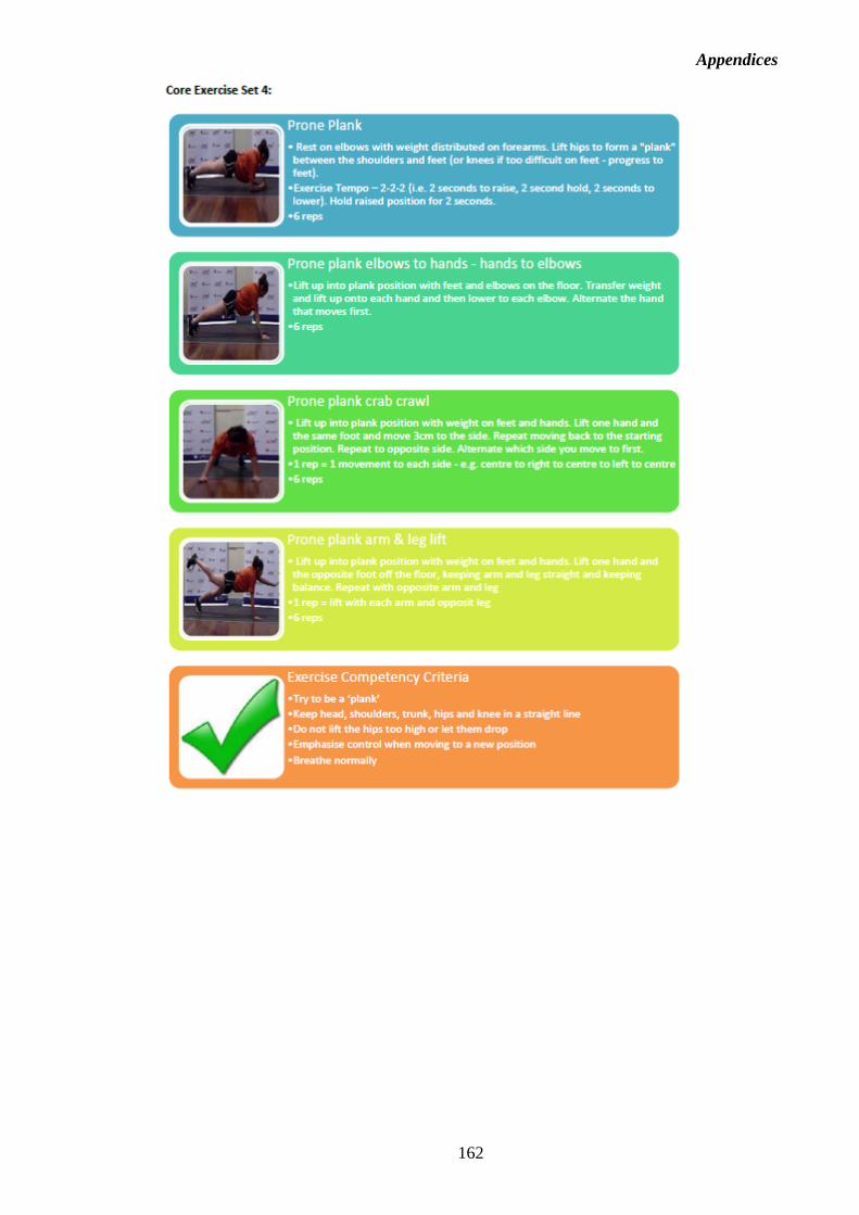
Appendices
162
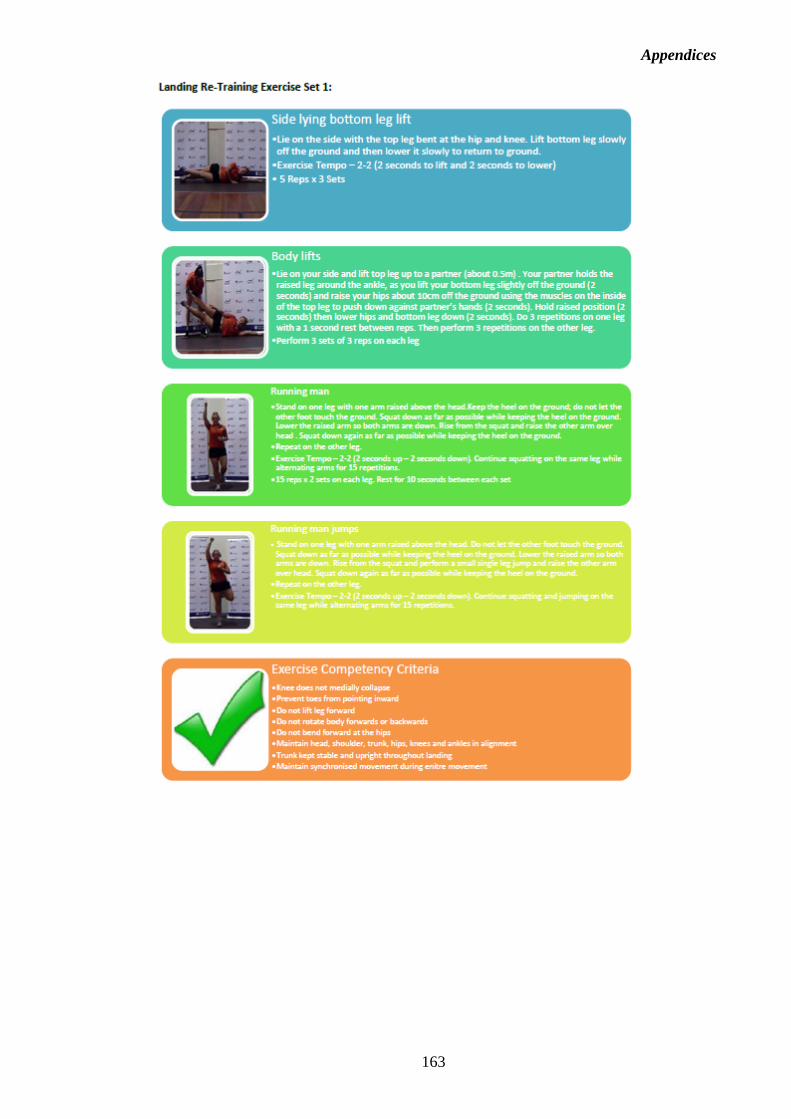
Appendices
163

Appendices
164

Appendices
165

Appendices
166

Appendices
167
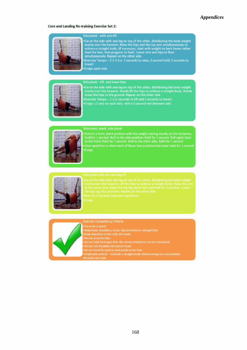
Appendices
168

Appendices
169

Appendices
170
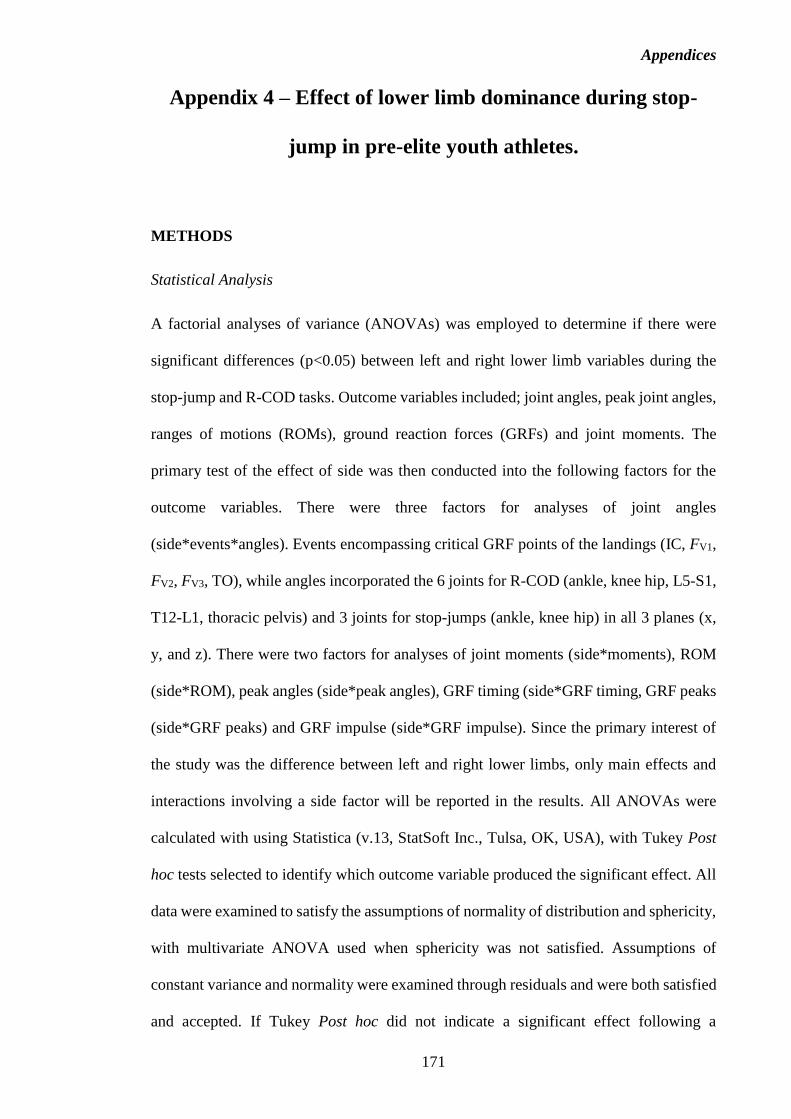
Appendices
171
Appendix 4 – Effect of lower limb dominance during stop-
jump in pre-elite youth athletes.
METHODS
Statistical Analysis
A factorial analyses of variance (ANOVAs) was employed to determine if there were
significant differences (p<0.05) between left and right lower limb variables during the
stop-jump and R-COD tasks. Outcome variables included; joint angles, peak joint angles,
ranges of motions (ROMs), ground reaction forces (GRFs) and joint moments. The
primary test of the effect of side was then conducted into the following factors for the
outcome variables. There were three factors for analyses of joint angles
(side*events*angles). Events encompassing critical GRF points of the landings (IC, FV1,
FV2, FV3, TO), while angles incorporated the 6 joints for R-COD (ankle, knee hip, L5-S1,
T12-L1, thoracic pelvis) and 3 joints for stop-jumps (ankle, knee hip) in all 3 planes (x,
y, and z). There were two factors for analyses of joint moments (side*moments), ROM
(side*ROM), peak angles (side*peak angles), GRF timing (side*GRF timing, GRF peaks
(side*GRF peaks) and GRF impulse (side*GRF impulse). Since the primary interest of
the study was the difference between left and right lower limbs, only main effects and
interactions involving a side factor will be reported in the results. All ANOVAs were
calculated with using Statistica (v.13, StatSoft Inc., Tulsa, OK, USA), with Tukey Post
hoc tests selected to identify which outcome variable produced the significant effect. All
data were examined to satisfy the assumptions of normality of distribution and sphericity,
with multivariate ANOVA used when sphericity was not satisfied. Assumptions of
constant variance and normality were examined through residuals and were both satisfied
and accepted. If Tukey Post hoc did not indicate a significant effect following a

Appendices
172
significant interaction of ANOVAs, univariate and independent t-tests were employed to
assess which variable was responsible for the interaction and effect size analyses,
respectively. Effects sizes (p) were defined as trivial (<0.0099), small (0.0099-0.0588),
moderate (0.0588-0.1379), and large (>0.1379) sizes (Richardson 2011).
RESULTS
Stop-Jump Kinematics
Joint angles showed no significant main effect of side (F1,87=1.87, p=0.18); however, a
significant interaction for side*events (F4,348=2.46, p=0.45), side*angles (F8,696=13.51,
p<0.001) and side*events*angles (F32,2784=1.73, p=0.01, Figure 1A) was present. Post
hoc tests showed when comparing the LEFT to the RIGHT, a greater grand mean across
all angles for the TO stage only (LEFT= -0.7±0.2°; RIGHT=0.1±0.2°; P<0.001) and
forefoot abduction across all events (LEFT= -12.4±0.8°; RIGHT=-6.7±0.6°; p<0.001). It
was revealed the LEFT when compared to the RIGHT displayed significantly greater
inversion at IC (p<0.05), FV1 (p<0.05) and FV2 (p<0.001), forefoot abduction across the
whole stance phase (IC p<0.001; FV1: p<0.001; FV2: p<0.001; FV3: p<0.001) and TO
(p<0.001), knee internal rotation at FV3 (p<0.001), with a decrease in knee flexion at TO
(p<0.05).

Appendices
173
Figure 1 Mean ± standard error (SE) of joint angles for left and right lower limbs (A)
joint angles (°) (B) joint angles range of motion (°) (C) peak joint angles (°)
during the horizontal landing phase of a stop-jump movement.

Appendices
174
Joint range of motion displayed a main effect of side (F1,87=47.88, p=0.01), and a
significant side*ROM interaction (F8,696=3087.43, p=0.01, Figure 1B). Post hoc analysis
identified an increase in LEFT when compared to the RIGHT in knee
adduction/abduction (p<0.001), knee internal/external rotation (p<0.01), hip
flexion/extension (p<0.001), hip adduction/abduction (p<0.001), hip internal/external
rotation ROM (p<0.01), with a decrease in ankle dorsiflexion/plantarflexion (p<0.001)
and knee flexion/extension (p<0.001).
A main effect of side was shown in peak joint angles (F1,87=7.07, p=0.01), with a
significant side*peak angle interaction (F17,1479=12.49, p<0.001, Figure 1C). Post hoc
analysis determined a significant difference with increased peak angles in LEFT
compared to the RIGHT eversion (p<0.001), inversion (p<0.001) and knee internal
rotation (p<0.05).
Stop-Jump Kinetics
There was no main effect of side was present for joint moments (F1,87=0.40, p=0.53);
however, a side*moment significant interaction was present (F17,1479=3.48, p<0.001,
Figure 2A). Post hoc analysis indicated a significant difference in RIGHT compared to
the LEFT hip extension moment (p<0.001).

Appendices
175
Figure 2 Mean ± standard error (SE) of joint kinetics for left and right lower limbs (A)
joint moments (BW) (B) peak GRFs (BW) (C) GRF timing (s) during the
horizontal landing phase of a stop-jump movement.

Appendices
176
A significant main effect of side was seen in peak GRF (F1,87=48.34, p=0.00), with a
side*peak force significant interaction (F4,348=11.89, p<0.01, Figure 2B). When
comparing LEFT versus RIGHT, a significant increase was identified in medial GRFs
(p<0.001), with a significant decrease found in lateral GRFs (p<0.001). Although no main
effect of timing was identified in GRF timing (F1,87=0.003, p=0.96), a side*GRF timing
interaction was evident (F7,609=3.78, p<0.001, Figure 2C). Post hoc analysis identified a
significant difference in LEFT compared to RIGHT with an increase in IC to peak anterior
force time (p<0.05). Although there was a main effect of side was identified in GRF
impulse (F1,87=6.09, p=0.02), there was no side*impulse interaction and no significant
differences identified in post hoc analysis.
R-COD Kinematics
A main significant effect of side was present in R-COD joint angles (F1,86=5.08, p=0.03),
with a side*angles interaction (F17,1462=8.77, p=0.00, Figure 3A). Post hoc analysis
determined significant differences when comparing the LEFT to the RIGHT with a
decrease in forefoot abduction (p<0.001) and an increase in T12-L1 lateral flexion
(p<0.001). A comparison of LEFT and RIGHT joint ROM did not display a main effect
of side (F1,64=0.45, p=0.50) and no other interaction was identified.
A significant main effect of side was displayed in peak joint angles (F1,86=4.41, p=0.04),
with a side*peak angles interaction (F29,2494=9.75, p=0.00, Figure 3Error! Reference
source not found.B). Post hoc analysis indicated significant differences in RIGHT
compared to LEFT with an increase identified in peak ankle inversion (p<0.001), peak

Appendices
177
T12-L1 left lateral flexion (p<0.001) and a decrease in T12-L1 right lateral flexion
(p<0.001).
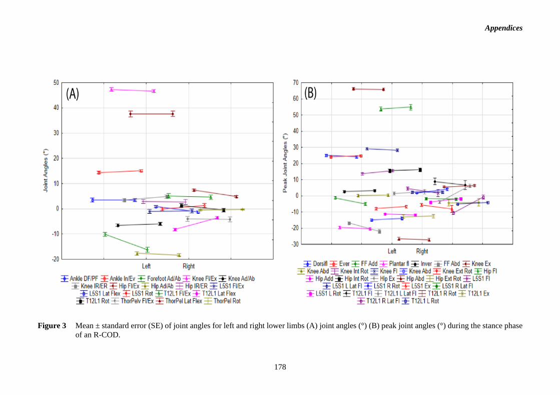
Appendices
178
Figure 3 Mean ± standard error (SE) of joint angles for left and right lower limbs (A) joint angles (°) (B) peak joint angles (°) during the stance phase
of an R-COD.
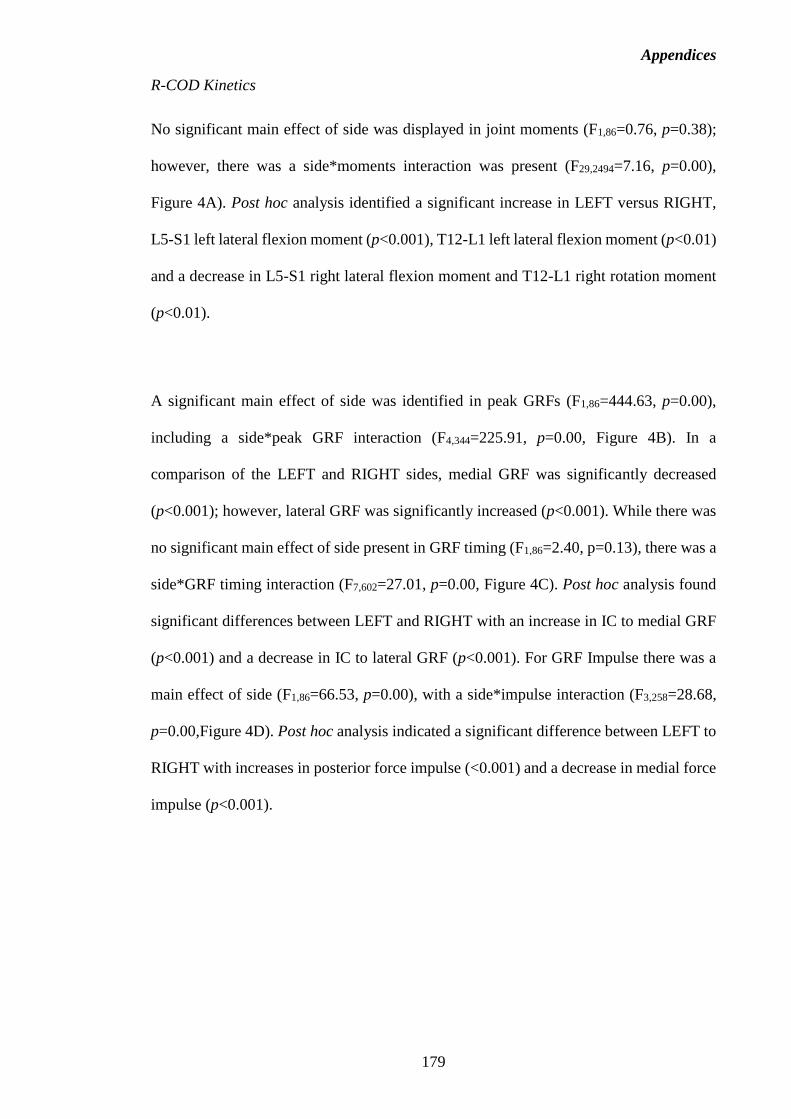
Appendices
179
R-COD Kinetics
No significant main effect of side was displayed in joint moments (F1,86=0.76, p=0.38);
however, there was a side*moments interaction was present (F29,2494=7.16, p=0.00),
Figure 4A). Post hoc analysis identified a significant increase in LEFT versus RIGHT,
L5-S1 left lateral flexion moment (p<0.001), T12-L1 left lateral flexion moment (p<0.01)
and a decrease in L5-S1 right lateral flexion moment and T12-L1 right rotation moment
(p<0.01).
A significant main effect of side was identified in peak GRFs (F1,86=444.63, p=0.00),
including a side*peak GRF interaction (F4,344=225.91, p=0.00, Figure 4B). In a
comparison of the LEFT and RIGHT sides, medial GRF was significantly decreased
(p<0.001); however, lateral GRF was significantly increased (p<0.001). While there was
no significant main effect of side present in GRF timing (F1,86=2.40, p=0.13), there was a
side*GRF timing interaction (F7,602=27.01, p=0.00, Figure 4C). Post hoc analysis found
significant differences between LEFT and RIGHT with an increase in IC to medial GRF
(p<0.001) and a decrease in IC to lateral GRF (p<0.001). For GRF Impulse there was a
main effect of side (F1,86=66.53, p=0.00), with a side*impulse interaction (F3,258=28.68,
p=0.00,Figure 4D). Post hoc analysis indicated a significant difference between LEFT to
RIGHT with increases in posterior force impulse (<0.001) and a decrease in medial force
impulse (p<0.001).

Appendices
180
Figure 4 Mean ± standard error (SE) of joint kinetics for left and right lower limbs (A) joint moments (BW), (B) peak GRF (BW), (C) GRF timing (s), and (D) impulse (BW) during the stance phase of a R-COD.

Appendices
181
Appendix 5 – Example Western Region Academy of Sport
Strength and Conditioning Program.

Appendices
182
Appendix 6 – Additional Figures for Chapter 3.
Figure 1 Mean ± standard error (SE) for joint angles (°) pre- and post-intervention during the stop-jump task for four intervention groups (No additional training (N), Core (C), Landing (L) and Core+ Landing (CL)).
Ankle dorsiflexion-plantarflexion (Ankle DF/PF), ankle inversion-eversion (Ankle In/Ev), forefoot adduction-abduction (Forefoot Ad/Ab), knee flexion-extension (Knee Fl/Ex), knee adduction-abduction (Knee Ad/Ab), hip adduction-abduction (Hip Ad/Ab), knee (Knee IR/ER), hip internal-external rotation (Hip IR/ER), L5-S1 flexion-extension (L5-S1 Fl/Ex), L5-S1 left(+)/right(-) lateral flexion (L5-S1 Lat Flex), L5-S1 right(+)/left(-) rotation (L5-S1 Rot), T12-L1 flexion-extension (T12-L1 Fl/Ex), T12-L1 left(+)/right(-) lateral flexion (T12-L1 Lat Flex), L5-S1 rotation right(+)/left(-) (T12-L1 Rot), Trunk_Ab-Pelvis flexion-extension (TrAb_Pelv Fl/Ex), Trunk_Ab-Pelvis right(+)/left(-) lateral flexion (TrAb_Pelv Lat Flex), and Trunk_Ab-Pelvis left(+)/right(-) rotation (TrAb_Pelv Rot).

Appendices
183
Figure 1 Mean ± standard error (SE) for (A) peak joint angle and (B) joint range of motion angles (°)pre- and post-intervention during the Stop-jump task for four intervention groups (No additional training (N), Core (C), Landing (L) and Core+ Landing (CL).

Chapter 3: Intervention programs alters stop-jump technique
184
Figure 2 Mean ± standard error (SE) for segment angles (°) pre- and post-intervention during the stop-jump task for four intervention groups (No additional training (N), Core (C), Landing (L) and Core+ Landing (CL)). Foot dorsiflexion-plantarflexion (Foot DF/PF), foot inversion-eversion (Foot In/Ev), foot adduction-abduction (Foot Ad/Ab), shank posterior-anterior inclination (Shank Post/Ant), shank adduction-abduction (Shank Ad/Ab), shank external-internal rotation (Shank Ex/In Rot) thigh posterior-anterior inclination (Thigh Post/Ant), thigh adduction-abduction (Thigh Ad/Ab), thigh external-internal rotation (Thigh ER/IR), pelvis extension-flexion (Pelvis Ex/Fl), pelvis right(+)/;eft(-) lateral tilt (Pelvis R/L Tilt), pelvis left(+)/right(-) rotation (Pelvis L/R Rot), Lumbar extension-flexion (Thoracic Ex/Fl), Lumbar right(+)/left(-) lateral flexion (Thoracic R/L Lat Flex), lumbar rotation left(+)/right(-) (Lumbar L/R Rot), thoracic Ex/Fl (Thoracic Ex/Fl), thoracic right(+)/left(-) lateral flexion (Thoracic R/L Lat Fl), thoracic left(+)/right(-) rotation (Thoracic L/R Rot), Trunk_Ab-Pelvis extension-flexion (TrunkAb Ex/Fl), Trunk_Ab-Pelvis right(+)/left(-) lateral flexion (TrunkAb R/L Lat Fl), and Trunk_Ab-Pelvis left(+)/right(-) rotation (TrunkAb Rot)..

Chapter 3: Intervention programs alters stop-jump technique
185
Figure 3 Mean ± standard error (SE) for peak net internal joint moments (normalised to body mass x height) pre- and post-intervention during the stop-jump task for four intervention groups (No additional training (N), Core (C), Landing (L) and Core+ Landing (CL).

Chapter 3: Intervention programs alters stop-jump technique
186
Figure 4 Mean ± standard error (SE) for (A) peak GRF (BW), (B) time to peak GRF (s) and (C) loading rate (BW·s-1) pre- and post-intervention during the stop-jump task for four intervention groups (No additional training (N), Core (C), Landing (L) and Core+ Landing (CL).
(A)
(B)
(C)
(C)

Chapter 3: Intervention programs alters stop-jump technique
187
Appendix 7 – Additional Figures for Chapter 4.
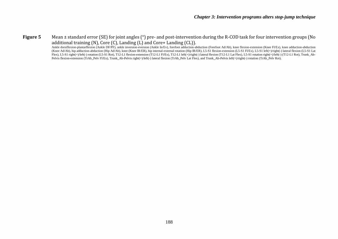
Chapter 3: Intervention programs alters stop-jump technique
188
Figure 5 Mean ± standard error (SE) for joint angles (°) pre- and post-intervention during the R-COD task for four intervention groups (No additional training (N), Core (C), Landing (L) and Core+ Landing (CL)). Ankle dorsiflexion-plantarflexion (Ankle DF/PF), ankle inversion-eversion (Ankle In/Ev), forefoot adduction-abduction (Forefoot Ad/Ab), knee flexion-extension (Knee Fl/Ex), knee adduction-abduction (Knee Ad/Ab), hip adduction-abduction (Hip Ad/Ab), knee (Knee IR/ER), hip internal-external rotation (Hip IR/ER), L5-S1 flexion-extension (L5-S1 Fl/Ex), L5-S1 left(+)/right(-) lateral flexion (L5-S1 Lat
Flex), L5-S1 right(+)/left(-) rotation (L5-S1 Rot), T12-L1 flexion-extension (T12-L1 Fl/Ex), T12-L1 left(+)/right(-) lateral flexion (T12-L1 Lat Flex), L5-S1 rotation right(+)/left(-) (T12-L1 Rot), Trunk_Ab-
Pelvis flexion-extension (TrAb_Pelv Fl/Ex), Trunk_Ab-Pelvis right(+)/left(-) lateral flexion (TrAb_Pelv Lat Flex), and Trunk_Ab-Pelvis left(+)/right(-) rotation (TrAb_Pelv Rot).

Chapter 3: Intervention programs alters stop-jump technique
189
Figure 6 Mean ± standard error (SE) for (A) peak joint angle and (B) joint range of motion angles (°)pre- and post-intervention during the R-COD task for four intervention groups (No additional training (N), Core (C), Landing (L) and Core+ Landing (CL).

Chapter 4: Intervention programs alters R-COD technique
190
Figure 7 Mean ± standard error (SE) for segment angles (°) pre- and post-intervention during the stop-jump task for four intervention groups (No additional training (N), Core (C), Landing (L) and Core+ Landing (CL)).
Foot dorsiflexion-plantarflexion (Foot DF/PF), foot inversion-eversion (Foot In/Ev), foot adduction-abduction (Foot Ad/Ab), shank posterior-anterior inclination (Shank Post/Ant), shank adduction-abduction (Shank Ad/Ab), shank external-internal rotation (Shank Ex/In Rot) thigh posterior-anterior inclination (Thigh Post/Ant), thigh adduction-abduction (Thigh Ad/Ab), thigh external-internal rotation (Thigh ER/IR), pelvis extension-flexion (Pelvis Ex/Fl), pelvis right(+)/;eft(-) lateral tilt (Pelvis R/L Tilt), pelvis left(+)/right(-) rotation (Pelvis L/R Rot), Lumbar extension-flexion (Thoracic Ex/Fl), Lumbar right(+)/left(-) lateral flexion (Thoracic R/L Lat Flex), lumbar rotation left(+)/right(-) (Lumbar L/R Rot), thoracic Ex/Fl (Thoracic Ex/Fl), thoracic right(+)/left(-) lateral flexion (Thoracic R/L Lat Fl), thoracic left(+)/right(-) rotation (Thoracic L/R Rot), Trunk_Ab-Pelvis extension-flexion (TrunkAb Ex/Fl), Trunk_Ab-Pelvis right(+)/left(-) lateral flexion (TrunkAb R/L Lat Fl), and Trunk_Ab-Pelvis left(+)/right(-) rotation (TrunkAb Rot)..

Chapter 4: Intervention programs alters R-COD technique
191
Figure 8 Mean ± standard error (SE) for peak net internal joint moments (normalised to body mass x height) pre- and post-intervention during the R-COD task for four intervention groups (No additional training (N), Core (C), Landing (L) and Core+ Landing (CL).

Chapter 4: Intervention programs alters R-COD technique
192
Figure 9 Mean ± standard error (SE) for (A) peak GRF (BW), (B) time to peak GRF (s) and (C) loading rate (BW·s-1) pre- and post-intervention during the R-COD task for four intervention groups (No additional training (N), Core (C), Landing (L) and Core+ Landing (CL).
(A)
(B)
(C)

Chapter 4: Intervention programs alters R-COD technique
193
Appendix 8 – Additional Figures for Chapter 5.
Figure 10 Mean ± standard error (SE) for joint angles (°) during the R-COD task for the three trunkab-pelvic range of motion groups. Ankle dorsiflexion-plantarflexion (Ankle DF/PF), ankle inversion-eversion (Ankle In/Ev), forefoot adduction-abduction (Forefoot Ad/Ab), knee flexion-extension (Knee Fl/Ex), knee adduction-abduction (Knee Ad/Ab), hip adduction-abduction (Hip Ad/Ab), knee (Knee IR/ER), hip internal-external rotation (Hip IR/ER), L5-S1 flexion-extension (L5-S1 Fl/Ex), L5-S1 left(+)/right(-) lateral flexion (L5-S1 Lat Flex), L5-S1 right(+)/left(-) rotation (L5-S1 Rot), T12-L1 flexion-extension (T12-L1 Fl/Ex), T12-L1 left(+)/right(-) lateral flexion (T12-L1 Lat Flex), L5-S1 rotation right(+)/left(-) (T12-L1 Rot), Trunk_Ab-Pelvis flexion-extension (TrAb_Pelv Fl/Ex), Trunk_Ab-Pelvis right(+)/left(-) lateral flexion (TrAb_Pelv Lat Flex), and Trunk_Ab-Pelvis left(+)/right(-) rotation (TrAb_Pelv Rot).
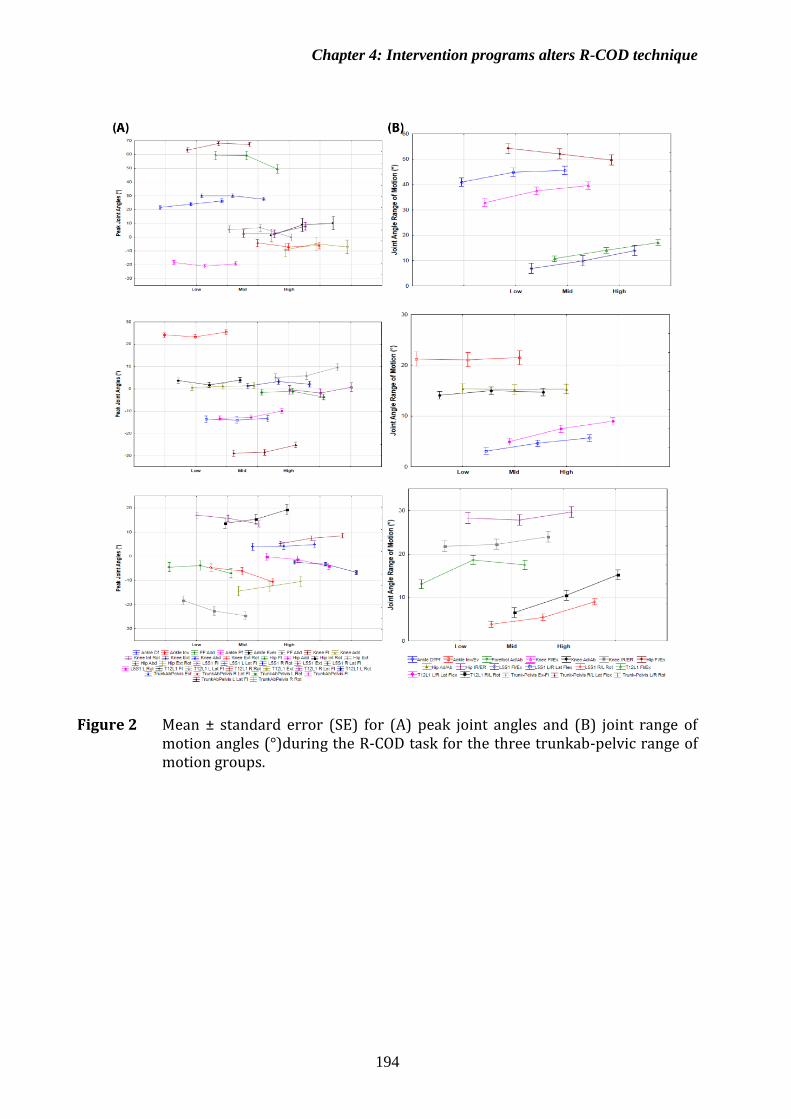
Chapter 4: Intervention programs alters R-COD technique
194
Figure 2 Mean ± standard error (SE) for (A) peak joint angles and (B) joint range of motion angles (°)during the R-COD task for the three trunkab-pelvic range of motion groups.
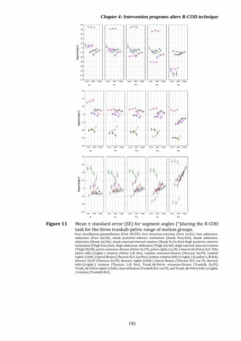
Chapter 4: Intervention programs alters R-COD technique
195
Figure 11 Mean ± standard error (SE) for segment angles (°)during the R-COD task for the three trunkab-pelvic range of motion groups. Foot dorsiflexion-plantarflexion (Foot DF/PF), foot inversion-eversion (Foot In/Ev), foot adduction-abduction (Foot Ad/Ab), shank posterior-anterior inclination (Shank Post/Ant), shank adduction-abduction (Shank Ad/Ab), shank external-internal rotation (Shank Ex/In Rot) thigh posterior-anterior inclination (Thigh Post/Ant), thigh adduction-abduction (Thigh Ad/Ab), thigh external-internal rotation (Thigh ER/IR), pelvis extension-flexion (Pelvis Ex/Fl), pelvis right(+)/;eft(-) lateral tilt (Pelvis R/L Tilt), pelvis left(+)/right(-) rotation (Pelvis L/R Rot), Lumbar extension-flexion (Thoracic Ex/Fl), Lumbar right(+)/left(-) lateral flexion (Thoracic R/L Lat Flex), lumbar rotation left(+)/right(-) (Lumbar L/R Rot), thoracic Ex/Fl (Thoracic Ex/Fl), thoracic right(+)/left(-) lateral flexion (Thoracic R/L Lat Fl), thoracic left(+)/right(-) rotation (Thoracic L/R Rot), Trunk_Ab-Pelvis extension-flexion (TrunkAb Ex/Fl), Trunk_Ab-Pelvis right(+)/left(-) lateral flexion (TrunkAb R/L Lat Fl), and Trunk_Ab-Pelvis left(+)/right(-) rotation (TrunkAb Rot)..

Chapter 5: Trunk-Pelvis ROM & Injury Risk in R-COD
196
Figure 12 Mean ± standard error (SE) for peak net internal joint moments (normalised to body mass x height) during the R-COD task for the three trunkab-pelvic range of motion groups..
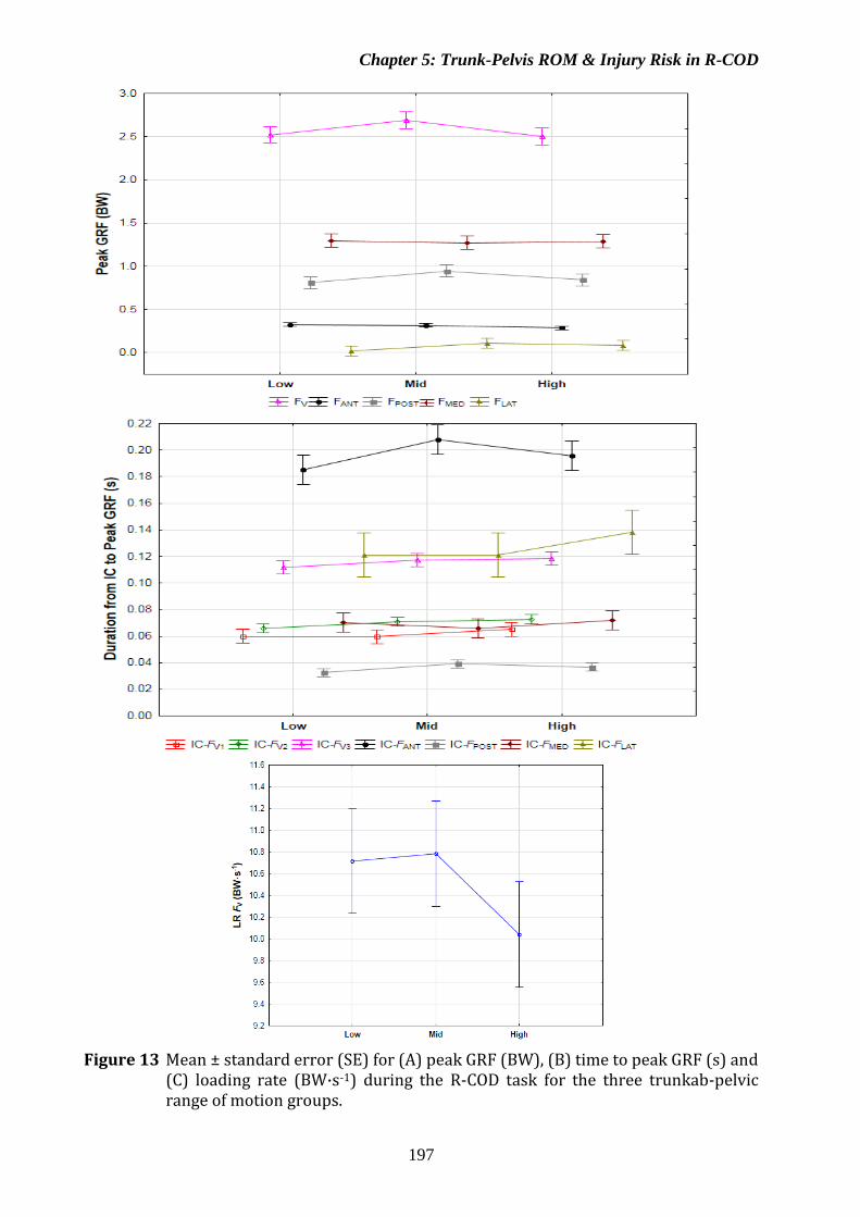
Chapter 5: Trunk-Pelvis ROM & Injury Risk in R-COD
197
Figure 13 Mean ± standard error (SE) for (A) peak GRF (BW), (B) time to peak GRF (s) and
(C) loading rate (BW·s-1) during the R-COD task for the three trunkab-pelvic range of motion groups.