lower eyelid pinch · pdf filethe pinch lower blepharoplasty can comfortably usurp the...
TRANSCRIPT

Lower Eyelid Pinch Blepharoplasty
�
LORNE K. ROSENFIELD
CHAPTER 32
32_N2e_Rosenfield_r5_bs_969-994.qxd:Volume1 9/28/10 4:10 PM Page 969
Reprinted with permission from Nahai F. The Art of Aesthetic Surgery: Principles and Techniques, 2nd edition. St. Louis: Quality Medical Publishing, 2010. Copyright © 2010 Quality Medical Publishing, Inc. All rights reserved.

When I examined my own results, I observed, not as infrequently as I would haveliked, two particular stigmata of a less than perfect result at the lower eyelid. First,lasting mild scleral show was evident, often preceded by weeks of overly optimisticeyelid taping. This 55-year-old woman, shown preoperatively and 1 year after a tra-ditional skin-muscle lower blepharoplasty, exhibits this telltale postoperative signof scleral show.
970 Part VI � Eyelid Surgery
The lower eyelid blepharoplasty embodies a classic surgical paradox worth re-visiting: the more one performs a particular surgery, the more respect it may
command. Whereas ignorance may be bliss, knowledge can be quite motivating. Anysurgeon who has critically assessed his or her skin-muscle flap lower blepharoplastyresults would heartily agree with this statement.
Second, residual crêpey skin was identified, most often after treatment of prodigiousfat herniation. This 48-year-old woman, shown preoperatively and 1 year after atraditional blepharoplasty, exhibits this “untreated” redundant skin.
I was compelled by these discomfiting observations to seek an effective solution—amodified procedure that would at once ensure optimal correction of the eyelid de-formities and yet maintain normal eyelid posture. And so was born the pinch bleph-aroplasty series. My personal experience, reported in 2005, confirmed the safety andefficacy of this approach.
32_N2e_Rosenfield_r5_bs_969-994.qxd:Volume1 9/28/10 4:10 PM Page 970

Although the true incidence of eyelid malposition after a traditional muscle flap bleph-aroplasty is not well defined, the plethora of articles on the subject attests to its per-sistence. Scleral show has been ascribed to multiple causes: excess skin removal, un-treated eyelid laxity, denervation of the orbicularis muscle, and scarring of the outeror middle lid lamellae. This postoperative problem may be considered subtle and in-deed is often not even acknowledged, but it represents what could also be seen as aglaring example of the “operated” look that we should all strive to avoid. As for theexcess skin left behind postoperatively, this problem has been equally neglected inthe literature.
Evolution of Technique
There have been many efforts to reduce the incidence of eyelid malposition follow-ing traditional blepharoplasty. As documented by Zarem and Resnick, one approachhas been to forego the skin incision entirely, thus preserving the integrity of the outerand middle eyelid lamellae, and to approach the eyelid instead through the conjunc-tiva only. Although the incidence of scleral show may be less with this approach,there can be a greater chance of untreated excess eyelid skin.
In another effort to avoid the skin incision and still treat the skin, skin resurfacingwith a chemical peel or laser, in conjunction with a transconjunctival approach, canindeed reduce the incidence of scleral show. However, unless the skin redundancy ismodest and the entire face is treated, resurfacing may not adequately treat the skinredundancy and can otherwise produce distracting lines of demarcation. Addition-ally, if these therapies penetrate too deeply, undesirable changes in eyelid posturemay still occur.
With a skin-muscle flap, a relatively conservative resection of the skin has alwaysbeen advocated, regardless of the extent of redundancy. Indeed, it is often impres-sive how little skin is actually removed despite such an aggressive flap dissection.And of course, in a patient with more significant excess skin, this conservatism hassurely led to inadequate treatment.
Another adjunctive technique to a blepharoplasty is the canthopexy, particularly ina patient with a lax eyelid. There is no question that this repair helps reduce the in-cidence of lid malposition, but the results have been frustratingly inconsistent. Wehave all seen scleral show after a traditional blepharoplasty despite the addition of acanthopexy, even when applied prophylactically. An inadequate canthopexy, coupledwith overaggressive skin resection, and the inciting factors of muscle denervationand middle lamellar scarring, probably explain this inconsistency. The deliberatepreservation of a wider orbicularis strip of muscle, when conducting a skin-muscleflap approach, may be salutary but has clearly not proved to be the complete answer.
971Chapter 32 � Lower Eyelid Pinch Blepharoplasty
32_N2e_Rosenfield_r5_bs_969-994.qxd:Volume1 9/28/10 4:10 PM Page 971
The idea of pinching the excess skin from the lower eyelid is not a new one; in 1973Parkes et al were the first to suggest the technique. This description, which predatedthe transconjunctival approach, attenuated its potential benefits by also describing thedivision of the underlying orbicularis muscle to retrieve the excess fat.
Then in 1992 Dinner et al published a case report on the ultimate combination ofthe skin pinch with the transconjunctival approach in a “no flap” technique. Ristow,in 1994 in Mimis Cohen’s textbook, included the concept of a direct skin resectionwith a measured and marked resection.
My impetus to revisit and refine a pinch blepharoplasty came from a personal com-munication with Glenn Jelks in 2000. The solution became even more lucid whenour decades-old standard approach to the upper eyelid blepharoplasty was consid-ered: we “pinch” the eyelid excess to determine the extent of the excision while ob-serving the effect on the eyelash and brow posture. Why not apply the same simplemetric to the lower eyelid? So was born the “pinch blepharoplasty” series.
Advantages
My personal experience of more than 400 pinch blepharoplasties confirms that thisapproach is indeed capable of producing better, more consistent results than the tra-ditional skin-muscle flap technique. This variation offers two distinctive advantages:more crêpey skin can be safely removed, and an aesthetic eyelid posture is secured.
This rewarding marriage of goals is primarily the result of the inherent accuracy ofthe pinch technique. The approach enables the surgeon to assess and define in realtime the prospective skin resection as well as carefully evaluate its effect on eyelidposture. The pinch technique avoids a heavy skin-muscle flap, which can otherwisecreate both worrisome vertical traction and more swelling. Additionally, the pinchblepharoplasty eliminates the usual violation of the orbicularis muscle and orbitalseptum, an action that could lead to denervation, scarring, and poor eyelid posture.
Another possible reason for the improved results is the often seen amelioration ofthe eyelid-cheek groove with a more youthful vertical shortening of the eyelid, per-haps secondary to the effacing effect of the significant skin resection.
These benefits translate into a more adaptable and consistent blepharoplasty. Thisadvantage is seen particularly and most gratifyingly in morphologically challengingpatients with a negative vector and poor lid posture; older patients with poor lidtone; patients with extensive skin or festoons; and younger patients with primary
972 Part VI � Eyelid Surgery
32_N2e_Rosenfield_r5_bs_969-994.qxd:Volume1 9/28/10 4:10 PM Page 972
skin redundancy and nominal excess fat. If there is asymmetrical skin between theeyelids, or even within one eyelid itself, the pinch can be tailored accordingly. Em-powered with this versatile tool, the surgeon can now treat the medial eyelid skin,a zone that was traditionally neglected, for an even more complete result. In addi-tion, because the skin excess is more thoroughly treated, the surgeon can avoid theneed for regional laser resurfacing of the eyelid, with its added period of healing andattendant, often distracting lines of demarcation.
The advantages of the pinch blepharoplasty can be doubled with a staged reappli-cation of the pinch to excise even more skin. This “repinch” can be accomplishedquite simply, with a local anesthetic. Thus it is feasible that essentially all crêpey skinat the lower eyelid can now be removed.
Indications and Contraindications
The pinch lower blepharoplasty can comfortably usurp the standard skin-muscleflap technique. Therefore this approach may be offered to the same group of pa-tients. In contradistinction to the standard technique, there is a productive breadthof application, depending on the extent of the patient’s problem. That is, all patientsare candidates, but some are better candidates than others. Although results will besuperior in all patients, defining the best and worst patient candidates for the pinchmost effectively illustrates the nuances of the technique.
973Chapter 32 � Lower Eyelid Pinch Blepharoplasty
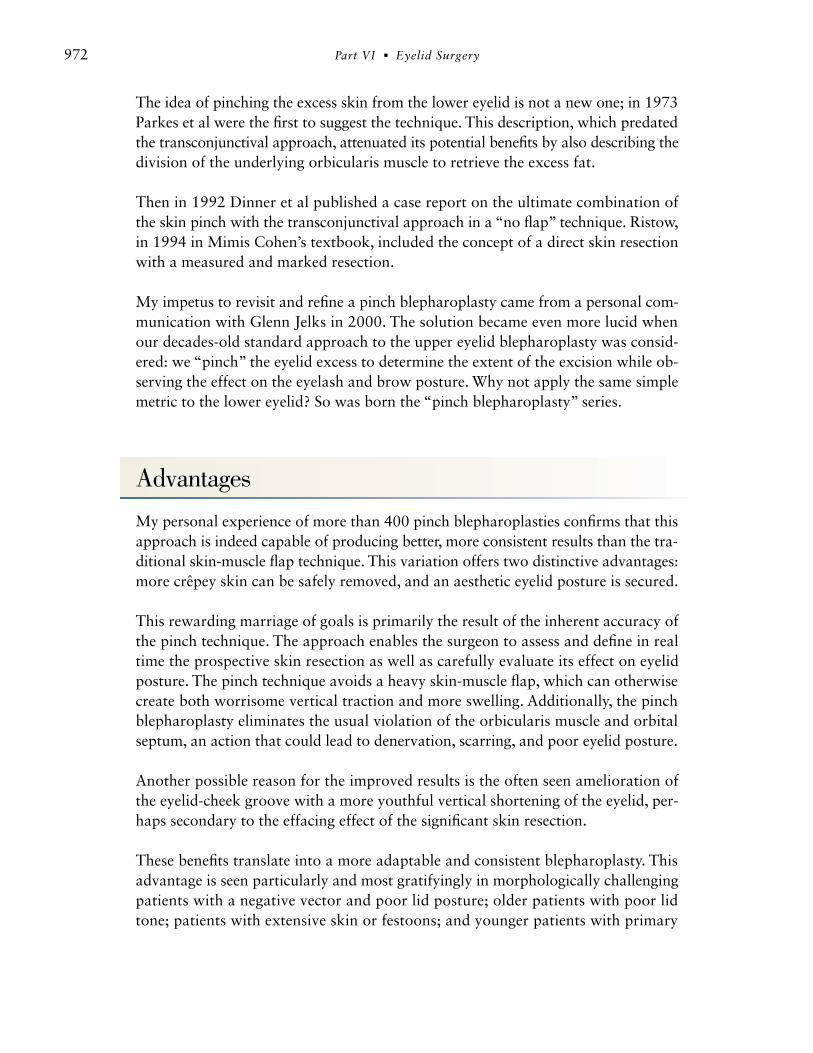
The ideal patient has abundant thin, crêpey skin, minimal excess fat, and morpholog-ically advantageous anatomy (such as high cheekbones and almond-shaped eyes).With the application of the pinch and a planned second pinch procedure, if neces-sary, the improvement achieved in these patients can be dramatic, simply becausethe surgeon is able to more fully treat the eyelid.
32_N2e_Rosenfield_r5_bs_969-994.qxd:Volume1 9/28/10 4:11 PM Page 973
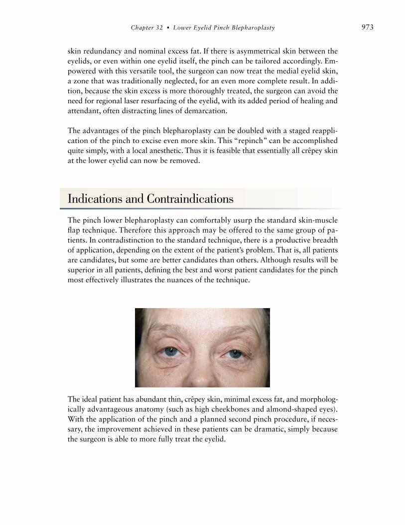
At the opposing end of the spectrum, the imperfect patient is one with thicker, sun-damaged skin, which clearly will not “pinch” well. In these patients, it is best to planthe surgery so that the skin will have the least distortion from local anesthetic andto apply the pinch conservatively. Just as with patients with very thin skin, a thick-skinned patient will equally benefit from a staged second pinch procedure.
Because a dramatic skin resection can be done with this approach, the surgeon mustbe cautious when applying the pinch to a patient with equally significant redundantfat, because some of this skin must remain to redrape the now less convex lowereyelid. Again, these patients can always undergo a simple second pinch procedureat a later date.
Pertinent Anatomy
The essence of the pinch technique embodies a key anatomic principle: to violatethe eyelid anatomy as little as possible, and if it is compromised by age or genetics,to repair it. Between the transconjunctival approach and the pinch, the pinch touchesand takes only what it needs to—the skin—leaving behind the critical neuromechan-ical support: the orbital septum and the orbicularis muscle and its attendant nerves.
As for the canthopexy, the key anatomic principle is to keep it simple. Because thelateral canthus truly comprises several components and is not easily identified as a dis-tinct structure, the canthopexy has to become a kind of maneuver “on faith“: youcan only really feel its presence. That is, rather than trying to dissect and visualizethe canthal tissues themselves, the surgeon need only grasp and pull whatever tissueis present in the area to know the “right stuff” has been captured. Then it simply be-comes a matter of manipulating this anchor thoughtfully to perform an individual-ized canthopexy.
974 Part VI � Eyelid Surgery
32_N2e_Rosenfield_r5_bs_969-994.qxd:Volume1 9/28/10 4:11 PM Page 974

Preoperative Assessment
History There are several critical historical points to be gleaned, including a history of eyedryness, tearing, or the need for drops; allergies; Graves’ disease; prior eyelid sur-gery; and previous injection of tissue fillers.
Physical ExaminationThe preoperative physical examination should include the same components as forany blepharoplasty. However, the difference is that because the pinch allows one tomodify the surgery to match the patient’s problem, the value of a detailed analysisis even more rewarding.
The critical elements to evaluate are listed in the box.
975Chapter 32 � Lower Eyelid Pinch Blepharoplasty
Critical Factors to Assess in the Physical Examination
� The extent of the redundant skin: If necessary, a second pinch procedure can beplanned in advance. This is particularly relevant in a patient with significant “active”skin: exaggerated wrinkling on animation.
� The extent of the redundant fat: Depending on the volume of excess, more or less skinshould be pinched to ensure that enough skin is left behind.
� The degree of lid laxity and attendant scleral show (as evaluated by a snap test): Thiscan be treated efficiently with a stitch canthopexy.
� The level of the lateral canthus (canthal tilt): The canthal position can be adjusted rel-atively precisely with a stitch canthopexy.
� The vector angle of the globe to the malar (positive/neutral/negative): This aids thesurgeon in the design of the best application of the pinch and canthopexy componentsof the surgery.
32_N2e_Rosenfield_r5_bs_969-994.qxd:Volume1 9/28/10 4:11 PM Page 975

976 Part VI � Eyelid Surgery
PhotographyThere are several tenets to be followed for photographic evaluation of the eyelidsand documentation of the results.
Prevent Parallax ErrorParallax refers to the apparent displacement or difference in the apparent positionof an object viewed along two different lines of sight, such as occurs because the lensof a camera and the viewfinder see the subject from a slightly different position. Thusphotographs must be taken with the patient and surgeon seeing “eye to eye”; thatis, there should be no height discrepancy that could distort the appearance of theposture of the lower eyelid in relation to the globe.
This problem can be manifested when either the patient is not level or the camerais not level. These effects can mask or exaggerate the actual eyelid position.
Patient tipped up Patient tipped down Patient level
Camera high Camera low Camera level
THE PARALLAX EFFECT
32_N2e_Rosenfield_r5_bs_969-994.qxd:Volume1 9/28/10 4:11 PM Page 976

Beware of the Moro Reflex
977Chapter 32 � Lower Eyelid Pinch Blepharoplasty
When a tighter photo of just the eyelids is to be taken, some patients, anticipatingthe flash’s effect, may widen their eyes dramatically. If this is noted, pulling the cam-era back to include a full-face picture should correct the distortion.
Follow the Dermatologist’s Credo
To accurately document the effects of the pinch procedure, progressively closer pic-tures of the face should be taken, from full face to just wrinkles at the lower eyelid.
Control the Patient’s EmotionsThere is a place for both active and passive photographs of a patient’s lower eyelid.Photos should be taken of the patient smiling to define the extent of muscle-skin excess.
Close-up with Moro
Full face without Moro
Close-up preoperative photo Close-up postoperative photo
32_N2e_Rosenfield_r5_bs_969-994.qxd:Volume1 9/28/10 4:11 PM Page 977

978 Part VI � Eyelid Surgery
Often orbicularis wrinkling is the patient’s primary concern, and any correction of thisproblem is a true measure of a technique’s efficacy.
On the other hand, a smile can camouflage an eyelid posture or a scleral show prob-lem.
Compare the Present With the PastThe patient should be asked to bring photographs from the past (such as college orwedding photos or well-focused candid photos). The insight gained from reviewingsuch historic images is invaluable not only for observing changes in the character-istics of the periorbital area but also in our attempt to recapture the patient’s char-acteristic expression or persona.
Patient EducationA patient with significant wrinkling must be informed that a second pinch blepharo-plasty should be considered, depending on the salutary effects of the first procedure.The goals and actions of a canthopexy should be explained, because the palpebralfissure will become more almond shaped.
Patient smiling preoperatively Patient smiling postoperatively
Patient’s smile hides scleral show Patient at rest with scleral show
32_N2e_Rosenfield_r5_bs_969-994.qxd:Volume1 9/28/10 4:11 PM Page 978
Planning and Technique
Particularly when first attempted, the pinch procedure is best conducted with infil-tration of as little local anesthetic as possible. With deeper sedation or general anes-thesia, it is both possible and ideal to use no local infiltration. This consideration iscritical, because it allows the surgeon to most accurately judge and pinch the skinexcess without the potential distortion caused by aggressive local infiltration.
Surgical Plan 1. If a local anesthetic must be used, place it in the lower eyelid in the region of the
future pinch.2. Conduct the upper eyelid blepharoplasty, leaving the wound open.3. Complete the transconjunctival portion of the lower blepharoplasty.4. Perform a stitch canthopexy through the upper-outer eyelid wound.5. Close the upper eyelid wound.6. Perform the lower eyelid pinch procedure and close the wound.
Marking
979Chapter 32 � Lower Eyelid Pinch Blepharoplasty
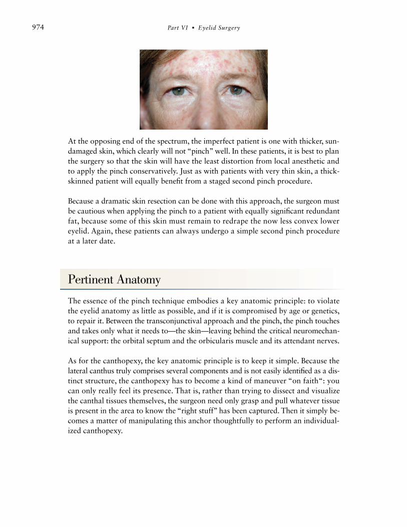
The first step is to mark a guiding “pinch line” along the lower eyelid, representingthe uppermost margin of the future excision. Medially, this line is usually placedwithin a few millimeters of the ciliary margin, continuing in a straight line laterally,deliberately leaving a progressively enlarging triangular island of intact skin of 4 to5 mm in minimum height below the lateral eyelid margin. This maneuver furtherdiscourages scleral show by lessening the purse-string–like scar contracture that mayoccur with a curvilinear incision hugging the ciliary margin. However, the latitudeof this line can be modified, depending on the location and quality of the excess skin.That is, the pinch line can be moved up or down on the eyelid to facilitate maximalskin removal. If the skin is noticeably thicker, the pinch line should be moved far-ther away from the ciliary margin to help prevent scleral show.
2232-1 Pinch
blepharoplasty
32_N2e_Rosenfield_r5_bs_969-994.qxd:Volume1 9/28/10 4:11 PM Page 979

Sequencing of ProceduresThe sequence of operative steps is critical. If an upper blepharoplasty is planned aswell, it should be conducted first, with wound closure delayed until after a can-thopexy but before the lower eyelid pinch. Thus the lateral upper eyelid wound isavailable for the canthopexy, and any lower eyelid skin redundancy that may betreated with closure of the upper eyelid takes place before the pinch procedure. Onthe other hand, if only a lower blepharoplasty is to be performed, to accomplish apexy a 10 to 15 mm counterincision may be made in the lateral upper eyelid alongthe path of an upper blepharoplasty.
Operative Technique
After the markings, the surgeon applies any local anesthetic early and judiciously tominimize the amount of local swelling and maximize its resolution. The transcon-junctival portion of the procedure is best conducted preceding a possible canthopexy,lest the surgeon have difficulty distracting or even undoing the tightened eyelid. Thecanthopexy should be performed before the lower eyelid pinch procedure, becausethe potential lateral lift of the pexy can treat some of the lower eyelid excess. As forthe lower eyelid, the excess fat is trimmed as indicated and the wound reapproxi-mated at its midsection with a 6-0 fast-absorbing catgut suture.
The upper eyelid portion of the blepharoplasty is then performed as premarked. Theexcess skin and as indicated, excess muscle, are resected. Then the underlying or-bital fat is treated, if necessary, with electrocautery and/or excision. Before closureof the upper eyelid wound, the lateral stitch canthopexy is performed.
I distinguish two kinds of canthopexy to consider and design: prophylactic and ther-apeutic. The choice is primarily determined by two factors: the position of the lat-eral canthus and the posture and tone of the lateral lower eyelid. If the lateral can-thus is at or above the horizon and/or the lower eyelid tone is mildly weak withoutscleral show, a prophylactic pexy is in order. If instead the lateral canthus is belowthe horizon and/or the lower lateral eyelid tone is weak with scleral show, a thera-peutic lateral canthopexy should be performed. Clearly, there is always a gray zonein this arbitrary categorization: there are patients with a normal neutral canthal cantwho could have an aesthetic improvement with a slight elevation of the lateral can-thus, and as such the canthopexy could be considered not only prophylactic but ther-apeutic as well. This nuance is particularly relevant in a patient who in youth hadan almond-shaped eye.
980 Part VI � Eyelid Surgery
32_N2e_Rosenfield_r5_bs_969-994.qxd:Volume1 9/28/10 4:11 PM Page 980

981Chapter 32 � Lower Eyelid Pinch Blepharoplasty
In either case, the procedure is conducted through the lateral aspect of an upper bleph -aroplasty incision. First, using the Bovie, a segment of approximately 1 cm of thesuperolateral inner orbital rim is exposed, with great care to preserve the integrityof the periosteum.
Then a subfascial/submuscular “tunnel” is created between the orbital rim and the lat-eral canthus using round-tipped iris scissors.
A very fine, curved mosquito clamp is passed down the tunnel, and its very tip isused to capture the lateral canthal tissue.
If a prophylactic canthopexy is planned, the mosquito clamp tip is passed up to theorbital rim to determine the ideal pexy location, which will at once modestly tightenthe lower eyelid and maintain the canthal angle. If a therapeutic pexy is desired, the
32_N2e_Rosenfield_r5_bs_969-994.qxd:Volume1 9/28/10 4:11 PM Page 981
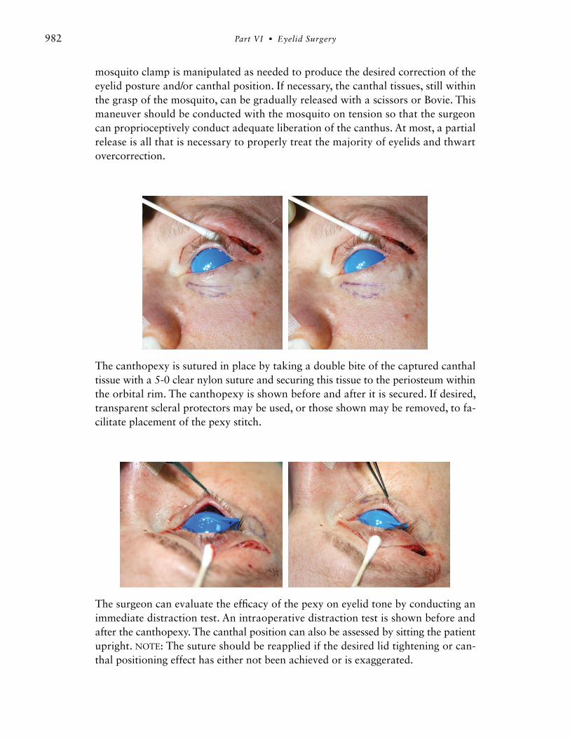
mosquito clamp is manipulated as needed to produce the desired correction of theeyelid posture and/or canthal position. If necessary, the canthal tissues, still withinthe grasp of the mosquito, can be gradually released with a scissors or Bovie. Thismaneuver should be conducted with the mosquito on tension so that the surgeoncan proprioceptively conduct adequate liberation of the canthus. At most, a partialrelease is all that is necessary to properly treat the majority of eyelids and thwartovercorrection.
982 Part VI � Eyelid Surgery
The canthopexy is sutured in place by taking a double bite of the captured canthaltissue with a 5-0 clear nylon suture and securing this tissue to the periosteum withinthe orbital rim. The canthopexy is shown before and after it is secured. If desired,transparent scleral protectors may be used, or those shown may be removed, to fa-cilitate placement of the pexy stitch.
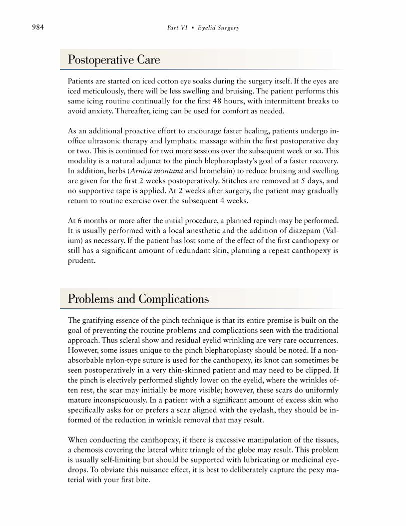
The surgeon can evaluate the efficacy of the pexy on eyelid tone by conducting animmediate distraction test. An intraoperative distraction test is shown before andafter the canthopexy. The canthal position can also be assessed by sitting the patientupright. NOTE: The suture should be reapplied if the desired lid tightening or can-thal positioning effect has either not been achieved or is exaggerated.
32_N2e_Rosenfield_r5_bs_969-994.qxd:Volume1 9/28/10 4:11 PM Page 982

Then, using a pair of fine Adson-Brown forceps, the subciliary skin is progressivelypinched along the premarked line into a standing “wall” of skin. The endpoint shouldbe the maximal effacement of wrinkled skin resting primarily below while preserv-ing a normal posture of the eyelid margin above.
983Chapter 32 � Lower Eyelid Pinch Blepharoplasty
With a forceps producing linear traction and a straight pair of scissors, the wall isamputated at its base.
The appropriate amount of skin has been removedif the wound edges are close to each other or justkissing. The wound may be opened to electrocoagu-late any small bleeding vessels and is then closed witha 7-0 running nylon suture. No taping or other sup-port of the eyelids is necessary.
Pinch Blepharoplasty
32_N2e_Rosenfield_r5_bs_969-994.qxd:Volume1 9/28/10 4:11 PM Page 983
Postoperative Care
Patients are started on iced cotton eye soaks during the surgery itself. If the eyes areiced meticulously, there will be less swelling and bruising. The patient performs thissame icing routine continually for the first 48 hours, with intermittent breaks toavoid anxiety. Thereafter, icing can be used for comfort as needed.
As an additional proactive effort to encourage faster healing, patients undergo in-office ultrasonic therapy and lymphatic massage within the first postoperative dayor two. This is continued for two more sessions over the subsequent week or so. Thismodality is a natural adjunct to the pinch blepharoplasty’s goal of a faster recovery.In addition, herbs (Arnica montana and bromelain) to reduce bruising and swellingare given for the first 2 weeks postoperatively. Stitches are removed at 5 days, andno supportive tape is applied. At 2 weeks after surgery, the patient may graduallyreturn to routine exercise over the subsequent 4 weeks.
At 6 months or more after the initial procedure, a planned repinch may be performed.It is usually performed with a local anesthetic and the addition of diazepam (Val-ium) as necessary. If the patient has lost some of the effect of the first canthopexy orstill has a significant amount of redundant skin, planning a repeat canthopexy isprudent.
Problems and Complications
The gratifying essence of the pinch technique is that its entire premise is built on thegoal of preventing the routine problems and complications seen with the traditionalapproach. Thus scleral show and residual eyelid wrinkling are very rare occurrences.However, some issues unique to the pinch blepharoplasty should be noted. If a non-absorbable nylon-type suture is used for the canthopexy, its knot can sometimes beseen postoperatively in a very thin-skinned patient and may need to be clipped. Ifthe pinch is electively performed slightly lower on the eyelid, where the wrinkles of-ten rest, the scar may initially be more visible; however, these scars do uniformlymature inconspicuously. In a patient with a significant amount of excess skin whospecifically asks for or prefers a scar aligned with the eyelash, they should be in-formed of the reduction in wrinkle removal that may result.
When conducting the canthopexy, if there is excessive manipulation of the tissues,a chemosis covering the lateral white triangle of the globe may result. This problemis usually self-limiting but should be supported with lubricating or medicinal eye-drops. To obviate this nuisance effect, it is best to deliberately capture the pexy ma-terial with your first bite.
984 Part VI � Eyelid Surgery
32_N2e_Rosenfield_r5_bs_969-994.qxd:Volume1 9/28/10 4:11 PM Page 984

985Chapter 32 � Lower Eyelid Pinch Blepharoplasty
Finally, if performed too aggressively, the canthopexy will produce an overly cor-rected result. Usually the effect will improve over time. On the other hand, if thepexy is not harnessed adequately, the patient may be left with a canthal or lid de-formity. The best preventive maneuver is to critically assess and redo the pexy if nec-essary in the first place.
Outcomes
The spirit of this modification of the lower blepharoplasty is defined by a triad ofmaneuvers: (1) the deliberate preservation of the eyelid’s integrity with a transcon-junctival incision, (2) the mindful alignment of the lower eyelid with respect to theglobe and canthus with the application of the canthopexy, and, pivotally, (3) the pro-ficient excision of the redundant skin with the mastery of the pinch technique.
These examples demonstrate the extent of skin excision possible with pinch tech-nique, averaging 8 to 12 mm. In addition, the patient usually has less bruising andswelling and heals more rapidly.
The typical early appearance of a postoperative patient is shown at 5 days and at 7 days. Together these efforts consistently deliver a salutary removal of redundantskin of the eyelid while still safeguarding its posture. In fact, with greater comfortperforming the stitch canthopexy, the surgeon can often go one step further and cre-
32_N2e_Rosenfield_r5_bs_969-994.qxd:Volume1 9/28/10 4:11 PM Page 985

986 Part VI � Eyelid Surgery
ate a subtle, more aesthetic shape to the otherwise normal lower eyelid. On the otherhand, particularly in a morphologically challenging patient, the canthopexy may de-finitively correct the attendant canthal deformity. Additionally, the application ofthe repinch procedure, best planned for before the first pinch is performed, has re-liably contributed to even more satisfying results.
Results
The following cases were chosen to demonstrate the power of the pinch proceduregiven various patient presentations—from the best to the more difficult blepharo-plasty candidates.
The Older Patient
This 68-year-old woman is shown preoperatively and 1 year after an upper andlower pinch blepharoplasty. Lower eyelid aging was corrected and a normal eyelidposture was ensured.
This 53-year-old woman with the classic stigmata of aging would previously haveundergone a routine blepharoplasty with CO2 laser or chemical resurfacing. Instead,upper and lower pinch blepharoplasties with prophylactic canthopexies were per-formed. She is seen preoperatively at age 53, in a historic photo at age 23, and post-operatively at age 54, with an attractive, youthful eyelid posture restored.
32_N2e_Rosenfield_r5_bs_969-994.qxd:Volume1 9/28/10 4:11 PM Page 986
The Younger Patient
987Chapter 32 � Lower Eyelid Pinch Blepharoplasty
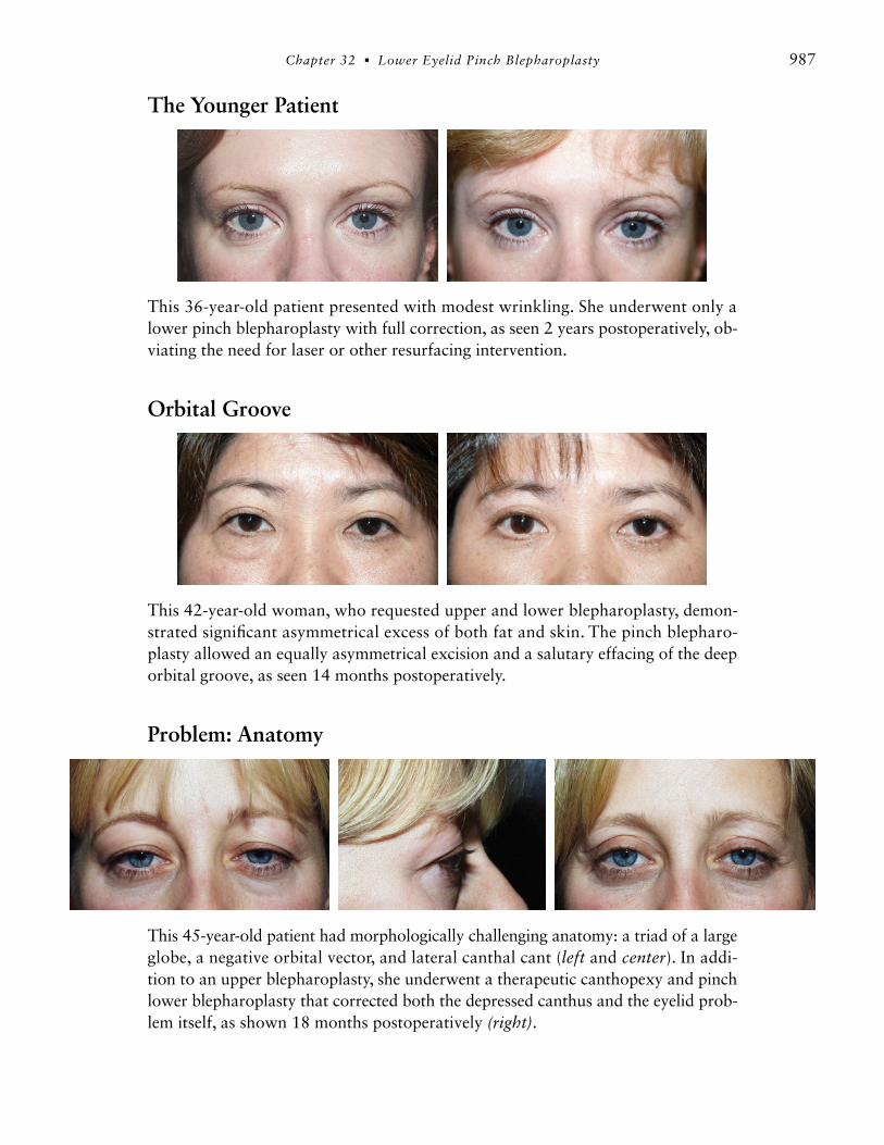
This 36-year-old patient presented with modest wrinkling. She underwent only alower pinch blepharoplasty with full correction, as seen 2 years postoperatively, ob-viating the need for laser or other resurfacing intervention.
Orbital Groove
This 42-year-old woman, who requested upper and lower blepharoplasty, demon-strated significant asymmetrical excess of both fat and skin. The pinch blepharo-plasty allowed an equally asymmetrical excision and a salutary effacing of the deeporbital groove, as seen 14 months postoperatively.
Problem: Anatomy
This 45-year-old patient had morphologically challenging anatomy: a triad of a largeglobe, a negative orbital vector, and lateral canthal cant (left and center). In addi-tion to an upper blepharoplasty, she underwent a therapeutic canthopexy and pinchlower blepharoplasty that corrected both the depressed canthus and the eyelid prob-lem itself, as shown 18 months postoperatively (right).
32_N2e_Rosenfield_r5_bs_969-994.qxd:Volume1 9/28/10 4:11 PM Page 987
988 Part VI � Eyelid Surgery
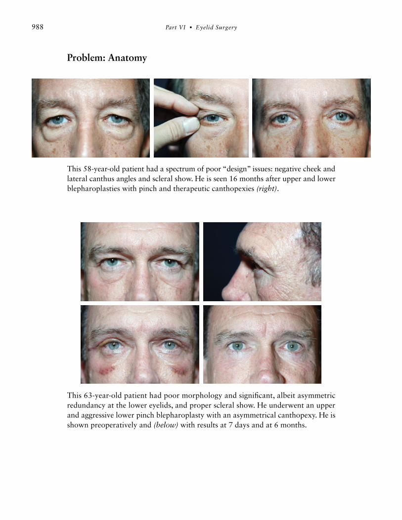
This 58-year-old patient had a spectrum of poor “design” issues: negative cheek andlateral canthus angles and scleral show. He is seen 16 months after upper and lowerblepharoplasties with pinch and therapeutic canthopexies (right).
This 63-year-old patient had poor morphology and significant, albeit asymmetricredundancy at the lower eyelids, and proper scleral show. He underwent an upperand aggressive lower pinch blepharoplasty with an asymmetrical canthopexy. He isshown preoperatively and (below) with results at 7 days and at 6 months.
Problem: Anatomy
32_N2e_Rosenfield_r5_bs_969-994.qxd:Volume1 9/28/10 4:11 PM Page 988

Problem: Redundant Skin
989Chapter 32 � Lower Eyelid Pinch Blepharoplasty
This 52-year-old woman had considerable excess crêpey skin. Two years after anupper and lower pinch blepharoplasty, she demonstrates essentially complete re-moval of the redundant skin; good eyelid posture has been maintained.
This 73-year-old woman was an ideal patient for a pinch blepharoplasty. The photoseries reveals the pathway to relatively complete repair of lower eyelid aging. Thepatient is shown intraoperatively, demonstrating the prodigious pinched wall of skinand wound, with preservation of an attractive eyelid posture. The 1-year postoper-ative view confirms the rewarding improvement.
32_N2e_Rosenfield_r5_bs_969-994.qxd:Volume1 9/28/10 4:11 PM Page 989
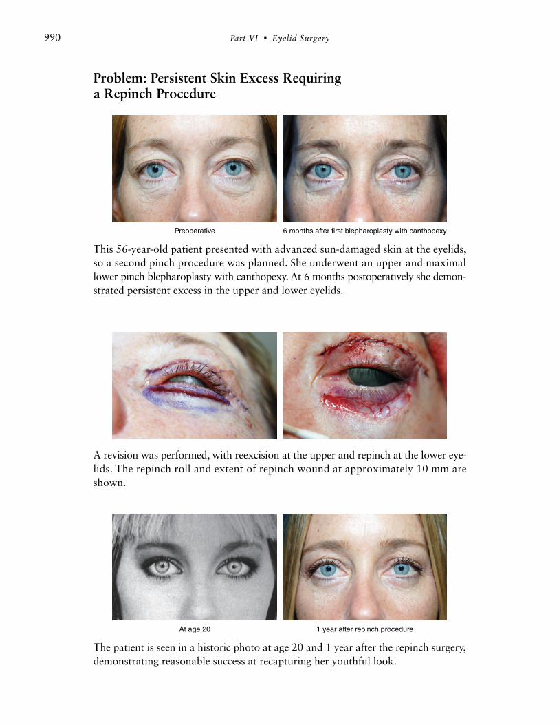
Problem: Persistent Skin Excess Requiring a Repinch Procedure
990 Part VI � Eyelid Surgery
This 56-year-old patient presented with advanced sun-damaged skin at the eyelids,so a second pinch procedure was planned. She underwent an upper and maximallower pinch blepharoplasty with canthopexy. At 6 months postoperatively she demon-strated persistent excess in the upper and lower eyelids.
A revision was performed, with reexcision at the upper and repinch at the lower eye-lids. The repinch roll and extent of repinch wound at approximately 10 mm areshown.
The patient is seen in a historic photo at age 20 and 1 year after the repinch surgery,demonstrating reasonable success at recapturing her youthful look.
Preoperative 6 months after first blepharoplasty with canthopexy
At age 20 1 year after repinch procedure
32_N2e_Rosenfield_r5_bs_969-994.qxd:Volume1 9/28/10 4:11 PM Page 990

Repinch Procedure
991Chapter 32 � Lower Eyelid Pinch Blepharoplasty
This 72-year-old woman illustrates the power of the repinch procedure. The stagedsurgery was performed at approximately 6 months. The patient is shown preoper-atively and 6 months after the first pinch procedure.
The intraoperative views demonstrate the second pinch roll, the wound kissing, andthe pinch wound.
The patient is shown 4 days after the second pinch procedure and 1 year later.
32_N2e_Rosenfield_r5_bs_969-994.qxd:Volume1 9/28/10 4:11 PM Page 991

This 68-year-old patient is seen in close-up views of progressive correction: preop-eratively, following the first pinch blepharoplasty, and following the repinch surgery,each result 6 months apart.
Concluding Thoughts
One could say that these modifications empower the surgeon to tame and commandgreater control of the lower eyelid. Personally, I have found the effects of this tech-nique quite gratifying: I have both a newfound serenity when approaching the lowereyelid with this more reliable means for preventing surgical stigmata and a satisfac-tion with the results because of the more comprehensive eyelid correction that can berealized.
The procedure’s advantages are particularly manifested when one is confronted bychallenging morphology. As an endorsement of the efficacy of the pinch blepharo-plasty, I have not performed a skin-muscle flap lower blepharoplasty in more than12 years. The pinch blepharoplasty has eliminated the need for lower eyelid resur-facing in most cases.
In addition, we have abolished the proverbial “therapeutic” postoperative taping. Inow perform more canthopexies intelligently and excise more skin confidently. Theresults of these efforts have included consistently faster patient recovery, more ac-curate canthal and eyelid positioning, and what is most important, significantly im-proved aesthetic outcomes.
992 Part VI � Eyelid Surgery
32_N2e_Rosenfield_r5_bs_969-994.qxd:Volume1 9/28/10 4:11 PM Page 992

Clinical Caveats
� The surgeon should infiltrate the local anesthetic as early and minimally as pos-sible. The less distortion of the tissues, the more accurate and facile the pinch ma-neuver will be. This is particularly germane at the start of a surgeon’s learningcurve. To this end, it is often helpful to sedate the patient more deeply just beforethe pinch procedure.
� The addition of a stitch canthopexy is a preoperative decision predicated on eval-uation of both the position of the lateral canthus and the posture and tone of thelower eyelid. The action of the canthopexy may be classified as either prophylac-tic (elderly, prominent globe, malar hypoplasia, or “negative” lateral canthal po-sition) or therapeutic (laxity or scleral show).
� Generally, if a patient is deemed to be a candidate for a lower blepharoplasty, atleast a prophylactic canthopexy should probably be performed. Thus almost everypatient now undergoes a canthopexy. In effect, the stitch canthopexy, when judi-ciously harnessed, empowers the surgeon to take full advantage of the pinch pro-cedure and perform the most complete removal of redundant skin.
� Both the pinch blepharoplasty and the stitch canthopexy techniques have a no-table learning curve. During this evolution, the surgeon should be aware that it isdeceivingly easy to “overpinch” or “overpexy.” As a layer of protection initially,it is prudent to pinch a few millimeters farther away from the lash margin com-pared with the traditional incision. This scar consistently heals imperceptibly. Also,when conducting a stitch canthopexy, the surgeon should have a low thresholdfor replacing the stitch if an overcorrection is perceived; it may not “settle” enoughon its own postoperatively. Likewise, the pexy should be reattempted if an under-correction is noted.
� The pinch portion of this technique can easily be repeated with the patient underlocal anesthesia to treat any residual crêpey skin.
993Chapter 32 � Lower Eyelid Pinch Blepharoplasty
32_N2e_Rosenfield_r5_bs_969-994.qxd:Volume1 9/28/10 4:11 PM Page 993
Annotated BibliographyDinner MI, Glassman H, Artz JS. The “no flap” technique for lower-lid blepharoplasty. Aes-
thetic Plast Surg 16:155-158, 1992.In a case report, the authors described the first lower blepharoplasty with a skin pinch incombination with the transconjunctival approach.
Parkes M, Fein W, Brennan HG. Pinch technique for repair of cosmetic eyelid deformities.Arch Ophthalmol 89:324-328, 1973.This is the first description in the medical literature of the pinch technique for skin re-moval. However, the approach negated some of the benefit of the pinch, because Parkesdivided the orbicularis muscle to access the fat.
Ristow B. Transconjunctival blepharoplasty. In Cohen M, ed. Mastery of Plastic and Recon-structive Surgery, vol 3. Boston: Little Brown, 1994. Ristow included the concept of a direct excision of skin in conjunction with a transcon-junctival blepharoplasty. Rather than pinching the redundant skin, it was measured,marked, and excised in a fashion akin to the traditional upper eyelid skin removal.
Suggested ReadingsJelks G. No touch blepharoplasty. In Cardoso de Castro C, ed. Midface Surgery. Philadel-
phia: Elsevier/Saunders, 2009. Kim EM, Bucky LP. Power of the pinch. Ann Plast Surg 60:532-537, 2008.Rosenfield LK. The pinch blepharoplasty revisited. Plast Reconstr Surg 115:1405-1412; dis-
cussion 1413-1414, 2005.Rosenfield LK. A safe technique with superior results. Aesthetic Surg J 27:199-203, 2007. Zarem HA, Resnick JI. Minimizing deformity in lower blepharoplasty: the transconjunc-
tival approach. Clin Plast Surg 20:317-321, 1993.
994 Part VI � Eyelid Surgery
32_N2e_Rosenfield_r5_bs_969-994.qxd:Volume1 9/28/10 4:11 PM Page 994