low-grade myofibroblastic sarcoma arising in fibroadenoma ... · low-grade myofibroblastic sarcoma...
TRANSCRIPT
CASE REPORT Open Access
Low-grade myofibroblastic sarcoma arisingin fibroadenoma of the breast-A casereport–Na-Hye Myong1* and Jun-Won Min2
Abstract
Background: Myofibroblastic sarcoma or myofibrosarcoma is a malignant tumor of myofibroblasts and known todevelop rarely in the breast, but its underlying lesion and tumor cell origin have never been reported yet.
Case presentation: A 61-year-old female presented with a gradually growing breast mass with well-demarcatedovoid nodular shape. The tumor was histologically characterized by fascicular-growing spindle cell proliferation withlarge areas of hyalinized fibrosis and focally ductal epithelial remnants embedded in myxoid stroma, mimicking afibroadenomatous lesion. It had frequent mitoses of 5–16/10 high-power fields, hemorrhagic necrosis, and focallypericapsular invasion. The spindle cells were diffusely immunoreactive for fibronectin, smooth muscle actin, andcalponin, which suggest a myofibroblastic origin. Multiple irregularly thickened vessels with medial or pericytic cellproliferation were found to be merged with the intrinsic tumor cells. The tumor could be diagnosed low-grademyofibroblastic sarcoma arising in an old fibroadenoma.
Conclusion: We report a case of a low-grade mammary myofibrosarcoma that showed a background lesion offibroadenoma first in the worldwide literature and suggest the pericytes or medial muscle cells of the intratumoralvessels as the cell origin of the myofibroblastic sarcoma.
Keywords: Breast, Fibroadenoma, Myofibroblasts, Pericytes, Sarcoma
BackgroundMyofibroblastic sarcoma or myofibrosarcoma (MFS) isa malignant tumor which is composed of or originatedfrom myofibroblasts. It has been known to arise mainlyat head and neck regions, although it could be found atthe extremities, trunk, and retroperitoneum. Mammarymyofibrosarcomas have been very rarely reported, withonly about 10 cases in the worldwide literature [1–7].The mammary myofibroblastic sarcomas in the litera-ture have revealed predominantly low-grade cytomor-phology rather than high-grade tumor mimickingmalignant fibrous histiocytoma (MFH) in the primarylesions. Thus, the main differential diagnoses of low-grade myofibrosarcoma (LG-MFS) in the breast rangefrom nodular fasciitis and myofibroblastoma to fibro-sarcoma, leiomyosarcoma, and cystosarcoma phyllodes
[7, 8]. However, there has never been reported aboutthe background lesion or cell origin of myofibroblastsin the worldwide literature, because all of the reportedcases reveal neither epithelial component nor morpho-logic evidence for the cell origin anywhere as in thepresent case. We report a case of mammary myofibro-sarcoma showing the background lesion of fibroaden-oma and the pericytic cell origin of myofibroblastictumor cells first in the worldwide literature.
Case presentationA 61-year-old woman visited a breast clinic at our hos-pital, because she palpated a left breast mass first8 months ago and felt that it has grown gradually in sizefrom that time on. She had no family history of breastcancer and her physical examination revealed no axillarylymph node enlargement. Her mammography revealed a3.5 cm-sized well-defined nodular mass with highdensity at 11 o’clock position of left breast, that wasradiologically diagnosed as fibroadenoma (Fig. 1). She
* Correspondence: [email protected] of Pathology, Dankook University College of Medicine, 119Dandae-ro, Dongnam-gu, Cheonan-si, Chungnam 31116, KoreaFull list of author information is available at the end of the article
© 2016 Myong and Min. Open Access This article is distributed under the terms of the Creative Commons Attribution 4.0International License (http://creativecommons.org/licenses/by/4.0/), which permits unrestricted use, distribution, andreproduction in any medium, provided you give appropriate credit to the original author(s) and the source, provide a link tothe Creative Commons license, and indicate if changes were made. The Creative Commons Public Domain Dedication waiver(http://creativecommons.org/publicdomain/zero/1.0/) applies to the data made available in this article, unless otherwise stated.
Myong and Min Diagnostic Pathology (2016) 11:33 DOI 10.1186/s13000-016-0480-8

underwent a core needle biopsy initially under the im-pression of fibroadenoma, but the mass was diagnosedas being suggestive of myofibroblastoma at that time.Then, the mass was excised completely by a lumpec-tomy, which grossly revealed a slightly lobulated andovoid nodular lesion, measuring 5×3×3 cm, and whitishyellow, solid, homogeneous, rubbery, and myxoid cutsurface. Microscopic examination disclosed predomin-antly spindle cells with whorling or fascicular arrange-ment, which had mildly pleomorphic fusiform nuclei,moderate amount of eosinophilic fibrillary cytoplasm,and indistinct cytoplasmic borders (Fig. 2). Mitotic activ-ity averaged 5–10 per 10 high-power fields (HPFs) but
was found focally increased up to 16/10HPFs (Fig. 3).The nodular mass showed the hemorrhagic necrosis inabout 5 % of entire tumor sections and a few foci ofpseudocystic degeneration (Fig. 4). Background stromawas variably fibromyxoid according to the cellularity,ranging from being myxoid or loosely collagenous in thecellular areas to densely collagenous in the hypocellularor acellular areas (Fig. 5). Densely collagenous areaamounted up to about 30 % and showed a few scatteredductal epithelial remnants of collapsed-linear or abort-ively oval shape. The peripheral portion of the massalso showed the ductal epithelial components embed-ded in the myxoid stroma, that suggests an ancientfibroadenomatous lesion (Fig. 6). Intriguingly, someintratumoral vessels revealed irregularly thickened wallswith proliferating medial muscle cells that merge withadjacent intrinsic spindled tumor cells (Fig. 7). Therewere no areas of highly pleomorphic tumor cells likethose of malignant fibrous histiocytoma. The tumorwas relatively well-encapsulated, but focally showed thepushing-type extension outside the capsule. Immuno-histochemical staining results of the tumor cells werediffusely positive for fibronectin, α-smooth muscleactin, calponin, and vimentin (Fig. 8). Immunoreactiv-ities for CD34, desmin, pancytokeratin, bcl-2, estrogenreceptor, and progesterone receptor were negative. Thepericytes or medial muscle cells of the small venuleswere also immunoreactive for smooth muscle actin(SMA) and calponin, but not for desmin. All those lightmicroscopic findings and immunohistochemical datasuggested a myofibroblastic sarcoma of probably pericyticor vascular medial cell origin, with the background of
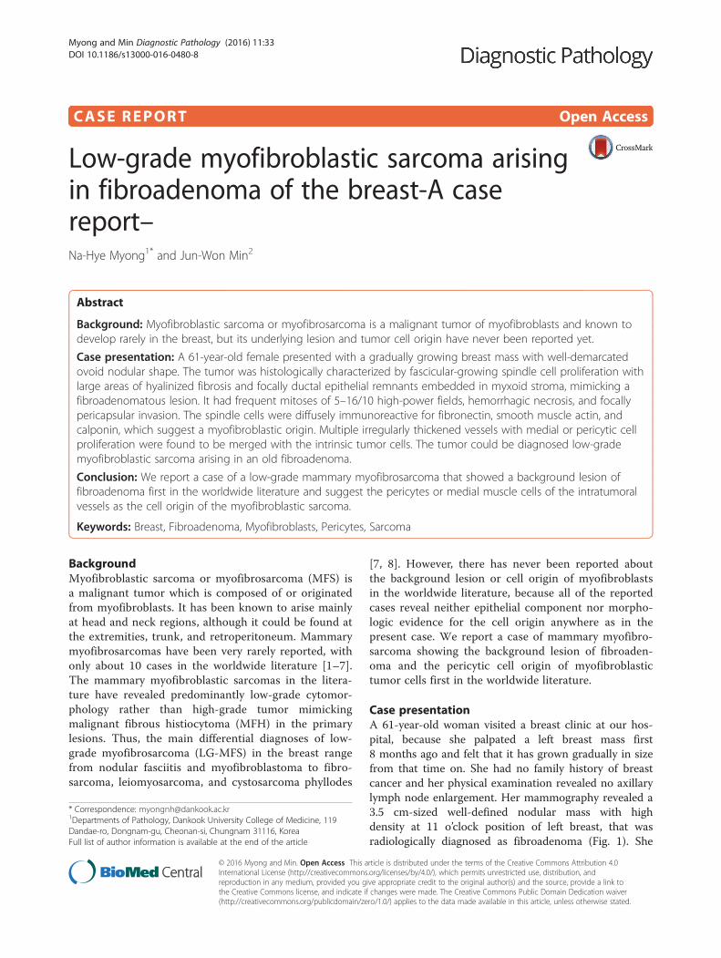
Fig. 1 Mammographic finding and low-powered microscopic view of the breast mass. a A mammographic image revealed a well-circumscribedand highly dense nodular mass at 11 o’clock position. b The mass was well-circumscribed and mostly encapsulated, with the slightly lobulatedmargin (H&E stain, ×12.5)
Fig. 2 Representative histologic findings of the tumor. It is mostlycomposed of spindle-shaped mesenchymal cells with vaguelyfasciculated growing pattern, elongated plump nuclei, pale eosinophiliccytoplasms, and small indistinct nucleoli (H&E stain, ×200)
Myong and Min Diagnostic Pathology (2016) 11:33 Page 2 of 7

ancient fibroadenomatous lesion. She got no additionalchemotherapy or therapeutic radiation after lumpectomy.Follow-up data for 2 years after the lumpectomy revealedno evidence of local recurrence or distant metastasis onthe mammogram, ultrasonography, Positron EmissionTomography, Magnetic Resonance Imaging of the liver,and abdominal Computed Tomography. Myofibroblastsare generally best identified in the granulation tissue butalso might be originated from vascular smooth musclecells or pericytes [9] or even from epithelial cells via
metaplasia [10]. It has been a matter of considerable con-troversy whether or not a true myofibroblast can be neo-plastic, because myofibroblasts can be found in thereactive conditions and benign neoplasms such as nodularfasciitis and fibromatosis. However, myofibroblastoma ofthe breast has been known as a distinctive benign mesen-chymal tumor, since a study on 16 cases at 1987 showedthat the myofibroblastoma cells are ultrastructurallydifferent from fibroblasts, smooth muscle cells, or myoe-pithelial cells and microscopically characterized by
Fig. 4 Two histologic findings indicative of degeneration in the tumor cells. a The tumor had focally hemorrhagic necrosis in about 5 % of theentire mass (H&E stain, ×200). b There was a few scattered foci of pseudocystic degeneration (H&E stain, ×200)
Fig. 3 A cytologic finding suggestive of malignancy in this case. a The tumor cells show high cellularity and frequent mitotic figures (arrow) (H&Estain, ×200). b In one high-powered microscopic field, two mitotic figures (arrows) are observed (H&E, stain, ×400)
Myong and Min Diagnostic Pathology (2016) 11:33 Page 3 of 7

uniformly slender and bipolar spindle cells haphazardlyarranged in fascicular clusters with hyalinized fibrosis [11].Thereafter, malignant tumors of myofibroblasts or thosewith myofibroblastic differentiation have been diagnosedin the breast by the presence of more aggressive histologicfindings such as frequent mitotic counts, infiltrativegrowth pattern, and necrosis compared to myofibroblas-tomas. They are characterized clinically by variablymalignant behavior ranging from recurrence to diffusepleuropulmonary metastasis [1–7]. Myofibroblastic
sarcoma or myofibrosarcoma (MFS) has been generallyknown to be identified best by their characteristic ultra-structural findings such as fibronectin extracellular fi-brils and their fibronexus junctions, but it has beenrecently reported that the fibronectin can be demon-strated consistently by an immunohistochemical stain-ing result alone [8]. Therefore, the present case could bediagnosed as MFS showing myofibroblastic differenti-ation with diffuse immunoreactivity for fibronectin.About 10 cases of mammary myofibroblastic sarcoma
Fig. 5 Variable stromal backgrounds according to the tumor cellularity. a Myxoid stroma is representatively found in moderately cellular tumorareas (H&E stain, ×100). b Slightly fibrous stromal background is diffusely noticed in the highly cellular areas (H&E stain, ×100). c Denselycollagenous tissue replaces about 30 % of the mass, showing a few sparsely entrapped ductal epithelial components (H&E stain, ×40)
Fig. 6 A peripheral region with fibroadenomatous remnants. a Round to ovoid ductal components are embedded in the myxoid cellular stroma(H&E stain, ×12.5). b The fibroadenomatous remnant shows still benign epithelial and mesenchymal components (H&E stain, ×40)
Myong and Min Diagnostic Pathology (2016) 11:33 Page 4 of 7

have been reported in the worldwide literature to thepresent, including the present case. The clinicopatho-logic features of only 8 cases among them are summa-rized for comparison in Table 1. Male to female ratiowas 2:8 and the duration between disease onset and itsdiagnosis ranged from 1 week to 8 months. Immunohis-tochmical demonstration for myofibroblasts was done
best by immunopositivities for SMA(100 %), vimen-tin(100 %), fibronectin(100 %), calponin(100 %), andbcl-2(50 %). Desmin, CD34, type IV collagen, laminin,h-caldesmon, and S100 protein were not immunoreac-tive in most cases. Four out of 8 cases had metastasis orrecurrence, with 1 metastatic and 1 recurrent case beingalive. Six cases (case 1–4, 6, 7) underwent ultrastructural
Fig. 7 Intratumoral small vessels showing the transition to the tumor cells. a A peripheral venule-like vessel reveals intraluminally medialcell proliferation (arrows) (H&E stain, ×40). b Another tumor vessel is irregularly thickened by medial cell proliferation (curved arrows) ofpericytic smooth muscle cells, which merge with the adjacent tumor cells (H&E stain, ×100)
Fig. 8 Immunohistochemical staining profiles favoring a myofibroblastic cell origin. a Diffuse fibronectin-immunoreactivity is found in the tumorcell cytoplasms (Diaminobenzidine, ×400). b Most tumor cell cytoplasms are immunoreactive for α-smooth muscle actin (Diaminobenzidine,×400). c There is a diffuse nuclear immunopositivity for calponin, a myofibroblastic differentiation marker (Diaminobenzidine, ×200). d Vimentin, ageneral mesenchymal marker is immunostained strongly in most tumor cells (Diaminobenzidine, ×400)
Myong and Min Diagnostic Pathology (2016) 11:33 Page 5 of 7

studies, that usually demonstrated their myofibroblastictumor cells with fibronectin extracellular fibrils and fibro-nexus junctions. However, any case report for mammaryMFS have not reported or commented the background le-sions such as fibroadenoma as like the present case. Also,any suggested cell origin of MFS has not been discussedyet within the tumor tissue in a worldwide literature.MFS is generally graded by two-tiered system as lowand high-grades rather than low, intermediate, and highgrades by mitotic count, nuclear pleomorphism, andnecrosis. The former two grades of MFS share to a de-gree histopathologic findings of low-grade fascicularspindle cell neoplasm, whereas high-grade MFS showdistinctively marked cellular pleomorphism mimickingMFH [8]. Therefore, our case could be diagnosed cyto-logically as low-grade myofibrosarcoma (LG-MFS). Themost representative myofibroblastic tumors of thebreast are myofibroblastoma and LG-MFS. LG-MFS isa predominantly spindle cell neoplasm that could showfrequent mitoses and focal necrosis, whereas benignmyofibroblastic tumors including mammary-type myo-fibroblastoma don’t reveal the two. In addition, LG-MFS of the breast may have a wide range of differentialdiagnoses such as leiomyosarcoma, fibrosarcoma, andcystosarcoma phyllodes with fibrosarcoma-like over-growth, but the most important differential diagnosis inthe present case is the last one. Both lesions can showepithelial entrapment within the tumor, but there areobvious differences in morphological, immunohisto-chemical, and ultrastructural study results for the sar-coma cell nature. The overgrown fibrosarcoma-likelesion arising in cystosarcoma phyllodes generally showcleft-like compressed epithelial components and the fi-broblastic features rather than myofibroblasts by theimmunohistochemical and ultrastructural studies. In
contrast, the present tumor revealed multifocally re-sidual linear or round ductal epithelial remnants mainlyin the hyalinized collagenous stromal backgrounds,mimicking histologically ancient fibroadenoma, andimmunohistochemically diffuse fibronectin reactivity inthe tumor cells. With regard to the sarcoma componentalone, the most important differential diagnoses mayinclude fibrosarcoma, leiomyosarcoma, and inflamma-tory myofibroblastic tumor. Fibrosarcoma is composedof malignant spindle cells showing fibroblastic differen-tiation which is different from myofibroblasts by noimmunohistochemical evidence of fibronectin, SMA,calponin. Leiomyosarcoma is also a malignant spindlecell tumor with smooth muscle features that are char-acterized by the presence of immunoreactivities for H-caldesmon, desmin, and occasionally keratin but noevidence of immunoreactivity for fibronectin. Thus,leiomyosarcoma and MFS can be differentiated bythose immunohistochemical staining results, in spite ofcommon immunoreactivity for SMA. Finally, inflamma-tory myofibroblastic tumor (IMT) can be differentiatedfrom MFS by its microscopic findings such as diffuseinflammatory infiltrates of lymphoplasma cells, eosino-phils, and histiocytes and no evidence of nuclear pleo-morphism or atypical mitotic figures. Also, IMT showsa relatively frequent immunoreactivity for ALK, an im-portant diagnostic marker for differential diagnosis withMFS. With the diffuse immunoreactivity for fibronec-tin, MFS can be differentiated from leiomyosarcomaand malignant fibrous histiocytoma, because the lattertwo use type IV collagen to connect to the extracellularmatrix [6]. Pericytes were discovered first by CharlesRouget and are generally known as mural cells or vas-cular smooth muscle cells because of their contractilefibers [12]. They are quite abundant on small venules
Table 1 Case Summary and Comparison of 8 Mammary Myofibrosarcomas in the Worldwide Literature (1997-2015)
Case No. Clinical Findings Pathologic Findings Immunohistochemical Findings Therapy and F/U data
Sex/age(Pre.y) Site, Size, DUR Grade, Necrosis, Mitosis Positive/Negative
1. F/55(1997) UOQ, 2- > 8 cm, 1 W High Gr, +, 10–45/10HPFs VM,SMA,FN;+/DSM,LMN,COLIV;− Mastectomy with LA. Died 11 Mwith meta.
2. M/60(1999) UOQ, 2.5 cm, 1 M Low Gr, +, 10/10HPFs VM,SMA;+/DSM,LMN,S100,CD34;− Mastectomy with LA. Alive 10Ywith recurrence
3. F/59(1999) NC, 2.3 cm, NC Low Gr, -, 6–15/10HPFs VM,SMA,FN;+/DSM,LMN,S100,CD34;− Mastectomy with LRT. NED 20 M
4. F/72(2001) NC, 3.4 cm, NC Low Gr, +, 2/10HPFs SMA,MSA,DSM:+/Cytokeratin:− Mastectomy. Died 12 M with meta.
5. F/51(2003) 4Qs, 22 cm, 3 M High Gr, + 8–35/10HPFs VM,SMA;+/S100,DSM,h-CAL,PCK;− Mastectomy with CRT. Alive 24 M
6. F/46(2005) UOQ, 2 cm, 6 M Low Gr, -, NC VM,SMA,FN;+/PCK,COLIV;− Mastectomy. NED 12 M
7. F/81(2011) NC, 4.2 cm, 2 W Low Gr, NC, many/10HPFs VM,SMA,bcl-2;+/DSM,S100,c-kit,CD34;− Mastectomy with RT. Alive 16 Mwith meta.
8a. F/61(2015) UOQ, 3–> 5 cm, 8 M Low Gr, +, 5–16/10HPFs VM,SMA,FN,calponin;+/DSM,CD34,p63,bcl-2;− Lumpectomy. NED 18 Ma: the present case, Pre.y presented year, DUR duration, F/U follow-up, UOQ upper outer quadrant, W week, Gr grade, HPF high-power field, VM vimentin, SMAsmooth muscle actin, FN fibronectin, DSM desmin, LMN laminin, COLIV collagen type IV, LA lypmphadnectomy, M month, meta. metastasis, Qs quadrants, Y year,NC no comment, LRT local radiation therapy, NED no evidence of disease, h-CAL h-caldesmon, PCK pancytokeratin, CRT chemoradiation therapy, RTradiation therapy
Myong and Min Diagnostic Pathology (2016) 11:33 Page 6 of 7

and arteries but are rather sparse on capillaries. Theyexihibit a number of characteristics consistent withmuscle cell activity and thus can express smoothmuscle actin. However, it is still not clear whether peri-cytes are smooth muscle cells or cells with smoothmuscle cell characteristics that can turn into smoothmuscle cells, which could suggest that pericytes andsmooth muscle cells represent phenotypic variants ofthe same lineage, or even have a distinct progenitor[12]. Therefore, pericytes share the smooth muscleactin with myofibroblast and thus have been consid-ered a possible origin of myofibroblastic neoplasm.The present case showed some intratumoral vessels ofsmall venule size to be irregularly thickened by muralcell proliferation of probably pericytic origin, whichmerged with the myofibroblastic sarcoma cells (Fig. 7).Another putative cell origin could be the uncommittedvimentin+/CD34+ fibroblast of mammary stroma, becauseit is thought to be capable of multidirectional differenti-ation into fibroblastic, myofibroblastic, leiomyomatous,adipocytic, osseous, and cartilaginous lines [13]. How-ever, it is less likely candidate of tumor cell origin inthis case, because the tumor cells don’t’ expressCD34-immunoreactivity and no evidence of transitionto mammary stromal cells.
ConclusionsA myofibroblastic sarcoma of the breast could developspontaneously by sarcomatous transformation of vascu-lar pericytes or medial muscle cells in a long-standingfibroadenoma. This is the first case report of low-grademyofibroblastic sarcoma showing a tumor cell originand a benign background lesion in the worldwideliterature.
ConsentWritten informed consent was obtained from the patientfor publication of this case report and any accompanyingimages. A copy of the written consent is available for re-view by the Editor-in-Chief of this journal.
AbbreviationsALK: Anaplastic lymphoma kinase; CD34: Cluster designation 34;h-caldesmon: High molecular weight isoform of caldesmon.
Competing interestsThe authors declare that they have no competing interests.
Authors’ contributionsNM carried out the histopathological and immunohistochemical studies,interpreted the data, designed, drafted, and edited the manuscript. JMprovided the clinical data and the consent form. Both authors read andapproved the final manuscript.
Authors’ informationNM (MD, PhD): Professor, Department of Pathology, Dankook UniversityCollege of Medicine. JM (MD, PhD): Assistant professor, Department ofSurgery, Dankook University College of Medicine.
Author details1Departments of Pathology, Dankook University College of Medicine, 119Dandae-ro, Dongnam-gu, Cheonan-si, Chungnam 31116, Korea. 2Surgery,Dankook University College of Medicine, Cheonan, Korea.
Received: 4 December 2015 Accepted: 9 March 2016
References1. Taccagni G, Rovere E, Masullo M, Christensen L, Eyden B. Myofibrosarcoma
of the breast: Review of the literature on myofibroblastic tumors and criteriafor defining myofibroblastic differentiation. Am J Surg Pathol. 1997;21:489–96.
2. Gocht A, Bosmuller H-C, Bassler R, Tavassoli FA, Moinfar F, Katenkamp D,et al. Breast tumors with myofibroblastic differentiation: Clinico-pathologicalobservations in myofibroblastoma and myofibrosarcoma. Pathol Res Pract.1999;195:1–10.
3. Fernando G-P, Jose JE, Miguel PS, Rosario V, Monica C-C. Myofibroblastictumors of the breast: A histologic spectrum with a case of recurrent malebreast myofibrosarcoma. Int J Surg Pathol. 1999;7:11–7.
4. Montgomery E, Goldblum JR, Fisher C. Myofibrosarcoma. A clinicopathologicstudy. Am J Surg Pathol. 2001;25:219–28.
5. Lucin K, Mustac E, Jonjic N. Letter to the editor. Breast sarcoma showingmyofibroblastic differentiation. Virchows Arch. 2003;443:222–4.
6. Morgan PB, Chundru S, Hatch SS, Hawkins HK, Adegboyega PA, Eltorky MA.Uncommon malignancies. Case 1. Low-grade myofibroblastic sarcoma ofthe breast. J Clin Oncol. 2005;23:6249–51.
7. Stark M, Hoffmann A, Xiong Z. Mammary myofibrosarcoma: case report andliterature review. Breast J. 2011;17:300–4.
8. Fisher C. Review article- Myofibroblastic malignancies. Adv Anat Pathol.2004;11:190–201.
9. Eyden BP, Ponting J, Davies H, Bartley C, Torgersen E. Defining themyofibroblast: normal tissues, with special reference to the stromal cellsof Wharton’s jelly in human umbilical cord. J Submicrosc Cytol Pathol.1994;26:347–55.
10. Balercia G, Bahn AK, Dickersin GR. Sarcomatoid carcinoma: an ultrastructuralstudy with light microscopic and immunohistochemical correlation of 10cases from various anatomic sites. Ultrastruct Pathol. 1995;19:249–63.
11. Wargotz ES, Weiss SW, Norris HJ. Myofibroblastoma of the breast –sixteencases of a Distinctive Benign Mesenchymal Tumor. Am J Surg Pathol.1987;11:492–502.
12. Bergers G, Song S. The role of pericytes in blood-vessel formation andmaintenance. Neuro Oncol. 2005;7:452–64.
13. Magro G, Michal M, Bisceglia M. Benign spindle cell tumors of themammary stroma: diagnostic criteria, classification, and histogenesis.Pathol Res Pract. 2001;197:453–66.
• We accept pre-submission inquiries
• Our selector tool helps you to find the most relevant journal
• We provide round the clock customer support
• Convenient online submission
• Thorough peer review
• Inclusion in PubMed and all major indexing services
• Maximum visibility for your research
Submit your manuscript atwww.biomedcentral.com/submit
Submit your next manuscript to BioMed Central and we will help you at every step:
Myong and Min Diagnostic Pathology (2016) 11:33 Page 7 of 7