long-term results for a retinal prosthesis show positive...
TRANSCRIPT

OCTOBER 2014 RETINA TODAY 105
COVER STORY
Long-Term Results for a Retinal Prosthesis Show
Positive OutcomesPostoperative results are encouraging, although much depends on the patient.
BY STANISLAO RIZZO, MD
The Argus II Retinal Prosthesis System (Second Sight Medical Products) consists of a camera worn on a pair of glasses that transmits electrical signals to a microchip with an array of 60 elec-
trodes implanted on a patient’s retina (Figure 1). The device first received approval for sale in the European Community in March 2011 for long-term intraocular implantation in patients with severe photoreceptor degenerations and was later approved in February 2013 by the US Food and Drug Administration for use in patients with retinitis pigmentosa (RP).
Although RP leads to photoreceptor degeneration, the inner retinal cells and nerve fiber layer are, for the most part, preserved. Electrical stimulation to the remain-ing cells of the retina provided by the electrodes of the Argus II device produces spots of light, called phosphenes, that are visible to users of the device. Users learn to interpret these patterns of light, thereby regaining some visual function. A single-center study on the 12-month outcomes of patients implanted with the device demon-strated extremely positive and encouraging results.1
PATIENT SELECTIONEligibility assessment and preoperative testing are argu-
ably the most essential parts of a successful outcome with the device. It is important to choose candidates with proper expectations and the willingness to put in the effort to relearn how to see. The postoperative reha-bilitation, which can be lengthy, is necessary for success, and compliance and realistic expectations are crucial.
We followed stringent inclusion and exclusion criteria at our center for our study. In addition to having RP, the patients had to be 25 years of age or older, have some visual memory, have no electroretinographic response, and have residual light perception. Exclusion criteria
included other ocular diseases that might interfere with proper device functioning or implantation, pregnancy or a desire to become pregnant, deafness, and uncontrolled systemic disease.
Figures Courtesy of Second Sight Medical Products
Figure 1. The Argus II Retinal Prosthesis System is surgically
implanted in and on the eye and includes an antenna, an
electronics case, and an electrode array.
A
B
106 RETINA TODAY OCTOBER 2014
COVER STORY
Of the 150 patients who expressed interest in partici-pating in the study, 15 were deemed appropriate candi-dates because of their preliminary visual acuity, health, visual memory, expectation for favorable compliance, and realistic expectations, and were invited for further screening.
The initial screening visit included a complete eye examination, retinal fundus photography, fluorescein angiography, optical coherence tomography (OCT), Goldmann full-field visual field testing, and ultrasound (A-scan) axial length measurement. Patients with high myopia were excluded because the cable used in the device will not fit in eyes with a long axial length. Only patients with an axial length between 20.5 and 26.0 mm were considered eligible.
After the screening process, 6 patients (5 men) were included in the study and were implanted with the Argus II Retinal Prosthesis System (Figure 2). All surgeries were performed between October 2011 and May 2012. The patients ranged in age from 30 to 59 years, and all patients had a visual acuity no better than light percep-tion. One patient was phakic and 5 patients were pseu-dophakic at the time of surgery. Additionally, 1 patient had cellophane maculopathy in both eyes.
PREOPERATIVE TESTINGBefore the surgery, all patients were administered the
square localization test, which measures a patient’s abil-ity to localize a white square on a black touch-screen monitor. The size of the square (7.3 cm) and the 100% contrast level on the computer screen remained the same, while the location of the square on the screen var-ied. The patient was positioned 30.5 cm from the screen and was asked to touch the middle of the white square a total of 40 times. The difference between the square’s center and where the patient touched was automatically calculated by the computer.
A direction-of-motion test was also performed. This test measures a patient’s ability to detect motion by
utilizing a white bar moving across a black computer screen. The width of the bar (3.7 cm), the 100% contrast, and the speed of the bar movement (2 seconds to tra-verse the screen) remained constant, with only the direc-tion of the bar motion varying. The subject was asked to indicate the direction the bar moved on a touch-screen. This test was performed 80 times, and the average differ-ence between the bar angle and the patient’s response was automatically computed.
POSTOPERATIVE TESTING RESULTSOne patient was lost to follow-up, most likely due to
unrealistic expectations despite the careful assessment process. The postoperative results are therefore based on the remaining 5 patients.
OCT imaging conducted postoperatively showed that the implant array was well positioned in all patients. Patient 1 was shown to have a posterior pole staphy-loma, and, as a result, some of the array was not in close contact with the retinal surface.
After implantation, patients went through an electron-ic fitting process that was generally completed 1 week after surgery. Mobility tests were administered in which all patients were able to locate a bright light on the ceil-ing and distinguish a 30–cm-wide dark stripe on the floor, along with using the device in everyday conditions.
The square localization test and direction-of-motion tests were readministered. Four of the 5 patients showed improvement 12 months after surgery in the square local-ization test, and 3 of the 5 showed improvement with direction of motion. All patients showed improvement in the Goldmann visual field, and Patient 2 was able to iden-tify gratings (grating visual acuity = 2.2 logMAR) in the operated eye when the device was switched on.
POSTOPERATIVE THERAPYIn order to attain optimum functional outcomes, it is
imperative for patients to adhere to ongoing postopera-tive visits. The device requires frequent programming
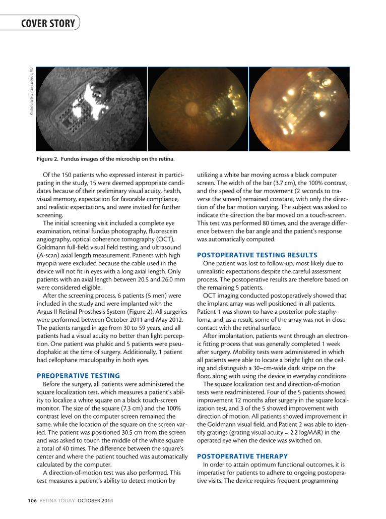
Figure 2. Fundus images of the microchip on the retina.
Phot
os Co
urte
sy St
anisl
ao Ri
zzo,
MD

OCTOBER 2014 RETINA TODAY 107
COVER STORY
and adjustments to fit the patient’s needs and changing visual ability. In the early postoperative period, the fitting specialist determines the lowest stimulus level needed for phosphene detection (electrical threshold) for each of the 60 electrodes. Because it is sometimes difficult for a patient to tell the difference between artificial retinal stimulation and naturally occurring phosphenes, reca-libration is sometimes necessary, especially as patients become more accustomed to the device and their stimu-lation thresholds change.
Once basic use is mastered, the devices are customized and programmed according to each patient’s individual needs. For example, a patient who spends a lot of time indoors might need a device set for low light conditions, whereas a patient who is often outdoors could antici-pate brighter lighting conditions. The customization and training sessions are lengthy and require time, energy, and compliance. Patients chosen for these devices must be able to handle the rigors of not only the actual sur-gery, but, all the necessary postoperative follow-ups, in order to have a successful outcome. As mentioned above, 1 patient dropped out of the study during the follow-up period, most likely due to the difficult postop-erative training requirements.
Using the Argus II Retinal Prosthesis System requires a team of experts: an ophthalmologist, retinal surgeon, and a rehabilitation specialist. Due to the already fragile psychological state of many RP patients, the addition of a psychologist in the postoperative period may be advisable. Future studies on retinal prostheses may wish to consider including a psychological evaluation in the enrollment criteria.
REAL-WORLD EXPERIENCESFor many patients, the possibility of seeing after being
blind for many years is exciting. Although the type of vision they will gain is very different from what they once had, it is something that many embrace. Many express their gratitude for the newfound independence and con-fidence the Argus II provides and describe the experience as “wonderful” and “life-changing.”2
Patients describe the Argus II as being most useful when there is perceptible contrast between images in the field of view. For example, 1 man explained how his grandchildren wear white shirts when they come to visit him to help boost the contrast between them and their surroundings. He cannot see their faces, but he can see their movement, something that brings him great joy.2
Argus II users might not regain their full sight, but with the device they can do things such as detect trees and other objects in their paths as they walk, the lines of a crosswalk, or a sidewalk curb. One patient detailed how
he will “scan” his surroundings, searching for the best light conditions that will provide the best contrast with the objects around him. The better the contrast, the better he can see. He also points out the importance of continuing with the follow-up visits once the device has been implanted, describing the visits as “fundamental for acquiring new skills.”3
It is necessary for a patient to scan what is in front of him or her because the visual field provided by the Argus II is only about 20°. Thus, a patient must scan his or her environment by moving the head from 1 side to the other in order to see the whole picture. This scan-ning movement is something users are taught during their postoperative visits.
Patients also seem to agree that the more they use the system, the better they get at both using the actual device and interpreting what they see. Barbara Campbell, a patient who was interviewed for a New York Times article in February 2013, commented on how the contin-ued use of the system improves her experience: “I see so much more now than when I first began to use it,” she said. “It’s only going to get better.”4
STATUS OF THE IMPLANTS IN THE UNITED STATES
In the United States, the first implants were done at the University of Michigan’s Kellogg Eye Center, a loca-tion chosen for its excellence, cutting-edge approach to medicine, and unparalleled commitment to patient care. The center is 1 of roughly a dozen that are cur-rently taking patient consultations. Procedures have also been performed at University of Southern California Eye Institute, Wills Eye Hospital, Duke University, and Toronto Western Hospital in Canada.
LOOKING TO THE FUTUREMany exciting developments are already under way
to provide enhanced features for the Argus II. System updates are available roughly every 3 to 6 months and can be performed when patients return for their regular follow-up visits. Upcoming updates include high acuity
”Once basic use is mastered, the devices are customized and programmed according to each
patient’s individual needs.”
(Continued on page 110)
110 RETINA TODAY OCTOBER 2014
COVER STORY
processing and facial tracking and recognition software. It is hoped that 1 day there will be a module for color perception.
With the facial recognition software installed and switched on, the camera will capture a high-resolution image; the location of a face within the image will be extracted, and a low-resolution image will then be sent to the visual implant. This update will allow patients to more easily recognize not just the fact that there is a person in front of them, but that person’s facial features as well.
Trials have begun with color perception software to test whether subjects blinded by outer retinal dystro-phies can consistently perceive different colored phos-phenes at the same time. In these tests, the electrodes of the devices of 4 blind patients were directly stimulated simultaneously with trains of cathodic-anodic pulses at different frequencies and intensities. The subjects report-ed the colors they distinguished after each round of stimulation. In total, 3 of the 4 patients perceived 7 dif-ferent color combinations: gray/yellow, brown/white, white/gray, white/blue, yellow/blue, white/yellow, and orange/white. One patient detected a gray/gray pattern, and 1 patient saw only white phosphenes.
CONCLUSIONThe implantation surgery for the Argus II device
appears to be safe, with no severe complications occur-ing during this trial. The postoperative results are very encouraging, although much depends on the patient. The assessment process is vital, as true success depends on the patient’s realistic expectations as to what the device can do and a willingness to participate in the rigorous postoperative therapy. There are many excit-ing developments on the horizon that will add to what is already a life-changing experience for patients and physicians alike. n
Stanislao Rizzo, MD, is director of U.O. Chirurgia Oftalmica, Ospedale Cisanello, Azienda Ospedaliero Universitaria Pisana in Pisa, Italy. He is a member of the Retina Today Editorial Board. He has no financial interests relevant to the content of this article. Dr. Rizzo may be reached at [email protected]
1. Rizzo S, Belting C, Cinelli L, et al. The Argus II Retinal Prosthesis: Twelve-month outcomes from a single-study center. Am J Ophthalmol.. 2014;157(6):1282-1290.2. YouTube. Argus II, Manchester user testimonial – artificial retina – bionic eye. www.youtube.com/watch?v=8AWIJdayKow. Accessed September 18, 2014.3. YouTube. Argus II Pisa user testimonial 1 - artificial retina - bionic eye. www.youtube.com/watch?v=BDYDlWOzSdQ. Accessed September 18, 2014.4. YouTube. The F.D.A. approves a bionic eye: Argus II device helps the blind. www.youtube.com/watch?v=WhYe6Redljw. Accessed September 18, 2014.
(Continued from page 107)