long qt mutations at the interface between kcnq1 helix c...
TRANSCRIPT

Jour
nal o
f Cel
l Sci
ence
RESEARCH ARTICLE
Long QT mutations at the interface between KCNQ1 helix C andKCNE1 disrupt IKS regulation by PKA and PIP2
Meidan Dvir1, Roi Strulovich2, Dana Sachyani2, Inbal Ben-Tal Cohen1, Yoni Haitin1, Carmen Dessauer3,Olaf Pongs5, Robert Kass4, Joel A. Hirsch2 and Bernard Attali1,*
ABSTRACT
KCNQ1 and KCNE1 co-assembly generates the IKS K+ current,
which is crucial to the cardiac action potential repolarization.
Mutations in their corresponding genes cause long QT syndrome
(LQT) and atrial fibrillation. The A-kinase anchor protein, yotiao
(also known as AKAP9), brings the IKS channel complex together
with signaling proteins to achieve regulation upon b1-adrenergic
stimulation. Recently, we have shown that KCNQ1 helix C interacts
with the KCNE1 distal C-terminus. We postulated that this interface
is crucial for IKS channel modulation. Here, we examined the yet
unknown molecular mechanisms of LQT mutations located at this
intracellular intersubunit interface. All LQT mutations disrupted
the internal KCNQ1–KCNE1 intersubunit interaction. LQT mutants
in KCNQ1 helix C led to a decreased current density and a
depolarizing shift of channel activation, mainly arising from impaired
phosphatidylinositol-4,5-bisphosphate (PIP2) modulation. In the
KCNE1 distal C-terminus, the LQT mutation P127T suppressed
yotiao-dependent cAMP-mediated upregulation of the IKS current,
which was caused by reduced KCNQ1 phosphorylation at S27.
Thus, KCNQ1 helix C is important for channel modulation by PIP2,
whereas the KCNE1 distal C-terminus appears essential for the
regulation of IKS by yotiao-mediated PKA phosphorylation.
KEY WORDS: Potassium channel, Long QT, KCNQ, KCNE,
Arrhythmia, IKS
INTRODUCTIONThe KCNQ (Kv7) subfamily of voltage-gated K+ channels (Kv)
comprises five members that play important functions in different
tissues including brain and heart (Jentsch et al., 2004). KCNQ1 a-
subunits can interact with any one of the five KCNE auxiliary b-
subunits, resulting in different channel functional characteristics
(Abbott et al., 2001; Nakajo and Kubo, 2011; Sun et al., 2012;
Wrobel et al., 2012). Co-assembly of KCNQ1 with KCNE1
generates the IKS K+ current that is vital for the proper
repolarization of the cardiac action potential (Barhanin et al.,
1996; Nerbonne and Kass, 2005; Sanguinetti et al., 1996).
Mutations in either KCNQ1- or KCNE1-encoding genes lead to
life-threatening cardiac arrhythmias causing long QT syndrome
(LQT) and atrial fibrillation (Peroz et al., 2008). The KCNQ1
subunit possesses a large C-terminus, which has been shown to be
important for channel gating, assembly and trafficking (Ghosh
et al., 2006; Haitin and Attali, 2008; Shamgar et al., 2006; Wiener
et al., 2008). The KCNQ C-terminus comprises proximal ahelices A and B that bind calmodulin (CaM), whereas the distal
coiled-coil helix C and helix D tetramerize, and crystallographic
studies have shown that helix D forms a tetrameric parallel
coiled-coil (Howard et al., 2007; Wiener et al., 2008).
The KCNQ C-terminus also appears to be a crucial region for
modulation by phosphatidylinositol-4,5-bisphosphate (PIP2), which
acts to stabilize the channel open state (Gamper and Shapiro, 2007).
Accordingly, the spontaneous rundown of IKS channels observed in
excised patches is markedly reduced by cytosolic application of
PIP2 (Loussouarn et al., 2003; Park et al., 2005). The site of PIP2
binding on KCNQ2–KCNQ4 channels has been suggested to be
located in the linker connecting helix A and B (Hernandez et al.,
2008); however, a recent work indicates that this AB helix linker is
not required for PIP2 regulation of KCNQ2 (Aivar et al., 2012). The
PIP2-binding site also remains elusive for KCNQ1. Recent studies
identified clusters of basic residues in KCNQ1 at the segment-2–
segment-3 and segment-4–segment-2 intracellular linkers and
proximal C-terminus as being involved in PIP2 binding (Thomas
et al., 2011; Zaydman et al., 2013). KCNE1 was found to increase
PIP2 sensitivity 100-fold over the KCNQ1 a-subunit alone and a
juxtamembranous site in the KCNE1 C-terminus is a key structural
determinant of PIP2 sensitivity (Li et al., 2011).
The KCNQ1 C-terminus also provides an interface for
interacting with signaling proteins (Haitin and Attali, 2008).
The scaffolding A-kinase anchor protein (AKAP) called yotiao or
AKAP9 brings the IKS channel complex together with protein
kinase PKA, protein phosphatase PP1, phosphodiesterase
PDE4D3 and adenylate cyclase AC9 (also known as ADCY9)
to achieve regulation following b-adrenergic stimulation (Chen
and Kass, 2005; Kurokawa et al., 2004; Li et al., 2012; Marx
et al., 2002; Terrenoire et al., 2009). The modulation is mediated
by the physical interaction of yotiao with the distal helix D, which
regulates IKS channel activity following phosphorylation or
dephosphorylation of S27 at the N-terminus of KCNQ1
(Kurokawa et al., 2004). S92 at the N-terminus of KCNQ1 is
also predicted to be phosphorylated (Lopes et al., 2007; Lundby
et al., 2013). We and others have found a direct interaction
between the C-termini of KCNQ1 and KCNE (Haitin et al., 2009;
Zheng et al., 2010). We have previously shown that the coiled-
coil helix C module of KCNQ1 is important for this interaction
with KCNE1 distal C-terminus, thereby revealing a new interface
between the two subunits (Haitin et al., 2009). We hypothesized
that the interface between KCNQ1 helix C and the KCNE1
C-terminus provides a strategic platform for IKS channel
1Department of Physiology & Pharmacology, Tel Aviv University, Tel Aviv, 69978,Israel. 2Department of Biochemistry & Molecular Biology, Tel Aviv University, TelAviv 69978, Israel. 3Department of Integrative Biology and Pharmacology,University of Texas Health Science Center, Houston, TX 77030, USA.4Department of Pharmacology, Columbia University, New York, NY 10027, USA.5Institut fur Physiologie, Universitat des Saarlandes, 66424 Homburg, Germany.
*Author for correspondence ([email protected])
Received 25 November 2013; Accepted 8 July 2014
� 2014. Published by The Company of Biologists Ltd | Journal of Cell Science (2014) 127, 3943–3955 doi:10.1242/jcs.147033
3943
Jour
nal o
f Cel
l Sci
ence
modulation. Here, we explored the yet unknown mechanisms ofLQT mutations located in this intracellular interface between the
IKS a- and b-subunits. LQT1 mutants in KCNQ1 helix C, S546L,K557E, R555H, R555C and R562M showed weaker interactionwith KCNE1 C-terminus. They displayed decreased currentdensity and a voltage right-shift of channel activation, which
could be accounted for by reduced PIP2 binding. In the distalKCNE1 C-terminus, the LQT mutation P127T showed weakerinteraction with KCNQ1 helix C and suppression of cAMP-
mediated IKS current upregulation that is caused by a reducedKCNQ1 phosphorylation at S27. Taken together, the data revealthat the intracellular interface between a- and b-subunits is
crucial for proper IKS channel function.
RESULTSIKS currents resulting from LQT5 mutations and truncationmutants at the KCNE1 C-terminusIn our quest to explore the mechanisms underlying LQTmutations located in the intracellular interface between KCNQ1
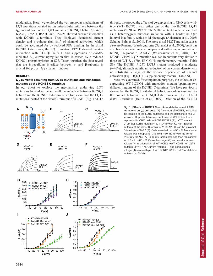
helix C and the KCNE1 C-terminus, we first examined the LQT5mutations located at the distal C-terminus of KCNE1 (Fig. 1A). To
this end, we probed the effects of co-expressing in CHO cells wild-type (WT) KCNQ1 with either one of the two KCNE1 LQT5
mutations V109I and P127T. The V109I mutant has been describedas a heterozygous missense mutation with a borderline QTcinterval in a family with a mild phenotype (Ackerman et al., 2003;Schulze-Bahr et al., 2001). The more distal P127T mutation causes
a severe Romano-Ward syndrome (Splawski et al., 2000), but it hasalso been associated in a certain proband with a second mutation inKCNQ1 segment 6, A341V (Westenskow et al., 2004). The
KCNE1 V109I LQT5 mutation resulted in currents very similar tothose of WT IKS (Fig. 1B,C,G,H; supplementary material TableS1). The KCNE1 P127T LQT5 mutant produced a moderate
(<40%), although significant, reduction of the current density withno substantial change of the voltage dependence of channelactivation (Fig. 1B,D,G,H; supplementary material Table S1).
Next, we examined, for comparison purposes, the effects of co-expressing WT KCNQ1 with truncation mutants spanning twodifferent regions of the KCNE1 C-terminus. We have previouslyshown that the KCNQ1 coiled-coil helix C module is essential for
the contact between the KCNQ1 C-terminus and the KCNE1distal C-terminus (Haitin et al., 2009). Deletion of the KCNE1
Fig. 1. Effects of KCNE1 C-terminus deletions and LQT5mutations on IKS currents. (A) A cartoon of KCNE1, indicatingthe location of the LQT5 mutations and the deletions in the C-terminus. Representative current traces of WT KCNQ1, co-expressed in CHO cells with WT KCNE1 (B), LQT5 mutantV109I (C), LQT5 mutant P127T (D) or with KCNE1 deletionmutants at the distal C-terminus D109–129 (E) or the proximalC-terminus D69–77 (F). Cells were held at 290 mV. Membranevoltage was stepped for 3 s from 250 mV to +60 mV (or to+100 mV for D69–77) in 10 mV increments and then repolarizedfor 1.5 s to 260 mV. Current–voltage (G) and conductance–voltage (H) relationships of WT KCNQ1+WT KCNE1 or LQT5mutants (n511–17). Current–voltage (I) and conductance–voltage (J) relationships of WT KCNQ1+WT KCNE1 or deletionmutants (n57–10).
RESEARCH ARTICLE Journal of Cell Science (2014) 127, 3943–3955 doi:10.1242/jcs.147033
3944
Jour
nal o
f Cel
l Sci
ence
distal C-terminus (D109–129) did not affect the current density(Fig. 1B,E,I). However, it produced a moderate right-shift of the
conductance–voltage relationship (+15 mV; Fig. 1J). In contrast,deletion of the KCNE1 proximal C-terminus (D69–77) resultedin a drastic reduction (<70%) of the current density accompaniedby a +39.1 mV right-shift of the conductance–voltage
relationship (Fig. 1B,F,I,J; supplementary material Table S1)and faster deactivation kinetics (tdeact5525655 ms andtdeact59167 ms for WT and D69–77, respectively;
mean6s.e.m., n57–10, P,0.001). This deletion in the KCNE1proximal C-terminus (D69–77) includes a site that was found tobe a key determinant of PIP2 sensitivity and is therefore expected
to impact on IKS channel gating (Li et al., 2011).
The KCNE1 P127T LQT5 mutation disrupts the interactionbetween KCNQ1 helix C and the KCNE1 C-terminus but doesnot affect the channel trafficking to the plasma membranePreviously, we have shown that deletion of the KCNQ1 helix C isnecessary and sufficient to abolish the physical interaction
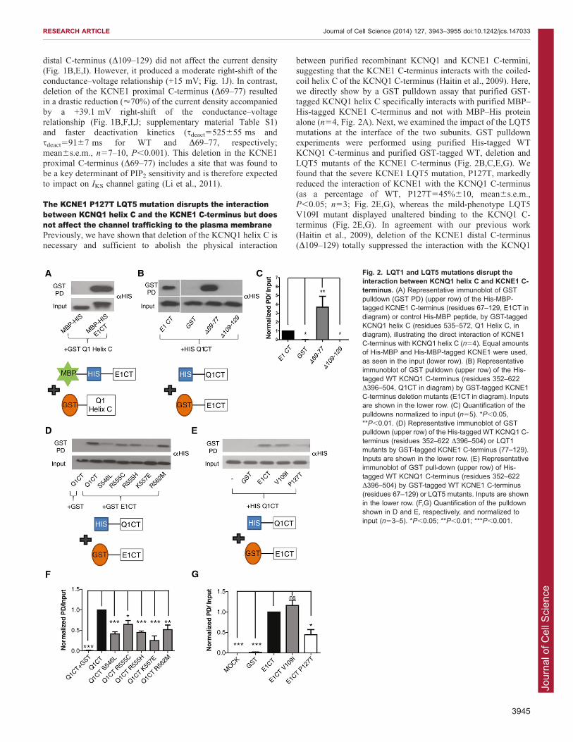
between purified recombinant KCNQ1 and KCNE1 C-termini,suggesting that the KCNE1 C-terminus interacts with the coiled-
coil helix C of the KCNQ1 C-terminus (Haitin et al., 2009). Here,we directly show by a GST pulldown assay that purified GST-tagged KCNQ1 helix C specifically interacts with purified MBP–His-tagged KCNE1 C-terminus and not with MBP–His protein
alone (n54, Fig. 2A). Next, we examined the impact of the LQT5mutations at the interface of the two subunits. GST pulldownexperiments were performed using purified His-tagged WT
KCNQ1 C-terminus and purified GST-tagged WT, deletion andLQT5 mutants of the KCNE1 C-terminus (Fig. 2B,C,E,G). Wefound that the severe KCNE1 LQT5 mutation, P127T, markedly
reduced the interaction of KCNE1 with the KCNQ1 C-terminus(as a percentage of WT, P127T545%610, mean6s.e.m.,P,0.05; n53; Fig. 2E,G), whereas the mild-phenotype LQT5
V109I mutant displayed unaltered binding to the KCNQ1 C-terminus (Fig. 2E,G). In agreement with our previous work(Haitin et al., 2009), deletion of the KCNE1 distal C-terminus(D109–129) totally suppressed the interaction with the KCNQ1
Fig. 2. LQT1 and LQT5 mutations disrupt theinteraction between KCNQ1 helix C and KCNE1 C-terminus. (A) Representative immunoblot of GSTpulldown (GST PD) (upper row) of the His-MBP-tagged KCNE1 C-terminus (residues 67–129, E1CT indiagram) or control His-MBP peptide, by GST-taggedKCNQ1 helix C (residues 535–572, Q1 Helix C, indiagram), illustrating the direct interaction of KCNE1C-terminus with KCNQ1 helix C (n54). Equal amountsof His-MBP and His-MBP-tagged KCNE1 were used,as seen in the input (lower row). (B) Representativeimmunoblot of GST pulldown (upper row) of the His-tagged WT KCNQ1 C-terminus (residues 352–622D396–504, Q1CT in diagram) by GST-tagged KCNE1C-terminus deletion mutants (E1CT in diagram). Inputsare shown in the lower row. (C) Quantification of thepulldowns normalized to input (n55). *P,0.05,**P,0.01. (D) Representative immunoblot of GSTpulldown (upper row) of the His-tagged WT KCNQ1 C-terminus (residues 352–622 D396–504) or LQT1mutants by GST-tagged KCNE1 C-terminus (77–129).Inputs are shown in the lower row. (E) Representativeimmunoblot of GST pull-down (upper row) of His-tagged WT KCNQ1 C-terminus (residues 352–622D396–504) by GST-tagged WT KCNE1 C-terminus(residues 67–129) or LQT5 mutants. Inputs are shownin the lower row. (F,G) Quantification of the pulldownshown in D and E, respectively, and normalized toinput (n53–5). *P,0.05; **P,0.01; ***P,0.001.
RESEARCH ARTICLE Journal of Cell Science (2014) 127, 3943–3955 doi:10.1242/jcs.147033
3945

Jour
nal o
f Cel
l Sci
ence
C-terminus (Fig. 2B,C). In contrast, deletion of the KCNE1proximal C-terminus (D69–77) significantly enhanced the
binding to the KCNQ1 C-terminus (by 2.560.2-fold, P,0.001,n55; Fig. 2B,C).
We sought to investigate the mechanisms underlying thedisrupted function of the severe KCNE1 LQT5 mutation P127T.
First, we examined whether defective channel trafficking couldaccount for the decreased current density observed with P127T.For this purpose, we transfected CHO cells with fluorescently
tagged WT and mutant subunits (KCNQ1–CFP and KCNE1–YFP) which we imaged using total internal reflectionfluorescence (TIRF) microscopy. To validate the TIRF
methodology, we used positive and negative controls. Theglycosylphosphatidylinositol (GPI)-linked fluorescent proteincitrine (GPI-citrine) was used as a positive control for plasma
membrane expression. GPI-citrine is known to express verystrongly at the plasma membrane of cells (Glebov and Nichols,2004). As shown in Fig. 3A, the GPI-citrine signal intensity washeavily detected under TIRF microscopy. Co-transfection of
KCNQ1–CFP and GPI-citrine revealed that KCNQ1–CFPcolocalized only slightly with GPI-citrine, reflecting the knownpartial processing of WT KCNQ1 to the plasma membrane
(Kanki et al., 2004). The substantial cytoplasmic retention of WTKCNQ1 is further illustrated by comparing the somewhat weakTIRF signal relative to the strong epi-fluorescence intensity
obtained from a cell expressing KCNQ1–CFP (Fig. 3B, upper
panels). The KCNQ1 LQT1 mutation G589D, located in thecoiled-coil helix D of the KCNQ1 C-terminus, was used as a
negative control, because this mutant has previously beendescribed as a mistrafficked channel that is heavily retained inthe endoplasmic reticulum (ER) (Kanki et al., 2004). Inagreement with this previous work, we found that the KCNQ1
G589D mutant exhibits a much weaker TIRF signal compared toWT KCNQ1 (13%64 of WT KCNQ1, mean6s.e.m., n516–25,P,0.001) (Fig. 3B,E). The robust ER retention of the G589D
mutant is further confirmed by the stronger signal obtained underepi-fluorescence relative to that revealed under TIRF microscopy(Fig. 3B). Importantly, the YFP-tagged KCNE1 LQT5 mutant
P127T showed comparable plasma membrane trafficking tothat of WT channels (n556–60; Fig. 3D,F), suggesting that thedecreased current density displayed by the LQT5 mutant does not
seem to be accounted for by trafficking defects. However, TIRFdoes not allow discrimination between the signals from theplasma membrane and those arising from the cortical ER, whichis especially relevant for some K+ channels (Fox et al., 2013).
Thus, an effect on trafficking cannot be totally excluded.
The P127T LQT5 mutation and D109–129 deletion at theKCNE1 distal C-terminus suppress the cAMP-mediatedupregulation of the IKS currentAs shown above (Fig. 3D,F), the decrease in current density
displayed by the KCNE1 P127T LQT5 mutant could not be
Fig. 3. LQT1 and LQT5 mutations do not affect the channel trafficking to the plasma membrane. (A) TIRF fluorescence images of CHO cells co-expressing CFP-tagged WT KCNQ1 and GPI-citrine. (B) Epi-fluorescence (left panels) and TIRF fluorescence (right panels) images of CHO cells expressingCFP-tagged WT KCNQ1+KCNE1 (upper panels) or CFP-tagged G589D LQT1 mutant+KCNE1 (lower panels). (C) TIRF fluorescence images of CHO cells co-expressing CFP-tagged helix C LQT1 mutants with KCNE1. (D) YFP-tagged WT KCNE1 or LQT5 mutant co-expressed with WT KCNQ1. Quantification of theTIRF fluorescent signals of CFP-tagged WT KCNQ1 and LQT1 mutants (E) and of YFP-tagged WTand LQT5 mutant (F) as normalized to membrane signal area(n516–60). ***P,0.001. Scale bars: 10 mm.
RESEARCH ARTICLE Journal of Cell Science (2014) 127, 3943–3955 doi:10.1242/jcs.147033
3946

Jour
nal o
f Cel
l Sci
ence
accounted for by trafficking defects. We hypothesized thatthe KCNE1 distal C-terminus could be important for cAMP-
dependent upregulation of the IKS current, and that mutations inthis region might disrupt the modulation. Thus, we tested whethercAMP-stimulation of the P127T KCNE1 mutant was similar tothat obtained with the WT IKS channel. CHO cells were
transfected with both WT KCNQ1 and WT yotiao, togetherwith WT KCNE1 or its mutants. Transfected CHO cells werestimulated by 200 mM cAMP and 0.2 mM okadaic acid, a PP1
and PP2A phosphatase inhibitor, added in the patch pipette (Marxet al., 2002). WT IKS currents were significantly upregulated bycAMP plus okadaic acid treatment (at +50 mV, 70%69
stimulation; mean6s.e.m., n57, P,0.01; Fig. 4A,B); however,no significant changes in deactivation kinetics and conductance–voltage relations were observed. In striking contrast, the P127T
LQT5 mutant completely lost its stimulation upon cAMP plusokadaic acid treatment (Fig. 4C,D). Likewise, the KCNE1 distalC-terminus deletion mutant D109–129 was unresponsive tocAMP plus okadaic acid stimulation (Fig. 4E,F). Interestingly,
the mild phenotype LQT5 V109I mutant was significantly
upregulated by cAMP plus okadaic acid treatment (at +50 mV,64%67 stimulation; n56, P,0.05; Fig. 4G,H).
P127T LQT5 mutation and deletion mutant D109–129 do notdisrupt the KCNQ1 interaction with yotiaoTo elucidate the possible mechanisms underlying the impaired
cAMP-mediated modulation in KCNE1 distal C-terminusmutants, we first examined whether the mutations P127T andD109–129 disrupted KCNQ1 interaction with yotiao. To verify
proper expression of the proteins in transfected HEK 293 cells,lysates were subjected to western blotting (supplementarymaterial Fig. S1A). Lysates from cells expressing both WT
KCNQ1 and WT yotiao, plus WT KCNE1 or its mutants weresubjected to immunoprecipitation with anti-KCNQ1 antibodies.Western blots probed with anti-yotiao antibodies, quantitatively
tested the interaction of KCNQ1 with yotiao, by normalizing theyotiao signal to the KCNQ1 signal intensity in the precipitate(supplementary material Fig. S1B). The results revealed thatwhen compared to WT KCNE1, there was no significant
difference in the ability of KCNQ1 to interact with yotiao when
Fig. 4. P127T LQT5 mutation and D109–129 deletion at theKCNE1 distal C-terminus suppress the cAMP-mediatedupregulation of IKS current. Representative current traces recordedfrom CHO cells co-expressing WT KCNQ1+WT yotiao and WTKCNE1 (A), KCNE1 P127T (C), KCNE1 D109–129 (E) and KCNE1V109I (G). Cells were held at 290 mV and stepped to +30 mV for 3 sand then repolarized to 240 mV for 1.5 s. Cells were recorded in theabsence (black traces) or presence (red traces) of 200 mMcAMP+0.2 mM okadaic acid. Current–voltage relationships of WTKCNQ1+WT yotiao and WT KCNE1 (B) or KCNE1 P127T (D) orKCNE1 D109–129 (F) or KCNE1 V109I (H). Voltage was stepped for3 s from 250 mV to +60 mV in 10 mV increments followed byrepolarization to 240 mV for 1.5 s. Red and black curves representrecordings with or without cAMP+okadaic acid in the patch pipette,respectively. (n56–7). Above +30 mV, the WT IKS and, above+20 mV, the KCNE1 V109I current upregulation induced bycAMP+okadaic acid were significant. *P,0.05, **P,0.01.
RESEARCH ARTICLE Journal of Cell Science (2014) 127, 3943–3955 doi:10.1242/jcs.147033
3947

Jour
nal o
f Cel
l Sci
ence
co-expressed with P127T LQT5 mutant or with KCNE1 distal C-terminus deletion mutant D109–129 (supplementary material Fig.
S1B,C).
The P127T LQT5 mutation and deletion mutant D109–129impair phosphorylation of KCNQ1 at S27 during cAMP-dependent stimulationNext, we set out to examine whether the KCNE1 distal C-terminus mutations P127T and D109–129 disrupted
phosphorylation of S27 in KCNQ1, in the presence of yotiao.Transfected HEK 293 cells expressing both WT KCNQ1 and WTyotiao, plus WT KCNE1 or its mutants were treated for 10 min
with 250 mM 8-(4-chlorophenylthio) adenosine 39,59-cyclicmonophosphate (8CPT), a membrane permeable analog ofcAMP and with 0.2 mM okadaic acid. Cell lysates were
subjected to SDS-PAGE followed by western blotting and blotswere probed with anti-KCNQ1, anti-yotiao and anti-KCNE1antibodies for validating protein expression in the complex andfor quantification purposes. Importantly, blots were also probed
with a specific antibody against phosphorylated S27 of KCNQ1to explore the extent of KCNQ1 phosphorylation at S27 underbasal and cAMP-stimulated conditions (Kurokawa et al., 2003).
Basal S27 phosphorylation was not significantly different whenWT KCNQ1 plus WT yotiao were co-expressed with either WTKCNE1 or its mutants. In the WT IKS channel complex co-
expressed with yotiao, cAMP-stimulation produced a significant2.6-fold increase in S27 phosphorylation (n55, P,0.0001;Fig. 5A,B). A comparable stimulation was found for the mild-
phenotype LQT5 mutation V109I, with a 2-fold increase inS27 phosphorylation upon cAMP treatment (n53, P,0.05;Fig. 5A,B). In contrast, for the P127T LQT5 mutation, the
increase in S27 phosphorylation following cAMP exposure waslower and not statistically significant (1.65-fold increase, n55,
P.0.05). Similarly, a weaker and non-significant stimulation ofS27 phosphorylation was obtained with the KCNE1 distal C-terminus deletion mutant D109–129 (1.7-fold increase, n55,P.0.05; Fig. 5A,B). When expressed as the relative cAMP- and
okadaic acid-stimulated phosphorylation, the effect of the V109Imutant appears to be larger than that of the deletion mutantD109–129, although not statistically different (Fig. 5C). For
comparison, we examined whether the LQT1 mutation K557E,residing in KCNQ1 helix C and whose defect involves weakerbinding to PIP2 (see below), could alter stimulation of S27
phosphorylation following cAMP treatment. Results show thatthe KCNQ1 mutant K557E co-expressed with both WT KCNE1and WT yotiao produced a significant 2.4-fold increase in S27
phosphorylation (n54, P,0.01; Fig. 5A,B). We conclude thatthe LQT5 mutation P127T disrupts yotiao regulation, whereas anunrelated LQT1 mutation (K557E) does not.
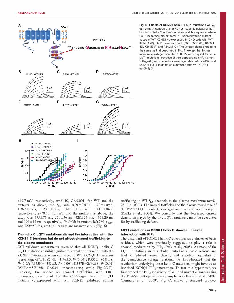
IKS currents resulting from LQT1 mutations at the KCNQ1helix CWe then studied the effects of co-expressing WT KCNE1 in CHO
cells with five LQT1 mutations, S546L, R555C, R555H, K557Eand R562M, located in the KCNQ1 helix C (Fig. 6A;supplementary material Table S1). All mutations markedly
reduced the current densities, which were paralleled by potentright-shifts of the voltage dependence of channel activation,slower activation kinetics and faster deactivation (for S546L,
R555C, R555H, K557E and R562M, respectively, 75%, 88%,69%, 99% and 57% inhibition of current density at +60 mV; theDV50 was +48 mV, +65.4 mV, +47.1 mV, +92.7 mV and
Fig. 5. LQT5 P127T mutant and deletion mutant D109–129 impair phosphorylation of KCNQ1 at S27 duringcAMP-dependent stimulation. (A) Representativeimmunoblots (IB) of HEK 293 cells lysates co-expressingyotiao, WT KCNQ1 and WT, mutated (V109I and P127T)or truncated (D109–129) KCNE1 from cells treated in theabsence and presence of 250 mM 8CPT+0.2 mM okadaicacid and immunoblots of HEK 293 cells lysates co-expressing yotiao WT KCNE1 and WT KCNQ1 or LQT1mutant K557E from cells treated in the absence andpresence of 250 mM 8CPT+0.2 mM okadaic acid. Blotswere probed with antibodies against phosphorylatedKCNQ1 S27 (first row from top), KCNQ1 (second row fromtop), yotiao (third row from top) and KCNE1 (fourth rowfrom top). (B) Quantification of phosphorylated KCNQ1S27 was calculated by dividing phosphorylated signal toKCNQ1 input (anti-KCNQ1 blot) for both unstimulated andcAMP-stimulated cells (250 mM 8CPT+0.2 mM okadaicacid) and normalized to stimulated WT KCNQ1+WTKCNE1+yotiao (n53–5). *P,0.05, ** P,0.01,***P,0.001; ns: not statistically significant. (C) Analternative quantification of phosphorylated KCNQ1 S27was expressed as the relative cAMP+okadaic acid-stimulated S27 phosphorylation where the extent ofinduced phosphorylation of WT IKS was equal to 1(n53–5). *P,0.05; ns: not statistically significant.
RESEARCH ARTICLE Journal of Cell Science (2014) 127, 3943–3955 doi:10.1242/jcs.147033
3948
Jour
nal o
f Cel
l Sci
ence
+40.7 mV, respectively, n55–10, P,0.001; for WT and themutants as above, the t1/2 was 0.9160.07 s, 1.2060.09 s,1.3660.07 s, 1.2860.07 s, 1.4060.11 s and 1.4160.06 s,
respectively, P,0.05; for WT and the mutants as above, thetdeact was 675676 ms, 550636 ms, 428626 ms, 460629 msand 194618 ms, respectively, P,0.05; in mutant R562M, tdeact
was 720650 ms, n56; all results are mean6s.e.m.) (Fig. 6).
The helix C LQT1 mutations disrupt the interaction with theKCNE1 C-terminus but do not affect channel trafficking tothe plasma membraneGST-pulldown experiments revealed that all KCNQ1 helix CLQT1 mutations exhibit significantly weaker interaction with the
KCNE1 C-terminus when compared to WT KCNQ1 C-terminus(percentage of WT: S546L541%65, P,0.001; R555C565%68,P,0.05; R555H545%63, P,0.001; K557E525%68, P,0.01;
R562M552%68, P,0.01; mean6s.e.m.; n53; Fig. 2D,F).Exploring the impact on channel trafficking with TIRFmicroscopy, we found that the CFP-tagged helix C LQT1mutants co-expressed with WT KCNE1 exhibited similar
trafficking to WT IKS channels to the plasma membrane (n58–25; Fig. 3C,E). The normal trafficking to the plasma membrane ofthe R555C LQT1 mutant is in agreement with a previous report
(Kanki et al., 2004). We conclude that the decreased currentdensity displayed by the five LQT1 mutants cannot be accountedfor by trafficking defects.
LQT1 mutations in KCNQ1 helix C showed impairedinteraction with PIP2
The distal half of KCNQ1 helix C encompasses a cluster of basic
residues, which were previously suggested to play a role inchannel modulation by PIP2 (Park et al., 2005). As most of theLQT1 mutations in this study neutralize a basic residue and
lead to reduced current density and a potent right-shift ofthe conductance–voltage relations, we hypothesized that themechanism underlying these helix C mutations might involve an
impaired KCNQ1–PIP2 interaction. To test this hypothesis, wefirst probed the PIP2 sensitivity of WT and mutant channels usingthe Dr-VSP voltage-sensitive phosphatase (Hossain et al., 2008;Okamura et al., 2009). Fig. 7A shows a standard protocol
Fig. 6. Effects of KCNQ1 helix C LQT1 mutations on IKScurrents. A cartoon of one KCNQ1 subunit indicating thelocation of helix C in the C-terminus and its sequence, whereLQT1 mutations are situated (A). Representative currenttraces of WT KCNE1 co-expressed in CHO cells with WTKCNQ1 (B), LQT1 mutants S546L (C), R555C (D), R555H(E), K557E (F) and R562M (G). The voltage-clamp protocol isthe same as that described in Fig. 1, except that highermembrane voltages of up to +160 mV were applied for someLQT1 mutations, because of their depolarizing shift. Current–voltage (H) and conductance–voltage relationships of WTandKCNQ1 LQT1 mutants co-expressed with WT KCNE1(n55–9) (I).
RESEARCH ARTICLE Journal of Cell Science (2014) 127, 3943–3955 doi:10.1242/jcs.147033
3949

Jour
nal o
f Cel
l Sci
ence
previously used to monitor WT IKS currents before and following
activation of Dr-VSP (Danio rerio VSP, also known as TPTE),where a +10 mV prepulse opens the channel, followed by a+100 mV voltage step that activates the phosphatase and then a
return to +10 mV (Kruse et al., 2012). When this protocol wasrepeatedly applied every 10 s for 2 min, activation of Dr-VSP ledto a 64%64 reduction of WT IKS currents (n55; Fig. 7A, right
panel, red trace), while in the absence of Dr-VSP a slight currentovershoot of 21%64 was observed (n55; Fig. 7A, left panel, redtrace). We obtained a relatively lower suppression of WT IKS
currents compared to that found by Kruse et al., because ourintracellular solution did not contain Mg2+ (see Materials andMethods) as we wanted to prevent a fast run-down of WT IKS
current, even in the absence of Dr-VSP. Indeed, Mg2+ has
previously been found to reduce the KCNQ currents byelectrostatic binding to the negative charges of PIP2,competitively reducing the amount of free PIP2 available for
interaction with the channels (Suh and Hille, 2007). However,this +10 mV prepulse protocol could not be applied for the fiveLQT1 mutants because they exhibit potent right-shifts of the
voltage dependence of activation, which prevents them beingactivated at +10 mV like WT IKS (see Fig. 6). To circumvent thisdifficulty, we applied repeatedly (every 5 s) a 2-s-long step pulse
to +100 mV in order to both open the channel and activate theDr-VSP. During the successive sweeps, the rate of current declinewould be expected to reflect the channel affinity for PIP2. Thus,
Fig. 7. LQT1 mutations in KCNQ1 helix Cexhibit greater sensitivity to the voltage-sensitive phosphatase Dr-VSP.(A) Representative current traces of WT IKS(KCNQ1+KCNE1) sensitivity to the voltagesensitive phosphatase Dr-VSP recorded bymeans of a tri-pulse protocol wheremembrane potential is first stepped to+10 mV (3 s, from 290 mV holding potential)to open the channel, followed by a +100 mVvoltage step (2 s) to activate Dr-VSP andthen return to +10 mV (3 s). This protocolwas repeatedly applied every 10 s for 2 min.Left panel shows WT IKS sensitivity toprotocol application in the absence of Dr-VSP at the first (black trace) and last sweeps(red trace). Right panel shows WT IKSsensitivity to protocol application in thepresence of Dr-VSP at the first (black trace)and last sweeps (red trace) (n55).(B,C,E,G,I,K) Representative current tracesof WT IKS, S546L, R555C, R555H, K557Eand R562M sensitivity to Dr-VSP recordedby one pulse protocol, where membranepotential is stepped to +100 mV (2 s, from290 mV holding potential) to both open thechannels and activate Dr-VSP. Shown arecurrent traces at time 0 (black trace) and attime 10 s (red trace). Kinetics of currentdecline for WT IKS and mutant channels areshown in the absence or presence of Dr-VSP. The comparison between WT IKS andthe five LQT1 mutants was quantified at anearly time point (10 s) before the current dropreached steady state and was expressed asa ratio of currents measured at time 10 sover that recorded at time 0. At 10 s, ratios ofcurrent decline were 0.7560.05 (n55),0.7460.02 (n57), 0.3260.01 (n53,P,0.01), 0.5460.06 (n54, P,0.05),0.5560.04 (n57, P,0.05) and 0.4860.04(n57, P,0.01) for WT, S546L, R555C,R555H, K557E and R562M, respectively.
RESEARCH ARTICLE Journal of Cell Science (2014) 127, 3943–3955 doi:10.1242/jcs.147033
3950

Jour
nal o
f Cel
l Sci
ence
assuming that the rate of PIP2 re-synthesis is roughly the same,the faster is the rate of current decrease, the lower the channel
affinity for PIP2. The kinetics of current decline for WT IKS andmutant channels were analyzed in the absence or presence of Dr-VSP (Fig. 7D,F,H,J,L). The comparison between WT IKS and thefive LQT1 mutants (+KCNE1) was quantified at an early time
point (10 s) before current drop reached steady state and wasexpressed as a ratio of currents measured at time 10 s over thatrecorded at time 0. Representative current traces are also shown
at time 0 (Fig. 7B,C,E,G,I,K, black trace) and at time 10 s(Fig. 7B,C,E,G,I,K, red trace). Except for mutant S546L, theresults indicate that mutants R555C, R555H, K557E and R562M
exhibited a significantly faster current decline compared to WTIKS [at 10 s, ratios of current decline were 0.7560.05 (n55),0.7460.02 (n57), 0.3260.01 (n53, P,0.01), 0.5460.06 (n54,
P,0.05), 0.5560.04 (n57, P,0.05) and 0.4860.04 (n57,P,0.01) for WT, S546L, R555C, R555H, K557E and R562M,respectively (all results are mean6s.e.m.)]. In the absence of Dr-VSP, WT IKS and mutant channels tended to show current
overshoot following this protocol (Fig. 7D,F,H,J,L). Next, weassessed whether the higher sensitivity to the Dr-VSP phosphatasedisplayed by the helix C LQT1 mutants arose from their weaker
binding to PIP2. To test this assumption, we performed pulldownexperiments of purified His-tagged WT and LQT1 mutant KCNQ1C-terminus using PIP2-coated agarose beads. We found all mutants
to exhibited significantly lower binding to PIP2 as compared to theWT KCNQ1 C-terminus [percentage of WT binding: 43%612 for
S546L, n53, P,0.01; 55%66 for R555C, n53, P,0.05; 34%67for R555H, n53, P,0.001; 17%66 for K557E, n53, P,0.001
and 14%65 for R562M, n53, P,0.001; Fig. 8A,B (all results aremean6s.e.m.)]. These results suggest that LQT1 mutations inKCNQ1 helix C, except S546L, have an impaired interaction withPIP2.
DISCUSSIONIn the present work, we provide novel insights into the molecular
mechanisms underlying LQT mutations located at a crucialintracellular interface of the subunits forming the cardiac IKS
channel and corresponding to the interaction between the KCNE1
distal C-terminus and the KCNQ1 coiled-coil helix C module(Haitin et al., 2009). Here, we show that LQT mutations locatedon both sides of this interface disrupted IKS channel function. On
the one hand, the LQT1 mutants in KCNQ1 helix C (R555C,R555H, K557E and R562M) showed reduced PIP2 binding and astrong right-shift of channel activation. On the other hand, theLQT5 mutation P127T in KCNE1 distal C-terminus, suppressed
yotiao-dependent cAMP-mediated upregulation of IKS currents byreducing KCNQ1 phosphorylation at S27 (Fig. 8C).
All members of the KCNQ family require the presence of the
membrane phospholipid PIP2 for proper function (Gamper andShapiro, 2007; Suh and Hille, 2005; Zhang et al., 2003). PIP2
stabilizes the open state of the cardiac IKS KCNQ1–KCNE1
channel, thereby slowing deactivation kinetics and producinga negative shift in the voltage dependence of activation
Fig. 8. LQT1 mutations in KCNQ1 helix C showimpaired binding to PIP2. (A) Representativeimmunoblot of PIP2 pulldown (PIP2 PD) by PIP2-coatedagarose beads of His-tagged WT KCNQ1 C-terminus orLQT1 mutants (upper row). Inputs are shown in the lowerrow. (B) Quantification of the pull-down normalized toinput (n53). *P,0.05; **P,0.01; ***P,0.001.(C) Cartoon summarizing the two different pathwaysinvolved in the mechanisms of LQT mutations located atthe interacting KCNE1 distal C-terminus and KCNQ1helix C module: one involving LQT1 mutants in KCNQ1helix C that impairs PIP2 interaction and one of the LQT5mutant P127T at the distal KCNE1 C-terminus, whichimpedes IKS regulation by yotiao-mediated PKAphosphorylation.
RESEARCH ARTICLE Journal of Cell Science (2014) 127, 3943–3955 doi:10.1242/jcs.147033
3951

Jour
nal o
f Cel
l Sci
ence
(Loussouarn et al., 2003; Park et al., 2005; Thomas et al., 2011).Different regions endowed with clusters of basic residues have
been proposed to interact with PIP2. Hernandez et al., identified aconserved cluster of basic residues within the linker connectinghelix A and B in the C-terminus of KCNQ2–KCNQ4 subunits(Hernandez et al., 2008). This basic cluster was suggested to
interact with PIP2 through electrostatic and hydrogen-bondingnetworks (Hernandez et al., 2008). However, a recent studyshowed that this linker is not required for PIP2 regulation of
KCNQ2 (Aivar et al., 2012). Others have identified, in both theKCNQ1 and KCNQ2 proximal C-terminus, a cluster of basicresidues just downstream of segment 6 (Telezhkin et al., 2013;
Thomas et al., 2011; Zhang et al., 2003). In KCNQ1, residues inthe segment-2–segment-3 and segment-4–segment-5 linkers havealso been proposed to be important for PIP2 interaction (Matavel
et al., 2010; Zaydman et al., 2013). In addition, basic residueshave been identified in coiled-coil helix C and have beensuggested to be important for PIP2 interaction (Matavel et al.,2010; Park et al., 2005). Notably, in the present study, the LQT1
mutations located in KCNQ1 helix C (R555C, R555H, K557E,R562M), impaired their physical interaction with KCNE1 C-terminus. In addition, these LQT1 mutants co-expressed with
WT KCNE1, showed significantly smaller current densities,depolarizing shifts of the conductance–voltage relationship andfaster deactivation kinetics. Such features are suggestive of
stabilization of the channel closed state.These properties are in line with those expected for an impaired
channel–PIP2 interaction, which led us to hypothesize that this
cluster of basic residues in KCNQ1 helix C might be involvedin PIP2 modulation. Using two complementary approaches, wefound that all LQT1 mutants exhibited significantly lowerbinding to PIP2 than WT as measured by pulldown experiments
and by the use of the voltage-dependent phosphatase Dr-VSP(Okamura et al., 2009). Our results indicate that upon activationof Dr-VSP, mutants R555C, R555H, K557E and R562M
exhibited a significant faster current decline compared to WTIKS, which likely reflects their lower affinity for PIP2. MutantS546L was an exception and displayed a similar sensitivity to Dr-
VSP as WT IKS, suggesting that it does not appear to interactfunctionally with PIP2. However, it is possible that, like mutantR539W, it interacts with cholesterol (Coyan et al., 2014).
Our results suggest that KCNQ1 helix C is crucial for PIP2
modulation (Fig. 8C). They also raise the question of thesignificance of multiple PIP2-interacting regions in KCNQ1. Itis possible that helix C does not bind PIP2 directly but rather
contributes allosterically to modulate PIP2 binding to a pocketmolded by multiple contact sites (Zaydman et al., 2013),including the conserved membrane regions and regions located
downstream segment 6.The macro-molecular scaffolding complex formed by KCNQ1
and KCNE1, together with yotiao, PKA, PP1, PDE4D3 and AC9,
is designed to achieve IKS channel regulation following b1-adrenergic stimulation and can be disrupted in various waysunder disease conditions (Chen and Kass, 2005; Kurokawa et al.,2004; Li et al., 2012; Marx et al., 2002; Terrenoire et al., 2009).
The LQT1 mutation A341V in KCNQ1 segment 6 has recentlybeen shown to preserve the KCNQ1–yotiao interaction but toreduce cAMP-induced KCNQ1 phosphorylation at S27 and
subsequent IKS current upregulation (Heijman et al., 2012). TheLQT5 mutation D76N in KCNE1 C-terminus spared the cAMP-mediated KCNQ1 phosphorylation at S27 but failed to upregulate
its resulting IKS current. In this study, we found that the LQT5
mutant P127T in KCNE1 distal C-terminus led to a significantdecrease in basal IKS currents, which were unable to be stimulated
upon cAMP exposure. In addition, the P127T mutation impairedthe physical interaction with KCNQ1 helix C. We showed thatneither defective channel trafficking nor an impaired KCNQ1–yotiao interaction could account for the compromised P127T
mutant channel function. In contrast, we found that mutant P127Tsignificantly decreased KCNQ1 phosphorylation at S27. To ourknowledge, the LQT5 mutation P127T is the first described
example of a KCNE1 mutant leading to impaired KCNQ1phosphorylation at S27 following cAMP stimulation (Fig. 8C).To date, only one LQT1 mutation A341V in KCNQ1 (Heijman
et al., 2012) has been found to disrupt KCNQ1 phosphorylationat S27, while preserving the KCNQ1–yotiao interaction.Interestingly, the LQT5 mutant P127T has been described both
as a single mutation (Splawski et al., 2000) and as beingadditionally associated with the LQT1 mutation A341V inanother proband who presented with syncope and longer QTcinterval than his parents (Westenskow et al., 2004). As
both mutations impair KCNQ1 phosphorylation at S27 andsubsequently prevent current increase upon cAMP stimulation,they might expose the affected individuals to severe clinical
phenotypes and increase their susceptibility to ventriculartachyarrhythmias under sympathetic stress conditions. Like S27,S92 has recently been found to be phosphorylated upon b-
adrenergic stimulation when the channel is localized at theplasma membrane, and this phosphorylation event resulted inincreased IKS current (Lundby et al., 2013). It will be interesting
in future work to examine the impact of the LQT5 mutant P127Ton the phosphorylation of S92 following cAMP stimulation.
KCNQ1 can be phosphorylated at S27 upon PKA activation,without the need for KCNE1 expression (Kurokawa et al., 2003;
Marx et al., 2002). However, in the absence of KCNE1, there isno transduction of PKA-mediated KCNQ1 phosphorylation(Kurokawa et al., 2003). An intact KCNE1 C-terminus has
been suggested to be crucial for cAMP-dependent upregulation ofIKS currents (Kurokawa et al., 2009), which reflects the crucialrole played by KCNE1 in coupling sympathetic b1-adrenergic
signaling to post-phosphorylation functional events in the cardiacIKS channel. Here, we mapped the KCNE1 effect to its distal C-terminus by showing that deletion of the KCNE1 C-terminus(D109–129) and the LQT5 mutation P127T depress KCNQ1
phosphorylation at S27 and subsequently prevent the cAMP-mediated increase in IKS currents. The molecular mechanism bywhich this transduction occurs is currently unknown. Previous
studies have shown a direct interaction between the KCNQ1proximal C-terminus with the KCNE1 C-terminus (Zheng et al.,2010). Using FRET and fluorescently labeled subunits, we have
previously shown that the interaction between the KCNE1 C-terminus and the KCNQ1 C-terminus in the channel complexshortens the distance between the KCNQ1 N- and C-termini
(Haitin et al., 2009). This is probably a crucial step for the yotiao-mediated transfer of PKA from the C-terminus to the N-terminusof KCNQ1 and the subsequent phosphorylation of S27. In adynamic FRET study, we also found that during IKS channel
opening, the KCNQ1 C-terminus and KCNE1 C-terminus drawcloser together and that this gated motion is suppressed by theLQT5 mutant D76N (Haitin et al., 2009), a mutation previously
shown to cause functional disruption of cAMP-mediatedstimulation of IKS current (Kurokawa et al., 2003). Thus, thestrategic location of the KCNE1 C-terminus interacting with
KCNQ1 helix C and the proximity of KCNQ1 N- and C-termini
RESEARCH ARTICLE Journal of Cell Science (2014) 127, 3943–3955 doi:10.1242/jcs.147033
3952
Jour
nal o
f Cel
l Sci
ence
and their gated motions might be pivotal for transducing thephosphorylation of KCNQ1 S27 into IKS current upregulation.
In summary, this work reveals the molecular mechanisms ofLQT mutations located at the interacting KCNE1 distal C-terminus and KCNQ1 helix C module. Our results show that thisintracellular interface is a crucial platform for transduction and
integration of two modulatory mechanisms, one involving thePKA-mediated increase in IKS currents following b-adrenergicstimulation, and one implying PIP2 modulation (Fig. 8C).
Interestingly, these two regulation pathways have recently beensuggested to undergo cross talk in such a way that KCNQ1sensitivity to PIP2 can be modulated by PKA activation (Choveau
et al., 2012; Matavel et al., 2010). Consequently, the LQTmutations examined in this study selectively disrupt either one ofthese two regulatory processes according to their respective
location in this important intersubunit interface.
MATERIALS AND METHODSMolecular biologyHuman KCNQ1 and KCNE1 were cloned into pCDNA3 vector to allow
eukaryotic expression. The LQT1 mutations in KCNQ1 (S546L, R555C/
H, K557E and R562M) and the LQT5 mutations in KCNE1 (V109I and
P127T) and KCNE1 C-terminus deletions (69–77 and 109–129) were
introduced using the PCR-based Quikchange site-directed mutagenesis
(Stratagene). For TIRF microscopy, KCNQ1 and KCNE1 were cloned
into pECFP-N1 and pEYFP-N1 vectors, respectively. All constructs were
verified by full DNA sequencing.
Production and purification of recombinant proteinsThe KCNQ1 C-terminus (residues 352–622), with deletion of the
intervening loop between helix A and B (residues 396–504) was
cloned into the multiple cloning site I of the pET-Duet vector (Novagen),
located downstream of a His8 tag and a tobacco etch virus (TEV)
protease site and the calmodulin-encoding gene in multiple cloning site
II. LQT1 mutations were introduced to the construct by standard PCR
techniques. KCNQ1 C-terminus constructs were co-purified with CaM as
previously described (Haitin et al., 2009; Wiener et al., 2008). The
KCNE1 C-terminus (residues 67–129 or 77–129) was amplified by PCR
with primers containing BamHI and EcoRI sites in the 59 and 39 flanking
regions, respectively. Digested PCR products were ligated into the
pGEX-3X vector (Pharmacia Biotech). The LQT5 mutations were
introduced to KCNE1, by standard PCR techniques (Quikchange).
TIRF microscopyFluorescence images of CHO cells were taken at room temperature by
TIRF microscopy. CHO cells were imaged 48 h after transfection at
room temperature (20 C–22 C). Images were taken with a Zeiss inverted
microscope (Axio Observer Z1) equipped with an alpha Plan fluar 1006oil immersion objective (NA 1.45). Fluorescent cells were excited with
the 458- or 514-nm line of an air-cooled argon-ion laser (LASOS 771)
with light intensities of 20%, 5% and 5% for CFP, YFP and GPI-citrine
fluorophores, respectively. Emission images were acquired with 485- and
575-nm filter sets (Zeiss, filter sets 51 and 53, respectively) at an
exposure time of 100 ms and gain of 400 with an EM CCD camera
(QUANT-EM 512SC) controlled by an AxioVision software version
4.8.2, that was also used for quantification.
Pulldown experimentsFor GST pulldown, 10 mg of purified WT or mutant GST-tagged KCNE1
C-terminus proteins were incubated with 50 mg of purified WT or mutant
His-tagged KCNQ1 C-terminus proteins in PBS (pH 7.4) containing 1%
Triton X-100 and 1 mM PMSF for 1.5 h at 4 C under constant rotation.
Alternatively, 50 mg of GST-tagged KCNQ1 helix C was incubated with
10 mg of MBP–His-tagged KCNE1 C-terminus in PBS (pH 7.4)
containing 0.5% Igepal and 1 mM PMSF. Input samples were taken
for quantification purposes. Equal amounts of glutathione–Sepharose
(GE Healthcare) were added. After incubation under rotation for 1 h at
4 C and three washes with PBS containing 1% Triton X-100 and 1 mM
PMSF, bound complexes were eluted using buffer B (containing in mM,
100 Tris-HCl pH 8, 150 NaCl and 20 glutathione). Eluates were boiled
together with Laemmli sample buffer. Detection of western blots was
performed using horseradish peroxidase (HRP)-conjugated anti-His
antibody and ECL. For PIP2 pulldown, 5 mg of purified WT or LQT1
mutants His-tagged KCNQ1 C-terminus proteins were incubated with
equal amounts of PIP2-coated agarose beads (Echelon Biosciences) for
2 h at room temperature in binding buffer containing (in mM): 10
HEPES pH 7.4, 150 NaCl and 0.25% Igepal. After sample centrifugation
at 600 g, three washes in binding buffer were performed and followed by
boiling with 46 sample buffer at 95 C. Samples were subjected to SDS-
PAGE and followed by western blotting. Input samples were taken for
quantification purposes. Detection was performed using HRP-conjugated
anti-His antibody and ECL.
ElectrophysiologyCHO cells were recorded in the whole-cell configuration of the patch-
clamp technique as previously described (Meisel et al., 2012). The
intracellular solution contained in mM: 130 KCl, 5 Na2ATP, 5 EGTA, 10
HEPES pH 7.3 (adjusted with KOH), and sucrose was added to adjust
osmolarity to 290 mOsm. The external solution contained (in mM): 140
NaCl, 4 KCl, 1.2 MgCl2, 1.8 CaCl2, 11 glucose, 5.5 HEPES, pH 7.3
(adjusted with NaOH), and sucrose was added to adjust osmolarity to
320 mOsm. Cells were held at 290 mV. Voltage was stepped for 3 s to
+30 mV followed by repolarization to 260 mV for 1.5 s every 30 s until
current stabilization. Then, to establish the current–voltage relations,
voltage was stepped for 3 s from 250 mV to +60 mV in 10 mV
increments followed by repolarization to 260 mV for 1.5 s. To activate
PKA, 200 mM cAMP (Sigma) plus 0.2 mM okadaic acid (Sigma) was
added to the patch pipette solution. For the treatment with cAMP, the
current–voltage relationships were analyzed as for the above, except that
the repolarization was at 240 mV.
Data analysis was performed using the Clampfit program (pClamp10,
Axon Instruments), Microsoft Excel and Prism 5.0 (GraphPad).
Conductance (G) was calculated as G5I/(V2Vrev). G was then
normalized to the maximal conductance. Activation curves were
fitted to a single Boltzmann distribution according to G/Gmax51/
{1+exp[(V502V)/s]}, where V50 is the voltage at which the current is
half-activated and s is the slope factor. All data are expressed as
mean6s.e.m. Statistically significant differences were assessed by
unpaired Student’s t-test for two sample assuming unequal variances
for comparing WT with mutant channels.
Cell culture and transfectionsChinese hamster ovary (CHO) and human embryonic kidney (HEK 293)
cells were maintained in Dulbecco’s modified Eagle’s medium (DMEM)
supplemented with 2 mM glutamine, 10% fetal calf serum and
antibiotics, incubated at 37 C in 5% CO2 as previously described
(Meisel et al., 2012). CHO cells were seeded on poly-L-lysine-coated
glass coverslips in a 24 multiwell plate and were co-transfected with
KCNQ1 and KCNE1 (0.5 and 1 mg, respectively) in the absence or
presence of Dr-VSP (2 mg), using Transit LT1-Transfection Reagent
(Mirus). In experiments involving cAMP stimulation, 0.5 mg of yotiao
was also co-transfected with KCNQ1 and KCNE1. Transfected cells were
visualized by co-transfection with the pEYFP-N1 vector (0.15 mg). For
TIRF imaging, CHO cells were seeded on poly-L-lysine-coated 1.5-mm
glass coverslips in a 12-well multiwall plate and transfected with 1 mg of
CFP-tagged WT or mutant KCNQ1 and 2 mg of YFP-tagged WT or
mutant KCNE1. For western blotting and immunoprecipitation, HEK 293
cells were seeded on 90-mm dishes and transfected using the calcium
phosphate method with 5 mg KCNQ1, 7 mg yotiao and 12 mg KCNE1.
Phosphorylation assays and immunoprecipitationsFor immunoprecipitations, HEK 293 cells were washed with ice-cold
PBS (with 1.5 mM CaCl2 and 1.5 mM MgCl2) and centrifuged for 8 min
at 600 g in 4 C. Pellets were frozen in liquid nitrogen and then lysed
RESEARCH ARTICLE Journal of Cell Science (2014) 127, 3943–3955 doi:10.1242/jcs.147033
3953

Jour
nal o
f Cel
l Sci
ence
under rotation for 1 h at 4 C in 300 ml of immunoprecipitation (IP) buffer
containing (in mM): 50 Hepes pH 7.4, 150 NaCl, 10 EDTA, 10% Glycerol,
1% Triton, 1 PMSF and protease inhibitor cocktail (Sigma). Lysates were
then centrifuged for 20 min at 10,000 g in 4 C and the supernatant was
collected. One mg protein of each cell lysate was incubated with 5 mg of
anti-KCNQ1 antibody (Alomone Labs) in 500 ml of IP buffer for overnight
at 4 C under rotation. The following day, equal amounts of protein-G–
agarose beads (Pierce) were added to the immune complexes and incubated
for 2 h at room temperature. Sample centrifugation at 2500 g for 3 min was
followed by three washes with 0.5 ml of IP buffer. The final wash was
performed with distilled water. Beads were then resuspended in 30 ml of 26sample buffer, denatured at 95 C for 5 min and samples were subjected to
SDS-PAGE and followed by western blotting. For phosphorylation of
KCNQ1 channels, HEK293 cells were treated with 250 mM 8CPT plus 0.2
mM okadaic acid in DMEM (without serum and antibiotics) for 10 min in
37 C. Cells were then lysed with IP buffer and lysates were treated with
sample buffer, heated for 5 min at 55 C and subjected to SDS-PAGE
and western blotting. Primary antibodies were anti-KCNQ1 (1:1000,
Alomone), anti-KCNE1 (1:200, Alomone Labs), anti-yotiao (1:1000,
Carmen Dessauer, University of Texas Health Science Center, Houston)
and anti-KCNQ1 phospho-S27 (1:250, Robert S. Kass, University of
Columbia, New York). Secondary antibodies were HRP-conjugated anti-
rabbit (1:10,000, Jackson laboratories).
AcknowledgementsWe thank Yasushi Okamura (Osaka University, Japan) and Koret Hirschberg (TelAviv University, Israel) for their kind gift of the Dr-VSP and GPI-citrine plasmids,respectively.
Competing interestsThe authors declare no competing interests.
Author contributionsM.D., R.S., D.S., I.B.T.C. performed the experiments and analyzed the data. Y.H.,C.D., O.P. and R.K. provided invaluable tools and help in this work. J.A.H. andB.A. designed the experiments. B.A. wrote the manuscript.
FundingThis work is supported by the Deutsch-Israelische Projektkooperation DIP fundfrom the Deutsche Gorschungsgemeinschaft [grant number AT119/1-1 to B.A,O.P. and J.A.H.]: the Israel Science Foundation [grant number ISF 1215/13]; andthe Fields Fund for Cardiovascular Research (to B.A.)
Supplementary materialSupplementary material available online athttp://jcs.biologists.org/lookup/suppl/doi:10.1242/jcs.147033/-/DC1
ReferencesAbbott, G. W., Butler, M. H., Bendahhou, S., Dalakas, M. C., Ptacek, L. J. andGoldstein, S. A. (2001). MiRP2 forms potassium channels in skeletal musclewith Kv3.4 and is associated with periodic paralysis. Cell 104, 217-231.
Ackerman, M. J., Tester, D. J., Jones, G. S., Will, M. L., Burrow, C. R. andCurran, M. E. (2003). Ethnic differences in cardiac potassium channel variants:implications for genetic susceptibility to sudden cardiac death and genetictesting for congenital long QT syndrome. Mayo Clin. Proc. 78, 1479-1487.
Aivar, P., Fernandez-Orth, J., Gomis-Perez, C., Alberdi, A., Alaimo, A.,Rodrıguez, M. S., Giraldez, T., Miranda, P., Areso, P. and Villarroel, A.(2012). Surface expression and subunit specific control of steady protein levelsby the Kv7.2 helix A-B linker. PLoS ONE 7, e47263.
Barhanin, J., Lesage, F., Guillemare, E., Fink, M., Lazdunski, M. and Romey,G. (1996). K(V)LQT1 and lsK (minK) proteins associate to form the I(Ks) cardiacpotassium current. (see comments). Nature 384, 78-80.
Chen, L. and Kass, R. S. (2005). A-kinase anchoring proteins: different partners,different dance. Nat. Cell Biol. 7, 1050-1051.
Choveau, F. S., Abderemane-Ali, F., Coyan, F. C., Es-Salah-Lamoureux, Z.,Baro, I. and Loussouarn, G. (2012). Opposite effects of the S4-S5 linker andPIP(2) on voltage-gated channel function: KCNQ1/KCNE1 and other channels.Front Pharmacol 3, 125.
Coyan, F. C., Abderemane-Ali, F., Amarouch, M. Y., Piron, J., Mordel, J.,Nicolas, C. S., Steenman, M., Merot, J., Marionneau, C., Thomas, A. et al.(2014). A long QT mutation substitutes cholesterol for phosphatidylinositol-4,5-bisphosphate in KCNQ1 channel regulation. PLoS ONE 9, e93255.
Fox, P. D., Haberkorn, C. J., Weigel, A. V., Higgins, J. L., Akin, E. J., Kennedy,M. J., Krapf, D. and Tamkun, M. M. (2013). Plasma membrane domainsenriched in cortical endoplasmic reticulum function as membrane proteintrafficking hubs. Mol. Biol. Cell 24, 2703-2713.
Gamper, N. and Shapiro, M. S. (2007). Regulation of ion transport proteins bymembrane phosphoinositides. Nat. Rev. Neurosci. 8, 921-934.
Ghosh, S., Nunziato, D. A. and Pitt, G. S. (2006). KCNQ1 assembly and functionis blocked by long-QT syndrome mutations that disrupt interaction withcalmodulin. Circ. Res. 98, 1048-1054.
Glebov, O. O. and Nichols, B. J. (2004). Lipid raft proteins have a randomdistribution during localized activation of the T-cell receptor. Nat. Cell Biol. 6,238-243.
Haitin, Y. and Attali, B. (2008). The C-terminus of Kv7 channels: a multifunctionalmodule. J. Physiol. 586, 1803-1810.
Haitin, Y., Wiener, R., Shaham, D., Peretz, A., Cohen, E. B., Shamgar, L.,Pongs, O., Hirsch, J. A. and Attali, B. (2009). Intracellular domainsinteractions and gated motions of I(KS) potassium channel subunits. EMBO J.28, 1994-2005.
Heijman, J., Spatjens, R. L., Seyen, S. R., Lentink, V., Kuijpers, H. J., Boulet,I. R., de Windt, L. J., David, M. and Volders, P. G. (2012). Dominant-negativecontrol of cAMP-dependent IKs upregulation in human long-QT syndrome type1. Circ. Res. 110, 211-219.
Hernandez, C. C., Zaika, O. and Shapiro, M. S. (2008). A carboxy-terminal inter-helix linker as the site of phosphatidylinositol 4,5-bisphosphate action on Kv7(M-type) K+ channels. J. Gen. Physiol. 132, 361-381.
Hossain, M. I., Iwasaki, H., Okochi, Y., Chahine, M., Higashijima, S.,Nagayama, K. and Okamura, Y. (2008). Enzyme domain affects themovement of the voltage sensor in ascidian and zebrafish voltage-sensingphosphatases. J. Biol. Chem. 283, 18248-18259.
Howard, R. J., Clark, K. A., Holton, J. M. and Minor, D. L., Jr (2007). Structuralinsight into KCNQ (Kv7) channel assembly and channelopathy. Neuron 53, 663-675.
Jentsch, T. J., Hubner, C. A. and Fuhrmann, J. C. (2004). Ion channels: functionunravelled by dysfunction. Nat. Cell Biol. 6, 1039-1047.
Kanki, H., Kupershmidt, S., Yang, T., Wells, S. and Roden, D. M. (2004). Astructural requirement for processing the cardiac K+ channel KCNQ1. J. Biol.Chem. 279, 33976-33983.
Kruse, M., Hammond, G. R. and Hille, B. (2012). Regulation of voltage-gatedpotassium channels by PI(4,5)P2. J. Gen. Physiol. 140, 189-205.
Kurokawa, J., Chen, L. and Kass, R. S. (2003). Requirement of subunitexpression for cAMP-mediated regulation of a heart potassium channel. Proc.Natl. Acad. Sci. USA 100, 2122-2127.
Kurokawa, J., Motoike, H. K., Rao, J. and Kass, R. S. (2004). Regulatory actionsof the A-kinase anchoring protein Yotiao on a heart potassium channeldownstream of PKA phosphorylation. Proc. Natl. Acad. Sci. USA 101, 16374-16378.
Kurokawa, J., Bankston, J. R., Kaihara, A., Chen, L., Furukawa, T. and Kass,R. S. (2009). KCNE variants reveal a critical role of the beta subunit carboxylterminus in PKA-dependent regulation of the IKs potassium channel. Channels(Austin) 3, 16-24.
Li, Y., Zaydman, M. A., Wu, D., Shi, J., Guan, M., Virgin-Downey, B. and Cui, J.(2011). KCNE1 enhances phosphatidylinositol 4,5-bisphosphate (PIP2)sensitivity of IKs to modulate channel activity. Proc. Natl. Acad. Sci. USA 108,9095-9100.
Li, Y., Chen, L., Kass, R. S. and Dessauer, C. W. (2012). The A-kinase anchoringprotein Yotiao facilitates complex formation between adenylyl cyclase type 9and the IKs potassium channel in heart. J. Biol. Chem. 287, 29815-29824.
Lopes, C. M., Remon, J. I., Matavel, A., Sui, J. L., Keselman, I., Medei, E.,Shen, Y., Rosenhouse-Dantsker, A., Rohacs, T. and Logothetis, D. E.(2007). Protein kinase A modulates PLC-dependent regulation and PIP2-sensitivity of K+ channels. Channels (Austin) 1, 124-134.
Loussouarn, G., Park, K. H., Bellocq, C., Baro, I., Charpentier, F. andEscande, D. (2003). Phosphatidylinositol-4,5-bisphosphate, PIP2, controlsKCNQ1/KCNE1 voltage-gated potassium channels: a functional homologybetween voltage-gated and inward rectifier K+ channels. EMBO J. 22, 5412-5421.
Lundby, A., Andersen, M. N., Steffensen, A. B., Horn, H., Kelstrup, C. D.,Francavilla, C., Jensen, L. J., Schmitt, N., Thomsen, M. B. and Olsen, J. V.(2013). In vivo phosphoproteomics analysis reveals the cardiac targets of b-adrenergic receptor signaling. Sci. Signal. 6, rs11.
Marx, S. O., Kurokawa, J., Reiken, S., Motoike, H., D’Armiento, J., Marks, A. R.and Kass, R. S. (2002). Requirement of a macromolecular signaling complexfor beta adrenergic receptor modulation of the KCNQ1-KCNE1 potassiumchannel. Science 295, 496-499.
Matavel, A., Medei, E. and Lopes, C. M. (2010). PKA and PKC partially rescuelong QT type 1 phenotype by restoring channel-PIP2 interactions. Channels(Austin) 4, 3-11.
Meisel, E., Dvir, M., Haitin, Y., Giladi, M., Peretz, A. and Attali, B. (2012).KCNQ1 channels do not undergo concerted but sequential gating transitions inboth the absence and the presence of KCNE1 protein. J. Biol. Chem. 287,34212-34224.
Nakajo, K. and Kubo, Y. (2011). Nano-environmental changes by KCNE proteinsmodify KCNQ channel function. Channels (Austin) 5, 397-401.
Nerbonne, J. M. and Kass, R. S. (2005). Molecular physiology of cardiacrepolarization. Physiol. Rev. 85, 1205-1253.
Okamura, Y., Murata, Y. and Iwasaki, H. (2009). Voltage-sensing phosphatase:actions and potentials. J. Physiol. 587, 513-520.
Park, K. H., Piron, J., Dahimene, S., Merot, J., Baro, I., Escande, D. andLoussouarn, G. (2005). Impaired KCNQ1-KCNE1 and phosphatidylinositol-4,
RESEARCH ARTICLE Journal of Cell Science (2014) 127, 3943–3955 doi:10.1242/jcs.147033
3954

Jour
nal o
f Cel
l Sci
ence
5-bisphosphate interaction underlies the long QT syndrome. Circ. Res. 96, 730-739.
Peroz, D., Rodriguez, N., Choveau, F., Baro, I., Merot, J. and Loussouarn, G.(2008). Kv7.1 (KCNQ1) properties and channelopathies. J. Physiol. 586, 1785-1789.
Sanguinetti, M. C., Curran, M. E., Zou, A., Shen, J., Spector, P. S., Atkinson,D. L. and Keating, M. T. (1996). Coassembly of K(V)LQT1 and minK (IsK)proteins to form cardiac I(Ks) potassium channel. Nature 384, 80-83.
Schulze-Bahr, E., Schwarz, M., Hauenschild, S., Wedekind, H., Funke, H.,Haverkamp, W., Breithardt, G., Pongs, O. and Isbrandt, D. (2001). A novellong-QT 5 gene mutation in the C-terminus (V109I) is associated with a mildphenotype. J. Mol. Med. 79, 504-509.
Shamgar, L., Ma, L., Schmitt, N., Haitin, Y., Peretz, A., Wiener, R., Hirsch, J.,Pongs, O. and Attali, B. (2006). Calmodulin is essential for cardiac IKS channelgating and assembly: impaired function in long-QT mutations. Circ. Res. 98,1055-1063.
Splawski, I., Shen, J., Timothy, K. W., Lehmann, M. H., Priori, S., Robinson,J. L., Moss, A. J., Schwartz, P. J., Towbin, J. A., Vincent, G. M. et al. (2000).Spectrum of mutations in long-QT syndrome genes. KVLQT1, HERG, SCN5A,KCNE1, and KCNE2. Circulation 102, 1178-1185.
Suh, B. C. and Hille, B. (2005). Regulation of ion channels by phosphatidylinositol4,5-bisphosphate. Curr. Opin. Neurobiol. 15, 370-378.
Suh, B. C. and Hille, B. (2007). Electrostatic interaction of internal Mg2+ withmembrane PIP2 Seen with KCNQ K+ channels. J. Gen. Physiol. 130, 241-256.
Sun, X., Zaydman, M. A. and Cui, J. (2012). Regulation of voltage-activated K(+)channel Gating by transmembrane b subunits. Front Pharmacol 3, 63.
Telezhkin, V., Thomas, A. M., Harmer, S. C., Tinker, A. and Brown, D. A.(2013). A basic residue in the proximal C-terminus is necessary for efficientactivation of the M-channel subunit Kv7.2 by PI(4,5)P(2). Pflugers Arch.
Terrenoire, C., Houslay, M. D., Baillie, G. S. and Kass, R. S. (2009). Thecardiac IKs potassium channel macromolecular complex includes thephosphodiesterase PDE4D3. J. Biol. Chem. 284, 9140-9146.
Thomas, A. M., Harmer, S. C., Khambra, T. and Tinker, A. (2011). Characterization ofa binding site for anionic phospholipids on KCNQ1. J. Biol. Chem. 286, 2088-2100.
Westenskow, P., Splawski, I., Timothy, K. W., Keating, M. T. and Sanguinetti,M. C. (2004). Compound mutations: a common cause of severe long-QTsyndrome. Circulation 109, 1834-1841.
Wiener, R., Haitin, Y., Shamgar, L., Fernandez-Alonso, M. C., Martos, A.,Chomsky-Hecht, O., Rivas, G., Attali, B. and Hirsch, J. A. (2008). TheKCNQ1 (Kv7.1) COOH terminus, a multitiered scaffold for subunit assembly andprotein interaction. J. Biol. Chem. 283, 5815-5830.
Wrobel, E., Tapken, D. and Seebohm, G. (2012). The KCNE tango – howKCNE1 interacts with Kv7.1. Front Pharmacol 3, 142.
Zaydman, M. A., Silva, J. R., Delaloye, K., Li, Y., Liang, H., Larsson, H. P., Shi,J. and Cui, J. (2013). Kv7.1 ion channels require a lipid to couple voltagesensing to pore opening. Proc. Natl. Acad. Sci. USA 110, 13180-13185.
Zhang, H., Craciun, L. C., Mirshahi, T., Rohacs, T., Lopes, C. M., Jin, T. andLogothetis, D. E. (2003). PIP(2) activates KCNQ channels, and its hydrolysisunderlies receptor-mediated inhibition of M currents. Neuron 37, 963-975.
Zheng, R., Thompson, K., Obeng-Gyimah, E., Alessi, D., Chen, J., Cheng, H.and McDonald, T. V. (2010). Analysis of the interactions between the C-terminalcytoplasmic domains of KCNQ1 and KCNE1 channel subunits. Biochem. J.428, 75-84.
RESEARCH ARTICLE Journal of Cell Science (2014) 127, 3943–3955 doi:10.1242/jcs.147033
3955