location and function of linker histones
TRANSCRIPT

news and views
nature structural biology • volume 5 number 12 • december 1998 1025
The crystal structure of the nucleosomecore particle has been solved to 2.8 Åresolution1 and the notion that nucleo-somes act as obstacles for the transcrip-tional machinery is well established inthe chromatin field2. However, both theprecise location of the linker histone inthe nucleosome and its function in tran-scription are still a matter of debate.Here, we briefly discuss these controver-sial issues, focusing mainly on threerecent articles: a report that presentsevidence regarding the location of thelinker histone on the nucleosome3; andtwo different interpretations of the roleof the linker histone in the regulation ofthe expression of the Xenopus 5S rRNAgenes4,5.
As its name suggests, the fifth histoneis associated with the linker DNA thatextends between nucleosomal core par-ticles. We will use the name ‘linker his-tone’ to include all the closely relatedsubtypes of H1 as well as the tissue spe-cific or developmental stage specificforms, making reference when necessaryto particular variants.
Outstanding amongst the many func-tions proposed for the linker histone areits ability to stabilize the nucleosomalparticle structure and its participationin the condensation of the ‘beads-on-a-string’ fiber into higher order struc-tures. This correlation between thepresence of linker histones on chro-matin and a compacted structure, whichlimits the access of components of thetranscriptional machinery, has led to thesuggestion that linker histones mayfunction as generalized repressors oftranscription6. More recently, it hasbeen proposed that they may regulatetranscription at a finer level by influenc-ing the accessibility of specific tran-scription factors to their binding siteson particular promoters7.
Location of the linker histoneA long held view of the most probablesite for the binding of the linker histoneto the nucleosome core particle is over
the dyad axis of symmetry, where theentering and exiting DNA duplexescross8. The high concentration of back-bone phosphates in this zone generates adense patch of negative charge, and thusa good binding site for the arginine- andlysine-rich protein by means of electro-static interactions. The linker histone ofmost organisms contains a central glob-ular domain rich in hydrophobic aminoacids, plus basic N-terminal and C-terminal tails that are unstructured insolution9. NMR10 and crystallographic11
description of the secondary structureof the globular domain of linker his-tones has led to the observation thattheir major DNA binding domain issimilar to that of the bacterial CAP andthe mammalian HNF-3 proteins12,which bind the major groove of DNAthrough a winged-helix motif.Structural studies also have revealed theexistence of a second potential DNAbinding site composed of a cluster ofhighly conserved basic amino acids onthe opposite face of the molecule11. Thisobservation explained several findings:(i) the ability of the protein to formtram-like structures composed of twoDNA molecules separated by a scaffoldof linker histone molecules13, (ii) thepreferential binding of the linker histoneto crossovers of double helical DNA14
and artificial four-way junctions15 overlinear double stranded DNA, and (iii) itsability to change the path of the linkerDNA so that it exits the nucleosome par-ticle as a ‘stem’16.
As a result of the linker histone bind-ing, two full turns of DNA are locked onthe surface of the histone octamer, cre-ating a 168-base pair micrococcal nucle-ase digestion intermediate, 20 base pairslonger than the 146 base pairs of DNAprotected by the nucleosome core17.Both DNA binding sites in the globulardomain are required for the formationof this digestion intermediate, suggest-ing that it requires the interaction of theprotein with two distinct regions of thenucleosomal DNA18.
Based on digestion studies of bulkchromatin, it was originally proposedthat the linker histone-dependent 168base pair micrococcal nuclease digestionintermediate (termed the chromato-some stop) resulted from the protectionof ~10 base pairs of additional DNA oneach end of the nucleosome core particleDNA19. More recently, analysis of nucle-osomes containing defined DNA
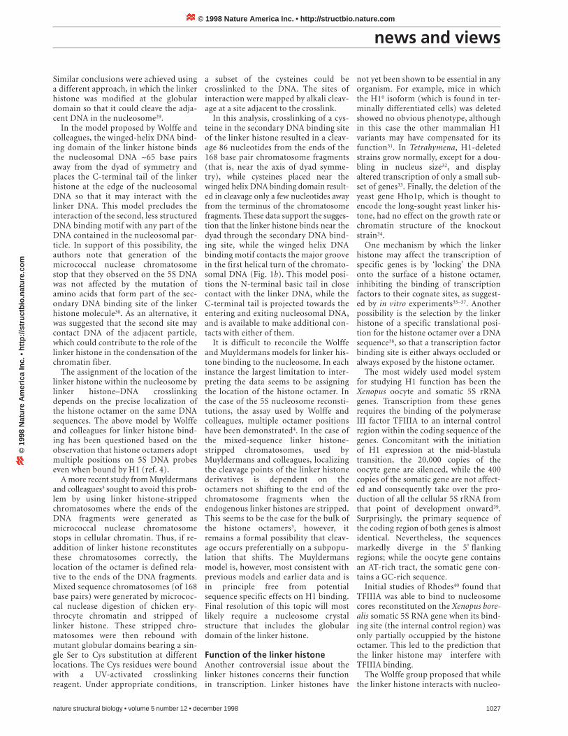
Fig. 1 Alternative hypotheses for the location ofH1 on the nucleosome. The histone octamer isshown in grey, the DNA in blue and the globulardomain of H1 in yellow. a, Off-dyad axis modelproposed by Wolffe and colleagues43 based onstudies of the X. borealis 5S gene. The globulardomain binds the major groove of the nucleoso-mal DNA 60–70 base pair from the dyad throughthe winged-helix motif, while the secondary DNAbinding site does not contact the DNA of thenucleosome. The C-terminal tail of the linker his-tone projects towards the linker DNA. b, Dyad-axis binding model postulated by Muyldermansand colleagues3 based on the analysis of bulknucleosomes. The globular domain binds theDNA at the dyad axis through the secondary DNAbinding site, while the winged-helix domainbinds either the entering or exiting DNA duplex-es. Both the N-terminal and C-terminal tails couldmake contacts with the linker DNA.
a
b
Location and function of linker histonesMarissa Vignali and Jerry L. Workman
The linker histones, H1 and its variant forms, have been implicated in the formation of higher orders ofchromatin structure and gene repression. Three recent manuscripts have reexamined the location of H1within the nucleosome particle and its function in the regulation of transcription.
© 1998 Nature America Inc. • http://structbio.nature.com©
199
8 N
atu
re A
mer
ica
Inc.
• h
ttp
://s
tru
ctb
io.n
atu
re.c
om

news and views
1026 nature structural biology • volume 5 number 12 • december 1998
sequences has suggested that this pro-tection is asymmetric. The protectionprovided by the linker histone in nucle-osomes reconstituted on the Xenopusborealis somatic 5S rRNA gene has beenreported to be of this type, such that 5base pairs are protected on one side and15 to the other side of the nucleosomalcore particle DNA20,21. Other studieshave suggested that the extra 20 basepairs protected by the linker histone arelocated entirely on one side of the nucle-osome core22,23. Asymmetry in H1 pro-tection of linker DNA is consistent withthe fact that the linker histone moleculeis intrinsically asymmetric. The basic N-terminal and C-terminal tails are verydifferent in length. The C-terminal tailcomprises almost 50% of the protein24.
The means by which the linker histonegenerates asymmetric nuclease protec-tion is dependent on how and where itbinds nucleosomal DNA. The observa-tion of DNase I protection at the dyadof the nucleosome25 led to a model in
which the linker histone binds close tothe dyad axis of the nucleosome. Adetailed analysis of the DNA sequence ofbulk nucleosomes27, supported by neu-tron scattering data26, suggested that thelinker histone may bind one terminus ofthe nucleosome and either the other ter-minus or the nucleosomal dyad. Thefirst alternative would generate an indi-rect protection of the dyad DNA andexplain the generation of the two chro-matosome stops. The second possibility,in which the dyad protection would bedirect, assumed that the second micro-coccal nuclease digestion stop would begenerated by the displacement of a corehistone tail upon linker histone binding.
In contrast, Wolffe and colleagues rea-soned that the asymmetric protectionfrom micrococcal nuclease that theyobserved on nucleosomes reconstitutedon the Xenopus borealis somatic 5S genecould result from off-dyad binding ofthe linker histone21. To identify wherethe linker histone may bind they utilized
broad specificity photoaffinity probesspecific for the major groove of DNA.The photoactive derivative and radioac-tive nucleotides were incorporated atdifferent positions along one half of thelength of the 5S rDNA molecule, whichwas then reconstituted into nucleo-somes including the globular domain ofthe linker histone. The subset of probesthat crosslinked to the globular domainof linker histone suggested that the link-er histone contacts the nucleosomalDNA at a region ~60–70 base pairs awayfrom the dyad axis28 (Fig. 1a). Based onthese data and their mapping of thelocation of the histone octamer, a modelwas proposed in which the globulardomain of the linker histone is boundinside the DNA gyres specifically on oneside of the 5S nucleosome particle.While the other half of the 5S nucleoso-mal DNA was not analyzed, this reportdid demonstrate linker histone DNAinteractions well away from the pro-posed dyad axis of the nucleosome core.
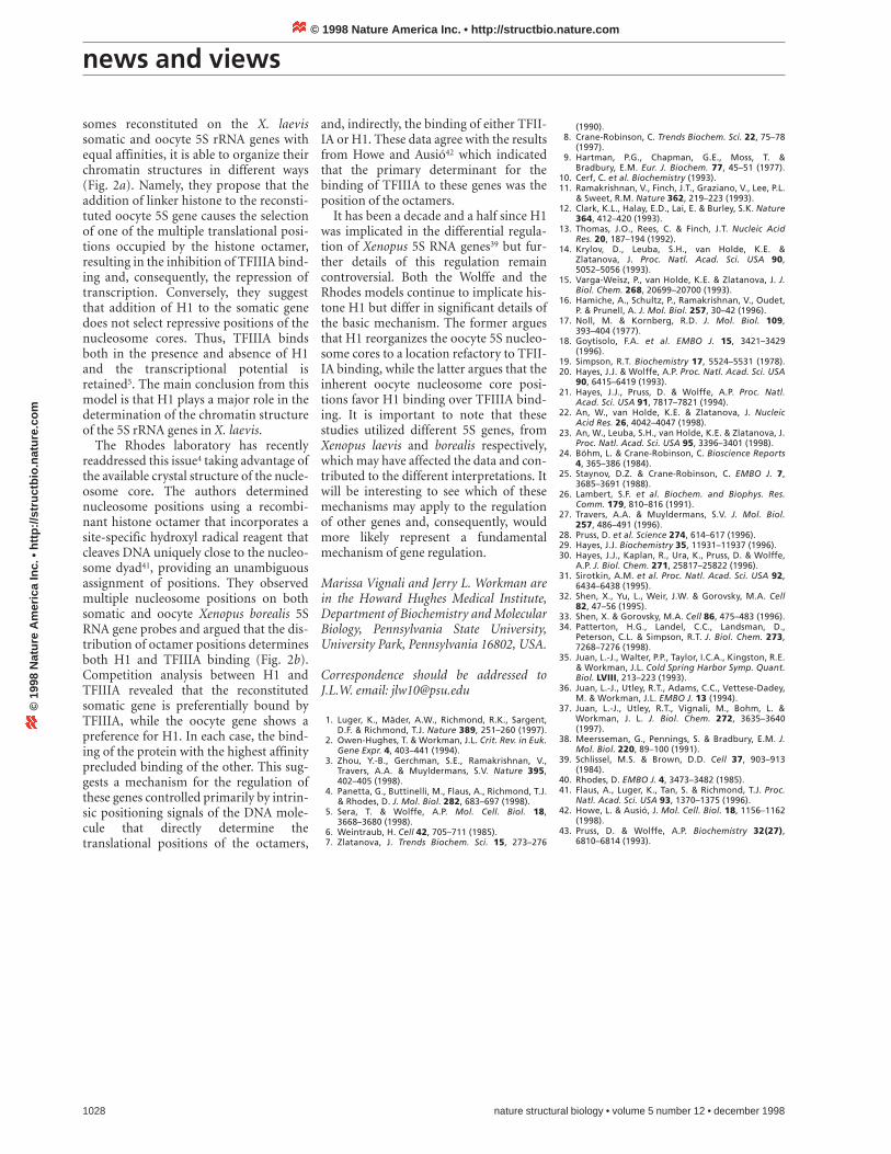
Fig. 2 Diagrams summarizing two alternative models for the effect of the linker histone on the positioning of histone octamers and the binding of TFIIIA to the 5S rRNA genes. The coding regions are shown in purple, the TFIIIA protein and binding site in pink and the linker histone (H1) is shown inyellow. The positions of the octamers and chromatosomes that allow TFIIIA binding are blue, while those that repress binding are orange. a, Themodel proposed by Wolffe and colleagues5 states that the histone octamer can adopt multiple alternative positions over the Xenopus laevis oocyte 5Ssequence, some of which allow TFIIIA binding. The addition of H1 relocates the octamers to a unique translational position that does not allow TFIIIAbinding. In the case of the somatic gene, TFIIIA can bind its cognate site both in the absence or presence of H1. b, The model proposed by Rhodes andcolleagues4 states that the octamers adopt multiple positions on both the Xenopus borealis 5S oocyte and somatic genes, only some of which allow TFIIIA binding. The addition of H1 does not affect these positions, while the addition of TFIIIA does. Different affinities of the reconstituted nucleo-somes results in the oocyte genes preferentially binding H1, (preventing TFIIIA binding), while the somatic gene preferentially binds TFIIIA.
a b
© 1998 Nature America Inc. • http://structbio.nature.com©
199
8 N
atu
re A
mer
ica
Inc.
• h
ttp
://s
tru
ctb
io.n
atu
re.c
om
news and views
Similar conclusions were achieved usinga different approach, in which the linkerhistone was modified at the globulardomain so that it could cleave the adja-cent DNA in the nucleosome29.
In the model proposed by Wolffe andcolleagues, the winged-helix DNA bind-ing domain of the linker histone bindsthe nucleosomal DNA ~65 base pairsaway from the dyad of symmetry andplaces the C-terminal tail of the linkerhistone at the edge of the nucleosomalDNA so that it may interact with thelinker DNA. This model precludes theinteraction of the second, less structuredDNA binding motif with any part of theDNA contained in the nucleosomal par-ticle. In support of this possibility, theauthors note that generation of themicrococcal nuclease chromatosomestop that they observed on the 5S DNAwas not affected by the mutation ofamino acids that form part of the sec-ondary DNA binding site of the linkerhistone molecule30. As an alternative, itwas suggested that the second site maycontact DNA of the adjacent particle,which could contribute to the role of thelinker histone in the condensation of thechromatin fiber.
The assignment of the location of thelinker histone within the nucleosome bylinker histone–DNA crosslinkingdepends on the precise localization ofthe histone octamer on the same DNAsequences. The above model by Wolffeand colleagues for linker histone bind-ing has been questioned based on theobservation that histone octamers adoptmultiple positions on 5S DNA probeseven when bound by H1 (ref. 4).
A more recent study from Muyldermansand colleagues3 sought to avoid this prob-lem by using linker histone-strippedchromatosomes where the ends of theDNA fragments were generated asmicrococcal nuclease chromatosomestops in cellular chromatin. Thus, if re-addition of linker histone reconstitutesthese chromatosomes correctly, thelocation of the octamer is defined rela-tive to the ends of the DNA fragments.Mixed sequence chromatosomes (of 168base pairs) were generated by micrococ-cal nuclease digestion of chicken ery-throcyte chromatin and stripped oflinker histone. These stripped chro-matosomes were then rebound withmutant globular domains bearing a sin-gle Ser to Cys substitution at differentlocations. The Cys residues were boundwith a UV-activated crosslinkingreagent. Under appropriate conditions,
nature structural biology • volume 5 number 12 • december 1998 1027
a subset of the cysteines could becrosslinked to the DNA. The sites ofinteraction were mapped by alkali cleav-age at a site adjacent to the crosslink.
In this analysis, crosslinking of a cys-teine in the secondary DNA binding siteof the linker histone resulted in a cleav-age 86 nucleotides from the ends of the168 base pair chromatosome fragments(that is, near the axis of dyad symme-try), while cysteines placed near thewinged helix DNA binding domain result-ed in cleavage only a few nucleotides awayfrom the terminus of the chromatosomefragments. These data support the sugges-tion that the linker histone binds near thedyad through the secondary DNA bind-ing site, while the winged helix DNAbinding motif contacts the major groovein the first helical turn of the chromato-somal DNA (Fig. 1b). This model posi-tions the N-terminal basic tail in closecontact with the linker DNA, while theC-terminal tail is projected towards theentering and exiting nucleosomal DNA,and is available to make additional con-tacts with either of them.
It is difficult to reconcile the Wolffeand Muyldermans models for linker his-tone binding to the nucleosome. In eachinstance the largest limitation to inter-preting the data seems to be assigningthe location of the histone octamer. Inthe case of the 5S nucleosome reconsti-tutions, the assay used by Wolffe andcolleagues, multiple octamer positionshave been demonstrated4. In the case ofthe mixed-sequence linker histone-stripped chromatosomes, used byMuyldermans and colleagues, localizingthe cleavage points of the linker histonederivatives is dependent on theoctamers not shifting to the end of thechromatosome fragments when theendogenous linker histones are stripped.This seems to be the case for the bulk ofthe histone octamers3, however, itremains a formal possibility that cleav-age occurs preferentially on a subpopu-lation that shifts. The Muyldermansmodel is, however, most consistent withprevious models and earlier data and isin principle free from potentialsequence specific effects on H1 binding.Final resolution of this topic will mostlikely require a nucleosome crystalstructure that includes the globulardomain of the linker histone.
Function of the linker histoneAnother controversial issue about thelinker histones concerns their functionin transcription. Linker histones have
not yet been shown to be essential in anyorganism. For example, mice in whichthe H10 isoform (which is found in ter-minally differentiated cells) was deletedshowed no obvious phenotype, althoughin this case the other mammalian H1variants may have compensated for itsfunction31. In Tetrahymena, H1-deletedstrains grow normally, except for a dou-bling in nucleus size32, and displayaltered transcription of only a small sub-set of genes33. Finally, the deletion of theyeast gene Hho1p, which is thought toencode the long-sought yeast linker his-tone, had no effect on the growth rate orchromatin structure of the knockoutstrain34.
One mechanism by which the linkerhistone may affect the transcription ofspecific genes is by ‘locking’ the DNAonto the surface of a histone octamer,inhibiting the binding of transcriptionfactors to their cognate sites, as suggest-ed by in vitro experiments35–37. Anotherpossibility is the selection by the linkerhistone of a specific translational posi-tion for the histone octamer over a DNAsequence38, so that a transcription factorbinding site is either always occluded oralways exposed by the histone octamer.
The most widely used model systemfor studying H1 function has been theXenopus oocyte and somatic 5S rRNAgenes. Transcription from these genesrequires the binding of the polymeraseIII factor TFIIIA to an internal controlregion within the coding sequence of thegenes. Concomitant with the initiationof H1 expression at the mid-blastulatransition, the 20,000 copies of theoocyte gene are silenced, while the 400copies of the somatic gene are not affect-ed and consequently take over the pro-duction of all the cellular 5S rRNA fromthat point of development onward39.Surprisingly, the primary sequence ofthe coding region of both genes is almostidentical. Nevertheless, the sequencesmarkedly diverge in the 5' flankingregions; while the oocyte gene containsan AT-rich tract, the somatic gene con-tains a GC-rich sequence.
Initial studies of Rhodes40 found that TFIIIA was able to bind to nucleosomecores reconstituted on the Xenopus bore-alis somatic 5S RNA gene when its bind-ing site (the internal control region) wasonly partially occuppied by the histoneoctamer. This led to the prediction thatthe linker histone may interfere withTFIIIA binding.
The Wolffe group proposed that whilethe linker histone interacts with nucleo-
© 1998 Nature America Inc. • http://structbio.nature.com©
199
8 N
atu
re A
mer
ica
Inc.
• h
ttp
://s
tru
ctb
io.n
atu
re.c
om
news and views
somes reconstituted on the X. laevissomatic and oocyte 5S rRNA genes withequal affinities, it is able to organize theirchromatin structures in different ways(Fig. 2a). Namely, they propose that theaddition of linker histone to the reconsti-tuted oocyte 5S gene causes the selectionof one of the multiple translational posi-tions occupied by the histone octamer,resulting in the inhibition of TFIIIA bind-ing and, consequently, the repression oftranscription. Conversely, they suggestthat addition of H1 to the somatic genedoes not select repressive positions of thenucleosome cores. Thus, TFIIIA bindsboth in the presence and absence of H1and the transcriptional potential isretained5. The main conclusion from thismodel is that H1 plays a major role in thedetermination of the chromatin structureof the 5S rRNA genes in X. laevis.
The Rhodes laboratory has recentlyreaddressed this issue4 taking advantage ofthe available crystal structure of the nucle-osome core. The authors determinednucleosome positions using a recombi-nant histone octamer that incorporates asite-specific hydroxyl radical reagent thatcleaves DNA uniquely close to the nucleo-some dyad41, providing an unambiguousassignment of positions. They observedmultiple nucleosome positions on bothsomatic and oocyte Xenopus borealis 5SRNA gene probes and argued that the dis-tribution of octamer positions determinesboth H1 and TFIIIA binding (Fig. 2b).Competition analysis between H1 andTFIIIA revealed that the reconstitutedsomatic gene is preferentially bound byTFIIIA, while the oocyte gene shows apreference for H1. In each case, the bind-ing of the protein with the highest affinityprecluded binding of the other. This sug-gests a mechanism for the regulation ofthese genes controlled primarily by intrin-sic positioning signals of the DNA mole-cule that directly determine thetranslational positions of the octamers,
and, indirectly, the binding of either TFII-IA or H1. These data agree with the resultsfrom Howe and Ausió42 which indicatedthat the primary determinant for thebinding of TFIIIA to these genes was theposition of the octamers.
It has been a decade and a half since H1was implicated in the differential regula-tion of Xenopus 5S RNA genes39 but fur-ther details of this regulation remaincontroversial. Both the Wolffe and theRhodes models continue to implicate his-tone H1 but differ in significant details ofthe basic mechanism. The former arguesthat H1 reorganizes the oocyte 5S nucleo-some cores to a location refactory to TFII-IA binding, while the latter argues that theinherent oocyte nucleosome core posi-tions favor H1 binding over TFIIIA bind-ing. It is important to note that thesestudies utilized different 5S genes, fromXenopus laevis and borealis respectively,which may have affected the data and con-tributed to the different interpretations. Itwill be interesting to see which of thesemechanisms may apply to the regulationof other genes and, consequently, wouldmore likely represent a fundamentalmechanism of gene regulation.
Marissa Vignali and Jerry L. Workman arein the Howard Hughes Medical Institute,Department of Biochemistry and MolecularBiology, Pennsylvania State University,University Park, Pennsylvania 16802, USA.
Correspondence should be addressed toJ.L.W. email: [email protected]
1. Luger, K., Mäder, A.W., Richmond, R.K., Sargent,D.F. & Richmond, T.J. Nature 389, 251–260 (1997).
2. Owen-Hughes, T. & Workman, J.L. Crit. Rev. in Euk.Gene Expr. 4, 403–441 (1994).
3. Zhou, Y.-B., Gerchman, S.E., Ramakrishnan, V.,Travers, A.A. & Muyldermans, S.V. Nature 395,402–405 (1998).
4. Panetta, G., Buttinelli, M., Flaus, A., Richmond, T.J.& Rhodes, D. J. Mol. Biol. 282, 683–697 (1998).
5. Sera, T. & Wolffe, A.P. Mol. Cell. Biol. 18,3668–3680 (1998).
6. Weintraub, H. Cell 42, 705–711 (1985).7. Zlatanova, J. Trends Biochem. Sci. 15, 273–276
1028 nature structural biology • volume 5 number 12 • december 1998
(1990).8. Crane-Robinson, C. Trends Biochem. Sci. 22, 75–78
(1997).9. Hartman, P.G., Chapman, G.E., Moss, T. &
Bradbury, E.M. Eur. J. Biochem. 77, 45–51 (1977).10. Cerf, C. et al. Biochemistry (1993).11. Ramakrishnan, V., Finch, J.T., Graziano, V., Lee, P.L.
& Sweet, R.M. Nature 362, 219–223 (1993).12. Clark, K.L., Halay, E.D., Lai, E. & Burley, S.K. Nature
364, 412–420 (1993).13. Thomas, J.O., Rees, C. & Finch, J.T. Nucleic Acid
Res. 20, 187–194 (1992).14. Krylov, D., Leuba, S.H., van Holde, K.E. &
Zlatanova, J. Proc. Natl. Acad. Sci. USA 90,5052–5056 (1993).
15. Varga-Weisz, P., van Holde, K.E. & Zlatanova, J. J.Biol. Chem. 268, 20699–20700 (1993).
16. Hamiche, A., Schultz, P., Ramakrishnan, V., Oudet,P. & Prunell, A. J. Mol. Biol. 257, 30–42 (1996).
17. Noll, M. & Kornberg, R.D. J. Mol. Biol. 109,393–404 (1977).
18. Goytisolo, F.A. et al. EMBO J. 15, 3421–3429(1996).
19. Simpson, R.T. Biochemistry 17, 5524–5531 (1978).20. Hayes, J.J. & Wolffe, A.P. Proc. Natl. Acad. Sci. USA
90, 6415–6419 (1993).21. Hayes, J.J., Pruss, D. & Wolffe, A.P. Proc. Natl.
Acad. Sci. USA 91, 7817–7821 (1994).22. An, W., van Holde, K.E. & Zlatanova, J. Nucleic
Acid Res. 26, 4042–4047 (1998).23. An, W., Leuba, S.H., van Holde, K.E. & Zlatanova, J.
Proc. Natl. Acad. Sci. USA 95, 3396–3401 (1998).24. Böhm, L. & Crane-Robinson, C. Bioscience Reports
4, 365–386 (1984).25. Staynov, D.Z. & Crane-Robinson, C. EMBO J. 7,
3685–3691 (1988).26. Lambert, S.F. et al. Biochem. and Biophys. Res.
Comm. 179, 810–816 (1991).27. Travers, A.A. & Muyldermans, S.V. J. Mol. Biol.
257, 486–491 (1996).28. Pruss, D. et al. Science 274, 614–617 (1996).29. Hayes, J.J. Biochemistry 35, 11931–11937 (1996).30. Hayes, J.J., Kaplan, R., Ura, K., Pruss, D. & Wolffe,
A.P. J. Biol. Chem. 271, 25817–25822 (1996).31. Sirotkin, A.M. et al. Proc. Natl. Acad. Sci. USA 92,
6434–6438 (1995).32. Shen, X., Yu, L., Weir, J.W. & Gorovsky, M.A. Cell
82, 47–56 (1995).33. Shen, X. & Gorovsky, M.A. Cell 86, 475–483 (1996).34. Patterton, H.G., Landel, C.C., Landsman, D.,
Peterson, C.L. & Simpson, R.T. J. Biol. Chem. 273,7268–7276 (1998).
35. Juan, L.-J., Walter, P.P., Taylor, I.C.A., Kingston, R.E.& Workman, J.L. Cold Spring Harbor Symp. Quant.Biol. LVIII, 213–223 (1993).
36. Juan, L.-J., Utley, R.T., Adams, C.C., Vettese-Dadey,M. & Workman, J.L. EMBO J. 13 (1994).
37. Juan, L.-J., Utley, R.T., Vignali, M., Bohm, L. &Workman, J. L. J. Biol. Chem. 272, 3635–3640(1997).
38. Meersseman, G., Pennings, S. & Bradbury, E.M. J.Mol. Biol. 220, 89–100 (1991).
39. Schlissel, M.S. & Brown, D.D. Cell 37, 903–913(1984).
40. Rhodes, D. EMBO J. 4, 3473–3482 (1985).41. Flaus, A., Luger, K., Tan, S. & Richmond, T.J. Proc.
Natl. Acad. Sci. USA 93, 1370–1375 (1996).42. Howe, L. & Ausió, J. Mol. Cell. Biol. 18, 1156–1162
(1998).43. Pruss, D. & Wolffe, A.P. Biochemistry 32(27),
6810–6814 (1993).
© 1998 Nature America Inc. • http://structbio.nature.com©
199
8 N
atu
re A
mer
ica
Inc.
• h
ttp
://s
tru
ctb
io.n
atu
re.c
om