local radiation protection rules - hospital … department/radiology/docs... · local radiation...
TRANSCRIPT
LOCAL RADIATION PROTECTION RULESDepartment of RadiologyQueen Mary Hospital
2 0 1 4Version October 2014
Preface
Local Radiation Protection Rules - Department of Radiology1. This set of Local Radiation Protection Rules is produced for the protection of staff, pa-
tients and members of the public in the Department of Radiology, Queen Mary Hospital.It is a simplified and tailored version the “Code of practice on Radiation Safety 2011" andincludes information and explanations on recommended procedures and possible riskspertaining to the safe use of radiation in the Department.
2. It is a requirement of all staff who might be exposed to ionizing radiations in this De-partment to read this "Local Radiation Protection Rules 2014 - Department of Radiol-ogy, Queen Mary Hospital” and to complete and sign the declaration record sheet at-tached at the end of this Local Rules stating that he/she has read and understood thecontents and is willing to observe them.
3. Staff are also reminded to read and observe the related sections which affect his/herwork and well being, in:
a) "Local Radiation Protection Rules - Nuclear Medicine Unit, Department of Radiol-ogy, Queen Mary Hospital”(HA-intranet:http://www3.ha.org.hk/qmh/department/Clinic%20Department/Radiology/patinfo1.htm),
b) "Code of Practice on Radiation Safety 2011” of the Hong Kong Hospital Authority(http://hkwc.home → OSH → Safety Manual)(HAHO level → HA Code of Practice onRadiation Safety 2011); and
c) HA Safety Manual (Chapter 9) Ionizing Radiation (http://hkwc.home → OSH →Safety Manual)(HAHO level → HA safety Manual: Chapter 9, Ionizing Radiation)
d)“Guidance Notes on Radiation Protection for Diagnostic Radiology” of the Radia-tion Health Unit, Department of Health, HKSAR (http://www.info.gov.hk/dh-rhu/english/html/Info_Pub.htm)
4. Copies of the Local Rules are available at the Department’s Library, Staff duty rooms, X-ray rooms, Senior Radiographers' offices and my office as well, for your reading.
Dr. Wendy Lam HKWC Service Director (DR)/ Chief of Service (DR), QMH15 September 2014

Table of Contents
CHAPTER CONTENTS PAGE
Chapter 1 Introduction 1
Chapter 2 Principles of Radiation Protection in Diagnostic Radiology 2
Chapter 3 Protection of Staff, Members of the Public and Women of Child Bearing Age 5
Chapter 4 General Measures for Radiological Protection 9
Chapter 5 Local Rules for General Radiography X-ray Rooms 12
Chapter 6 Local Rules for Mobile Radiography 15
Chapter 7 Local Rules for OT Radiography 17
Chapter 8 Local Rules for Fluoroscopy and Angiography / Interventional X-ray Rooms 19
Chapter 9 Local Rules for CT Rooms 24
Chapter 10 Local Rules for Mammography Rooms 26
Chapter 11 Roles of personnel on Radiation Safety 28
Chapter 12 Contingency Plans and Contact Persons in case of reporting of radiation Hazard
30
Appendix I Radiation Safety for Performing Radioembolization in Interventional Radiology Facility in DR/QMH
31
Appendix II Fetal absorbed dose of some common diagnostic procedures 41
Appendix III Typical effective doses, equivalent periods of natural background radiation and life-time fatal cancer risks from diagnostic medical exposures
42
Appendix IV Doses in Interventional Procedures 44
Appendix V 醫療輻射劑量比較圖 46
Appendix VI Flowchart on Radiological Investigation for Women of Child-bearing Age 48
Appendix VII Flowchart on Monthly Return of TLD Badges of Radiographers in DR, QMH 50
Appendix VIII Designation of Radiation Areas for Radiology Department at Block H-1, Block K-3 and A&E X-Ray & CT Scan Unit
52
Appendix IX List of RPS, DR, QMH 56
Appendix X Staff declaration recorda) Medical Staffb) Radiographic staffc) Nursing Staff
58
Chapter 1
Introduction
1.1 These Local Rules are produced in accordance with the 'Code of Practice on Radiation 2011' (Code of Practice). They include information and explanations on recommended pro-cedures and possible risks which may be encountered in the Department by staff, patients and members of the public who may be exposed to ionizing radiations.
1.2 The administrative responsibility for the protection measures set out in the Code rests, in this Hospital lies with the Hospital Authority (HA), and in the case of persons em-ployed by service agents which provided services to HA hospitals, their employers in ad-dition to the HA.
1.3 The Radiation Protection Adviser (RPA) is appointed by the Chief Executive of the HA. The RPA should normally be a certified medical physicist with appropriate experi-ence. The Chief of Service (COS) or Head of Unit may in consultation with the RPA ap-point one or more of his/her staff as Radiation Protection Supervisor (RPS) to assist him/her on radiation safety and protection measures.
1.4 The ultimate responsibility for the local observance of the protection measures within a Unit, however, rests on the Head of that Unit. In the first instance any matter concerning protection should be referred to the Radiation Protection Supervisor of the Unit involved.
1.5 It is a requirement that every radiation worker should read these Local Rules and those sections of the Code of Practice which affect his/her work and well being. The radia-tion worker should complete, sign and return the declaration form to the Radiation Protec-tion Supervisor (RPS) stating that he/she has read and understood the contents, and is willing to observe them.
D e p a r t m e n t o f R a d i o l o g y / Q M H!
1
Chapter 2
Principles of Radiation Protection in Diagnostic Radiology
2.1 Protection in diagnostic radiology is based on the following principles:
a)Radiological examination should be carried out only if it is likely that the informa-tion obtained will be of benefit to the patient or will improve the overall health of the population;
b)The irradiation of the patient during X-ray examination should be no greater than is necessary to result in a satisfactory examination and care should be taken to re-duce to a minimum the irradiation of particularly sensitive tissues, such as the fe-male breast∗, red bone marrow, lung and gonads;
c)Shielding for primary and secondary radiation should be close to the equipment or patient;
d)X-ray equipment should be used only when there is adequate protection∗∗ for all persons in all surrounding areas;
∗Between the age of 10 and 45 the female breast is more radiosensitive than is implied by the ICRP weighting factor of 0.12 which is an average value for males and females.
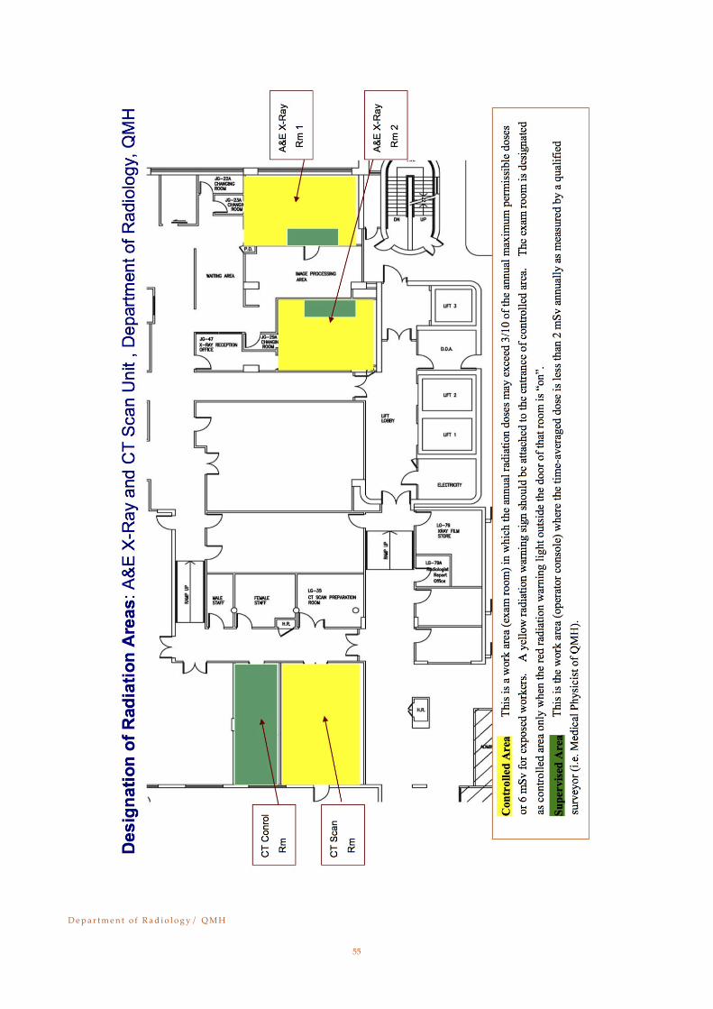
∗∗"Adequate protection” is defined as protection which is intended to ensure that doses received are as low as reasonably achievable and in any case do not exceed 3/10 of the dose limits which are appro-priate for the category of persons exposed. When shielding is provided the instantaneous dose rate outside the shield should not exceed 3 !Svh-1 except for radiation beam therapy when the time-averaged dose rate should not exceed 3 !Svh-1. (Reg. 17, the Radiation (Control of irradiating Appara-tus) Regulations, Cap 303, Laws of Hong Kong).
2.2 Every individual using ionizing radiation has a duty to protect himself/herself and others (including patients) from any radiation hazards arising from his/her work.
2.3 For radiation protection purposes, the hazards may be segregated into two classes:- a) External hazards
These arise from radiation sources outside the body. They can be controlled by: i) Limiting the exposure time.ii) Keeping at a distance as far as possible from the source.iii)Using suitable shielding.
b) Internal hazards These arise when radioactive materials enter the body through inhalation, inges-tion or absorption through the mouth, skin or wound. They can be controlled by:
i) Containment of the radioactive materialii) Good house keeping and cleanliness.iii)Use of least radio-toxic and the smallest activity if possible
D e p a r t m e n t o f R a d i o l o g y / Q M H!
2

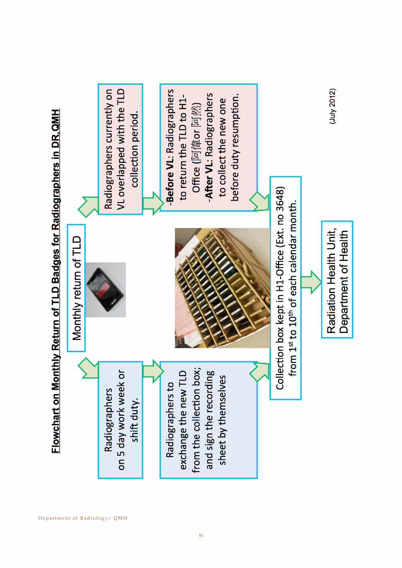
2.4 The TLD badge or ionization dosimeters, when provided, must be worn at all times when on duty. It should be worn on the trunk at chest or at waist height. In addition, ring badges are to be worn if the unprotected hands and forearms come in close proximity to the beam. TLD badge should be returned monthly and loss of TLD reported as soon as possible. (Refer to Appendix VII on TLD return flowchart including arrangement before and after vacation leave.)
2.5 When a protective apron is worn and the unprotected parts of the body are likely to be exposed to more than 1/10 of their dose limits, one or more dosemeters should be placed at collar level or other body parts outside the apron where it may best monitor the dose to the exposed parts of the body. Special devices for checking finger doses would be pro-vided in circumstances. Care should be taken to avoid any dosemeter from becoming wet or damaged.
2.6 All areas where there could be an ionizing radiation hazard or contaminations should be monitored at regular interval, and the results recorded by the Physicist.
2.7 The dose limits for occupationally exposed workers aged 18 years or above are given in Table 1 (page 4). The dose limits apply to the sum of the dose equivalent received from external sources during working hours and the committed dose equivalent due to internal sources entering into the body during the course of work.
2.8 Women of reproductive capacity have a special responsibility in that they must imme-diately inform either the administrative superior (such as COS, DM, RPS) or the Head of their unit as soon as they know that they are pregnant so that steps can be taken to ensure that the dose to the fetus does not exceed the maximum permissible level specified in Ta-ble 1.
D e p a r t m e n t o f R a d i o l o g y / Q M H!
3
Diagnostic X-ray procedures should only be carried out when there is a real need.
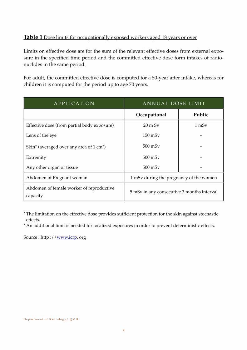
Table 1 Dose limits for occupationally exposed workers aged 18 years or over
Limits on effective dose are for the sum of the relevant effective doses from external expo-sure in the specified time period and the committed effective dose form intakes of radio-nuclides in the same period.
For adult, the committed effective dose is computed for a 50-year after intake, whereas for children it is computed for the period up to age 70 years.
APPLICATION ANNUAL DOSE LIMITANNUAL DOSE LIMIT
Occupational Public
Effective dose (from partial body exposure) 20 m Sv 1 mSv
Lens of the eye 150 mSv -
Skin∗ (averaged over any area of 1 cm2) 500 mSv -
Extremity 500 mSv -
Any other organ or tissue 500 mSv -
Abdomen of Pregnant woman 1 mSv during the pregnancy of the women1 mSv during the pregnancy of the women
Abdomen of female worker of reproductive capacity
5 mSv in any consecutive 3 months interval5 mSv in any consecutive 3 months interval
* The limitation on the effective dose provides sufficient protection for the skin against stochastic effects.
* An additional limit is needed for localized exposures in order to prevent deterministic effects.
Source : http ://www.icrp. org
D e p a r t m e n t o f R a d i o l o g y / Q M H!
4
Chapter 3Protection of staff, Members of the Public and Women of child bearing age
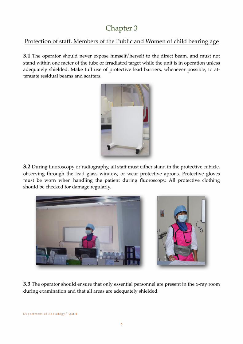
3.1 The operator should never expose himself/herself to the direct beam, and must not stand within one meter of the tube or irradiated target while the unit is in operation unless adequately shielded. Make full use of protective lead barriers, whenever possible, to at-tenuate residual beams and scatters.
3.2 During fluoroscopy or radiography, all staff must either stand in the protective cubicle, observing through the lead glass window, or wear protective aprons. Protective gloves must be worn when handling the patient during fluoroscopy. All protective clothing should be checked for damage regularly.
3.3 The operator should ensure that only essential personnel are present in the x-ray room during examination and that all areas are adequately shielded.
D e p a r t m e n t o f R a d i o l o g y / Q M H!
5
3.4 Make sure the doors of the x-ray room are closed before making any exposure during examination and equipment testing. It may be appropriate to lock certain X-ray room doors, to prevent unauthorized entry when the door is remote from the operator and out-side the immediate field of view.
3.5 Where an x-ray room is used for more than one radiology procedure at a time, take adequate protective measures to ensure there is no significant additional exposure, either of one patient from radiography of another or of staff from examination in which they themselves are not engaged.
3.6 The radiographer supervising the examination is responsible for checking the operat-ing conditions such as patient position, radiological technique selected and cassette load-ing etc. before making exposure.
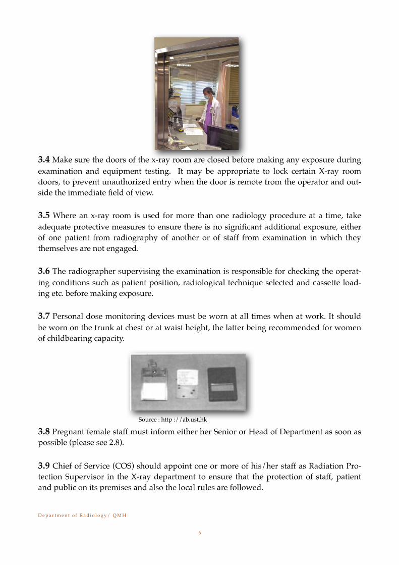
3.7 Personal dose monitoring devices must be worn at all times when at work. It should be worn on the trunk at chest or at waist height, the latter being recommended for women of childbearing capacity.
Source : http ://ab.ust.hk
3.8 Pregnant female staff must inform either her Senior or Head of Department as soon as possible (please see 2.8).
3.9 Chief of Service (COS) should appoint one or more of his/her staff as Radiation Pro-tection Supervisor in the X-ray department to ensure that the protection of staff, patient and public on its premises and also the local rules are followed.
D e p a r t m e n t o f R a d i o l o g y / Q M H!
6
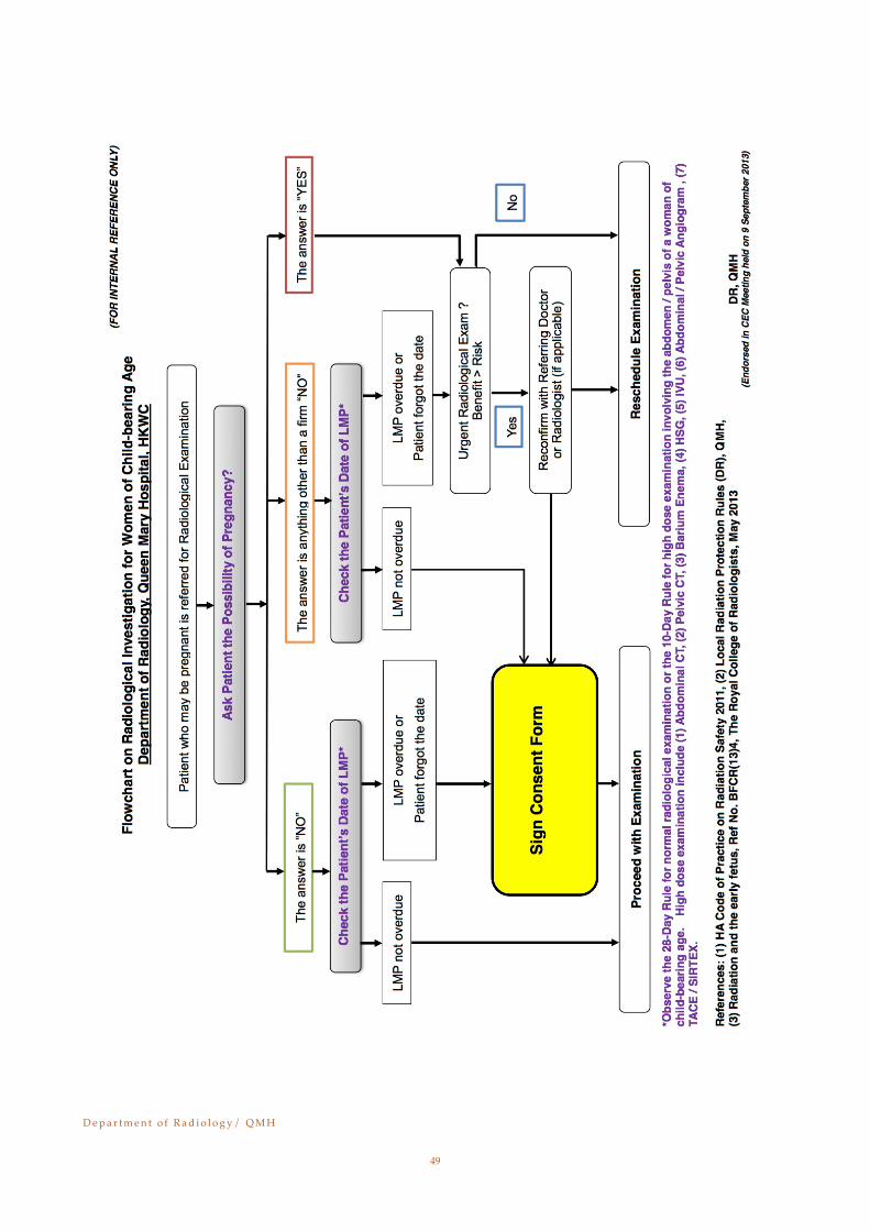
3.10 X-ray examination for women of childbearing age(Refer to Appendix VI- Flowchart on Radiological Investigation for Women of Child-bearing Age)3.10.1 All female patients of childbearing age should be asked whether there is any possibility of pregnancy. When the patient states that there is no possibility of preg-nancy, she should be asked the date of commencement of L.M.P. This should be re-corded and the examination could proceed. For high dose examinations involving abdomen/ pelvis of a women of child bearing age, the examination should be per-formed within 10 days from the date of commencement of L.M.P.
3.10.2 There are two rules to, be observed for females of childbearing age whose pregnancy cannot be excluded:
a)*(28-day-rule) - If she is not sure about her possibility of pregnancy or preg-nancy could not be excluded, she should be asked whether her menstrual pe-riod is overdue. If it is not overdue, then observe the 28-day-rule for normal radiological examinations. The patient should be asked to sign a consent form prior to proceeding with the examination. If the L.M.P. is overdue or she has forgot the date of L.M.P., then the radiographer should reconfirm with the re-ferring clinician or Radiologist whether to proceed with the examination or not. If it is considered as an urgent radiological examination that the benefits have out-weighted the risks, the examination could be proceeded after con-sent form has been signed, otherwise, the examination should be rescheduled or cancelled.
b)*(10-day-rule) - If she is not sure about her possibility of pregnancy or preg-nancy could not be excluded, she should be asked whether her menstrual pe-riod is overdue. If it is not overdue and the date of L.M.P. is within 28 days, then proceed with the examination except for high dose procedures (such as abdominal CT, pelvic CT, barium enema, and any other special x-ray exami-nations likely to deliver a dose of tens of mGy to the conceptus), in which case
D e p a r t m e n t o f R a d i o l o g y / Q M H!
7
the examination may be postponed to the early part of the menstrual cycle - the "limited return to the 10-day-rule”.
(Refer to the attached table in Appendix-II for Fetal Absorbed Dose of some common diagnostic procedures). Attention should be paid to ensure minimization of expo-sure to any embryo or fetus that may be present, whether or not the woman is known to be pregnant.
3.10.3 If a female patient can confirm that she is pregnant and her menstrual period has been clearly missed (also the 10-day-rule & 28-day-rule cannot apply), then any decision to proceed with the examination should be taken by the referring physician in its clinical necessity (e.g. urgent x-ray examinations when benefits are likely to far outweigh any small risk from the irradiation). The examination could be proceeded after the consent form has been signed. Attention should be paid to ensure minimiza-tion of exposure to any embryo or fetus.
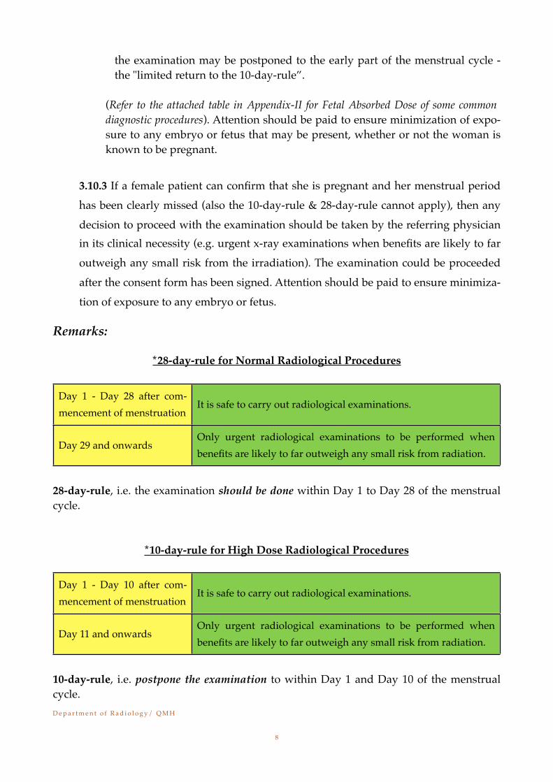
Remarks:
*28-day-rule for Normal Radiological Procedures
Day 1 - Day 28 after com-mencement of menstruation
It is safe to carry out radiological examinations.
Day 29 and onwardsOnly urgent radiological examinations to be performed when benefits are likely to far outweigh any small risk from radiation.
28-day-rule, i.e. the examination should be done within Day 1 to Day 28 of the menstrual cycle.
*10-day-rule for High Dose Radiological Procedures
Day 1 - Day 10 after com-mencement of menstruation
It is safe to carry out radiological examinations.
Day 11 and onwardsOnly urgent radiological examinations to be performed when benefits are likely to far outweigh any small risk from radiation.
10-day-rule, i.e. postpone the examination to within Day 1 and Day 10 of the menstrual cycle.
D e p a r t m e n t o f R a d i o l o g y / Q M H!
8
Chapter 4
General Measures for Radiological Protection
4.1 Personal monitoringa)The personal monitoring for classified persons or persons working under a written
scheme of work is required to be dealt with by a dosimetry service approved by the Radiation Board.
b)Where exposure is from external sources (other than low-energy beta emitters with no significant bremsstrahlung emission), personal monitoring should be by means of one or more dosimeters worn on an appropriate part or parts of the body.
c)The length of each monitoring period should depend on the doses likely to be re-ceived during the period. Dosimeters should be returned promptly after use for dose assessment and replaced with new ones. Each dosimeter is normally worn for 1 month, but periods ranging from 2 weeks to 3 months can be appropriate in cer-tain circumstances. For TLD badge from the Radiation Health Unit, it should be re-turned monthly. (Refer to Appendix VII on TLD return flowchart including arrangement before and after vacation leave.)
d)Persons who are issued with a dosimeter should wear it as instructed all the time while they are at work. Care should be taken to prevent the dosimeter, while not being worn, from being exposed inadvertently to ionizing radiation or subject to other conditions, e.g. heat, which could affect the assessment of doses. A dosimeter should normally be worn on the trunk at chest or waist height; it may then be in-terpreted as monitoring the dose to the whole body.
e) If a person is wearing a protective apron, then in addition to a dosimeter worn on the trunk under the apron, one or more dosimeters should be worn on the unpro-tected parts of the body if there are likely to be contributions greater than 1/10 of the effective dose equivalent from their exposure. This is unlikely to occur unless individual organs (or eyes) are exposed to more than 1/10 of their dose limits.
4.2 Persons undergoing examination with ionizing radiations
4.2.1 Examinations or treatment directly associated with illness or injury
a) All diagnostic procedures including exposure to radiation for medical purposes may carry some personal risk. The direct or indirect irradiation of patients' gonads may constitute a risk to future generations and in pregnancy there may also be a risk to the fetus. It is important therefore that only those medical exposures that are necessary should be undertaken. Alternative methods of obtaining the re-
D e p a r t m e n t o f R a d i o l o g y / Q M H!
9

quired diagnostic information should be considered, for example, by the use of non-ionizing radiation.
b)A radiological examination or investigation should be initiated only by a registered medical practitioner responsible for the care of the patient or by the radiologist or nuclear medicine specialist to whom the patient is referred.
c)A person who requests an examination should be satisfied that it is necessary, tak-ing into consideration the benefits expected from the examination and the radiation dose involved. They should ascertain first whether there are records of previous examinations which are relevant to the proposed examination. When an examina-tion is requested, the clinical indications, the provisional diagnosis, and the infor-mation required should be stated.
d)To reduce unnecessary examinations, administrative services should provide for the ready availability of previous films and for the rapid transfer on request of films or copies from one establishment or practice to another. Access to reports, filed in patients' records, can help to avoid or limit the need for further examina-tions. Results of investigations with radionuclides should also be made available.
e)If the person requested to undertake the examination has any doubt about its ad-visability or about the nature of the examination required, the matter should be re-solved by consultation between the medical officers responsible for the clinical and radiological care of the patient. Case discussions attended by clinicians, radiolo-gists, consultants in nuclear medicine and other staff, as appropriate; provide an opportunity for the critical assessment of the value and possible hazards of pro-posed x-ray examinations and diagnostic radionuclide investigations.
f) To reduce the necessity for repeat examinations, clinicians and nursing staff should co-operate with the radiology and nuclear medicine services to ensure that patients are adequately prepared before the examinations. This is particularly important for x-ray examinations of the abdomen or pelvis.
g)Fluoroscopy should not be requested if the same information could be obtained by radiography. Alternative methods not involving ionizing radiation should be con-sidered for locating metallic foreign bodies at operations.
h)If specific radiographic projections are requested, these should be kept to the nec-essary minimum. In general, the details of particular examinations including the nature and number of projections to be taken will be determined by the radiologist.
i) It is particularly important to establish that the proposed examination of a woman who is (or who may be) pregnant is necessary where there is a risk that a fetus may be irradiated; also that the same diagnostic information could not be obtained by a
D e p a r t m e n t o f R a d i o l o g y / Q M H!
10
different method carrying less risk. The request form should state that the woman is or may be pregnant so that particular care can be taken during the examinations.
j) Before any examination involving the abdomen or pelvis of a woman of childbear-ing age, an enquiry should be made about possible pregnancy. She should be treated as pregnant, and the guidance in paragraph i followed if her menstrual pe-riod is overdue or clearly missed, unless there is information indicating the absence of pregnancy. In some cases a pregnancy test may be advisable.
4.2.2 Examinations not directly associated with illness or injury
a)Screening programmes, e.g. chest radiographs, should be undertaken only if the expected medical benefits to the individuals examined, and to the population as a whole, exceed the economic and social costs, including the risks associated with the radiation dose involved. Since benefits are not always the same for all members of the population, screening should normally be limited to particular groups.
b)Some types of examination result in benefits which are shared by the person who is examined, the employer and a third party (e.g. an insurer). Such an examination should be requested only on specific medical advice and if it is expected to show net benefit to the subject. It should not be requested if the results of a previous ex-amination, giving the required information, could be obtained. A record of the ex-amination should be kept.
D e p a r t m e n t o f R a d i o l o g y / Q M H!
11
Chapter 5
Local Rules for General Radiography X-ray Rooms
5.1 No examination should be undertaken without an authorized request for the investi-gation.
5.2 Exposure factors and the selection of receptor (e.g. Horizontal or Vertical) should be checked by the operator on each occasion before an examination is performed.
5.3 Constant potential generator can be used to improve quality of the x-ray beam with a greater proportion of high-energy photons and result in less skin dose to patient and same imaging information.
5.4 Periodic survey of beam quality of all x-ray equipment should be conducted as part of a quality assurance program.
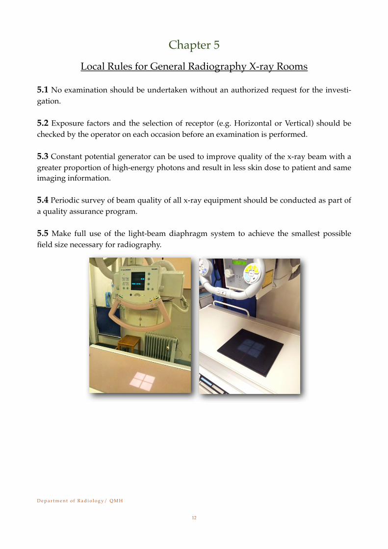
5.5 Make full use of the light-beam diaphragm system to achieve the smallest possible field size necessary for radiography.
D e p a r t m e n t o f R a d i o l o g y / Q M H!
12
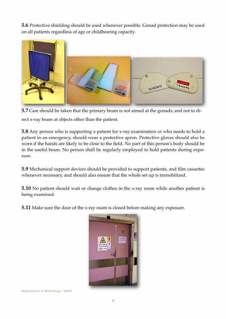
5.6 Protective shielding should be used whenever possible. Gonad protection may be used on all patients regardless of age or childbearing capacity.
5.7 Care should be taken that the primary beam is not aimed at the gonads; and not to di-
rect x-ray beam at objects other than the patient.
5.8 Any person who is supporting a patient for x-ray examination or who needs to hold a patient in an emergency, should wear a protective apron. Protective gloves should also be worn if the hands are likely to be close to the field. No part of this person's body should be in the useful beam. No person shall be regularly employed to hold patients during expo-sure.
5.9 Mechanical support devices should be provided to support patients, and film cassettes whenever necessary, and should also ensure that the whole set up is immobilized.
5.10 No patient should wait or change clothes in the x-ray room while another patient is being examined.
5.11 Make sure the door of the x-ray room is closed before making any exposure.
D e p a r t m e n t o f R a d i o l o g y / Q M H!
13
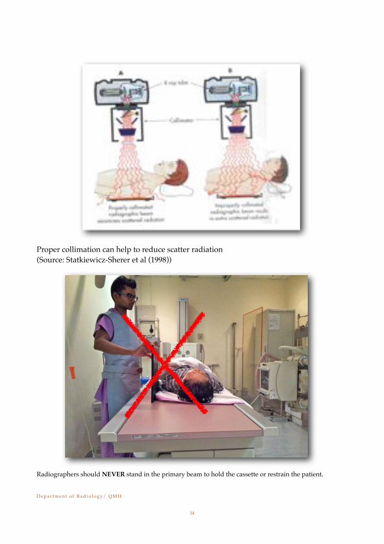
Proper collimation can help to reduce scatter radiation(Source: Statkiewicz-Sherer et al (1998))
Radiographers should NEVER stand in the primary beam to hold the cassette or restrain the patient.
D e p a r t m e n t o f R a d i o l o g y / Q M H!
14
Chapter 6
Local Rules for Mobile Radiography
6.1 The Radiographer should stand as far as possible from the tube and patient during exposure, and should wear a protective apron, or step behind an adequate shield.
6.2 The Radiographer must wear a protective apron when standing within 2 meters of the x-ray mobile machine; and will preferably wear a protective apron when standing 2 me-ters away from the x-ray mobile machine.
6.3 During radiography with mobile equipment, an oral warning must be given to allow non-essential staff to leave the vicinity of the tube and patient. If possible, do not make an exposure when any unprotected personnel is standing within 2 meters of the x-ray mobile machine. In addition, do check visually that no visitors are behind curtains.
6.4 Protective aprons and gonad shields should be kept with the mobile equipment and used whenever appropriate.
6.5 Whenever using a horizontal beam on wards, always ensure that patients in adjacent beds are not irradiated unnecessary.
6.6 For radiography with mobile equipment, the x-ray tube focal spot to patient skin dis-tance should never be less than 30 cm.
6.7 The Radiographer should use the 2D barcode system for patient identification during mobile radiography with an aim to control the risk of wrong patient identification which may result in unnecessary irradiation to the affected patient.
6.8 Film cassette should be stored in the cabinet provided with the mobile unit, but not outside, to avoid fogging.
6.9 Local Rules 3.10 on page 7 and 8 also apply in this chapter.
D e p a r t m e n t o f R a d i o l o g y / Q M H!
15
D e p a r t m e n t o f R a d i o l o g y / Q M H!
16
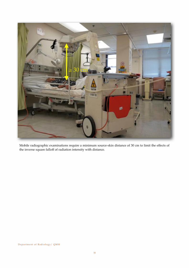
Mobile radiographic examinations require a minimum source-skin distance of 30 cm to limit the effects of the inverse square falloff of radiation intensity with distance.
≥ 30 cm
Chapter 7
Local Rules for OT Radiography
7.1 Radiation warning signs should be placed on the doors of the theatre room during fluoroscopy and radiography.
7.2 The mobile C-arm fluoroscopy equipment should only be operated by authorized per-sonnel with valid license for operation of irradiating apparatus.
7.3 Fluoroscopy should not be undertaken if the same information can be obtained by plain radiography.
7.4 Limit fluoroscopy time to the minimum commensurate with good diagnostic results.
7.5 Always use smallest possible collimated field size to reduce patient dose in fluoros-copy, make use of the circular iris collimator if available.
7.6 The operator should keep the X-ray tube at maximum and the image intensifier at a minimum distance from patient.
7.7 In prolonged procedures, the operator should reduce the dose to the irradiated skin by changing the angulation of the beam and fluoroscopy pulse rate, providing that it is ap-propriate and not degrading the image quality.
7.8 Designated dose reduction programs should be applied to paediatric patients. 7.9 Staff should wear lead apron and thyroid shield to reduce absorbed dose. 7.10 Lead glasses could be used to protect the lens of eyes.
7.11 A manually reset, cumulative timing device (5 to 10 min.) which will either sound an alarm, to turn off the fluoroscopy equipment when the total exposure reaches a certain previously determined limit should be used during all fluoroscopic examination.
7.12 The radiographer should ensure that the C-arm of mobile fluoroscopy C-arm equip-ment is positioned with the X-ray tube underneath the patient and keeping the image in-tensifier closer to the patient whenever it is appropriate and possible. When the C-arm is operating in the lateral plane (or near to horizontal), the operators (e.g. surgeons, nurses and other staff who work close to the patient and C-arm) should stand on the image inten-sifier side and keep a distance away from the image intensifier if it is possible and appro-
D e p a r t m e n t o f R a d i o l o g y / Q M H!
17
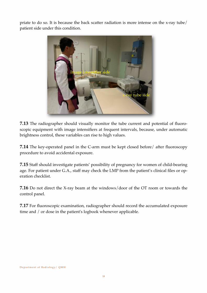
priate to do so. It is because the back scatter radiation is more intense on the x-ray tube/ patient side under this condition.
7.13 The radiographer should visually monitor the tube current and potential of fluoro-scopic equipment with image intensifiers at frequent intervals, because, under automatic brightness control, these variables can rise to high values.
7.14 The key-operated panel in the C-arm must be kept closed before/ after fluoroscopy procedure to avoid accidental exposure.
7.15 Staff should investigate patients’ possibility of pregnancy for women of child-bearing age. For patient under G.A., staff may check the LMP from the patient’s clinical files or op-eration checklist.
7.16 Do not direct the X-ray beam at the windows/door of the OT room or towards the control panel.
7.17 For fluoroscopic examination, radiographer should record the accumulated exposure time and / or dose in the patient's logbook whenever applicable.
D e p a r t m e n t o f R a d i o l o g y / Q M H!
18
x-ray tube side
Image intensifier side
Chapter 8
Local Rules for Fluoroscopy and Angiographic/ Interventional X-ray Rooms
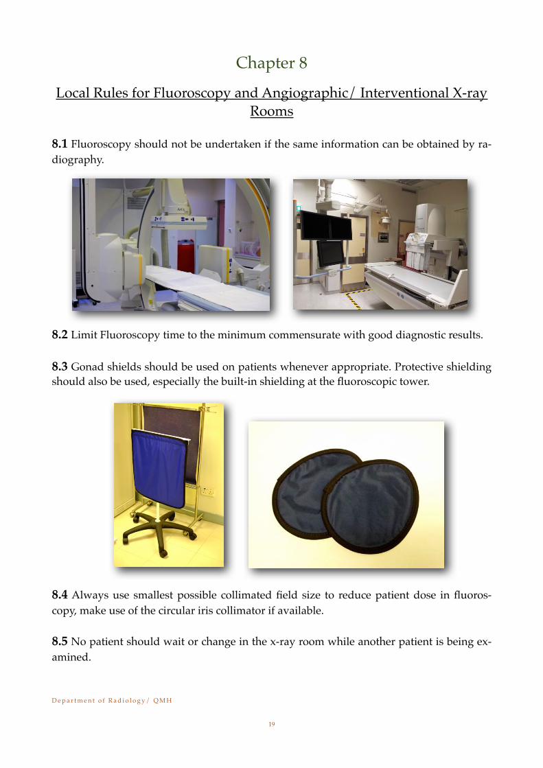
8.1 Fluoroscopy should not be undertaken if the same information can be obtained by ra-diography.
8.2 Limit Fluoroscopy time to the minimum commensurate with good diagnostic results.
8.3 Gonad shields should be used on patients whenever appropriate. Protective shielding should also be used, especially the built-in shielding at the fluoroscopic tower.
8.4 Always use smallest possible collimated field size to reduce patient dose in fluoros-copy, make use of the circular iris collimator if available.
8.5 No patient should wait or change in the x-ray room while another patient is being ex-amined.
D e p a r t m e n t o f R a d i o l o g y / Q M H!
19
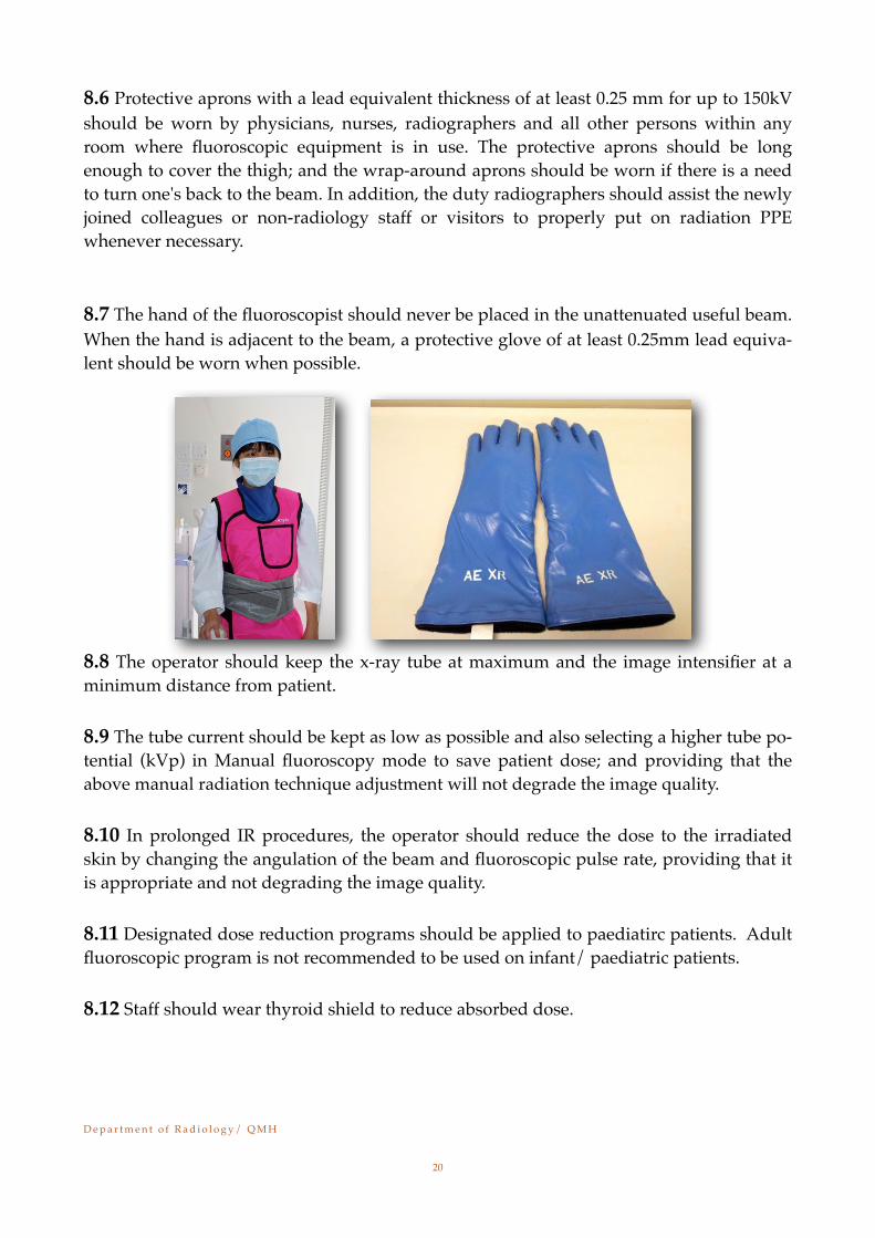
8.6 Protective aprons with a lead equivalent thickness of at least 0.25 mm for up to 150kV should be worn by physicians, nurses, radiographers and all other persons within any room where fluoroscopic equipment is in use. The protective aprons should be long enough to cover the thigh; and the wrap-around aprons should be worn if there is a need to turn one's back to the beam. In addition, the duty radiographers should assist the newly joined colleagues or non-radiology staff or visitors to properly put on radiation PPE whenever necessary.
8.7 The hand of the fluoroscopist should never be placed in the unattenuated useful beam. When the hand is adjacent to the beam, a protective glove of at least 0.25mm lead equiva-lent should be worn when possible.
8.8 The operator should keep the x-ray tube at maximum and the image intensifier at a minimum distance from patient.
8.9 The tube current should be kept as low as possible and also selecting a higher tube po-tential (kVp) in Manual fluoroscopy mode to save patient dose; and providing that the above manual radiation technique adjustment will not degrade the image quality.
8.10 In prolonged IR procedures, the operator should reduce the dose to the irradiated skin by changing the angulation of the beam and fluoroscopic pulse rate, providing that it is appropriate and not degrading the image quality.
8.11 Designated dose reduction programs should be applied to paediatirc patients. Adult fluoroscopic program is not recommended to be used on infant/ paediatric patients.
8.12 Staff should wear thyroid shield to reduce absorbed dose.
D e p a r t m e n t o f R a d i o l o g y / Q M H!
20
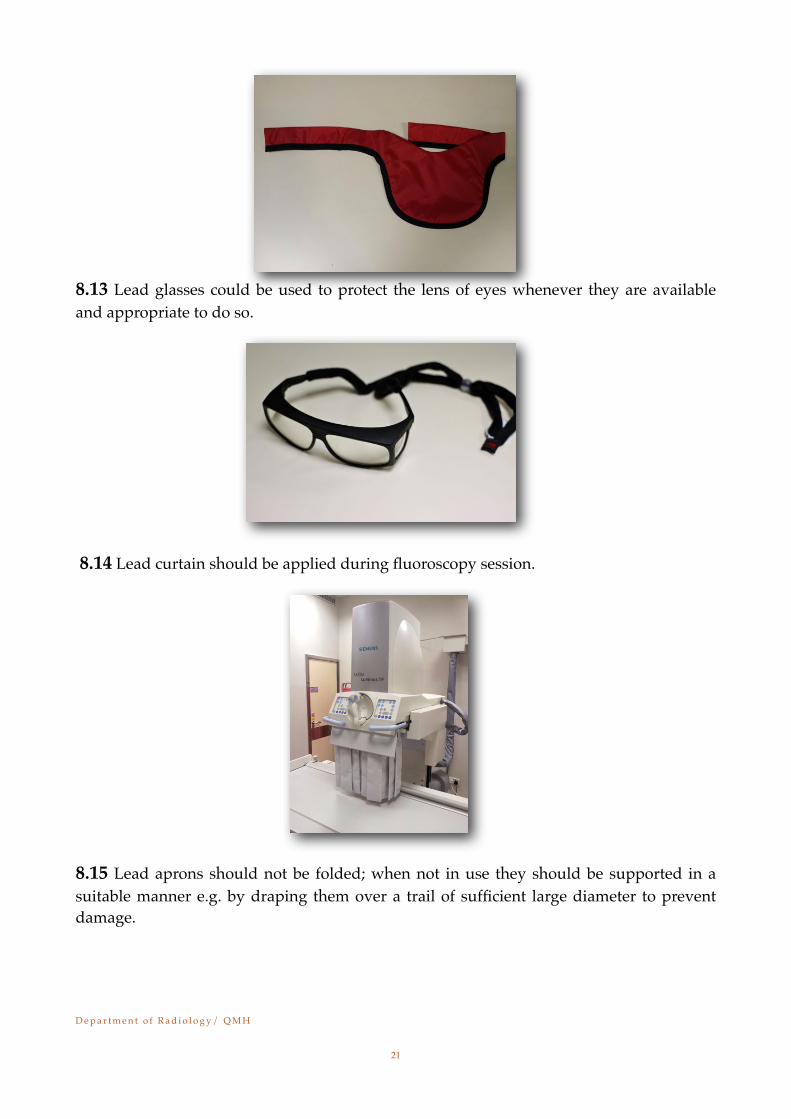
8.13 Lead glasses could be used to protect the lens of eyes whenever they are available and appropriate to do so.
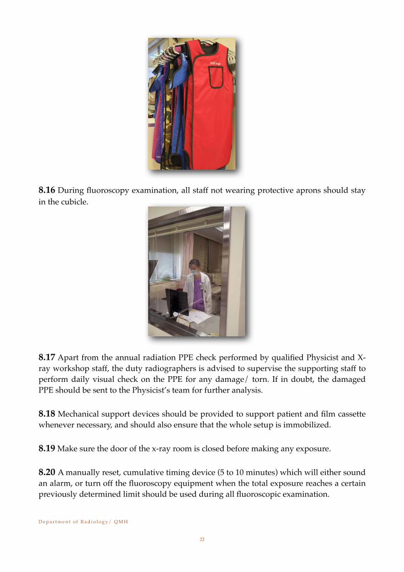
8.14 Lead curtain should be applied during fluoroscopy session.
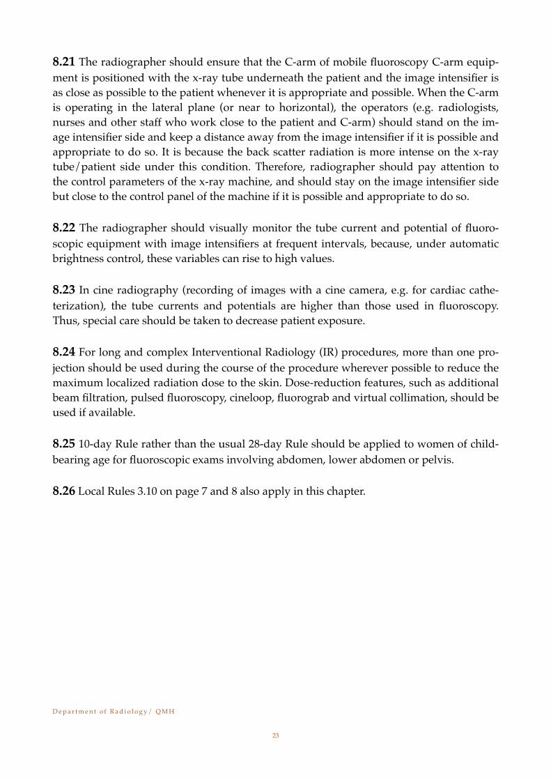
8.15 Lead aprons should not be folded; when not in use they should be supported in a suitable manner e.g. by draping them over a trail of sufficient large diameter to prevent damage.
D e p a r t m e n t o f R a d i o l o g y / Q M H!
21
8.16 During fluoroscopy examination, all staff not wearing protective aprons should stay in the cubicle.
8.17 Apart from the annual radiation PPE check performed by qualified Physicist and X-ray workshop staff, the duty radiographers is advised to supervise the supporting staff to perform daily visual check on the PPE for any damage/ torn. If in doubt, the damaged PPE should be sent to the Physicist’s team for further analysis.
8.18 Mechanical support devices should be provided to support patient and film cassette whenever necessary, and should also ensure that the whole setup is immobilized.
8.19 Make sure the door of the x-ray room is closed before making any exposure.
8.20 A manually reset, cumulative timing device (5 to 10 minutes) which will either sound an alarm, or turn off the fluoroscopy equipment when the total exposure reaches a certain previously determined limit should be used during all fluoroscopic examination.
D e p a r t m e n t o f R a d i o l o g y / Q M H!
22
8.21 The radiographer should ensure that the C-arm of mobile fluoroscopy C-arm equip-ment is positioned with the x-ray tube underneath the patient and the image intensifier is as close as possible to the patient whenever it is appropriate and possible. When the C-arm is operating in the lateral plane (or near to horizontal), the operators (e.g. radiologists, nurses and other staff who work close to the patient and C-arm) should stand on the im-age intensifier side and keep a distance away from the image intensifier if it is possible and appropriate to do so. It is because the back scatter radiation is more intense on the x-ray tube/patient side under this condition. Therefore, radiographer should pay attention to the control parameters of the x-ray machine, and should stay on the image intensifier side but close to the control panel of the machine if it is possible and appropriate to do so.
8.22 The radiographer should visually monitor the tube current and potential of fluoro-scopic equipment with image intensifiers at frequent intervals, because, under automatic brightness control, these variables can rise to high values.
8.23 In cine radiography (recording of images with a cine camera, e.g. for cardiac cathe-terization), the tube currents and potentials are higher than those used in fluoroscopy. Thus, special care should be taken to decrease patient exposure.
8.24 For long and complex Interventional Radiology (IR) procedures, more than one pro-jection should be used during the course of the procedure wherever possible to reduce the maximum localized radiation dose to the skin. Dose-reduction features, such as additional beam filtration, pulsed fluoroscopy, cineloop, fluorograb and virtual collimation, should be used if available.
8.25 10-day Rule rather than the usual 28-day Rule should be applied to women of child-bearing age for fluoroscopic exams involving abdomen, lower abdomen or pelvis.
8.26 Local Rules 3.10 on page 7 and 8 also apply in this chapter.
D e p a r t m e n t o f R a d i o l o g y / Q M H!
23

Chapter 9
Local Rules for CT Rooms
9.1 During "warm up" and detector calibration procedures, persons should not be allowed to enter or remain in the examination room. Make sure the doors to examination room are closed or locked to prevent unauthorized entry.
9.2 Protection should be provided for those who stay in the CT room during clinical pro-cedure.
9.3 Operator should remain at the control panel when high voltage is applied to the x-ray tube whenever the CT examination is in progress.
9.4 In view of the potential for high patient doses, CT examinations should only be carried out after there has been proper clinical justification for the examination of each individual patient. Examinations on children require a different level of justification to that for adults, since such patients are at greater risk from radiation than are adults. Always apply the paediatric protocols when imaging paediatric patients, for which the scan and techni-cal parameters are tailored to the age and size of the patients, the body region of interest and the clinical question. CT examinations should not be performed on the abdomen or pelvis of pregnant patients without overriding clinical indications and particular attention to low dose techniques. When clinically appropriate, the alternative use of safer non-ionising techniques (such as ultrasound and magnetic resonance imaging (MRI) where available) or of low dose X-ray techniques should be considered.
D e p a r t m e n t o f R a d i o l o g y / Q M H!
24
9.5 Operators should ensure that the minimum number of CT slices necessary to obtain the required diagnostic information are acquired. This is particularly important when scanning contiguous volumes using pre-set scanning protocols.
9.6 The parameters of standard scanning protocols should be modified according to the size of the particular patient to deliver the lowest dose consistent with the required image quality.
9.7 Slice increment (axial scanning) or pitch (helical scanning) together with beam collima-tion should be chosen with regard to the z-axis sensitively (imaged slice width) and low contrast detectability required, whilst maintaining the lowest practicable patient dose.
9.8 Care should be taken to minimize exposure to the eyes of the patient. Dose to the lens tissue can often be substantially reduced by angulation of the gantry to exclude the eyes from the primary beam during head examinations.
9.9 The necessity for the use of contrast agents should be assessed to reduce the number of regions that are rescanned with contrast.
9.10 Under no circumstance should the X-ray tube be energised for CT fluoroscopy when the person carrying out the examination is not looking at the monitor.
9.11 Physicians and radiologists should ensure that patients are not irradiated unjustifia-bly.
9.12 With reference to [1]HA Clinical Guidelines, continuous monitoring of the patient with implantable cardiac devices like pacemakers and implantable cardioverter-defibrillator (ICD) by pulse oximetry during the scan is necessary when the device is in or immediately adjacent to the scan ranges. If the device will be in the CT scanning beam for more than 4 seconds continuously (e.g. in CT-guided biopsy/perfusion CT in the vicinity of the implantable device), pre- and post- CT scanning programming should be per-formed.
9.13 10-day Rule rather than the usual 28-day Rule should be applied to women of child-bearing age for CT exams involving abdomen, lower abdomen and/or pelvis.
9.14 Local Rules 3.10 on page 7 and 8 also apply in this chapter. Reference
[1] Clinical Recommendation on Possible Malfunction of Implantable Cardiac Devices Caused by Computed Tomo-graphy (CT) Scanning. 10 May 2013
D e p a r t m e n t o f R a d i o l o g y / Q M H!
25

Chapter 10
Local Rules for Mammography
10.1 No examination should be undertaken without an authorized request for the investi-gation.
10.2 All staff must stand behind the protective lead screens during exposures.
10.3 ALARA principle shall be observed when performing mammographic examination.
10.4 For patients of child-bearing age, radiographer must check her L.M.P. (28-day rule to be applied) and/or sign the consent form before the mammographic examination. If preg-nancy of patient is confirmed, mammography should be cancelled or proceeded with modifications after the duty radiologist had duly discussed with the referring physician. In case the examination is decided to be proceeded, consent form must be signed by the concerned patient. Proper radiation protection e.g. abdominal shield and thyroid shield should be applied whenever applicable.
10.5 For mammography screening program, examination is performed on asymptomatic patients aged ≧40 while for diagnostic imaging mammography is performed on patients age ≧35.
10.6 With reference to the annual screening protocol, the clerical staff would check the date(s) of previous mammogram appointments if any and recorded this piece of informa-tion on the mammogram request form. Based on the information, the radiographer would then issue the appropriate mammogram appointment date to the patient.
10.7 For mammographic examination on young patient who is aged ≦35, ultrasound breast examination is usually performed first. Half set of mammogram would only be added if diagnostically indicated.
10.8 Thorough explanation about the mammographic examination including the use of compression and the positioning technique to the patient is essential to facilitate the pro-duction of high quality images with reasonably low radiation dose and to avoid unneces-sary repeat.
10.9 Quality Assurance program should be in place to ensure that the mammographic sys-tems are regularly tested to achieve optimal performance. The test results should be well documented, monitored and evaluated by appropriate personnel. Equipment faults should be properly recorded.
D e p a r t m e n t o f R a d i o l o g y / Q M H!
26
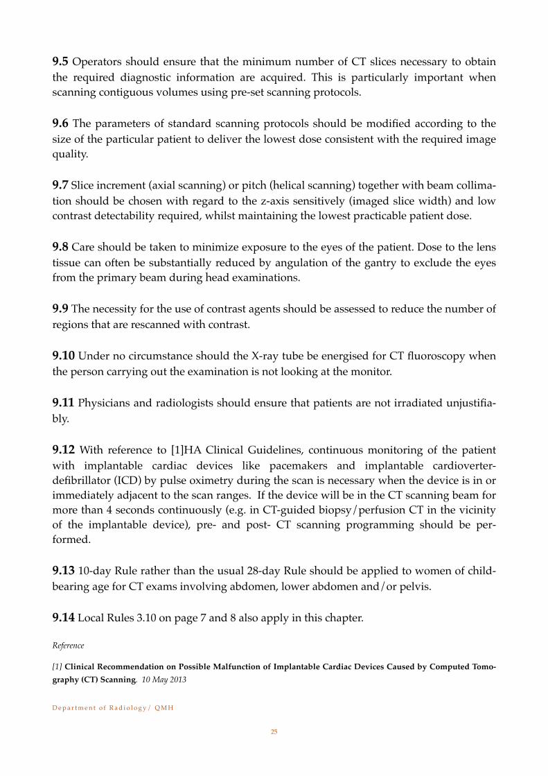
10.10 Reject analysis should be conducted as part of Quality Assurance program.
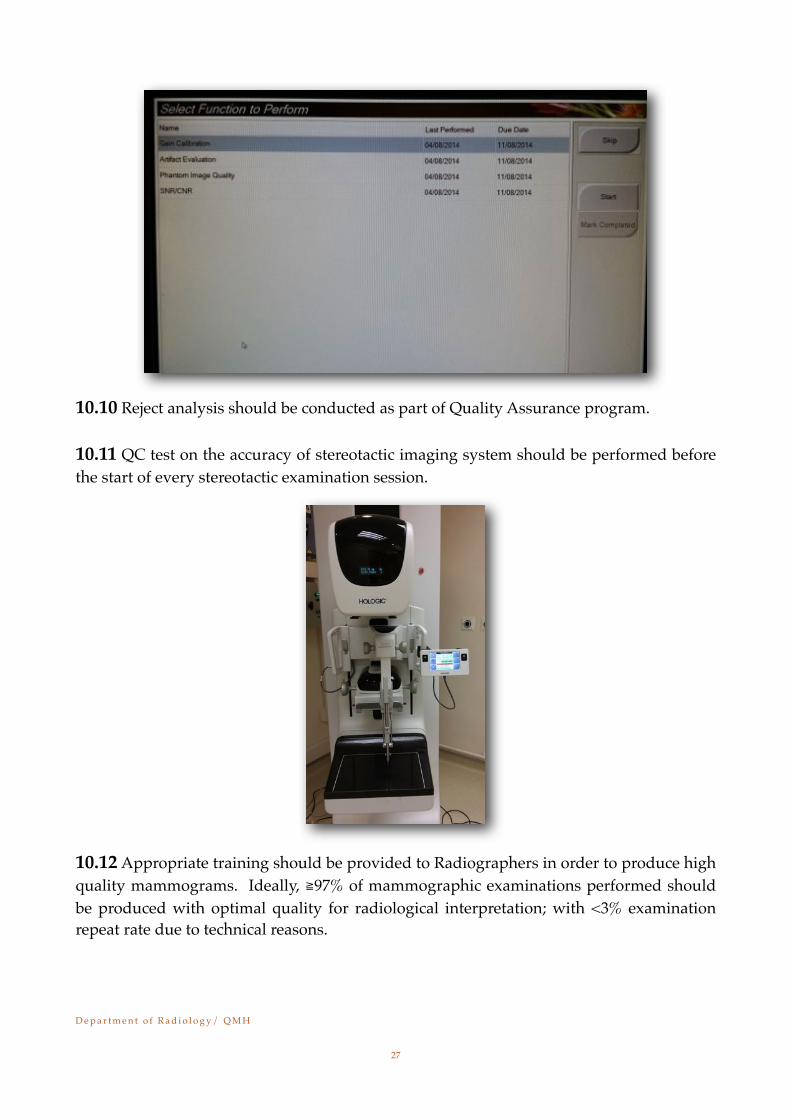
10.11 QC test on the accuracy of stereotactic imaging system should be performed before the start of every stereotactic examination session.
10.12 Appropriate training should be provided to Radiographers in order to produce high quality mammograms. Ideally, ≧97% of mammographic examinations performed should be produced with optimal quality for radiological interpretation; with <3% examination repeat rate due to technical reasons.
D e p a r t m e n t o f R a d i o l o g y / Q M H!
27
Chapter 11
The roles of personnel on radiation safety
11.1 Cluster Radiation Protection Adviser (RPA)a)Advise the CCE, HCE and COS/Head of Unit on radiation safety and protection.b)Advise the CCE, HCE and COS/Head of Unit on matters related to the compliance
with the Code of Practice. c)To conduct, as and when requested by the CCE, HCE or COS/Head of Unit, inves-
tigations on incidents involving over-exposure and other matters related to radia-tion safety and protection.
11.2 COS/ Head of Unit (Local Controlling Authority)a) Responsible for the radiation safety of the staff, patients and members of the pub-
lic in the department.b)Responsible for matters related to the compliance with the Code of Practice in his/
her department/unit.c)To arrange for medical surveillance and radiation monitoring of staff.d)To arrange for all relevant records to be kept.e)In consultation with the RPA, appoint suitable RPS.f) To notify the RPS as soon as a pregnancy has been declared in a member of the staff
so that necessary precautions can be taken to ensure that the fetal dose is kept be-low the relevant limit.
g)To obtain the necessary approval and licences from the relevant authorities.h)To report to the HCE, RPA and Committee on Radiation Safety on incidents involv-
ing over-exposure, new radiation work, change of the nature of radiation work and other matters which may affect the radiation safety of the department/unit.
11.3 Radiation Protection Supervisor (RPS)a)To draw up local radiological protection rules.b)To see that the instructions and requirements of the Code of Practice and Local
Rules are observed in his/her department/unit/section.c)To see that any personal monitoring devices issued are used in the correct manner.d)To instruct departmental staff on safe working practices and any system of work in
force in his/her department/unit/section.e)To establish and maintain operational procedures which ensure that staff and pa-
tient exposures are kept as low as reasonably practicable.f) To ensure that there is adequate protection to cover all changes in procedure, new
procedures and new equipment.g)To report to the COS/Head of Unit and, if necessary, the RPA any incident such as
infringement of Local Rules, a suspected over-exposure, equipment malfunction involving radiation hazard, etc.
D e p a r t m e n t o f R a d i o l o g y / Q M H!
28
h)To report to the RPA via the COS/Head of Unit any new procedures and/or iso-topes being used which may have radiation safety implications, and any deteriora-tion in the state of protection in his/her department/unit/section.
i) Maintain adequate supplies of protective equipment.j) Keep records of incoming and outgoing radioactive sources.k)Supervise storage and disposal of radioactive wastes and keep records.l) Handle declaration forms.m)Monitor the working areas.n)Assist the COS/Head of Unit or the RPA in carrying out investigation on incidents
involving over-exposure and on other matters related to radiation safety and pro-tection.
11.4 Staffa)Should take care not to expose himself/herself or any other person to ionizing ra-
diation to an extent greater than is necessary for the purposes of his/her work, and exercise reasonable care while carrying out such work.
b)Should make full and proper use of any personal protective equipment provided.c)Should follow the procedures as laid down in the Local Rules, and consult the RPS
in case of doubt.d)When any female staff is confirmed to be pregnant and need to work around the
radiation controlled area, her supervisor should be informed as soon as possible. Precautionary measures should be taken to minimize the occupational exposure in consideration of the strict dose limit of fetus. Change of duty at non-radiation area may be considered during the pregnancy of staff.
D e p a r t m e n t o f R a d i o l o g y / Q M H!
29
Chapter 12!Contingency Plans!!
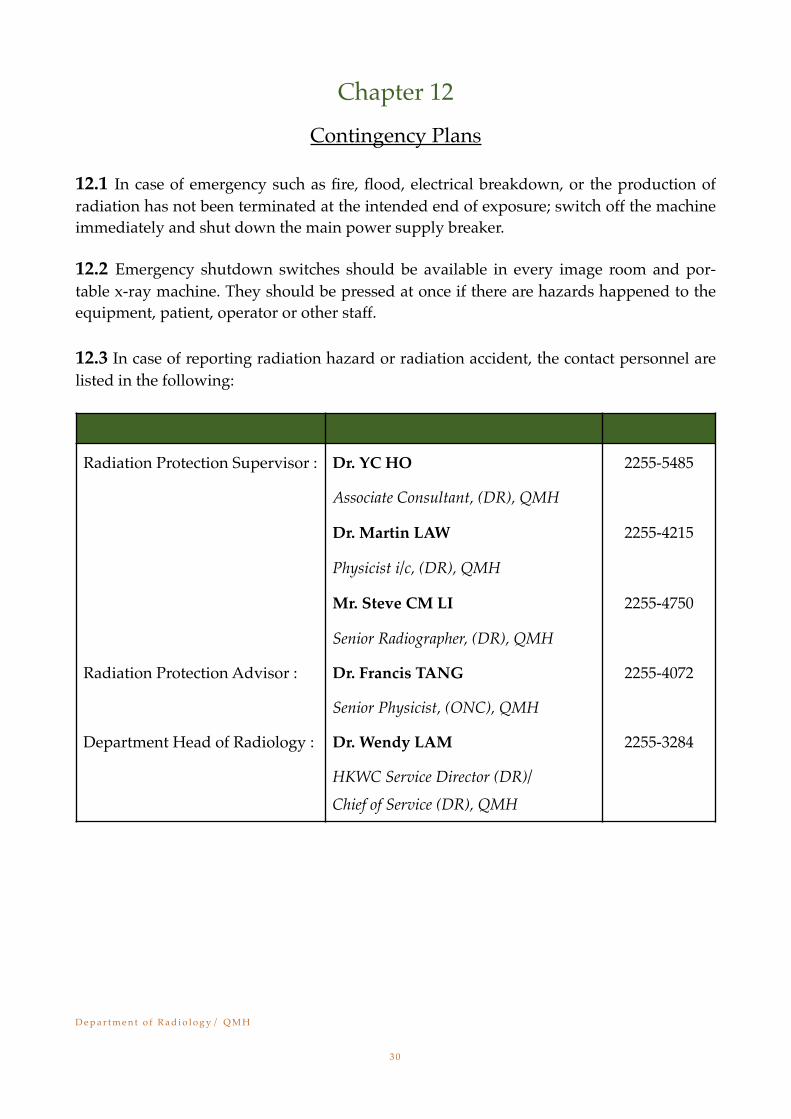
12.1 In case of emergency such as fire, flood, electrical breakdown, or the production of radiation has not been terminated at the intended end of exposure; switch off the machine immediately and shut down the main power supply breaker.!!12.2 Emergency shutdown switches should be available in every image room and por-table x-ray machine. They should be pressed at once if there are hazards happened to the equipment, patient, operator or other staff.!!12.3 In case of reporting radiation hazard or radiation accident, the contact personnel are listed in the following:!!
! !!!
Radiation Protection Supervisor : Dr. YC HO 2255-5485
Associate Consultant, (DR), QMH
Dr. Martin LAW 2255-4215
Physicist i/c, (DR), QMH
Mr. Steve CM LI 2255-4750
Senior Radiographer, (DR), QMH
Radiation Protection Advisor : Dr. Francis TANG 2255-4072
Senior Physicist, (ONC), QMH
Department Head of Radiology : Dr. Wendy LAM 2255-3284
HKWC Service Director (DR)/ Chief of Service (DR), QMH
D e p a r t m e n t o f R a d i o l o g y / Q M H ! ! !"3 0
Appendix I
Radiation Safety for Performing Radioembolization in Interventional Radiology Facility in DR/QMH
In addition to radiation safety to routine interventional procedures using X-ray, radiation safety procedures for radioembolization to liver cancer patients using unsealed source of 99mTc-MAA and 90Y-microspheres have been implemented for every patient session using these radiation sources. The aim is to minimize the possibility of radiation contamination and to summarize the procedures to handle radiation contamination if contamination does occur (Appendix A). Because of the significantly higher radioactivity of 90Y-microspheres used in the actual implant, a procedures list, including radiation protection setup, has been followed as a guidelines (Appendix B). Some safety measures to minimize the exposure from the unsealed radiation source to interventional radiologists are also implemented ac-cording to the ALARA principle.
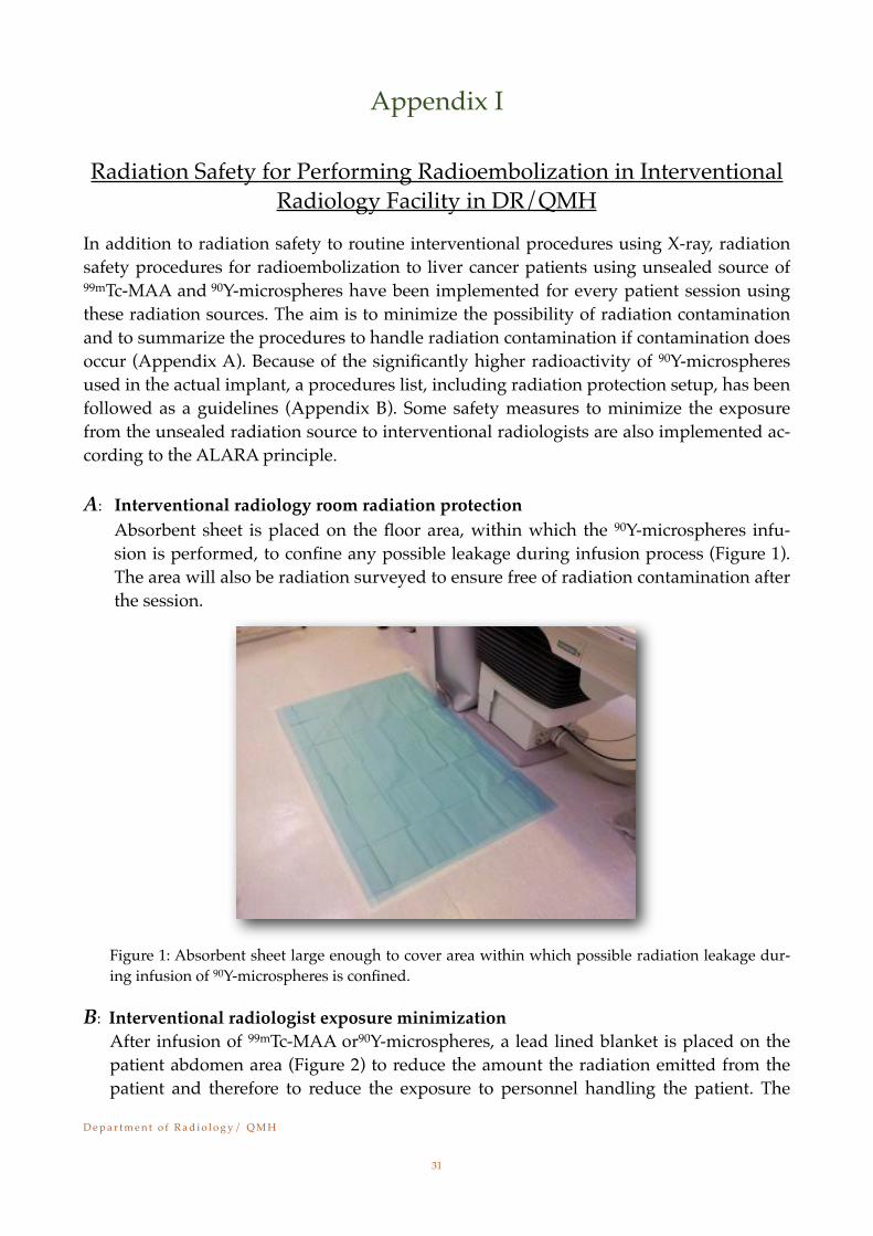
A:! Interventional radiology room radiation protectionAbsorbent sheet is placed on the floor area, within which the 90Y-microspheres infu-sion is performed, to confine any possible leakage during infusion process (Figure 1). The area will also be radiation surveyed to ensure free of radiation contamination after the session.
Figure 1: Absorbent sheet large enough to cover area within which possible radiation leakage dur-ing infusion of 90Y-microspheres is confined.
B: Interventional radiologist exposure minimizationAfter infusion of 99mTc-MAA or90Y-microspheres, a lead lined blanket is placed on the patient abdomen area (Figure 2) to reduce the amount the radiation emitted from the patient and therefore to reduce the exposure to personnel handling the patient. The
D e p a r t m e n t o f R a d i o l o g y / Q M H!
31
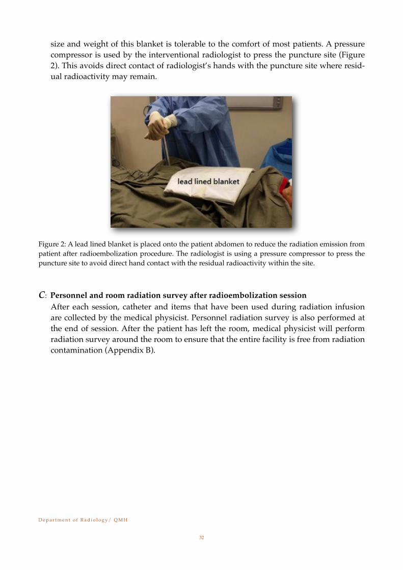
size and weight of this blanket is tolerable to the comfort of most patients. A pressure compressor is used by the interventional radiologist to press the puncture site (Figure 2). This avoids direct contact of radiologist’s hands with the puncture site where resid-ual radioactivity may remain.
Figure 2: A lead lined blanket is placed onto the patient abdomen to reduce the radiation emission from patient after radioembolization procedure. The radiologist is using a pressure compressor to press the puncture site to avoid direct hand contact with the residual radioactivity within the site.
C: Personnel and room radiation survey after radioembolization session! After each session, catheter and items that have been used during radiation infusion
are collected by the medical physicist. Personnel radiation survey is also performed at the end of session. After the patient has left the room, medical physicist will perform radiation survey around the room to ensure that the entire facility is free from radiation contamination (Appendix B).
D e p a r t m e n t o f R a d i o l o g y / Q M H!
32

Supplement A:Procedures to perform minor radiation decontamination in Interven-
tional Radiology/DR/QMH
A:! Decontamination materials available (Figure A1)
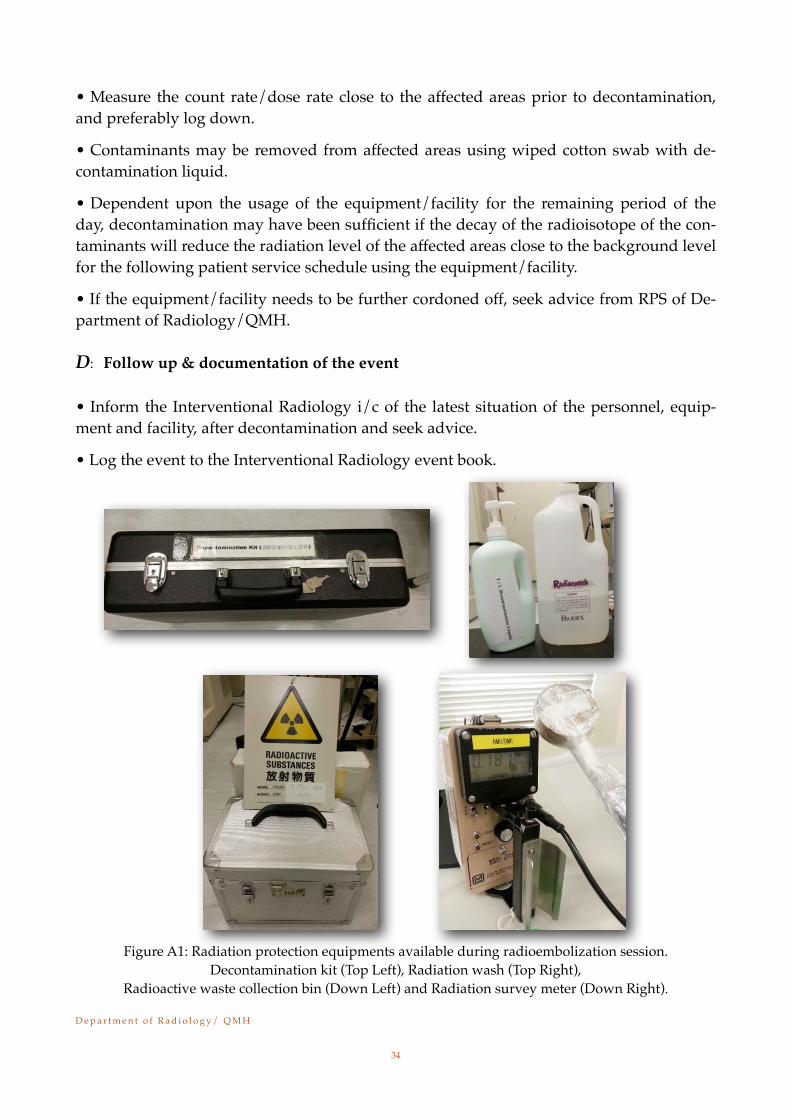
• In every radioembolization session, decontamination kit, radiation survey meter and waste collection bins are available at the vicinity of the Interventional Radiology room.
B:! Personnel (staff or patient) decontamination
• Gloves and overshoes must be put on before dealing with the spill.
• Radiation survey the personnel from head to toes (both anterior and posterior) with the radiation survey meter. If the patient is injected with radiopharmaceutical, wiped samples from the patient affected areas are collected and radiation surveyed to determine if patient is radiation contaminated. The meter readings from these wiped samples should be com-pared with background radiation level to know the degree of contamination.
• If contamination is on hands and/or feet, moist cotton swab with radiation wash may be used to wipe off majority of the contaminants. This procedure may be repeated from outer to inner affected areas to reduce the contamination level to an acceptable level (about 2 to 3 times of background radiation level for contaminants of 99mTc isotope), before washing thoroughly with soap and tap water. If water sink basin is available nearby, running tap water to the affected areas is a good alternative (taking care to avoid spread of contamina-tion and water splashing to the eyes).
• If the personnel is suspected of internal contamination, wiped samples from ears, nos-trils and mouth are collected for counting (and/or isotope identification) analysis. Consult medical physicist team for radioisotope identification and the quantity of contamination.
• If radiation contamination is on clothing, clean clothes are changed and any contami-nated clothing and personal articles should be put in plastic bag for radiation survey. Take note of the personnel privacy.
• If it is determined that the personnel has to take a shower for body decontamination, showering facility for decontamination use is available in NMU at 3rd Level, Cancer Center/QMH.
• All contaminated wastes are collected to store in NMU/DR/QMH. The items must be surveyed for residual activity prior to release by Radiation Protection Supervisor (RPS) or his delegates.
C:! Equipment/facility decontamination
• Locate the contamination spot(s). Dry the affected areas with absorbing paper. D e p a r t m e n t o f R a d i o l o g y / Q M H!
33
• Measure the count rate/dose rate close to the affected areas prior to decontamination, and preferably log down.
• Contaminants may be removed from affected areas using wiped cotton swab with de-contamination liquid.
• Dependent upon the usage of the equipment/facility for the remaining period of the day, decontamination may have been sufficient if the decay of the radioisotope of the con-taminants will reduce the radiation level of the affected areas close to the background level for the following patient service schedule using the equipment/facility.
• If the equipment/facility needs to be further cordoned off, seek advice from RPS of De-partment of Radiology/QMH.
D:! Follow up & documentation of the event
• Inform the Interventional Radiology i/c of the latest situation of the personnel, equip-ment and facility, after decontamination and seek advice.
• Log the event to the Interventional Radiology event book.
Figure A1: Radiation protection equipments available during radioembolization session. Decontamination kit (Top Left), Radiation wash (Top Right),
Radioactive waste collection bin (Down Left) and Radiation survey meter (Down Right).
D e p a r t m e n t o f R a d i o l o g y / Q M H!
34
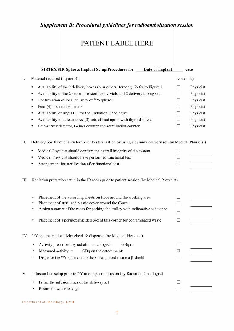
Supplement B: Procedural guidelines for radioembolization session
SIRTEX SIR-Spheres Implant Setup/Procedures for Date-of-implant case
I. Material required (Figure B1)Material required (Figure B1) Done by
Availability of the 2 delivery boxes (plus others: forceps). Refer to Figure 1 Physicist Availability of the 2 sets of pre-sterilized v-vials and 2 delivery tubing sets Physicist Confirmation of local delivery of 90Y-spheres Physicist Four (4) pocket dosimeters Physicist Availability of ring TLD for the Radiation Oncologist Physicist Availability of at least three (3) sets of lead apron with thyroid shields Physicist Beta-survey detector, Geiger counter and scintillation counter Physicist
II. Delivery box functionality test prior to sterilization by using a dummy delivery set (by Medical Physicist)Delivery box functionality test prior to sterilization by using a dummy delivery set (by Medical Physicist)Delivery box functionality test prior to sterilization by using a dummy delivery set (by Medical Physicist)Delivery box functionality test prior to sterilization by using a dummy delivery set (by Medical Physicist)
Medical Physicist should confirm the overall integrity of the system
Medical Physicist should have performed functional test
Arrangement for sterilization after functional test
III. Radiation protection setup in the IR room prior to patient session (by Medical Physicist)Radiation protection setup in the IR room prior to patient session (by Medical Physicist)Radiation protection setup in the IR room prior to patient session (by Medical Physicist)Radiation protection setup in the IR room prior to patient session (by Medical Physicist)
Placement of the absorbing sheets on floor around the working area
Placement of sterilized plastic cover around the C-arm
Assign a corner of the room for parking the trolley with radioactive substance
Placement of a perspex shielded box at this corner for contaminated waste
IV. 90Y-spheres radioactivity check & dispense (by Medical Physicist)90Y-spheres radioactivity check & dispense (by Medical Physicist)90Y-spheres radioactivity check & dispense (by Medical Physicist)90Y-spheres radioactivity check & dispense (by Medical Physicist)
Activity prescribed by radiation oncologist = GBq on
Measured activity = GBq on the date/time of:
Dispense the 90Y-spheres into the v-vial placed inside a β-shield
V. Infusion line setup prior to 90Y-microsphere infusion (by Radiation Oncologist) Infusion line setup prior to 90Y-microsphere infusion (by Radiation Oncologist)
Prime the infusion lines of the delivery set
Ensure no water leakage
D e p a r t m e n t o f R a d i o l o g y / Q M H!
35
PATIENT LABEL HERE
VI. Patient implant setup (by Radiation Oncologist and possibly assisted by Medical Physicist)Patient implant setup (by Radiation Oncologist and possibly assisted by Medical Physicist)Patient implant setup (by Radiation Oncologist and possibly assisted by Medical Physicist)Patient implant setup (by Radiation Oncologist and possibly assisted by Medical Physicist)
Ensure patient site no occlusion after connection to patient catheter
V-vial containing 90Y-spheres securely placed inside the delivery box
Insert the C-line and D-line needle into the v-vial
Manually perform infusion
Disconnect the patient catheter after implant completion
VII. Radiation monitoring after implant by Medical PhysicistRadiation monitoring after implant by Medical Physicist
Place the patient catheter into the delivery box after catheter removal
Radiation monitoring for every personnel involved in patient administration. This should be carried out outside the IR room with the use beta survey detector
Place all items used in the implant into the Perspex waste box
Take the delivery box and perspex box back to Hot-Lab of COD for decay
Physicist should label the container with the name of isotope & date
Physicist should radiation monitor the IR room after patient leave the room
Collect and record all pocket dosimeters and/or ring TLDs, if any.
VIII. Post-therapy issues Post-therapy issues
Instruction of radiation protection issues for patient in hospital and at home
Appointment for post-therapy nuclear medicine scan if needed
Room survey and collection of contaminated items after patient discharge from hospital
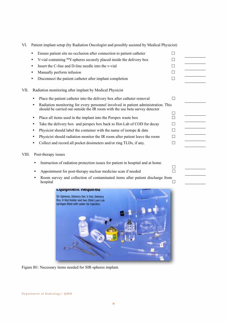
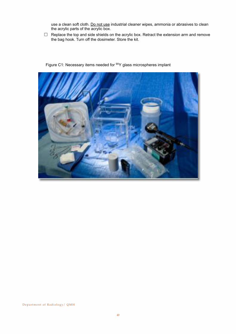
Figure B1: Necessary items needed for SIR-spheres implant.
D e p a r t m e n t o f R a d i o l o g y / Q M H!
36
Supplement C: Procedural guidelines for radioembolization session: Resin micro-spheres
Patient Name Number
Date of Treatment
TheraSphere® Lot Number Labeled Quantity (GBq)
Catheter Information: (Place catheter sticker in the space provided below)
1. Items Required for TheraSphere® Administration (Figure C1):
" Patient prescription for TheraSphere® (signed Written Directive) " Ionization survey meter " Geiger-Mueller (GM) contamination meter " Spill kit " A floor drape applied under the cart in the angiography suite " A sterile drape placed on the cart
Place the following items on the draped cart:
2. Administration Set Priming
" Open the Administration Set packaging and remove the Administration Set and 20 mL empty vial.
" Insert the white non-vented spike (CLEAR CAP) into the saline bag. Hang the saline bag on the hook.
" Insert the white vented spike (BLUE CAP) into the empty 20 mL vial. " Remove the (RED RUBBER) shield cap from the needle injector assembly. Place the needle
injector assembly on a sterile surface. " If the 20 mL syringe is marked ‘VacLok’, turn the syringe plunger fully clockwise to ensure it
is unlocked. (This step may not be required for alternate syringe.) " Slowly fill and discharge the syringe to remove air from the Administration Set tubing and
syringe. Continue priming vigorously with full pressure until there are no bubbles in the lines and there are continuous streams of saline flowing out of both needle holes in the needle injector assembly.
" Fill the syringe when priming is complete.
3. Dose Vial Preparation " Lift the TheraSphere® dose vial in its lead pot and tilt the lead pot back and forth to 90 de-
grees to wet any microspheres on the vial septum. Tap the bottom of the lead pot firmly on a hard surface. Place the lead pot into the pot holder in the acrylic box base.
" Remove the lead pot lid and place it upside down on a non-sterile surface.
D e p a r t m e n t o f R a d i o l o g y / Q M H!
37

" Use a hemostat to remove the purple seal from the top of the dose vial acrylic shield. Dis-card the seal in the Nalgene waste container.
" Use a sterile adhesive strip to remove the dose vial acrylic shield plug. Discard the plug and sterile adhesive strip in the Nalgene waste container.
" Use an alcohol swab and a hemostat to swab the dose vial septum. Discard the swab in the Nalgene waste container.
" Record the dosimeter initial reading from the dose vial (mR/h).
Dose Vial (mR/h)
" Measure and record the initial radiation field for the patient, using an ionization survey meter.
4. Final Assembly
" Close the pinch clamp on the outlet tubing near label ‘E.’ " Place the empty 20 mL vial in the holder on the acrylic box and push the relief valve tube
into gripper clip ‘A.’ " Insert the needle injector assembly into the acrylic dose vial shield. Press on the GREEN
cap to lock it in place. You will hear or feel a click or snap. " Place the inlet tubing through slot ‘B’ in the acrylic box. Place the outlet tubing through slot
‘D’ in the acrylic box. Loop the tubing around the side and place the fitting into the holder at ‘C.’
" Clamp the priming line at label ‘C’ with a hemostat (or equivalent). " Push the YELLOW tabs on the needle injector assembly all the way down, locking the nee-
dles into the dose vial. You will hear or feel a click or snap at the bottom of travel. " Ensure that the side shield is installed on the acrylic box. Place the top shield on the acrylic
box with the sloped shield towards slot ‘D.’ Ensure that the tubing is not pinched or kinked. " Move the cart close to the patient. Lower the bed to lowest position. " Place a sterile towel under the extension arm holder ‘E,’ and under holder ‘C.’ " Place a sterile towel across the gap between the acrylic box and the patient. " The Interventional Radiologist (IR) will flush the infusion catheter to ensure flow. Replace the
infusion catheter if it is damaged or does not have satisfactory flow. Do not use a catheter extension or extra fittings. Replace the catheter if it is too short.
" Disconnect the outlet tubing labeled ‘E’ from the priming tubing at holder ‘C.’ Firmly connect the outlet tubing ‘E’ to the catheter.
" Place the catheter connection into the slotted holder ‘E’ at the end of the extended arm. Outlet tubing ‘E’ must be above the holder, with the infusion catheter hanging vertically be-low.
" The IR will verify the infusion catheter position. Release the pinch clamp from the outlet tub-ing. Dents in the tubing may be reduced by rolling outlet tubing with fingers.
5. TheraSphere® Administration
ATTENTION: Beta radiation fields can be very high during microsphere transfer. Stand behind beta shielding OR MAINTAIN DISTANCE.
" Record the starting time of the administration: " Infuse TheraSphere® Y-90 glass microspheres using steady pressure on the syringe plunger.
Infuse continuously until syringe is empty (≥ 20 cc per minute).
NOTE: If the infusion pressure is over 30 psi, excess fluid will drip into the vented 20 mL vial. If this occurs, reduce the pressure being applied on syringe until no flow is seen going
D e p a r t m e n t o f R a d i o l o g y / Q M H!
38
into the vented vial. If the syringe flow is <20 cc per minute (i.e. appropriate to the flow of the native vessel), this may decrease the delivery efficiency of the administration system and result in higher residual waste.
" Observe the outlet line and catheter for proper operation. If a problem is observed, inform the team and take corrective action.
" Re-fill syringe for subsequent flushes by pulling back the syringe plunger. A minimum of 3 flushes (60 cc total) are recommended. Continue flushes until the desired dosimeter reading is achieved.
" Record the number of flushes completed:
" Record the time that administration was completed: " Record the dosimeter final reading:
Dose Vial (mR/h)
" Measure and record the final radiation field for the patient using an ionization survey meter.
6. Disassembly
" Cut the inlet line at indicated position. " Remove the acrylic box top shield and side shield. " The IR will remove the infusion catheter from the patient and lift the catheter connection out
of the extended holder ‘E.’ Do not disconnect the catheter from the outlet tubing. Use care to control the tip of the infusion catheter and guide catheter as these may be contaminated with microspheres. Use gauze, a small towel, or hemostat to handle the catheters for radiation protection. Any item that has come in contact with microspheres is considered contami-nated.
" Place all contaminated waste into the Nalgene waste container (in its beta shield), including the following:
• " Infusion and guide catheters with attached tubing and towels/gauze• " Dose vial with attached Needle Injector Assembly • Lift the lead pot and dump out the dose vial• " Contaminated items such as gauze, towels, and gloves
" Cap the Nalgene waste container and place the acrylic lid on the beta shield. Remove for measurements to determine percent delivery and for disposal.
" Use a GM contamination meter to check IR’s hands for contamination. " Survey all staff leaving the room with the GM contamination meter.
7. Cleanup and Waste Disposal
" Use GM contamination meter to check for contamination on the cart, lead pot, equipment, and the areas under the catheter connection and cart.
NOTE: Radiation from fluoroscopy, the patient, and the waste container will affect the ability to detect and measure contamination.
" Decontaminate and/or dispose of items as appropriate. " As required, clean the TheraSphere® acrylic box with water, mild soap and a clean soft cloth.
Alcohol wipes may be used (minimize alcohol contact with glued joints – alcohol degrades the glue over an extended time). Chlorine (bleach) disinfectants are also acceptable. Always
D e p a r t m e n t o f R a d i o l o g y / Q M H!
39
use a clean soft cloth. Do not use industrial cleaner wipes, ammonia or abrasives to clean the acrylic parts of the acrylic box.
" Replace the top and side shields on the acrylic box. Retract the extension arm and remove the bag hook. Turn off the dosimeter. Store the kit.
������������������������� ������ � ������� ���� � ���������������������� � ��������
D e p a r t m e n t o f R a d i o l o g y / Q M H!
40
Appendix II
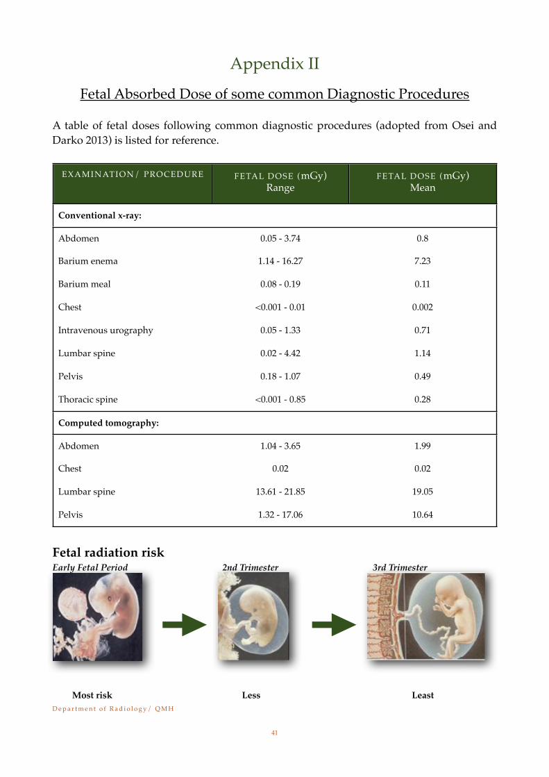
Fetal Absorbed Dose of some common Diagnostic Procedures
A table of fetal doses following common diagnostic procedures (adopted from Osei and Darko 2013) is listed for reference.
EXAMINATION/ PROCEDURE FETAL DOSE (mGy)Range
FETAL DOSE (mGy)Mean
Conventional x-ray:
Abdomen 0.05 - 3.74 0.8
Barium enema 1.14 - 16.27 7.23
Barium meal 0.08 - 0.19 0.11
Chest <0.001 - 0.01 0.002
Intravenous urography 0.05 - 1.33 0.71
Lumbar spine 0.02 - 4.42 1.14
Pelvis 0.18 - 1.07 0.49
Thoracic spine <0.001 - 0.85 0.28
Computed tomography:
Abdomen 1.04 - 3.65 1.99
Chest 0.02 0.02
Lumbar spine 13.61 - 21.85 19.05
Pelvis 1.32 - 17.06 10.64
Fetal radiation risk Early Fetal Period 2nd Trimester 3rd Trimester
D e p a r t m e n t o f R a d i o l o g y / Q M H!
41
Most risk Less Least

Appendix III
Typical effective dose, equivalent periods of natural background radiation and lifetime fatal cancer risks from diagnostic medical
exposures
DIAGNOSTIC
PROCEDURE
TYPICAL EFFEC-
TIVE DOSES (mSv)
EQUIVALENT PE-RIOD OF NATURAL BACKGROUND RA-
DIATION 1
LIFETIME ADDI-TIONAL RISK OF FA-
TAL CANCER PER
EXAMINATION 2
X-ray examinations:
Limbs and joints (except hip) 0.001 [0.002 - 0.1] < 1 days 1 in a 2 million
Teeth (single bitewing) < 1.5 days 1 in a few million
Teeth (panoramic) 1.5 days 1 in 2 million
Chest (single PA film) 0.02 [0.007 - 0.05] 2.5 days 1 in a million
Skull 0.1 [0.03 - 0.22] 12 days 1 in 200,000
Cervical spine (neck) 0.2 [007 - 0.3] 3.5 weeks 1 in 100,000
Hip 0.7 [0.18 - 2.71] 13 weeks 1 in 29,000
Thoracic spine 1 [0.6 - 1.4] 4 months 1 in 20,000
Pelvis 0.6 [0.2 - 1.2] 2.4 months 1 in 33,000
Abdomen 0.7 [0.04 - 1.1] 2.8 months 1 in 29,000
Lumbar spine 1.5 [0.5 - 1.8] 6 months 1 in 14,000
Barium swallow 8 months 1 in 13,000
IVU (kidneys and bladder) 3 [0.7 - 3.7] 12 months 1 in 6700
Barium meal 16 months 1 in 6700
Barium follow 16 months 1 in 6700
Barium enema 8 [2.0 - 18.0] including fluoroscopy
2.7 years 1 in 2500
CT head 2 [0.9 - 4.0] 8 months 1 in 10,000
CT chest 7 [4.0 - 18.0] 2.3 years 1 in 2900
CT abdomen 8 [3.5 - 25] 2.6 years 1 in 2500
CT pelvis 6 [3.3 - 10] 2 years 1 in 3400
D e p a r t m e n t o f R a d i o l o g y / Q M H!
42
DIAGNOSTIC
PROCEDURE
TYPICAL EFFEC-TIVE DOSES (mSv)
EQUIVALENT PE-RIOD OF NATURAL BACKGROUND RA-
DIATION 1
LIFETIME ADDI-TIONAL RISK OF FA-
TAL CANCER PER
EXAMINATION 2
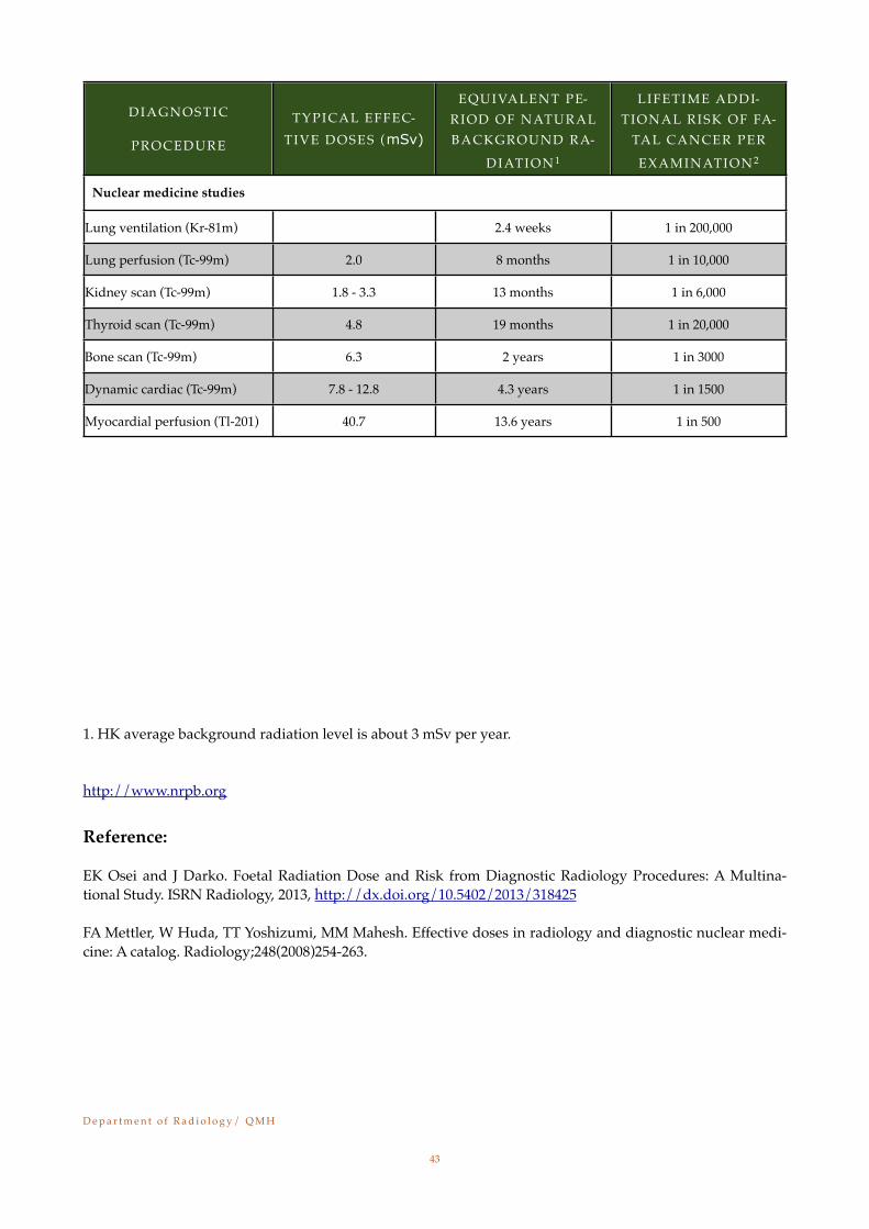
Nuclear medicine studies
Lung ventilation (Kr-81m) 2.4 weeks 1 in 200,000
Lung perfusion (Tc-99m) 2.0 8 months 1 in 10,000
Kidney scan (Tc-99m) 1.8 - 3.3 13 months 1 in 6,000
Thyroid scan (Tc-99m) 4.8 19 months 1 in 20,000
Bone scan (Tc-99m) 6.3 2 years 1 in 3000
Dynamic cardiac (Tc-99m) 7.8 - 12.8 4.3 years 1 in 1500
Myocardial perfusion (Tl-201) 40.7 13.6 years 1 in 500
1. HK average background radiation level is about 3 mSv per year.
http://www.nrpb.org
Reference:
EK Osei and J Darko. Foetal Radiation Dose and Risk from Diagnostic Radiology Procedures: A Multina-tional Study. ISRN Radiology, 2013, http://dx.doi.org/10.5402/2013/318425 FA Mettler, W Huda, TT Yoshizumi, MM Mahesh. Effective doses in radiology and diagnostic nuclear medi-cine: A catalog. Radiology;248(2008)254-263.
D e p a r t m e n t o f R a d i o l o g y / Q M H!
43
Appendix IV
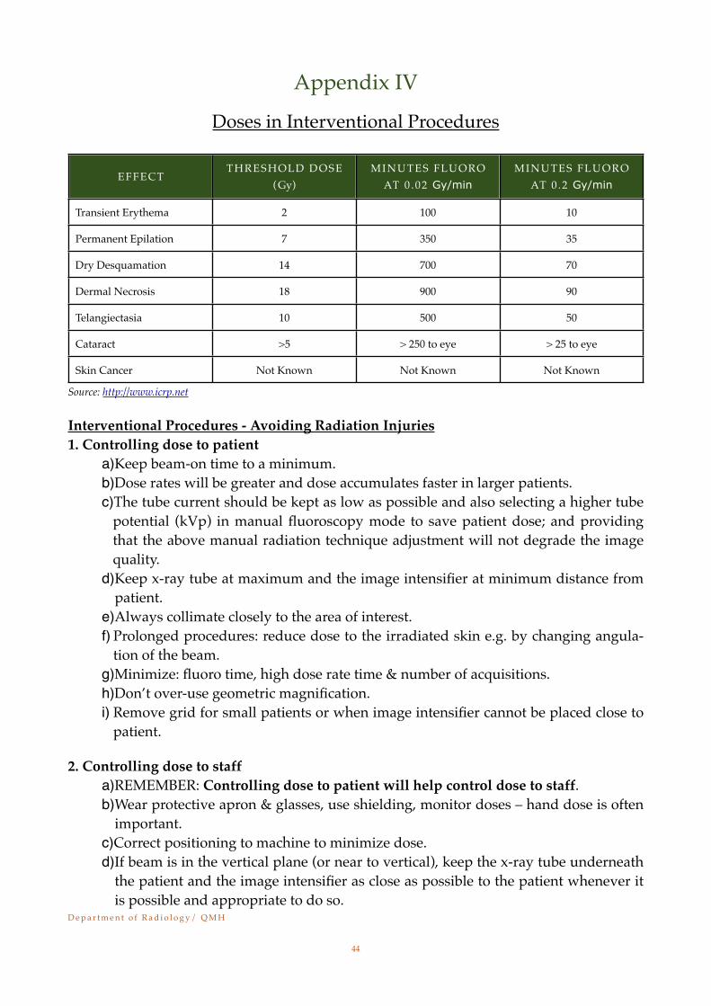
Doses in Interventional Procedures
EFFECTTHRESHOLD DOSE
(Gy)MINUTES FLUORO
AT 0 .02 Gy/minMINUTES FLUORO
AT 0 .2 Gy/min
Transient Erythema 2 100 10
Permanent Epilation 7 350 35
Dry Desquamation 14 700 70
Dermal Necrosis 18 900 90
Telangiectasia 10 500 50
Cataract >5 > 250 to eye > 25 to eye
Skin Cancer Not Known Not Known Not Known
Source: http://www.icrp.net
Interventional Procedures - Avoiding Radiation Injuries 1. Controlling dose to patient
a)Keep beam-on time to a minimum.b)Dose rates will be greater and dose accumulates faster in larger patients. c)The tube current should be kept as low as possible and also selecting a higher tube
potential (kVp) in manual fluoroscopy mode to save patient dose; and providing that the above manual radiation technique adjustment will not degrade the image quality.
d)Keep x-ray tube at maximum and the image intensifier at minimum distance from patient.
e)Always collimate closely to the area of interest. f) Prolonged procedures: reduce dose to the irradiated skin e.g. by changing angula-
tion of the beam.g)Minimize: fluoro time, high dose rate time & number of acquisitions.h)Don’t over-use geometric magnification.i) Remove grid for small patients or when image intensifier cannot be placed close to
patient.
2. Controlling dose to staffa)REMEMBER: Controlling dose to patient will help control dose to staff.b)Wear protective apron & glasses, use shielding, monitor doses – hand dose is often
important. c)Correct positioning to machine to minimize dose.d)If beam is in the vertical plane (or near to vertical), keep the x-ray tube underneath
the patient and the image intensifier as close as possible to the patient whenever it is possible and appropriate to do so.
D e p a r t m e n t o f R a d i o l o g y / Q M H!
44
e)If beam is in the horizontal plane (or near to horizontal), the operators (operators can be clinicians, nurses and other staff who work close to the patient and C-arm) should stand on the image intensifier side and keep a distance away from the im-age intensifier if it is possible and appropriate to do so. It is because the back scatter radiation is more intense on the x-ray tube/patient side under this condition. Therefore, radiographer should pay attention to the control parameters of the x-ray machine; and should stay on the image intensifier side but close to the control panel of the machine if it is possible and appropriate to do so.
3. Other factors in controlling dosesa)Ensure all staff are appropriately trained.b)Use dedicated interventional equipment with correct specification. c)Ensure comprehensive maintenance and quality assurance programmes are in
place.d)Obtain advice from a qualified radiation expert.
4. Informed consent and recordsa)Patients are entitled to know the risks of radiation injury if likely to be high.b)A written record should be kept if skin doses are estimated to be >3 Gy (1 Gy for
repeated procedures). c)Not all skin reactions are due to radiation; e.g. contrast medium allergy.
5. Follow-upa)Radiation skin injury may be present later, but the association would not be con-
sidered if there is no documentation.b)All patients with estimated skin doses of 3 Gy should be followed up 10-14 days
after exposure. c)A system to identify repeat procedures should be set up.
D e p a r t m e n t o f R a d i o l o g y / Q M H!
45

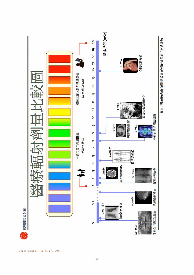
Appendix V
醫療輻射劑量比較圖
D e p a r t m e n t o f R a d i o l o g y / Q M H!
46
D e p a r t m e n t o f R a d i o l o g y / Q M H!
47
Appendix VI
Flowchart on Radiological Investigation for Women of Child-bearing Age
D e p a r t m e n t o f R a d i o l o g y / Q M H!
48
D e p a r t m e n t o f R a d i o l o g y / Q M H!
49

Appendix VII
Flowchart on Monthly Return of TLD Badges for Radiogra-phers in DR, QMH
D e p a r t m e n t o f R a d i o l o g y / Q M H!
50
D e p a r t m e n t o f R a d i o l o g y / Q M H!
51
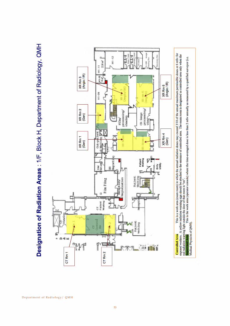
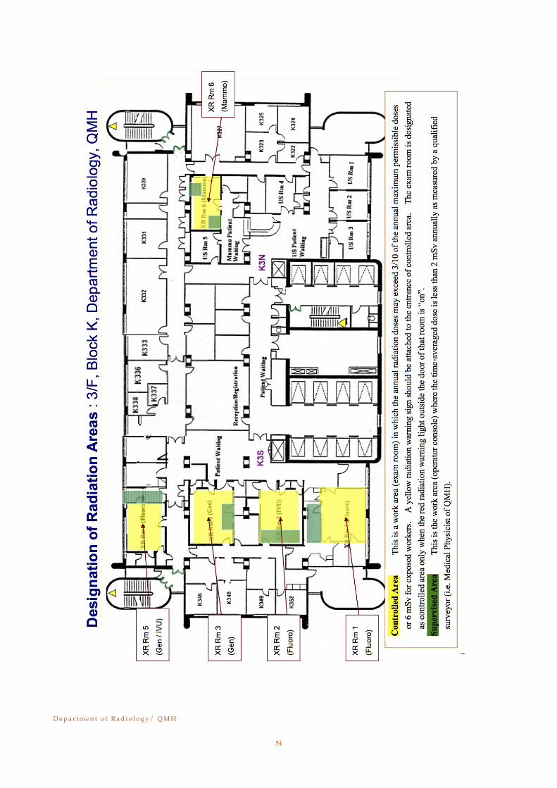
Appendix VIII
Designation of Radiation Areas for Radiology Department at Block H-1, Block K-3 and A&E X-ray & CT Scan unit
D e p a r t m e n t o f R a d i o l o g y / Q M H!
52
D e p a r t m e n t o f R a d i o l o g y / Q M H!
53
D e p a r t m e n t o f R a d i o l o g y / Q M H!
54
D e p a r t m e n t o f R a d i o l o g y / Q M H!
55

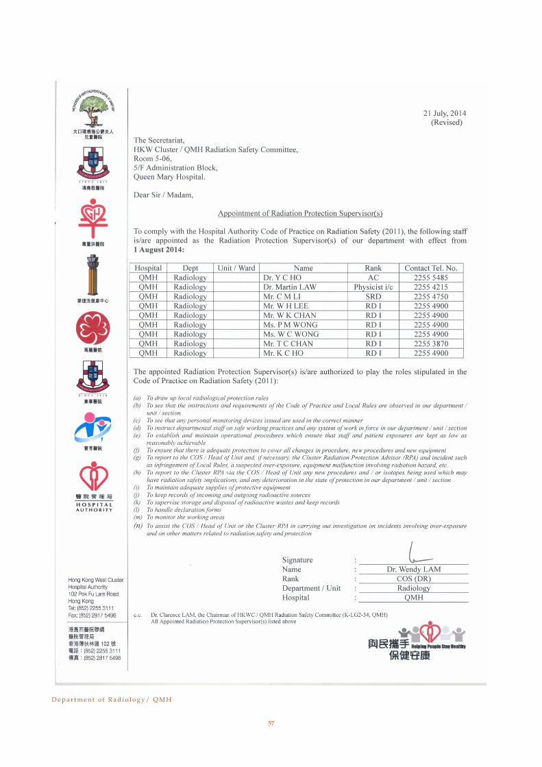
Appendix IX
List of RPS, DR, QMH
D e p a r t m e n t o f R a d i o l o g y / Q M H!
56
D e p a r t m e n t o f R a d i o l o g y / Q M H!
57

Appendix X
a) Medical Staff Declaration Record: 1Please sign your name in the below form to indicate that you have read and understood the contents of this “Local Radiation Protection Rules”, and is willing to observe them.
NAME ( IN BLOCK LETTER) RANK SIGNATURE DATE
1
2
3
4
5
6
7
8
9
10
11
12
13
14
15
16
17
18
19
20
D e p a r t m e n t o f R a d i o l o g y / Q M H!
58

Appendix X
a) Medical Staff Declaration Record: 2Please sign your name in the below form to indicate that you have read and understood the contents of this “Local Radiation Protection Rules”, and is willing to observe them.
NAME ( IN BLOCK LETTER) RANK SIGNATURE DATE
21
22
23
24
25
26
27
28
29
30
31
32
33
34
35
36
37
38
39
40
D e p a r t m e n t o f R a d i o l o g y / Q M H!
59

Appendix X
b) Radiographic Staff Declaration Record: 1Please sign your name in the below form to indicate that you have read and understood the contents of this “Local Radiation Protection Rules”, and is willing to observe them.
NAME ( IN BLOCK LETTER) RANK SIGNATURE DATE
1
2
3
4
5
6
7
8
9
10
11
12
13
14
15
16
17
18
19
20
D e p a r t m e n t o f R a d i o l o g y / Q M H!
60

Appendix X
b) Radiographic Staff Declaration Record: 2Please sign your name in the below form to indicate that you have read and understood the contents of this “Local Radiation Protection Rules”, and is willing to observe them.
NAME ( IN BLOCK LETTER) RANK SIGNATURE DATE
21
22
23
24
25
26
27
28
29
30
31
32
33
34
35
36
37
38
39
40
D e p a r t m e n t o f R a d i o l o g y / Q M H!
61

Appendix X
b) Radiographic Staff Declaration Record: 3Please sign your name in the below form to indicate that you have read and understood the contents of this “Local Radiation Protection Rules”, and is willing to observe them.
NAME ( IN BLOCK LETTER) RANK SIGNATURE DATE
41
42
43
44
45
46
47
48
49
50
51
52
53
54
55
56
57
58
59
60
D e p a r t m e n t o f R a d i o l o g y / Q M H!
62

Appendix X
b) Radiographic Staff Declaration Record: 4Please sign your name in the below form to indicate that you have read and understood the contents of this “Local Radiation Protection Rules”, and is willing to observe them.
NAME ( IN BLOCK LETTER) RANK SIGNATURE DATE
61
62
63
64
65
66
67
68
69
70
71
72
73
74
75
76
77
78
79
80
D e p a r t m e n t o f R a d i o l o g y / Q M H!
63

Appendix X
b) Radiographic Staff Declaration Record: 5Please sign your name in the below form to indicate that you have read and understood the contents of this “Local Radiation Protection Rules”, and is willing to observe them.
NAME ( IN BLOCK LETTER) RANK SIGNATURE DATE
81
82
83
84
85
86
87
88
89
90
91
92
93
94
95
96
97
98
99
100
D e p a r t m e n t o f R a d i o l o g y / Q M H!
64

Appendix X
c) Nursing Staff Declaration Record: 1Please sign your name in the below form to indicate that you have read and understood the contents of this “Local Radiation Protection Rules”, and is willing to observe them.
NAME ( IN BLOCK LETTER) RANK SIGNATURE DATE
1
2
3
4
5
6
7
8
9
10
11
12
13
14
15
16
17
18
19
20
D e p a r t m e n t o f R a d i o l o g y / Q M H!
65