local and systemic immune responses in rats …
TRANSCRIPT

TTHHEE MMEEDDIICCAALL JJOOUURRNNAALL OOFF BBAASSRRAAHH UUNNIIVVEERRSSIITTYY
1MSc, Technology Institute, Basrah 2 PhD, Department of Biology, College of Science, University of Basrah, Basrah, Iraq
LOCAL AND SYSTEMIC IMMUNE RESPONSES IN RATS INFECTED WITH GIARDIA LAMBLIA
Entehaa Abd-Al-Zahra1, Wafaa Sadoon Shani2, Maha Khalil Al-Malak2
ABSTRACT Objectives: This study was performed to determine the local (tissue) and systemic immune responses in rats infected with Giardia lamblia. Materials and methods: Rattus noruigicus rats aged 2 months were inoculated with 103 Giardia lablia cyst. After 7 and 14 days of infection samples (blood and small intestine) collected from both control and infected rats. Local immune response was assessed by counting the inflammatory cells in small intestine, mast cell number and lymph node diameter. Whearas systemic immune response was carried out by measurement of total and differential WBC count, phagocytic activity of polymphonuclear cells by NBT stain and histamine levels in blood plasma by ELISA kit. Results: Results showed an infiltration of inflammatory cells in all parts of small intestine and an increasing in lymph nodules diameter. Also, there is an infiltration of mast cells. Results of systemic immune response revealed a high significant differences in total and differential WBC count between two studied groups. Moreover, there was an increasing in phogocytic activity of polymorphonuclear cells and histamine plasma levels. Conclusion: Recent data indicated that there is a local and systemic immune response during acute phase of Giardia lamblia infection in experimental animals. INTRODUCTION
iardia lamblia is an intestinal protozoan parasite that causes intestinal malabsorption and diarrhea (giardiasis)
in humans and other mammals worldwide.[1] Giardiasis has been reported to mimic inflammatory bowel disease in man.[2] It is unknown whether variation in host responses to the parasite, differences among parasite genotypes, or both, are responsible for the differences seen in clinical outcomes.[3] Giardia infection in mice is usually self-limiting, which indicates the presence of an effective host defense.[4] Immunological and non immunological factors have different levels of importance during the course of a Giardia infection. Immunity to Giardia has been shown to occur in two major phases in mice: an early B-cell independent phases in the first two weeks after infection, followed by an antibody–dependent phase.[5,6] In addition most individuals effectively control infection within a few weeks,[3] although the precise immune mechanisms responsible for eliminating parasites are unknown. It also remains unclear whether symptomatic disease is due to immune mediated pathology or parasite derived factors or both. So, this study was designed to analyze the local or (tissue) and systemic immune response in rats infected with giardiasis in the first and second weeks after infection especially the presence of inflammatory cells in blood and intestine mast cells and histamine levels also the
response of gut associated lymphoid tissue (GALT). MATERIALS AND METHODS Samples collection Fecal samples were collected from patients with diarrhea from Al-Basrah General Hospital, Health Centers and Private Laboratories during a period between (Sept. 2008 to March 2009). All collected samples preserved in a chilled sucrose until reaching to the laboratory for examination. Stool samples examination The samples were examined with direct microscopical observation by using physiological normal saline to identify G. lamblia trophozoites and cysts. Cysts purification and enumeration Cystic stage of G.lamblia was purified with some modifications[7-9] with some modifications. Cysts enumerate by using neubaure chamber under oil objective lens. The sample was diluted with normal saline to (103) cyst for each rat. Rats inoculation Rattus noruvigicus rats were used in this study aged two months and (250–300gm) in weight. All animals were examined to be sure there is
G

_________________________________________________________________________________________________MJBU, VOL 30, No.1, 2012
61
no an infection, then divided into two groups, infected group which inoculated with (103) cyst according to Buret et al.[10] While, control group inoculated with physiological normal saline. Intestinal and blood samples collection After 7 and 14 days of infection, infected and control animals were anesthetized with chloroform and blood samples were collected from heart and divided into two portions (first portion preserved with EDTA tubes and used for total and differential white blood cell count. Plasma isolated after blood centrifugation which used for histamine measurement, while (second portion) of blood preserved in heparin for study of neutrophil function or phagocytosis by using nitroblue tetrazolium test. Small intestine, (duodenum, jejunum and ileum) from control and infected animals were resected and divided into small pieces washed and fixed with formalin (10%). Local or tissue immune response This study include the following studies: Rate of inflammatory cells infiltration Inflammatory cells were counted in both infected and control animals in (7,14) day after infection. The results expressed as number of cells/mm2 according to Yang et al.[11] in each part of small intestine .Tissues prepared for light microscopy examination according to Luna.[12]
Specimens were fixed with formalin (10%), washed, dehydrated and then embedded with paraffin wax then sections (5µm) were done and stained with haematoxylin and eosin to study the local immune response. Response of gut associated lymphoid tissue (GALT) Diameters of lymph nodules associated with small intestine were measured as mean±SD in all pieces of small intestine (duodenum, jujenum and ileum) related to (15) infected rats and (10) control rats. Counting of mast cells infiltration Number of mast cells in each part of small intestine related to (experimental animals was counted in (7, 14) day after infection. The mean number was counted in each (10) villi/crypt unit according to Scott et al.[13], Vallance et al.[14]
and McDermot et al.[15] All sections were stained with specific stain (Toulidin blue). Study of systemic immune response This study was carried out by the measurement of total white blood cells count, differential white blood cells, the phagocytosis activity of polymorphonuclear cells (PMN) by using nitroblue tetrazolium (NBT), and measurement of histamine level in blood plasma. Counting of total white blood cells number The total number of white blood cells was done according to Lewis et al.[15]
Differential white blood cells count Blood smears related to infected and control animals were prepared and stained with Leishman stain according to Lewis et al.[16]
Determination of phagocytosis activity of PMNs in blood According to Murata et al.[17] and Metcalf et al. [18] the phagocytosis activity was determined. Blood collected from (60) infected and (30) control rats after (7,14) days post infection. 0.5 ml of blood mixed with 0.5 ml of mixture from NBT-Cl stain and Tris-Hcl solution in heparinized tubes, then transfer to incubator at (37C°) for (1 hr). The specimens centrifuged and the supernantant used for absorption measurement by spectrophotometer on (515 nm) and this compare with the absorption related to control rats. Measurement of histamine level in blood plasma Histamin Bioassay Kit was used to measure the histamine level in blood plasma by using the method of enzyme-linked immunosorbent assay (competitive ELISA), Blood plasma which prepared from (16) infected and (10) control rats were used. The test was began by adding 50 µl from histamine enzyme (HRP) to (50 µl) of blood plasma in each well then incubated for 45 min at room temperature. After incubation each well washed with washing buffer, then add 150 µl from TMB substrate and incubated at room temperature for 30 minute. The optical density (OD) was read by the microplate ELISA reader at 450 nm. Then the concentrations of histamine in studied sample were calculated by plotting a

MJBU, VOL 30, No.1, 2012___________________________________________________________________________________________________
62
standard curve for standards (which supplemented with a kit).
Examination and photography All the slides were examined by light microscope (Olympus) under different magnification and photograph by microscope with digital camera.
Statistical analysis Analysis of variance (ANOVA test) was used to
clarified the statistical differences by using minitab program version 11.[19]
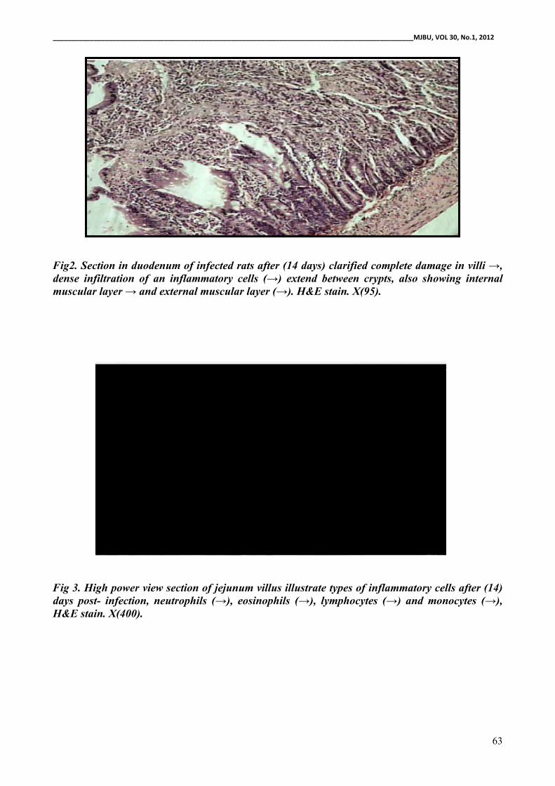
RESULTS Immune response study Local immune response Rate of inflammatory cells infiltration The study showed significant increase (p<0.01) in the numbers of inflammatory cell of infected rats in comparison with control while, their number less in ileum region followed by duodenum and jejunum ( Figure-1,2,3; Table-1).
Fig 1. Section in duodenum showing dense infiltration of an inflammatory cells after (7 days) post – infection→, crypts elongation→, also showing sinternal muscular layer→ and external
muscular layer. H&E stain. X (95).
_________________________________________________________________________________________________MJBU, VOL 30, No.1, 2012
63
Fig2. Section in duodenum of infected rats after (14 days) clarified complete damage in villi →, dense infiltration of an inflammatory cells (→) extend between crypts, also showing internal muscular layer → and external muscular layer (→). H&E stain. X(95).
Fig 3. High power view section of jejunum villus illustrate types of inflammatory cells after (14) days post- infection, neutrophils (→), eosinophils (→), lymphocytes (→) and monocytes (→), H&E stain. X(400).

MJBU, VOL 30, No.1, 2012___________________________________________________________________________________________________
64
Table 1. lnfiltration of neutrophils, eosinophils, lymphocytes and monocytes of infected rats with G. lamblia in comparison with control (values are expressed as mean±SD) on (7,14) days post infection.
Tissue type Period /Days
Studied groups
rat numbers
Mean numbers of inflammatory cells ±SD
Neutrophile Eosinophile Lymphocyte Monocyte
Duodenum
7 Infected 16 76.46 ± 18.90a 64.83±14.12a 199.92 ±44 4.926±7.586a
Control 8 0.61 ±1.93b 0.61 ±1.93b 2.44±4.27b 0.00 ±0.00b
14 Infected 16 83.93± 21.83a 74.22±15.5a 204.89±31.75a 6.445±8.730a
Control 8 0.61 ±1.93b 0.61 ±1.93b 4.27±2.44b 0.00±0.00b
Jejunum
7 Infected 16 76.94±13.54 a
68.61±16.14a
185±4.12b 4.185±6.488a
Control 8 0.61 ±1.93b 0.00 0± .00b 1.83±4.12b 0.00 ±0.00a
14 Infected 16 81.44±15.62a 73.44±20.27a 194.44±42.09a 4.24±6.50a
Control 8 0.61 ±1.93b 0.00±0.00b 4.12±1.83b 0.00±0.00a
Ileum
7 Infected 16 71.59±25.40a 55.48 ±19.13a 161.66 ± 35.49a
2.45±4.27a
Control 8 0.00±0.00b 0.00±0.00b 1.22±2.58b 0.00±0.00b
14 Infected 16 79.74±26.85 a 72.56±19.32a 165.39±40.18a 2.67±5.08a
Control 8 0.00± 0.00b 0.00±0.00b 1.22±2.58b 0.00±0.00b
• Similar letters referred no significant differences on p>0.05 • Different letters referred to significant differences on p<0.05
Mean rate of inflammatory cells reached (76.6± 18.9 cell) in duodenum for neutrophils, (199.9± 44.8) of lymphocyte cells and (64.8±14.12) of eosinophils cells after (7 day) of infection in comparison to control (Table-1). After (14 days) of infection the rate of inflammatory cells showed significant increase when compared with control rats while the monocytes number
did not show any changes in number among infected animals in comparison to control after (7,14) days post infection (Figure-3). Results of inflammatory proliferation both in jejunum and ileum related to infected rats after (7,14) days post infection revealed a significant increase in all inflammatory cells (neutrophils, eosinophils and lymphocytes) (Figure-4,a,b,c).
Fig 4. Small intestine of control rats showing general architecture. A‐ Section in duodenum showing the mucosal layer composed of leaf shape villi (→) and crypts of leiburkuhn
at the base (→). H&E stain. X(95).

_________________________________________________________________________________________________MJBU, VOL 30, No.1, 2012
65
B‐ Section in jejunum, finger like villi (→) and crypts of leiburkuhn (→). H&E stain. X(120).
C‐ Section in ileum illustrate short villi (→), crypts (→) and muscularis mucosa (→). H.E stain. X(95). The study not showed any changes in various region of small intestine of control animals (Table-1).
Mean infiltration of mast cells Results clarified that giardiasis cause an increase in rate of mast cells numbers with
significant difference (p<0.01) in (duodenum, jejunum, ileum) after (7,14) day post-infection. (Figure-5, 6; Table-2) when compared with control animals which show no mast cells infiltration.

MJBU, VOL 30, No.1, 2012___________________________________________________________________________________________________
66
Fig 5. Longitudinal section in duodenum villus showing mast cell infiltration (→) after (14) days post infection. Toulidin blue stain. X(480).
Fig 6. Tissue section in jejunum illustrate mast cells in villus connective tissue (→), the parasite well visible (→) after (14 days) post- infection. Toulidin blue stain. X(400). Table 2. Clarified mean infiltration of mast cells in an infected rats with G. lamblia in comparison with control after (7 and 14) days post infection.
Days Mean number of mast cell in small intestine ± SD
Duodenum Jejunum Ileum Infected Control Infected control Infected control
Animals number 15 10 15 10 15 10
7 38.7
±10.242 a
0.1 ±0.316
B
31.70 ±7.319
a
0 ±0 b
18.3 ±6.832
a
0 ±0 a
14 39.6
±12.825 a
0.1 ±0.316
b
32.8 ±8.456
a
0 ±0 b
19.1 ±8.517
b
0 ±0 b
(Values expressed as mean ± SD). • Similar letters referred no significant differences on p>0.05 • Different letters referred high significant differences on p<0.01

_________________________________________________________________________________________________MJBU, VOL 30, No.1, 2012
67
Number of these cells were more in duodenum and jejunum in comparison with their number in ileum, the mean rate reached to (38.7±10.4, 31.70±7.31 cells) in duodenum, jejunum and (18.3±6.3) cells in ileum in 7 day after infection. Also the mean rates of mast cells infiltration after 14 day post infection show the same pattern with high significant differences. The results didn’t show any significant differences (p>0.05) in mean number of mast cells when
compared in infected animals after 7,14 day post-infection. Response of gut associated lymphoid tissue (GALT) Results of present study related to (GALT) response in intestine infected with giardiasis concluded an increasing in lymph nodules diameter (Table-3).
Table 3. Illustrate mean of lymph nodules diameter in infected animals and control after (7 and 14) day post infection (values expressed as mean ± SD).
Days
Mmean number of lymph nodules diameter (µ) in different parts of small intestine ± SD
Duodenum Jejunum Ileum
Infected Control Infected Control Infected Control
Animals number 15 10 15 10 15 10
7 39.793 ±8.095
a
6.91 ±7.396
b
35.667 ±6.174
a
0 ±0 b
59.267 ±10.107
a
24.02 ±7.933
b
14 46.313 ±5.819
c
8.36 ±7.633
b
38.553 ±4.441
a
0 ±0 b
74.45 ±24.02
c
28.6 ±7.85
b • Similar litters referred no significant differences on p>0.05 • Different litters referred to significant differences on p<0.01
The mean diameter of lymph nodule in infected duodenum is (39.74±8.095) µm and (46.31±5.819) µm in compare to control
(7.39±6.91), (8.36±7.633)µm with high significant difference (p<0.01) in 7,14 day after infection respectively (Figure-7).
Fig 7. Photomicrograph of duodenum showing single lymph nodule composed of cortex (→) and germinal centre (→) penetrated by lymphocytes in (14 day) after infection. H&E. stain. X(95)
MJBU, VOL 30, No.1, 2012___________________________________________________________________________________________________
68
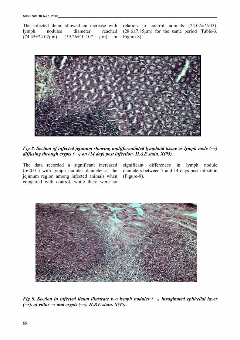
The infected ileum showed an increase with lymph nodules diameter reached (74.45±24.02µm), (59.26±10.107 µm) in
relation to control animals (24.02±7.933), (28.6±7.85µm) for the same period (Table-3, Figure-8).
Fig 8. Section of infected jejunum showing undifferentiated lymphoid tissue as lymph node (→) diffusing through crypts (→) on (14 day) post infection. H.&E stain. X(95).
The data recorded a significant increased (p<0.01) with lymph nodules diameter at the jejunum region among infected animals when compared with control, while there were no
significant differences in lymph nodule diameters between 7 and 14 days post infection (Figure-9).
Fig 9. Section in infected ileum illustrate two lymph nodules (→) invaginated epithelial layer (→). of villus → and crypts (→). H.&E stain. X(95).

_________________________________________________________________________________________________MJBU, VOL 30, No.1, 2012
69
Systemic immune response Total white blood cells count The results showed highly significant difference (p<0.01) in mean number of white blood cells in the infected animals (13265±1673) and controls (6186±1842) after 7 days of infection.
Also the same difference was noticed after 14 days post-infection while there was no significant difference showed when infected animals compared with each other after 7 and 14 days post-infection (Table-4).
Table 4. Illustrate mean number of total white blood cells count in infected rats with G. lamblia and control in (7 and 14) day post infection. (values expressed as mean ± SD).
Days Studied groups No. of animals Mean Range ± SD SE
7 Infected 15 13265
a 11150-17655
±1673 432.076
Control 11 6186 b 9450-4000 ±1842 555.488
14 Infected 15 14084
c 11350-19100
±2255 582.386
control 11 6777 d 9600-4650 ±1939 584.740
• Similar letters referred no significant differences on p>0.05 • Different letters referred high significant differences on p<0.01
Differential white blood cells count Data on (Table-6) clarify an increasing percentage rate of counting cells (lymphocytes and eosinophils) which reached (59.75±9.98), (6.65±2.20) in infected rats after 7 days post infection in comparison with control animals. The same increase in percentage rate of these cells recorded after 14 day post infection with significant difference (p<0.01). The results also
clarified the decrease in percentage rate of neutrophils and monocytes with significant difference (p<0.01) in relation to control after (7, 14) days, the rate reached to (29.35±9.32) for neutrophils, (1.450±1.48) for monocytes after 7 days of infection in comparison to control (58.65±2.39) for neutrophils and (10±1.44) for monocytes (Table-5).
Table 5. Mean percentage of differential white blood cells in infected rats and control after (7,14 days) post infection.(values expressed as mean±SD).
Days
Studied groups
Animals number
Mean of cells percentage ± SD Neutrophils Lymphocyte Eosinophils Monocytes Basophils
7
Infected 20 29.350 ±9.320
a
59.750 ±9.989
A
6.650 ±2.207
a
2.750 ±1.482
a
0.600 ±0.502
a
Control 20 58.650 ±2.390
b
31.300 ±2.736
B
4.450 ±1.468
b
5.100 ±1.447
b
0.500 ±0.513
a
14
Infected 20 17.450 ±10.318
c
79.150 ±11.246
C
7.550 ±2.762
a
1.450 ±1.572
c
0.300 ±0.470
a
control 20 58.650 ±2.390
b
31.300 ±2.736
B
4.450 ±1.468
b
5.100 ±1.447
b
0.500 ±0.513
a • Similar litters referred no significant differences on p> 0.05 • Different litters referred high significant differences on p<0.01

MJBU, VOL 30, No.1, 2012___________________________________________________________________________________________________
70
There were no change in basophils number in infected animals (0.60±0.50) compare with control (0.50±0.51) after 7 days of infection. Highly significant differences in lymphocytes number showed in infected animals after (7,14) days post infection. The basophils didn’t show any significant difference (p>0.5) when infected groups compared with each other on (14 days) post infection and the eosinophils didn’t show any significant differences among infected animals (Table-5).
Determination of phogacytic activity of PMN in blood Results of the study dealing with phagocytic activity of PMN cells showed high significant difference (p<0.01) in optical density rate which reflect the NBT stain reduction among infected rats (0.64±0.37), (0.735±0.28) and control (0.07±0.05), (0.076±0.07) after 7, 14 day post infection respectively. In spite there was no significant difference in absorption when compared between infected animals after 7 and 14 day of infection (Table-6).
Table 6. Optical density mean of (NBT) stain by neutrophils cells in both infected rats and control. After (7, 14 day) post infection. (values expressed as mean ±SD).
Days Studied groups No. of animals Mean of optical density Range ± SD SE
7 Infected 60 0.640
a 0.02-1.62 ±0.3773 o.048
Control 30 0.070 b 0.002-0.2 ±0.0515 9.311
14 Infected 60 0.759
a 0.07-1.54 ±0.2888 0.037
control 30 0.076 b 0.01-0.3 ±0.0744 0.013
• Similar letters referred no significant differences on P> 0.05 • Different leitters referred high significant differences on P<0.01.
Measurement of histamine level in blood plasma Systemic immune response was indicated an increase of plasma histamine level with high significant difference (p<0.01) among infected animals (1.78±0.50), (3.067±0.61) and control
animals (0.826±0.), (1.33±0.16) after 7 and 14 day of infection respectively (Table-7). The plasma histamine level related to infected animals after 14 days of infection showed high significant difference (p<0.01) comparative with first period (7days) of infection.
Table 7. Illustrate histamin level in blood plasma of infected rats with G. lamblia and control measured by ELISA technique after (7, 14) day post infection. (values expressed as mean±SD).
Days Studied groups No. of animals Mean ng\ml Range ± SD SE
7 Infected 16 1.78
a 0.98-2.84 ±0.505 0.122
Control 10 0.826 b 0.82-0.83 ±0.005 2.041
14 Infected 16 3.067
c 1.72-4.03 ±0.615 0.149
control 10 1.333 b 0.82-0.83 ±0.160 0.065
• Similar litters referred no significant differences on P> 0.05 • Different litters referred high significant differences on P<0.01.

_________________________________________________________________________________________________MJBU, VOL 30, No.1, 2012
71
DISCUSSIONThe immune response against G. lamblia parasite plays an important and active role during infection.[20] The results of related studies interested in local immune response represent a significant infiltration within small intestine lamina propria and this may be due to the importance of these cells in innate and adaptive immune response against parasites (were these cells either engulf the parasite or secrete reactive substances to kill it in addition to different mechanisms participating in parasite elimination.[21] Inflammatory cells (neutrophils , eosinophils, monocytes) infiltrated in all parts of small intestine (duodenum, jejunum, ileum) after (7, 14) days post infection and this may be related to fact that these cells considered as a first line of defense against pathogen , because they are an important source for cytokines (IL-1ß, TNF-α) which responsible for the switching on the early inflammation.[22] Neutrophils and eosinophiles infiltration showed the same model in all parts of small intestine and in two studied period whearas monocyte didn’t show the same model because their increasing don’t revealed any significant differences in comparison with control group and the most acceptable explanation that the infection still acute, and this is similar to Kaplan et al.[23] whom indicated the phagocytic activity of these cells with the aid of antibodies. Activation of inflammatory cells (Neutrophils, eosinophils) in acute phase represent a most important event during a defense process whearas the IL.8 play a significant role in this activation and attraction of neutrophiles to damaged area of small intestine.[24] Highly significant infiltration of lymphocytes was also illustrated in all studied parts of small intestine infected with giardiasis after (7,14) days post infection. Some result was also indicated by Vinayak et al.[25] when they recorded heavy lymphocyte infiltration in lamina propria. Moreover Biez[26] confirmed the T. cells and IL-6 action (during first two weeks of infection) in Giardia expulsion and elimination of infection. Recent work showed mast cells accumulation in infected small intestine and this clearly demonstrate an importance of these cells in elimination and control of infection. The presence of parasite act as an activator of mast cells which then lead to degranulation of their granules and with a mast
cell dependent process the cholycytokinin was secreted resulting in smooth muscle contractility, then coupled contractions with nitric oxide-mediated muscle relaxation, promote intestinal transiet and parasite elemination.[3] D'anchino et al.[27] and Zhou et al.[28] were also recorded the importance of mast cells during giardiasis because these cells are a source of histamine, leukotriens and serotonin which facilitate the Giardia and helminthes expulsion.[29] Our data reffered to an increasing in lymph nodules diameter (which is a compartment of GALT) in an infected rat in comparison with control group and this may be suggested that there is an induction of cellular inflammatory response because these nodules are the source of T-cells.[30] In addition, to stimulation of local immune response which was proved in present study, systemic immune response takes place and was documented as a significant increase in leukocytes total number in all experimental animals compared to control group. This give an indicator to cellular immune response against circulating antigens which released from parasite during acute infection and this result is more parallel with the proved thought which indicate the importance of cellular immune response during the first two weeks of infection or (acute infection) in comparison with the humoral immune response which present during the chronic phase of giardiasis.[31] The same results also recorded by Al-Kubassi[32] during Entamoeba histolytica infection. Data of differential WBC count was clarified a significant decrease (P<0.01) in neutrophils and monocytes number in all infected groups in comparison with control group and this results may be occurred as a result of cell filtration and migration from blood to an infected area ( lamina propria and mucosal layer). These results also agreed with Chadee and Meerrvitcho[33] Boirivant et al.[34] clarified the role of these cells in immunity against E.histolytica and their ability to kill the vegetative phase of the parasite, in addition to its toxic activity and lymphokines production which share in events of immune response. An increase with significant difference (P<0.01) in eosinophils number in all infected rats in comparison with control group was recorded. This result resembling with study done by Al-

MJBU, VOL 30, No.1, 2012___________________________________________________________________________________________________
72
Kubassi[31] on amoebiasis patients because these cells effective against parasite. Basophilic cells did not show any changes in their number in all infected rats (after 7 and 14) day post–infection and this related to their role through chronic infection. Phagocytic activity is other studied an important factor which support the systemic immune response and was done by polymorphonuclear cells (PMN), that ingested the vegetative phase in vitro, present study revealed the significant phagocytic activity in all infected rats, at (7,14) day post infection. These results are acceptable with other studies suggested that his activity act as defense mechanism against the parasite.[35] Measurement of phagocytic activity of polymorphonuclear cells in blood was determined by NBT stain reduction and this results agreed with Hill and Phol[36] who used the NBT stain reduction and formazan granules formation as an indicator for oxidative burst. The study also indicated an increasing of histamine level in blood plasma as a marker to systemic immune response by using ELISA test, which showed high significant difference in histamine level in all rats infected with giardiasis in comparison to control group, this due to mast cell increasing which is proved in recent work because the histamine which secreted from mast cell represent as an important factor in intestinal canal[37] and increased the blood vessels permeability[38] which in turn increase cells migration to infected region especially neutrophiles and monocytes which proved in recent results through an elevation in number of inflammatory cells in lamina propria and at the same time decreasing of its number in blood stream (specially neutrophiles and monocytes). Serna et al.[39] was illustrated that intestinal movement increased after treatment with histamine and this lead to parasite expulsion and controlling the infection. Elevated level of histamine which represent as an indicator to mast cells response may be shared in giardiasis pathophysiology in addition to its participating in parasite expulsion.[40] Our data referred to an increasing in histamin level after (14) days post infection more than 7 days post infection and this is the time of mast cells activity increasing which recorded in this study which clarified the
important of mast cells and histamine in parasite expulsion in order to control the infection. REFERENCES 1. Adam RD. Biology of Giardia lamblia. Clin.
Microbiol. 2001; Rev. 14(3):447-475. 2. Gunasekaran TS and Hassal E. Giardiasis
mimicking inflammatory bowel disease. J. Pediat. 1992; 120: 424-426.
3. Li E, Zhao A, Shea-Donohue T and Singer S M. Mast cell-mediated changes in smooth muscle contractility during mouse giardiasis. Infect. Immun. 2007; 75: 4514-4518.
4. Faubert G. Immune response to Giardia lamblia. Clin. Microbiol. 2000; Rev. 13(1): 35-54.
5. Eckmann L. Mucosal defenses against Giardia. Parasite. Immunol. 2003; 25(5): 259-270.
6. Li E, Zhou P and Singer SM. Neuronal nitric oxide synthase is necessary for elimination of Giardia lamblia infections in mice. J. Immunol. 2006; 176: 516-521.
7. Roberts-thompson IC, Stevens DP, Mahmoud AAF and Warren KS. Giardiasis in the mouse: an animal model. Gastroenterol. 1976;71:57-61
8. Sheffield HG and Bjorvatan B. Ultrastructure of the cyst of Giardia lamblia. Am. J. Trop. Med. Hyg. 1977; 26(1):23-30.
9. Bingham AK and Meyer EA. Giardia excystation can be induced in vitro in acidic solution. Nature (London). 1979; 277: 301-30.
10. Buret A, Gall DG and Olson ME. Effect of murine giardiasis on growth, intestinal morphology and diasaccharidase activity. J.Parasitol. 1990; 76(3): 407-409.
11. Yang P, Berin MC, Yu L and Perdue MH. Mucosal pathophysiology and inflammatory changes in the late phase of the intestinal allergic reaction in the rat. Am. J. Pathol.2001; 158(2): 681-690.
12. Luna LG. Manual of histologic staining methods of armed forces institute of pathology. 3rd edition. McGraw-Hill book company. 1968.
13. Scott K G E, Logan R, Klamer GM, Teoh DA and Buret AG (2000). Jejunal brush border microvillous alterations in Giardia muris-infected mice: role of T lymphocytes and interleukin 6. Infect. Immun. 2000; 68 (6): 3412 -3418.
14. Vallance BA, Blennerhassett PA, Huizinga JD and Collins SM(2001). Mast cell-independent impairment of host defense and muscle contraction in T. spiralis infected W/Wv mice. Am .J .Physiol. Gastrointest. Liver. Physio. 2001; 280:640-648.
15. McDermott JR, Bartram RE, Knight PA, Miller HRP Garrod DR and Grencis RK. Mast cell disrupt epithelial barrier function during enteric nematode infection. 2003; 100(13): 7761-7766.
16. Lewis SM, Bain BJ and Bates I. Dacie and Lewis Practicle Hematology. 9th ed. Churchill Leviugstone.2001.
17. Murata H, Takahashi H and Matsumoto H. NBT reduction activity of peripheral blood phagocytes in calves untreated or exposed to some stressors. Bull.Natl.Inst.Anim.Health. 1985; No.88:17-24.
18. Metcalf JA, Gallin JI, Nausseef WM and Root RK. Laboratory manual of neutrophil function. Raven .Press book, New York. 1986:100.

_________________________________________________________________________________________________MJBU, VOL 30, No.1, 2012
73
19. AL-Rawi KM and Khalaf–Allah AM. Designation and analysis of agricultural experiments. Mousel university. 1980:27.
20. Gaétan F. Immune response to Giardia duodenalis. Clinical microbiology reviews, Vol. 1, 2000; (1): 35-54.
21. Mc Alindon M.E and Mahida YR. Pro-inflammatory cytokines in inflammatory bowel disease. Aliment. Pharmacol. Thermacol. Ther. 1996; 10:72-74.
22. Ebenfelt,A, Lundqvist H, Dahlgren C and Lundberg C. Neutrophils in mucosal secretion are functionally active. Clin. Exp. Immunol. 1996; 106:404-409.
23. Kaplan BS, Uni S, Aikawa M and Mahmoud AAF. Effecter mechanism of host resistance in murine giardiasis: specific IgG and IgA cell-mediated toxicity. J. Immunol. 1985; 134: 1975-1981.
24. Seydel K B, Zang T, Champion G A, Fichtenbaum C, Swanson PE, Tzipori S, Griffiths JK and. Stanley SL. Cryptosporidium parvum infection of human intestinal xenografts in SCID mice induces production of human tumor necrosis factor alpha and interleukin-8. Infect. Immun. 1998; 66:2379-2382.
25. Vinayak VK, Khanna R and Kum K. Kinetics of intraepithelium and lamina propria lymphocyte responses during Giardia lamblia infection in mice. Microb. Pathog. 1991; 10: 343-350.
26. Biez M, Dai M, Welle M, Gottstein B and Müller N. Interlekin-6- deficient mice are highly susceptible to Giardia lamblia infection but exhibit normal intestinal immunoglobulin A responses against the parasite. Infect.Immun. 2003; 71(3): 1569-1573.
27. D'Anchino M, Orlando D and De Feudis L. Giardia lamblia infections become clinically evident by eliciting symptoms of irritable bowel syndrome. J. Infect. 2002; 45:169-172.
28. Zhou P, Li E, Zhu N, Robertson J, Nash T and Singer SM. Role of interleukin-6 in the control of acute and chronic Giardia lamblia infections in mice. Infect. Immun. 71:1566-1568.
29. Lee,T.D.; Swieter,M. and Befus,A.D. (1986). Mast cell responses to helminth infection. Parasitol Today. 2003; 2:186-191.
30. Elgert, K.D. Immunology: Understanding the immune system. 2nd ed. Wiley-Blackwell. 2009:48-53.
31. Langford TD, MP Housley, M Boes, J Chen, MF Kagnoff, FD Gillin and L Eckmann. Central importance of immunoglobulin A in host defense against Giardia spp. Infect. Immun. 2002; 70:11-18.
32. Al-Kubassi ABH. Immunological, epidemiological study of patients infected with Entamoeba histolytica . Ph.D. thesis. Coll. Sci. Al-Mustansiriya Univ., 2002.
33. Chadee K and Meerovitch E. The pathogenesis of experimentally induced amebic liver abscess in the gerbil (Meriones Unquiculatus) Am. J. Pathol. 1984; 117:71-80.
34. Boirivant M, Fuss IJ, Chu A and Strober W. Oxazolone colitis: a murine model of T helper cell type 2 colitis treatable with antibodies to interleukin-4.J. Exp. Med. 1998; 188:1929.
35. Franca-Botelho AC, Honorio-Franca AC, Franca EL, Gomes MA and Costa-Cruz JM. Phagocytosisof Giardia lamblia trophozoites by human colostral leukocytes. 2006; 95(4): 438-443.
36. Hill DR and Phol R. Ingestion of Giardia lamblia trophozoites by murine peyer’s patch macrophages. Infect. Immun. 1990; 85(1): 3202-3207.
37. Rangachari PK. Histamine: mercurial messenger in the gut. Am. J. physiol. 1992; 262:G1-G3.
38. Huang C, Friend DS, Qiu WT, Wong GW, Morales G, Hunt J and Stevens RL. Induction of a selective and persistent extravasation of neutrophils into the peritoneal cavity by tryptase mouse mast cell protease 6. J.Immunol., 1998;160: 1910–1919.
39. Serna H, Porras M and Vergara P. Mast cell stabilizer ketotifen [4-(1-methyl-4-piperidylidene)-4h-benzo [4,5] cyclohepta [1,2-b]thiophen-10(9H)-one fumarate] prevents mucosal mast cell hyperplasia and intestinal dysmotility in experimental Trichinella spiralis inflammation in the rat. J. Pharmacol. Exp. Ther. 2006; 319:1104-1111.
40. Di Prisco MC, Hagel I, Lynch NR, Jiménez, JC, Rojas R, Gil M and Mata E. Association between giardiasis and allergy. An allergy asthma. Immunol. 1998; 81(3):261–265.