liposomal doxorubicin in the treatment of aids-associated kaposi`s sarcoma · various forms of...
TRANSCRIPT

Liposomal doxorubicin in the treatment of AIDS-associated Kaposi's sarcoma:
c1inical, histological and cell biological evaluation
M. StürzI (1)(0 >, C. Zietz (2), B. Eisenburg (11, F.-D. Goebel (3\ R. Gillitzer (4),
P.H. Hofschneider (I) and J.R. Bogner (3)
(IJ Max-Planck-Instiful für Biochemie, Ab/ei/ung Virnsforschung, Am Klopferspirz 180. 82152 Martinsried (Germany),
(1) Pathologisches Institut der Ludwig-Maximilians-Universität München. Tho/kirchnerstr. 36, 80337 München (Germany),
(J) Medizinische Poliklinik der Ludwig-Maximilians-Universität München. Pettenkoferstrasse 80, 80336 München (Germany), and
(4) Klinik und Poliklinik für Haut- und Geschlechtskrankheiten der Universität Würzburg, losel-SchneideT-Slr. 2. 97080 Würzburg (Germany)
SUMMARY
AIOS-8ssociated Kaposi's sarcoma (KS) in eight patients was treated with the svstemic appUcation of liposomal doxorubicin (20 mg/m2 per cycle), After six cycles of treatment a significant regression of KS was observed in all patients. Tumour volume was reduced trom 556 ± 635 mm 3 before therapy to 42 ± 134 mm3 after therapy as determined by ultresonographv of selected tumours. Histological examination r\vealed a reductlon of tumour-lika structures end the absence of KS spindie cells in involved arees after therapy.ln vitro experiments with KS-derived ceU cultures, which most likeIy raprennt tha KS spindie cels, suggastad that liposomal doxorubicin may cause ragression of KS via two different mechanisms: liI by highly specific inhibition of KS spindia cell proliferation and (11) by induction of monocyte chemoattractant protein-1 expression in KS spindie cells. which mav result in incre8sad recruitmant of phagocytic cells (monocytes/macrophagesl into the lesions. A cooperative actten of both mechanisms may explain the high afficacy of liposomal doxorubicin in tha treatment of AIDS-KS.
Key-words: AIDS. Kaposi's sarcoma. Doxorubicin, Liposome; Therapy, Spindie cells, Monocyte chemoattractant protetn-l.
lNTRODUCnON
Kaposi's sarcoma (KS) is the most common tumour associated with the acquired immunodeficiency syndrome (AIDS) and occurs in more than 25 0)'0 of all reported cases (Haverkos et al., 1985;
Received February 23, 1994.
(.) Corresponding author.
Safai et al., 1985). Histologically. KS lesions are characterized by prominent vascularization and numerous spindle-shaped cells. These so-called spindIe cells are considered to be the tumour cells of KS (Ackerman and Gottlieb, 1988).

Various forms of treatment of KS have been described. The appropriatc treatment is selectcd according to the clinicaI presentation of the disease. Small, IocaIly restricted lesions are treated with electrodessication and curettage, cryotherapy, radiotherapy. electron-beam therapy, or by surgical excision (StickJer and Friedman-Kien, 1991). More severe forms of the disease, presenting with multiple disseminated. lesions and involvement of visceraJ organs, are treated with systemie ehemotherapy (StickJer and Friedman-Kien, 1991). Thc agents used include doxorubicin, bleomycin, vinblastine, vineristine and etoposide (VP-16), employed either as monotherapy or in combination (Stickler and Friedman-Kicn, 1991). Besides chemotherapy, biologic response modifiers such as interferon-lI are systemicaJly employed aecording to the hypo thesis that these agents will prevent tbe immunosuppression following combination chemotherapy (Stickler and Friedman-Kien, 1991).
Until recently, all forms of systemie therapy of KS werc hampered cither by severe side effects (ehemotherapy) or by low response rates (biologie response modifiers). Progress in chemotherapy was obtained by the availability of long-circulating "Stea1th" liposomes loaded with doxorubicin. In an animaJ model it has been reported that the prolonged cireulation time of liposomal doxorubicin accounts for the superior therapeutic effectiveness (Vaage el 0/., 1992). Phannacological data revealed a long plasma half-life, increased accumulation in tumour tissue and a decrease in uptake by tissues such as liver, spleen and bone marrow (Vaage er a/., 1992; Papahadjopulos el 01., 1991; Baly el 0/., 1990) .
Tbe objectives of our study were (i) to determine the efficacy of liposomal doxorubicin in the treatment of KS, (ö) to compare tbc histological presentalion of KS before and after therapy and (üi) to investigate the molecular mechanisms which might account for the high efficacy of liposomal doxorubicin in the treatment of KS.
MATERIALS AND METHODS
Patients
Eigbt patients with biopsy-proveD advanced AIDS-KS, presenting witb progressive disseminated cutaneous lesions
AIDS .. acquired immunodeficiency syndrome. AIDS-FIB .. AIDS-derived fibroblast . AIDS-KS - AIDS-a.ssociated KS. FOS - foetal bovine serum. HSMC - human smooth musde cell .
including oedema of the face or limbs or oraIlesions, were included in Ihis study. Indusion criteria were age ;<l= 18 years, Kamofsky status> 50 O'J'o, white blood cell count> 2,OOO/ tJl, haemoglobin > 10 g/ dl and platelets > 50,000/ 1'1. Exclusion criteria were acute opportunistic infections, non-Hodgkins lymphoma, systcmic chemotherapy, radiation of KS within 4 weeks prior 10 entry into the triaJ, major psychiatrie illness, cardiac fajlure and anthracydine intolerance. All patients gave wrinen informed consent. The trial was approved by the institutional review board of the Medizinische Poliklinik, University of Munich, Germany.
The baseline evaluation consisted of a medical history and a physica1 examination. Indicator lesions were evaluated by sonographie measurements of romour volume in all patients as described previously (Bogner et 01., 1993a and b) . At least one indicator lesion accessible for cutaneous ultrasound examination was required ir palients presented with oral or visceral lesions.
Clinica.l assessment and ultrasound measurements were performed at the end of each cyde within 48 h before administration of the next dose. Overall response was determined .after six cycles of treatment.
KS-staging was performed according to the tumour/ immune system/ systemic illness (fIS) system as published by the AIDS Clinica1 Trials Group (ACTG) (Krown et 01., 1989).
Response criteria were applled as recommended by the AcrG (Krown et al., 1989). A complete response (CR) was defined as the absence of any detectable residual disease, including tumour-associated oedema. In the case of rcmaining macular lesions, a biopsy documenting the histological absence of tumour-like structures was required.
A partial response (PR) was defined as either at least a 50 '70 decrease in the sum of the areas of all previously existing lesions lasting for at least 4 wecks or at least a 75 % decrease in nodularity of alI previously existing lesions. The rating of PR was only pennitted if no new skin or orallesions appeared and if KS-related oedema did not worsen.
Stable disease (SO) was any response not meeting the criteria for CR, PR or progressive disease.
Progressive disease (PD) was deftned as any occurrence of new Icsions or the increase of more than 25 0'/0 in the size of previously exisling lesions, or the increase of oedema or effusions.
Treatment scbedule
Liposomal encapsulated doxorubicin was administered intravenously at a dosage of 20 mg/ m I every three weeks
HUVEC - human umbilical vein endothelial cell. KS '" Kaposi's sarcoma. KSC .. AIDS-KS-derived cell cuhure. MCP-I monocyte ehernoattraetant protein-1. N-FlB - normal fibroblast. PAS periodic acid and Schiffs rtalent. TIS "" tumour/ immune system/ systemic iUness (system).

at the outpatient dink of the Medizinische Poliklinik. University of Munich. German)'.
Liposomal doxorubiein (SteaJth liposomal doxorubiein) was supplied by Liposome Technology Inc., Menlo Park. CA. USA.
Histologie" prootdures
Skin punch biopsies were performed for histological evaluation of KS lesions. Immediately after removal, biopsies were transferred to a solution of freshly prepared " '10 paraformaldehyde in phosphate-buffered saline (PBS). The procedure for dehydration and paraffin-embedding was performed as described by Stürz! et 01. (1992). Subsequently, haematoxylin-eosin, elastic van Gieson, PAS and prussian blue stained paraffin sections were examined.
Cell euUure
AIDS-KS-derived cell cullures (KSC), AIDS-derived fibroblasts (AIDS-FIB). normal fibroblasts (N-FIB) and human smooth rnusde cells (HSMC) were cultivated as described (Roth et 01 .• 1988). The following KSC were used in these studies: M7/3, MS/I. M812 and M8col". AIDSFIB were derived from an uninvolved area of the skin of the patients M7 and M5. N-FIß were derived from a skin biopsy of an HIV -negative man. Human umbilical vein endothelial cells (HUVEC) were purchased from Laevosan/Vienna and CI.Iltivated according 10 the instructions of the supplier. Suspension cultures of the m)'elomonOC}'lic ceilline U937 (ATCC. Wakersville, MD) were maintained in RPMI-I640 supplemented with IO '10 FBS.
Prollfention assays
In order 10 anal)'se the inhibitory effect of liposomal doxorubicin on proliferation of cultivated cells, 3 x 10· cells of HSMC or 3 x 10' cells of KSC, AIDS-FIß, N-FIB and HUVEC were seeded into 24-multiwell plates and cultivated in the appropriate medium supplemented with 10 '10 FBS for live da)'s (one da)' for HlNEC). After this incubation. all cultures reached confluency with an average cel! number of 106 cells/well. Subsequently, cells were washed in DMEM and incubated in DMEM/IO OJo FBS with various concentrations of liposomal doxorubicin. Nonadherent U937 cells were seeded with an initial density of 106 cells/well in RPMI-I640 supplemented with 10 '10 FBS and various conccntrations of liposomal doxorubicin. After 18 h of incubation with liposomal doxorubicin. 1 "Ci lH-thymidine was added to each weil for " h. Finali)', cells were harvested. and the incorporated radioactivity was determined by scintillation counting. Each experiment was carried out in triplicate and reproduced at least twice, The mean values were ca1culated.
RNA prepara(JOD and Northera blol analysis
Cells were grown to confluency. incubated for thrte days in DMEM/IO 010 FBS. Subsequently, the medium was replaced by fresh medium. and after 12 h of incubatjon, 10 ng/ml Iiposomal doxorubicin was added in
DMEM/IO OJa FBS er DMEM/O '1/0 FBS. At different time points after the addition of liposomal doxorubicin. total RNA was isolated using the acid phenol method as described (Sambrook et 01 .• 1989). Isolated RNA was subjected to Northern analysis with a cDNA probe which was radioactively labelIed using a random priming kit (Boehringer, Mannheim).
RESULTS
Eight patients were enrolled in a clinical study in order to examine the efficacy of Iiposomal doxorubicin in the treatment of AIDS-KS, The mean age of the patients was 39.6± 8,5 years. Classification of KS according to TlS stages (KIown e/ol,. 1989) was Tl (poor risk) and 11 (CD4 count below 2OO/fLI) in all patients. and SI (opportunistic infections before KS) in 6 patients. Oral and/or gastrointestinal KS were present in 6 and 2 patients, respectively. One patient had proven pulmonary KS.
Skin punch biopsies were performed prior to therapy for histological evaluation of KS diagnosis (fig. JA). Six cases showed typical histomorphological features of the plaque or the late plaque stage of KS. Two patients exhibited the criteria of an earl)' nodulae stage of KS. Numerous bizaere-shaped, thin-walled vessels (fig. lA, white arrow) and the typical KS spindle ceUs (fig. lA, black arrow), which were focally arranged in short fascicles, were obvious in all sections examined. Tumour volume was 556±635 ~ (range 22 to 2,204) as determined by ultrasonography.
After six cycles of treatment, the.,average turnour volume was reduced to 42 ± 134 jJJ (range 0 to 624; p < 0.01, paired / test). Response was rated by the criteria mentioned in "Materials and Methods". One patient showed complete response; partial response was observed in seven patients. No patients showed progression or stable disease while on therapy. Lesions that, prior to therapy, bad been clinicaUy and ultrasonographicaUy proven to be KS plaque or nodular stage showed a histological picture completeIy different from that expected in such cases after treatment with liposomal doxorubicin. The typical KS spindie ceDs were DO longer seen. neither in the upper (fig. IB) nor in the deeper part of the epidermis (fIg. lC). Especially in the upper third orthe dermis, there was a variable number of small vascular structures (fig. IB), sometimes resembling an early patch KS lesion. In the deeper part of the dermis, abundant siderophages were most evident (fig. lC). No evidence of necrosis or prominent haemorrhagia could be detected in any of the cases.
Tbe clinical study reported above demonstrated the high efficacy of liposomal doxorubicin in the treatment of AlDS-KS, Only minor side effects were observed in the patients enrolled. In order to investigate whether the rarity of adverse effects may be

explained by an increased sensitivity of the KS spindie cells to liposomal doxorubicin in companson with other ceUs, we performed proliferation assays in vitro (fig. 2). In these experiments similar concentrations of liposomal doxorubicin were used as were calculated to be present in the serum of KS patients during the treatment (10 ng/ml-l "g/ml). At these eoneentrations, proliferation of KS-derived cells, whieh exhibit features similar to those of KS spindie cells in vivo (Stürzl et 01., 1992), was stongly inhibited (fig. 2A). Proliferation of endothelial cells (fig. 2D), monocytes (fig. 2E) and smooth muscle cells (fig. 2F) was c1early inhibited to a lesser extent. Qnly the proliferation of fibroblasts which were established either from biopsies of an urunvolved area of the skin of AJDS-KS patients (fig. 2B) or from healthy persons (fig. 2C) was inhibited with equal efficacy as was the proliferation of KS-derived cells. Tbe differenees in growth inhibition were greatest at a concentration of 10 ng/ml of liposomal doxorubiein (fig. 3). At this concentration. DNA synthesis of KS-derived cells (fig. 3, open square), normal fihroblasts (fig. 3, open circle) and AIDS fibroblasts (fig. 3, black square) was less than 29.8 % of the values obtained from the untreated controls. By contrast, DNA synthesis of monocytes (fig. 3, open triangle), human smooth muscle cells (fig. 3, black triangle) and human endothelial cells (fig. 3, black circle) was still more than 79 % of the untreated controls. The increased inhibitory effect (~ 2.S-fold) of Iiposomal doxorubicio 00 DNA synthesis of KSderived cells and fibroblasts in comparison with its effect on other cell types may be an important reason for the high efficacy of liposomal doxorubicin in the treatment of KS.
Tbe absence of KS spindIe ce1ls after the treatment of patients with liposomal doxorubicin, without detectable necrosis, suggested that phagocytotic cells (monocytes/maerophages) may be involved in tumour regression. Therefore, we examined the effect of Iiposomal doxorubicin on the expression of
monocyte chemoattractant protein-l (MCP-l) by Northern analysis. lnterestingly, expression of this monocyte chemoattractant is strongly induced in KSderived cells between 0.5 and 8 h after exposure to liposomal doxorubicin in concentrations of 10 ng/ml (fig. 4). As shown above, monocytes are less sensitive to Iiposomal doxorubicin than are KS-derived cells (fig. 3). Therefore, during treatment of AIDSKS with liposomal doxorubicin, monocytes may still be able to respond to MCP-I expression of KS eells by increased migration into the tumour. Removal of eylostatie KS spindie cells by the phagocytotie capability of monocytes/macrophages may explain the absence of the KS spindie eells in KS lesions subsequent to treatment with Iiposomal doxorubicin (fig. 1).
D1SCUSSION
We report on a significant reduction of AIDS~KS lesioris in eight patients after treatment with liposomal doxorubicin. Subsequent histological examination of areas where tumour regression was observed revealed an absence of KS spindie cells and a significant reduction of tumour-Iike struetures. Adverse effects such as nausea, stomatitis and constipation. which were reported dunng therapy with free doxorubicin (Fisch] el 01., 1993), were rare events during therapy with liposomal doxorubicin. None of the adverse effccts was severe enough to terminate chemotherapy in the group of patients reported here. This indicates that liposomal doxorubicin is a powerful chemotherapeutic agent for the treatment of AIDS-KS.
lncreased pharmacological efficaey of liposomal doxorubiein in eomparison with free doxorubicin may explain the success of this treatment (Vaage el 01., 1992; Papahadjopulos et 01., 1991; Bally el 01 .• 1990). Stealth liposomes, which were used in this study, reveal decreased uptake by the mononuclear
Fi:g. 1. Histological examination of KS lesions before and after treatment with liposomal doxorubicin.
A) Histologica1 seetion ora KS biopsy berore treatment with liposomal doxorubicin. Within [his plaque of KS are an inereased number of relatively small, sometimes dilated, bizarre-shaped, [hinwalled vessels situated around pre-existing vascular structures in the enlire reticular dermis (white arrow). These spaces are lined by inconspicous endothelial eells without atypia, and there are also increased numbers of spindIe cells (black arrow), monocytes/histiocytes and plasma cells (baematoxylin-eosin stain; magnification x 130).
Band q Histologica1 seetion of a KS lesion after treatment with liposomal doxorubicin. The cbaraeteristic pattern of a KS plaque is no longer seen. Vascular structures, eleft-like spaces and spindie ceils are rcduced or absent (8: haematoxylin-eosin stain; magnification x 130). Especially in tbc dcepcr part of tbe epidermis. abundant siderophages were seen. No evidence of necrosis or pronounced haemorrhagia could be detected (C: iron stain with prussian blue, magnification x 130).

, j • " I ' , . ;1 •
, .... l .. .>
- \~~
I .- , . I " , I
, • , ) ,
~f , , ',.
•
I • (
• • J
.. '&" .... . " , ( . -:. ..,.r :, ~ ~.
~. I
I , , , ,
• .., \ ' .. I ,
\'''1 ' .
~ .. J ~
, , I ;
" t' ~I, •• ( , • , 1, I
~;
, !'-... "
'. .j' "\ ' , , ~t • "
, -I I , ~
I . - .f, \1' I , ' , ..... ' ., '< J
.. ,t , f • ( ,
" I
P'" , • ·:.l, ."', .. . , ...
~ , . \ , . 'i . , .
" ~ ,. /
'N / , " \
I " I . ~~-~ ~
, f
r ~j!! \' • '.~ I ' . •
j , , , ~' l • I •
, . • ,
\ ~ ~ • J -.
"

~ , .. • • 0 -• a .. 0
A
n , .. • • 0 -• a .. 0
D
KS-DERIVED CELLS AIDS-KS FIBROBLASTS NORMAL FIBROBLASTS
~ ~ , ~ , .. .. • • • • 0 0
" --• • a a
" .. .. 0 0
• , 2 3 • , • 3 • , • 3
B C BUVEC U937 BSMC .. "
~ ~ , , .. . • :- '0
• • 0 0 - -• • a a .. .. 0 0
, • 3 • , •• 3 • , • • E F
Fig.2. Growth inhibition of different cell types by IiposomaJ doxorubidn.
Cells were grown in 24-multiwell plates until a density of 106 cells/well was reached. SubsequentIy. cells wert: incubated in the respective medium supplemented with 10 '10 FBS and with various conoentratiollS of liposomaJ doxorubicin. After an 18-h incubation with liposomal doxorubicin, 1 "Ci 3H_thymidinc was added to each weil for 4 h. Finally, cells were harvested, and the incarporated radioactivity was determined by scintillation counting. Each experiment was carried out in triplicate. Thc mean values were calculated and are shown in the figure. Thc following concentrations of liposomal doxorubicin were used: column I = positive contral without liposomal doxorubicin. column 2 = 1 "gImi, column 3 z: 100 ng/ml and column 4 = 10 ng/ml.
HSMC = human smooth muscle cells; HUVEC "" human umbilica1 vein endothelial cells: U937 = myelomoDocytic cellline.
•
•
pbagocyte system (reticuIoendothelial system), resulting in prolonged circulation half-lives of the liposomes in the body in comparison with other formulations of liposomes (Allen el al., 1991). The high vascularization of KS lesions, in combination with an abnormal permeability of capillarics, may result in a proportionally bigher liposome deposition
in twnour tissue as compared with that in other tissues, which is concomitant with increased drug deposition (Vaage et 01., 1992; Rahman et 01., 1990). Therefore, lower toxicity was anticipated as comM
pared with conventional doxorubicin (Fischl et al .• 1993).
In addition, we could show in this study that
"0 -t( ' 00
~ ~
" ... 80 11 .. ..; 60 .~ -
. ~ 0 .. '0
'" - 20
" .. 0
0 10 100 1000
lIpolomal do"orublcln (n./ml)
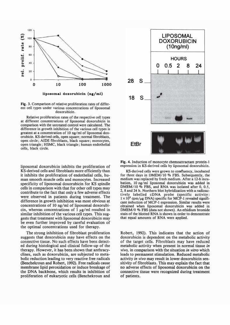
Fig. 3. Comparison of relative proliferation rates of differ· ent cell types under various concentrations cf liposomal
doxorubicin .
Relative proliferation rates of the respective cell types at different concentrations cf IiposomaJ doxorubicin in comparison with the untreated contral wert ca1cu]ated. Tbc difference in growth inhibition cf the various cell types is greatest al a conccntration cf 10 nglml cf liposomal doxorubicin. KS-derived cells, open square; normal fibroblasts, open circ1e i AIDS fibroblasts, black square; monocytes. open triangle ; HSMC, black triangle ; human endothelial cells, black cirele.
liposomal doxorubiein inhibits the proliferation of KS-derived ceUs and fibroblasts more efficienUy than it inhibits the proliferation of endothelial cells, human smooth muscle cells and monocytes. Increased specificity of liposomal doxorubicin for KS spindie cells in comparison with that for other cell types may contribute to the fact that only a few adverse effects were observed in patients during treatment. The difference in growth inhibition was most obvious at concentrations of 10 ng/ ml of liposomal doxorubiein, whereas concentrations of I f'g / ml resulted in similar inhibition of tbe various cell types . This suggests that treatment with liposomal doxonibiein may be even further improved by careful evaluation of the optimal concentrations uSed for therapy.
The strong inhibition of fibroblast proliferation suggests that doxorubicin may bave effects on the connective tissue. No such effeets have been detected during histological and clinical follow-up of the tberapy. However, it has been shown tbat anthracydines, such as doxorubicin, are subjected to metabolie reduction leading to very reactive free radieals (Benchekroun and Robert, 1992). Free radicals cause membrane lipid peroxidation or induce breakage of tbe DNA backbone, whicb results in inhibition of proliferation of eukaryotic ceUs (Bencbekroun and
28 S
18 S
EtBr
LlPOSOMAL DOXORUBICIN
(10ng/ml)
HOURS
o 0.5 2 8 24
, Oll
-
Flg. 4 . Induction of monocyte chemoattractant protein-] expression in KS-derived ceUs by liposomal doxorubicin.
KS-dcrived cells were grown to confluency. incubated for thr« days in OMEM/ lO "10 FBS. Subsequently, the medium was replaced by fresh medium. After a 12·h incubation, 10 ng/ ml liposomal doxorubicin was added in OMEM/ IO "10 FBS, and RNA was isolated after 0, 0.5, 2, 8 and 24 h. Nonhern blot hybridization wirh a radioactively labelIed cONA probe (specific activity : I x 10' cpm/~g DNA) specific for MCP-I reveaJed significant induetion of MCP-l expression. Similar results were obtained when liposomal doxorubicin was added in DMEM/ O "10 FBS (data not shown). An ethidiwn bromide stain of the biotted RNA is shown in order to demonstrate that equal amounts of RNA were applied.
Robert, 1992). This indicates tbat the action of doxorubicin is dependent on the metabolie activity of the target cells. Fibroblasts may have reduced metaboüc activity when present in normal tissue in vivo, in comparison witb tbe situation in vi/ro which leads to permanent stimulation. Reduced metaboüc activity in vivo may result in lower doxorubicin sensitivity of fibroblasts . This may explain tbe fact that no adverse effeets of liposomal doxorubicin on the conneetive tissue were reeognized during treatment of patients.

The histological follow~up of therapeutic regres~ sion of KS lesions demonstrated the absence of the KS spindie cells in involved areas. Areas where le~
sions in a plaque or nodular stage were present be~ fore therapy reveaIed either significant reduction or complete absence of tumour-like structures. Necrosis or pronounced haemorrhagia were not observed. The removaI of the KS spindie cells without deteetable necrosis suggests that, besides the cytostatic effeet of liposomal doxorubicin. further mechanisms may be involved in tumour regression during therapy. In this eontext two mechanisms can be imagined: (i) migration of KS spindie eells out of the lesion and (ii) involvement of phagoeyT.es which may contribute to the removaI of necrotic spindie cells under cytoslasis.
Support for the second mechanism was obtained by the observation that liposomaI doxorubicin in concentrations of 10 ng/mJ induces the expression of monoeyte ehemoattraetant protein-! in KS-derived cells. These concentrations of liposomal doxorubiein cause eomplete cyT.ostasis of KS-derived cells, whereas monocytes are only marginally affected. These data suggest that inereased migration of phagocytotic eells (e,g. siderophages) may indeed contribute to KS regression during treatment with Jiposomal doxorubiein.
In summary, we provide evidence that KS spindie cells are more sensitive than other cells to the CyT.ostatic effects of liposomal doxorubicin. Together with the increased drug delivery into KS lesions, which has been suggested by earlier reports, this finding may explain the high efficacy of liposomal doxorubicin in the treatment of AIOS-KS.
ACkDowledu:emeDls
We would Like to lhank T. COUIIS and C. Hohenadl (MbPlanck-lnstilU! fUr Biochemie, Martiruried, Gcrmany) (or linguistic revision of Ihe manuscript.
We graterully acknowledge ÜJac financiaJ suppon was provided by Ihe editorial board or the Münchner Medizinische Wochenzeitschrift. In addition, lhis work was supponed by grants from the Bundcsministerium für Forschung und Technologie (II.(JJJ-87, 1lI-002-89 and OI-ZU-86(7). M.S. was sponsored by the Irene Vogeler Award for Cancer Research.
La doxorubicine en liposomes dans le traitement du sarcome de Kaposi lie au SIDA;
evaluation clinique, histologique et biologique ceUulaire
Huit sujets atteints de sarcome de Kaposi (SK) associe au SIDA (SK*SIDA) ont ete traites par des liposomes porteurs de doxorubicine, a raison de
20 mg/m 2/perfusion. Apres 6 perfusions adminis* trees a des intervalles de 3 semaines, on observe une regression du sarcome chez tous les sujets. Le volume tumoraI, evalue par ultrasons, est passe de 556 ±635 a 42± 134!L1. L'anaIyse histologique revele une reduction des structures de type tumoral et une absence de cellules SK fusiformes dans les zones tumorales en involution apres ce traitement. L'etude in vitro, sur des cultures de SK, montre que deux mecanismes peuvent ~tre impliques dans I'action reductrice de la doxorubicine: (I) une forte inhibition specifique de Ja proliferation des cellules fusiformes et (2) une induction de I'expression de la proteine-I chimioattractive monocytaire dans les cellules fusiformes du SK, pouvant accroitre Je recru* tement des cellules phagocytaires (monocytes/macrophages) dans les lesions. Une synergie d'action de ces deux. mecanismes peut expliquer la grande efficacite de la doxorubieine en liposomes dans Je traitement du SK-SIDA.
Mols*cJes: SIOA, Sarcome de Kaposi, Doxorubicine, Liposome; Traitement, Cellules fusiformes, Proteine-) monocytaire.
References
Ackennan, A.B. & Gottlieb, G.J. (1988), Atlas ofthe gross and microscopic features, in "Kaposi's sarcoma: a text and atlas" (G.J. Gottlieb & A.B. Ackennan) (pp. 29-33). Lea & Febiger, Philadelphia.
Allen, T.M., Hansen, C., Martin , F., Redemann, C. & Yau-Young, A. (1991), Liposomes containing synthetic lipid derivatives of poly(ethylene glycol) show prolonged circulation half-Jives in vivo. Biochim. Biophys. Acta, 1066, 29-36.
Bally, N.B. , Nayar, R., Masin, n., Cullis, P.R. & Mayer, L.D. (1990), Studies on the myelosuppressive activity of doxorubiein entrapped in liposomes. Cancer Chemother. Pharmacol., 27, 13-19.
Benchekroun, M.N. & Robert, J. (1992), Measurement of doxorubicin-indueed lipid peroxidation under eonditions that detennine cytotoxieity in cultured tumor eeUs. Anal. Biochem., 201, 32&.330.
DOiner, J.R., Held, ·M. & Goebel, F.n. (1993a). Culaneous ultrasound for evaluation of Kaposi sareoma. J. AIDS, 6, 530-531.
Dogner, J .R., Zietz, C., Held, M. & Goebel, F.n. (l993b), Ultrasound as a tool to evaluate remission of cutaneous Kaposi sarcoma. AIDS, 7, 1081-1085.
FischI, M.A .• Krown, S.E., O'Doyle. K.P .• Mitsuyasu, R., Miles, S., Wernz, J.C., Volberding, P.A., Kahn, J ., Groopman, J.E., Feinberg, J., Woody, M. & the AIDS Clinical Trials Group (1993), Weekly doxorubicin in the treatment of patients with AIDS-related Kaposi's sareoma. J. AIDS, 6. 259-264.
Haverkos. H.W., Drotman, D.P. & Morgan, M. (1985), PrevaJence of Kaposi's sarcoma among patients with AIDS. N. Engl. J. Med .• 312. I.Sl8 .
Krown. S.E .• Metroka, C. & Wernz, J .C. (1989), Kapo-

si's sarcoma in the acquired immune deficiency syndrome: a proposal for uniform evaluation, response, and s!aging cri!eria. J. Clin. Oncol., 7, 1201-1207.
Papahadjopulos, 0., Gabizon, A., Mauhay, K., Huang, S.K., Lee, K.-D., Woodle, M.C., Lasic, 0.0., Redemann, C. & Martin, F.J. (1991), Sterically ~tabilized liposomes: improvements in pharmacokinetics and antitumor therapeutic erticacy. Proc. Natl. Acad. Sci. USA, 88, 11460-11464,
Rahman, A., Treat, J. & Roh,l.K. (1990), A phase I clinical !rial and pharmacokinetic evaluation of liposomeencapsula!ed doxorubicin. J. CUn. Oncol., 8, 1093-llDO.
ROlh, W.K., Werner, S., Risau, W., Remberger, K. & Hofschneider, P.H. (1988), Cultured, AIDS-related Kaposi's sarcoma cells express endothelial ceU markers and are weakly malignant in vj/ro. Int. J. Cancer, 42,767-713.
Safai, B., Johnson, K.G., Myskowski, P.L., Koziner, B.,
Vang, S.Y., Cunningham-Rundles, S., Godbold, I.H. & Dupont, B. (l98S), The natural bistory of Kaposi's sarcoma in the acquired immunodeficiency syndrome. Ann. intern. Med .• 103,744-715.
Sambrook, I., Fritsch, E.F. & Maniatis, T. (1989), Mo1ecular cloning: a !aborato!'}' manual, second edition. Cold Spring Harbor Laboratory, New York,
Stickler, M.C. & Friedman-Kien, A.E. (1991), Kaposi's S3!coma. Clin. Dermatol,. 9, 39-47.
Stürzl, M., Roth, W.K., Brockmeyer, N.H., Zietz, c., Speiser, B. & Hofschneider, P.H. (1992), Expression of platelet-derived growth factor and its receptor in AIDS-related Kaposi's sareoma in vivo 5Uggests paraerine and autoerine mechanisms of tumor maintenanee. Proc. Natl. Acad. Sei. USA, 89, 7046-7050.
Vaage, I., Mayhew, E., Lasic, D. & Martin, F. (1992), Therapy of primary and metastatic mouse mammary carcinoma with doxorubicin encapsulated in longcirculating liposomes. Inl. J. Cancer, Si, 942-948.