lipoprotein(a) upregulates abca1 in liver cells via …a) and ldl were quantified by measuring the...
TRANSCRIPT

1
Lipoprotein(a) upregulates ABCA1 in liver cells via scavenger receptor-B1 through its
oxidised phospholipids.
Monika Sharma1, Anne Von Zychlinski-Kleffmann
1, Carolyn M. Porteous
1, Gregory T.
Jones2, Michael J.A. Williams
3, and Sally P.A. McCormick
1*.
1Department of Biochemistry, University of Otago, Dunedin, New Zealand;
2Department of
Surgical Sciences, and 3Department of Medicine, Dunedin School of Medicine, University of
Otago, Dunedin, New Zealand.
* To whom correspondence should be addressed: Sally McCormick, Department of
Biochemistry, University of Otago, 710 Cumberland Street, Dunedin 9016, New Zealand.
Phone: (64 3) 479 7840; Fax: (64 3) 479 7866; E-mail: [email protected]
Running Title: Lp(a) upregulates ABCA1.
Abbreviations: ABCA1, ATP-binding cassette 1; CVD, cardiovascular disease; HDL, high
density lipoprotein; LDL, low density lipoprotein; Lp(a), lipoprotein(a); LXRα, liver X
receptor α; oxPL, oxidised phospholipids; PPARγ, peroxisome proliferator activated receptor
γ; SR-B1, scavenger receptor-B1.
by guest, on May 4, 2019
ww
w.jlr.org
Dow
nloaded from
2
Abstract:
Elevated levels of lipoprotein(a) [Lp(a)] are a well established risk factor for developing
cardiovascular disease (CVD). While Lp(a) levels are thought to be independent of other
plasma lipoproteins, some trials have reported a positive association between Lp(a) and high
density lipoproteins (HDL). Whether Lp(a) has a direct effect on HDL is not known. Here we
investigated if Lp(a) had any effect on the ATP-binding cassette 1 (ABCA1) pathway of HDL
production in liver cells. Incubation of HepG2 cells with Lp(a) upregulated the peroxisome
proliferator activated receptor γ (PPARγ) protein by 1.7 fold and the liver X receptor α
(LXRα) protein by 3 fold. This was accompanied by a 1.8 fold increase in ABCA1 protein
and a 1.5 fold increase in cholesterol efflux onto apolipoprotein A1 (apoA1). We showed that
Lp(a) was internalised by HepG2 cells, however, the ABCA1 response to Lp(a) was mediated
by the selective uptake of oxidised phospholipids (oxPL) from Lp(a) via the scavenger
receptor-B1 (SR-B1) and not by Lp(a) internalisation per se. We conclude that there is a
biological connection between Lp(a) and HDL through the ability of Lp(a)'s oxPL to
upregulate HDL biosynthesis.
Supplementary keywords: ABCA1, cholesterol efflux, HDL, lipoprotein(a), nuclear
receptor/LXR, nuclear receptor/PPAR, scavenger receptor-B1.
by guest, on May 4, 2019
ww
w.jlr.org
Dow
nloaded from

3
Introduction:
Lipoprotein(a) [Lp(a)] consists of a low density lipoprotein (LDL) linked by a disulphide
bond to apolipoprotein(a) [apo(a)] (1). Elevated levels of Lp(a) (>50 mg/dl) are an
independent risk factor for developing cardiovascular disease (CVD) (2). Lp(a) levels show a
strong correlation with oxidised phospholipids (oxPL) in plasma, more so than LDL (3).
Indeed the apo(a) moiety has been shown to bind oxPL (4). Oxidised lipids activate a number
of key molecules involved in atherosclerosis including the peroxisome proliferator activated
receptor γ (PPARγ) and liver X receptor α (LXRα) transcription factors (5). PPARγ and
LXRα upregulate scavenger receptors on macrophages to promote foam cell formation in the
artery but also upregulate the ATP binding cassette 1 (ABCA1) protein to promote
cholesterol efflux (5). In liver cells, PPARγ and LXRα promote ABCA1-mediated cholesterol
efflux to stimulate high density lipoprotein (HDL) formation (6-8).
Most but not all clinical studies show Lp(a) levels to be independent from that of other
plasma lipoproteins (2, 9). Detecting associations with Lp(a) is difficult since plasma levels
vary hugely within any given population (over a 1000 fold) and are skewed in distribution
towards low levels (2, 10, 11). A positive association between HDL and Lp(a) levels has been
reported in different epidemiological studies (12, 13, 14) but whether Lp(a) has any direct
biological effect on HDL is unknown. A recent study has documented that introduction of
low levels of Lp(a) into mice results in an increase in the HDL fraction (15). Past studies have
shown that low density lipoprotein (LDL) induces ABCA1-mediated cholesterol efflux in
both macrophage (16) and endothelial cells (17). As Lp(a) is structurally similar to LDL, one
might predict that it may also promote ABCA1-mediated intracellular cholesterol efflux and
could therefore promote HDL production.
The liver is the major site for Lp(a) catabolism (18) and HDL production (8). The cell surface
receptors responsible for Lp(a) uptake in the liver are not completely understood. Members of
the low density lipoprotein receptor (LDLR) family have been implicated in Lp(a) uptake.
by guest, on May 4, 2019
ww
w.jlr.org
Dow
nloaded from

4
The LDLR has been associated with Lp(a) uptake in familial hypercholesterolemia patients
(19) but definite proof of Lp(a) uptake by the LDLR in the liver is lacking. The very low
density lipoprotein receptor (VLDLR) is known to internalise Lp(a) (20), but is not expressed
by the liver. Kostner and colleagues reported that a significant amount of Lp(a) uptake in the
liver is mediated by the asialoglycoprotein receptor (21), however, subsequent in vivo
experiments have questioned this (18). A recent publication demonstrated enhanced uptake of
Lp(a) in cell lines and in transgenic mice overexpressing scavenger receptor-B1 (SR-B1) in
the liver providing evidence that SR-B1 is a receptor for Lp(a) (22).
Here we investigated whether Lp(a) had any effect on the ABCA1-mediated pathway of
cholesterol efflux in liver cells in order to establish if there was a biological connection
between Lp(a) and HDL. Our results showed that Lp(a) stimulates ABCA1 via activation of
PPARγ and LXRα resulting in a functional effect of enhanced cholesterol efflux to apoA1.
This prompted us to further investigate the role of SR-B1 and Lp(a) holoparticle uptake in
promoting the ABCA1 response.
by guest, on May 4, 2019
ww
w.jlr.org
Dow
nloaded from
5
MATERIALS AND METHODS
Lp(a) isolation
LDL and Lp(a) were isolated from a pooled plasma sample made from equal amounts of
plasma obtained from five healthy individuals with Lp(a) levels >50 mg/dL after informed
consent. Ethical approval for the study was granted by the Lower Regional South Ethics
committee. The lipid levels and apo(a) isoform sizes of the five samples are given in Table 1.
A pooled plasma sample was used to avoid any bias from using Lp(a) containing just one
apo(a) isoform. LDL and Lp(a) was isolated from the pooled plasma using a combination of
density ultracentrifugation (23) and fast protein liquid chromatography (FPLC) (24). Briefly,
4 ml of plasma was adjusted to a density of 1.063 g/ml with KBr and the adjusted plasma
overlaid with 1.063 g/ml KBr solution and subject to density ultracentrifugation in a Beckman
NVTi65 rotor for 3 hours at 10C at 60,000 rpm. The top layer containing VLDL and LDL
was fractionated by FPLC to isolate LDL from VLDL. The remaining plasma was adjusted to
a density of 1.12 g/ml with KBr and subjected to density ultracentrifugation at 100,000 rpm
for 4 hours at 10oC using a Beckman TL-100.3 ultracentrifuge. The top layer containing the
1.063-1.12 g/ml density fraction was fractionated by FPLC to isolate Lp(a) from HDL and
residual plasma proteins. Isolation of LDL and Lp(a) was also performed using 25 µM
butylated hydroxytoulene (BHT) and 0.26 mM EDTA throughout the process to establish the
effect on oxidised lipid content on both lipoproteins as measured by the TBARS assay. The
purity of the isolated LDL and Lp(a) was checked by lipoprotein electrophoresis on LIPO +
Lp(a) Hydragels using the HYDRASYS agarose gel electrophoresis system (Sebia, Norcross,
GA). Gels were stained with Fat Red 7B and subject to western blotting with anti-apo(a),
anti-apoB and anti-apoA1 antibodies. Lp(a) and LDL were quantified by measuring the total
protein content of each using the Qubit® Protein Assay kit (Life Technologies, Carlsbad, CA).
by guest, on May 4, 2019
ww
w.jlr.org
Dow
nloaded from

6
Cell culture
Hepatocellular carcinoma (HepG2) cells and human hepatoma (Hep3B) cells were obtained
from American Type Culture Collection (Manassas, VA). Cells were maintained in Advanced
Dulbecco's Modified Eagle Medium (DMEM) supplemented with 10% foetal bovine serum
(Bio International, Auckland, New Zealand), 2 mM L-glutamine, 0.25 µg/mL amphotericin
B, 100 U/mL penicillin, and 100 µg/mL streptomycin (Invitrogen, Carlsbad, CA) at 37°C in a
humidified environment with 5% CO2. HepG2 and Hep3B cells were checked for apo(a)
expression by western blot on cell lysate and media which showed the absence of apo(a) in
both. Twenty four hours after seeding HepG2 cells at 5 x 10⁵
density, cells were treated with
either purified Lp(a) or LDL at 1 μg/ml, 5 μg/ml and 10 μg/ml for 12 hours in DMEM
containing 1% serum. Hep3B cells were seeded at 2.5 x 10⁵
density and treated with purified
Lp(a) at 5 μg/ml for 12 hours. The trypan blue dye exclusion test (25) was performed to check
the viability of cells after Lp(a) treatment and we found the viability to be 70-80% with no
significant effect of Lp(a) concentration.
RT-PCR
Total RNA was extracted from Lp(a)-treated HepG2 cells using the RNeasy Mini Kit
(Qiagen, Venlo, Limburg) as per manufacturer’s instructions. On-column DNase digestion
was performed using the RNase free DNase Set (Qiagen). cDNA was synthesised from 1 μg
RNA using the first strand cDNA synthesis kit from Roche (Basel, Switzerland). Quantitative
RT-PCR was performed using the KAPA SYBR® FAST Universal 2X qPCR Master Mix
(Kapa Biosystems, Wilmington, MA) and cDNA as the template on a LightCycler® 480
(Roche). Specific primers spanning exonic regions in target genes, ABCA1, LXRα, PPARγ
and reference genes, β-2 microglobulin (β2M) and glyceraldehyde-3-phosphate
dehydrogenase (GAPDH) were designed (see supplementary Table 1 for primer sequences).
Expression of each target gene was quantified using Ct values after normalising to the two
reference genes.
by guest, on May 4, 2019
ww
w.jlr.org
Dow
nloaded from
7
Western blot
HepG2 cells were harvested in RIPA buffer (50 mM Tris-HCl pH-7.8, 150 mM NaCl, 0.1%
SDS, 0.5% sodium deoxycholate, 1% Triton X-100) supplemented with complete mini
protease inhibitors (Roche). Cell lysates containing 40 µg protein were resolved on 4%
polyacrylamide gels for apo(a), 7.5% gels for ABCA1 or on 10% gels for LXRα, PPARγ and
actin. Proteins were transferred to nitrocellulose membrane. After blocking with 5% skim
milk, membranes were incubated with primary antibodies specific for ABCA1 (ab7630),
LXRα (ab28478), PPARγ (ab27649) (all from Abcam, Cambridge, England), actin (A5060
from Sigma, St. Louis, MO) and the apo(a)-specific antibody, a5-hrp (26) at 4oC overnight.
After incubation with the appropriate secondary antibody, membranes were developed with
ECL reagents and imaged with a Fujifilm LAS3000 (R&D systems, Minneapolis, MN). The
quantification of blots was done using the ImageQuant TL software (Amersham Biosciences,
Piscataway, NJ) with proteins normalized against actin.
TBARS assay
The concentration of lipid oxidation products in the LDL and Lp(a) preparations was
measured using the thiobarbituric acid reactive substance (TBARS) (27) assay which detects
malondialdehyde (MDA) generated from oxidised lipids. The TBA-MDA adduct was
measured by fluorometric analysis (excitation: 544 nm; emission: 590 nm) in LDL and Lp(a)
samples using a POLARstar Optima (BMG Labtech, Offenburg, Germany).
Promoter activity assay
ABCA1 promoter activity was checked by luciferase assay (28). HepG2 cells were
transfected with 1 μg of the pGL4.10 luciferase reporter vector containing 700 bp of the
ABCA1 promoter region (from -1 to -699 bp) and 10 ng of internal control renilla vector
(phRL-SV40) for 48 hours using FuGENE® HD transfection reagent (Roche). Forty eight
hours after transfection, HepG2 cells were treated with 1 μg/ml, 5 μg/ml and 10 μg/ml Lp(a)
for 12 hours in serum free DMEM. Cell lysates were assayed for renilla and firefly luciferase
by guest, on May 4, 2019
ww
w.jlr.org
Dow
nloaded from

8
activity using the Dual Luciferase® Reporter Assay System (Promega, Madison, WI) as per
manufacturer’s instructions. Firefly luciferase measurements were normalised to renilla
luciferase measurements.
Cholesterol efflux assay
Cholesterol efflux assays on HepG2 cells were performed as described previously (29). Cells
were seeded in 12 well plates at 2 x 10⁵ cells per well. After 24 hours, cells were labelled with
0.5 μCi/ml of [1,2 3H (N)]-cholesterol in DMEM for another 48 hours. After incubation, cells
were treated with Lp(a) at 1 μg/ml, 5 μg/ml and 10 μg/ml concentrations for 12 hours in
serum free DMEM followed by treatment with 20 μg/ml purified apoA1 for 2 hours. Cells in
the plate were lysed with 0.1M NaOH and Optiphase Hisafe II scintillation fluid (Perkin
Elmer) was added to the medium and cell lysates. Tritium decay over 5 minutes in the
medium and cell lysates were measured as disintegrations per minute (dpm) using a liquid
scintillator analyzer (Perkin Elmer, Boston, MA). Non-specific efflux, in the absence of
apoA1, was determined and subtracted from each experimental measurement. Cholesterol
efflux was calculated using the following equation:
Cholesterol efflux = dpm (media with apoA1) - dpm (media without apoA1) / dpm (cells +
media) x 100
Immunohistochemistry
Lp(a) treated HepG2 cells were fixed with 4% paraformaldehyde and subsequently incubated
with AlexaFluor 594 Wheat Germ agglutinin (Invitrogen) membrane stain (5 µg/mL) for 10
minutes at room temperature. Cells were permeabilized and blocked with 3% goat serum in
0.1% Triton X-100 for 30 minutes at room temperature followed by an overnight incubation
with an apo(a)-specific monoclonal antibody, a5 (26) at 4oC. Cells were incubated with an
anti-mouse IgG AlexaFluor 488 (Invitrogen) for 2 hours at room temperature. Coverslips
were mounted with ProLong® Gold Antifade reagent with DAPI (Invitrogen). Images were
obtained using a Olympus FluoView™ FV1000 confocal microscope with the Argon (488
by guest, on May 4, 2019
ww
w.jlr.org
Dow
nloaded from

9
nm) and HeNe (633 nm) lasers. A series of images along the z axis at 0.5 μm step size from
top to bottom of the cell were collected. Image analysis and statistics were done using the
Olympus FluoView™ FV1000 Image Examiner software.
Antibody blocking
The SR-B1 receptor was blocked with 5 μg/ml of anti-SR-B1 antibody (Novus Biological,
Littleton, Colorado) preincubated with HepG2 cells for 3 hours at 37οC prior to Lp(a)
treatment. The SR-B1 receptor was also blocked with an inhibitor, BLT-1 (Merck Millipore,
Darmstadt, Germany) preincubated with HepG2 cells at 100 μM for 1 hour at 37οC prior to
Lp(a) treatment. Apo(a) internalisation was blocked with an anti-apo(a) polyclonal antibody
(Wako, Richmond, Virginia) preincubated with 5 μg/ml Lp(a) for 1 hour at room temperature
prior to incubation with HepG2 cells. In addition to blocking with anti-apo(a), the oxPL on
Lp(a) were blocked with 5 μg/ml of the E06 antibody (Avanti Polar Lipids, Alabaster,
Alabama) prior to Lp(a) treatment. RNA and protein was extracted from treated cells and
analysed for the ABCA1, PPARγ and LXRα mRNA and protein levels as described above.
Fluorescent lipid and protein labelling for uptake assay
Surface lipids in Lp(a) were labelled with Fast Dil-C18 (Invitrogen) by incubating 0.3 mg of
Dil with 100 μg of Lp(a) overnight at 37οC and removing unincorporated Dil on a Sephadex-
G-25 column. HepG2 cells were treated with 5 μg/ml of Dil-Lp(a) for 12 hours with and
without anti-SR-B1 and anti-apo(a) blocking. After 12 hours, cells were lysed in lysis buffer
(0.1% SDS and 0.1M NaOH) and the lysate quantified for Dil fluorescence. Protein labelling
of Lp(a) was done using an apo(a)-specific primary antibody, a5 (26) in combination with an
AlexaFluor 488 IgG secondary antibody. HepG2 cells were treated with 5 μg/ml of Lp(a) for
12 hours with and without anti-SR-B1 and anti-apo(a) blocking. After 12 hours, cells were
fixed, permeabilized and probed against anti-apo(a) followed by the AlexaFluor 488
secondary antibody and fluorescence quantified using the POLARstar Optima (BMG
Labtech, Offenburg, Germany).
by guest, on May 4, 2019
ww
w.jlr.org
Dow
nloaded from

10
Statistical Analysis
Data are expressed as mean ± SEM. Differences between means were analyzed by the
unpaired Student’s t-test.
by guest, on May 4, 2019
ww
w.jlr.org
Dow
nloaded from

11
RESULTS
Characterisation of purified Lp(a)
Agarose gel electrophoresis of the Lp(a) and LDL purified from pooled plasma showed
single bands in the Lp(a) and LDL position after staining with Fat Red7B (Supplementary
Fig. 1A). The Lp(a) band was detected by both anti-apo(a) (Supplementary Fig. 1B) and anti-
apoB antibodies (Supplementary Fig. 1C) while the LDL band was only detected by the anti-
apoB antibody (Supplementary Fig. 1C). Neither preparation showed contamination from
other apoB-containing lipoproteins (Supplementary Fig. 1C). A western blot with an anti-
ApoA1 antibody (Supplementary Fig. 1D) also showed no contamination with HDL.
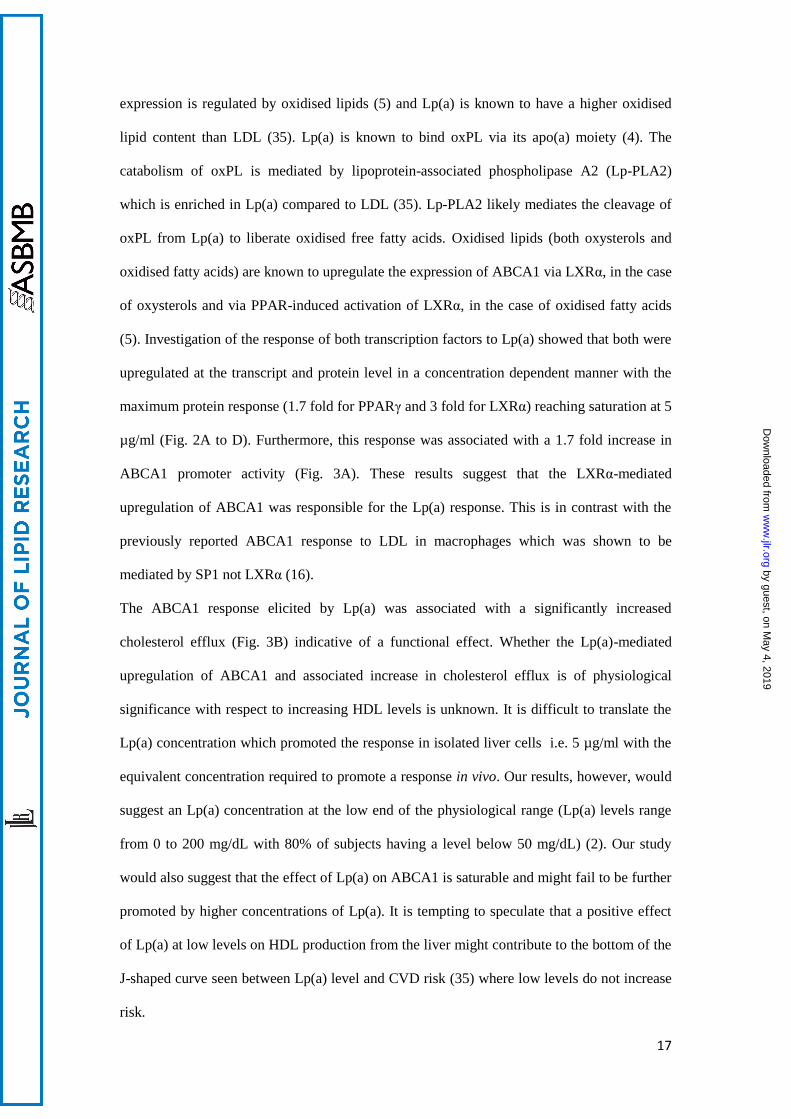
Lp(a) induces ABCA1 expression
Treatment of HepG2 cells with 1 to 10 μg/ml of purified Lp(a) for 12 hours resulted in a
significant increase in ABCA1 mRNA levels up to 4.0 fold at 5 μg/ml and 2.8 fold at 10
μg/ml (Fig. 1A). ABCA1 protein levels were increased significantly by 1.3 fold at 1 μg/ml
and 1.8 fold at 5 and 10 μg/ml Lp(a) (Fig. 1B). Treatment of HepG2 cells with LDL also
stimulated ABCA1 expression but not to the extent seen by Lp(a) and not in a concentration
dependent manner with ABCA1 protein levels showing a 1.2 fold increase at 1, 5 and 10
μg/ml (Fig. 1C). We hypothesised that the greater response of ABCA1 to Lp(a) compared to
LDL may be related to a higher oxidised lipid content. A TBARS assay to assess the MDA
content of the purified Lp(a) and LDL samples showed the MDA content of Lp(a) to be
significantly higher than that of LDL (206 nM versus 164 nM respectively (Fig. 1D).
Addition of BHT and EDTA throughout the purification process gave a reduced MDA
content in both lipoproteins albeit still higher in Lp(a) (34 nM versus 14 nM respectively (Fig.
1E). This indicates that MDA is mainly a measure of oxidised lipids generated in the
purification process rather than innate content.
by guest, on May 4, 2019
ww
w.jlr.org
Dow
nloaded from

12
Lp(a) induced ABCA1 expression is under LXR regulation and stimulates cholesterol
efflux
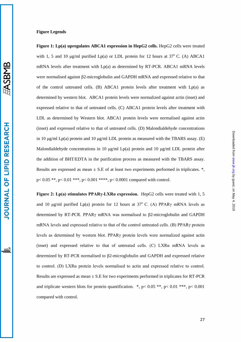
To study the mechanism underlying the regulation of ABCA1 expression upon Lp(a)
treatment, we measured the transcript levels of the PPARγ and LXRα transcription factors
which are known to upregulate ABCA1. The PPARγ transcript showed a significant increase
up to 2 fold at 5 μg/ml and 10 μg/ml Lp(a) (Fig. 2A). PPARγ protein levels were increased to
1.7 fold at 5 μg/ml and 1.3 fold at 10 μg/ml Lp(a) (Fig. 2B). The LXRα transcript was
significantly elevated by 2.3 fold at 5 μg/ml and 1.7 fold at 10 μg/ml Lp(a) (Fig. 2C). This
was associated with a significant increase in LXRα protein levels up to 3 fold at 5 μg/ml and
1.7 fold at 10 μg/ml (Fig. 2D). To further confirm the response of liver cells to Lp(a), we
repeated the experiments shown in figures 1A-B and 2A-D in another hepatoma cell line,
Hep3B. Incubation of Hep3B cells with 5 μg/ml Lp(a) also increased the ABCA1, PPARγ
and LXRα transcripts and proteins showing a similar (albeit lesser fold) response to that seen
in the HepG2 cell line (Supplementary Fig. 2).
A luciferase promoter assay was performed to see if the ABCA1 promoter was activated by
Lp(a). The LXR agonist T0901317 (2 μM) was included as a positive control. ABCA1
promoter activity was significantly increased up to 1.7 fold at 5 μg/ml Lp(a) and 1.4 fold at 10
μg/ml Lp(a) (Fig. 3A). To study whether the upregulation in the ABCA1 pathway by Lp(a)
had any functional significance, we performed cholesterol efflux assays on the treated cells.
Lp(a) treatment at 5 μg/ml promoted a 1.5 fold increase in cholesterol efflux onto apoA1 as
compared to untreated cells. An increase in efflux was also mediated by 1 and 10 μg/ml Lp(a)
but the increase was less than that seen at 5 μg/ml (Fig. 3B).
Internalisation of Lp(a) by HepG2 cells
To check if Lp(a) was being internalised, cell lysates from treated cells were subjected to
western blots with an anti-apo(a) monoclonal antibody (Fig. 4A). This showed the presence
of multiple apo(a) bands (resulting from the multiple apo(a) isoforms in the pooled plasma
by guest, on May 4, 2019
ww
w.jlr.org
Dow
nloaded from
13
used for Lp(a) isolation) indicative of Lp(a) uptake by cells. To confirm uptake further,
confocal microscopy of treated cells was performed to visualise apo(a) within the cells (Fig.
4B). The apo(a) signal (green) was concentrated within the bounds of the cells stained by the
WGA membrane-specific stain (red) giving rise to large areas of colocalisation (indicated as
yellow in the merged image). Z stacking of the merged confocal image was performed to
confirm that apo(a) was found throughout the cell and not just at the cell surface (Fig. 4C).
This showed the apo(a) signal (green) colocalised with the WGA signal (red) throughout the
volume of the cell confirming Lp(a) internalisation. As the cells were permeabilized to allow
internalisation of the anti-apo(a) antibody, the WGA stain was also internalised leading to
staining of membranous structures throughout the cell.
SR-B1 mediates the ABCA1 response to Lp(a) through oxPL
It has recently been reported that SR-B1 is a receptor for Lp(a) (22). Therefore, we
investigated whether the ABCA1 response to Lp(a) was mediated by the SR-B1 receptor by
using an antibody blocking approach. Preincubation of cells with an anti-SR-B1 antibody
before Lp(a) treatment significantly reduced the ABCA1 response to Lp(a) treatment both at
the transcript and protein level (Fig. 5A and B). Furthermore, the upregulation of the PPARγ
and LXRα transcripts and proteins was significantly reduced in response to the anti-SR-B1
antibody (Fig. 5C-F). In contrast, preincubation with an anti-apo(a) to block whole Lp(a)
particle uptake (assuming that this didn't also effect the SR-B1-mediated uptake) had no
effect on ABCA1 transcript and protein levels with levels remaining similar to that observed
in Lp(a) treated HepG2 cells without antibody (Fig. 5A, 5B). The levels of the PPARγ and
LXRα transcripts were reduced with anti-apo(a) treatment (Fig. 5C and E) but not to the
extent seen with anti-SR-B1 treatment and the proteins levels of both PPARγ and LXRα were
unaffected by the anti-apo(a) antibody treatment (Fig. 5D and F). Interestingly, a combination
of anti-apo(a) and the E06 antibody which binds the oxPL found in Lp(a) particles resulted in
a significant decrease in the levels of ABCA1 transcript and protein, PPARγ transcript and
by guest, on May 4, 2019
ww
w.jlr.org
Dow
nloaded from

14
LXRα transcript and protein levels (Fig. 6). Indeed the decreases were similar to that seen
with blocking SR-B1 (Fig. 5).
SR-B1 mediates Lp(a) lipid but not protein uptake
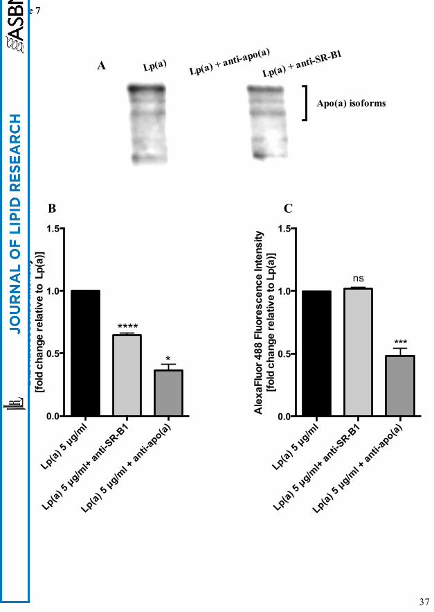
To confirm if both the anti-SR-B1 and anti-apo(a) antibodies had indeed blocked Lp(a)
uptake, we visualised Lp(a) uptake by western blot of cell lysates with an anti-apo(a)
antibody (Fig. 7A). The SR-B1 antibody did not block the uptake of Lp(a) whereas the anti-
apo(a) antibody completely blocked Lp(a) uptake. These results suggest that the ABCA1
response to Lp(a) might be mediated by selective lipid uptake from Lp(a) via SR-B1 and not
whole particle uptake. To investigate this further, we labelled Lp(a) with Dil to label surface
lipids and an anti-apo(a) antibody in combination with AlexaFluor 488 to label the protein
component and then determined the effect of the SR-B1 antibody on lipid and protein uptake.
The relative fluorescence intensity of Dil in cell lysates was reduced by 35% with anti-SR-B1
blocking (Fig. 7B) but the relative fluorescence of the protein component of Lp(a) showed no
change in fluorescence intensity (Fig. 7C) indicating that the SR-B1 antibody was only
affecting lipid transfer and not protein uptake. Treatment with the anti-apo(a) antibody, on the
other hand, did block both lipid and protein uptake from Lp(a) into HepG2 cells as indicated
by a significant reduction in Dil and AlexaFluor 488 relative fluorescence (Fig. 7B and C)
which we interpreted as a blocking of whole particle uptake.
To further confirm the role of SR-B1 in mediating the ABCA1 response to Lp(a), we also
treated cells with the SR-B1 inhibitor, BLT-1. BLT-1 also significantly reduced the ABCA1
protein response (Supplementary Fig. 3A). Furthermore, the relative fluorescence intensity of
Dil in cells pretreated with BLT-1 before incubation with Dil-labelled Lp(a), was reduced by
35% (Supplementary Fig. 3B). In contrast, the relative fluorescence of Lp(a) protein in cells,
detected with an anti-apo(a) antibody and AlexaFluor 488, showed no change in fluorescence
intensity with BLT-1 treatment (Supplementary Fig. 3C). The results obtained with BLT-1
by guest, on May 4, 2019
ww
w.jlr.org
Dow
nloaded from

15
reiterated the results obtained with the SR-B1 antibody showing that SR-B1 was mediating
selective lipid transfer from Lp(a) and not protein uptake.
by guest, on May 4, 2019
ww
w.jlr.org
Dow
nloaded from

16
Discussion
Our study has uncovered a new connection between Lp(a) and HDL by demonstrating that
Lp(a) upregulates the HDL production pathway in liver cells. We show that this effect is due
to transfer of oxPL from Lp(a) via the SR-B1 receptor leading to an upregulation of the
PPARγ-LXRα-ABCA1 pathway and resulting in an enhanced cholesterol efflux to apoA1.
This effect is specific to SR-B1 and independent of Lp(a) holoparticle uptake by liver cells.
Lp(a) has proven to be an important risk factor for developing CVD. Most trials report Lp(a)
as having an effect on CVD independent of other plasma lipoproteins (2). Kinetics studies
show that Lp(a) is metabolised differently to that of other apoB-containing lipoproteins (30)
and proteomics studies report Lp(a) to have a unique protein composition (31). There are
obvious connections between Lp(a) and LDL as both molecules share similar functional
properties with respect to their binding to structural proteins (32) and receptors from the
LDLR family (33, 34). A connection between Lp(a) and HDL is less obvious but there is
some evidence that Lp(a) levels show a positive association with HDL in some populations
(12, 13, 14). Here we aimed to investigate if there was a direct connection between Lp(a) on
HDL by testing the affect of Lp(a) on the ABCA1 mediated pathway of HDL production.
HepG2 cells were chosen for this study since the expression of ABCA1 in the liver is the
major contributor to HDL production (8). Lp(a) was purified from a pooled plasma sample to
avoid bias from working with just one or two apo(a) isoforms. We used LDL isolated from
the same pooled sample as a control for these studies as it had been previously reported that
LDL increases ABCA1 activity in macrophages (16). Incubation of HepG2 cells with Lp(a)
showed a significant upregulation in ABCA1 at both the transcript and protein level reaching
a saturable effect by 5 µg/ml (Fig. 1A and B). In comparison, incubation of HepG2 cells with
LDL showed the ABCA1 protein to be only modestly increased with no effect of
concentration (Fig. 1C).
As Lp(a) showed a more robust response in upregulating ABCA1 compared to LDL, we
considered that this might be due to a higher oxidised lipid content of Lp(a). ABCA1
by guest, on May 4, 2019
ww
w.jlr.org
Dow
nloaded from
17
expression is regulated by oxidised lipids (5) and Lp(a) is known to have a higher oxidised
lipid content than LDL (35). Lp(a) is known to bind oxPL via its apo(a) moiety (4). The
catabolism of oxPL is mediated by lipoprotein-associated phospholipase A2 (Lp-PLA2)
which is enriched in Lp(a) compared to LDL (35). Lp-PLA2 likely mediates the cleavage of
oxPL from Lp(a) to liberate oxidised free fatty acids. Oxidised lipids (both oxysterols and
oxidised fatty acids) are known to upregulate the expression of ABCA1 via LXRα, in the case
of oxysterols and via PPAR-induced activation of LXRα, in the case of oxidised fatty acids
(5). Investigation of the response of both transcription factors to Lp(a) showed that both were
upregulated at the transcript and protein level in a concentration dependent manner with the
maximum protein response (1.7 fold for PPARγ and 3 fold for LXRα) reaching saturation at 5
µg/ml (Fig. 2A to D). Furthermore, this response was associated with a 1.7 fold increase in
ABCA1 promoter activity (Fig. 3A). These results suggest that the LXRα-mediated
upregulation of ABCA1 was responsible for the Lp(a) response. This is in contrast with the
previously reported ABCA1 response to LDL in macrophages which was shown to be
mediated by SP1 not LXRα (16).
The ABCA1 response elicited by Lp(a) was associated with a significantly increased
cholesterol efflux (Fig. 3B) indicative of a functional effect. Whether the Lp(a)-mediated
upregulation of ABCA1 and associated increase in cholesterol efflux is of physiological
significance with respect to increasing HDL levels is unknown. It is difficult to translate the
Lp(a) concentration which promoted the response in isolated liver cells i.e. 5 µg/ml with the
equivalent concentration required to promote a response in vivo. Our results, however, would
suggest an Lp(a) concentration at the low end of the physiological range (Lp(a) levels range
from 0 to 200 mg/dL with 80% of subjects having a level below 50 mg/dL) (2). Our study
would also suggest that the effect of Lp(a) on ABCA1 is saturable and might fail to be further
promoted by higher concentrations of Lp(a). It is tempting to speculate that a positive effect
of Lp(a) at low levels on HDL production from the liver might contribute to the bottom of the
J-shaped curve seen between Lp(a) level and CVD risk (35) where low levels do not increase
risk.
by guest, on May 4, 2019
ww
w.jlr.org
Dow
nloaded from

18
We hypothesized that a receptor-mediated uptake of Lp(a) would be necessary to elicit the
effect on ABCA1. Western blots of lysates from Lp(a)-treated cells showed the presence of
multiple apo(a)-sized isoforms suggesting uptake of all isoforms present (Fig. 4A).
Immunohistochemistry studies showed Lp(a) to be present throughout the liver cells
colocalising with membranous fractions throughout the cells as shown by Z-stacking (Fig.
4C). Various receptors have been reported to bind Lp(a) including LDLR (33), LRP (34),
megalin (36), VLDLR (20), as well as asialoglycoprotein receptors (ASGPR) (21),
plasminogen receptors (37) and more recently SR-B1 (22). Neither VLDLR nor megalin are
expressed by the liver (20, 36) and evidence suggests Lp(a) is a reasonably poor ligand for
LDLR and LRP (34). As the LXR-ABCA1 pathway is activated by oxidised lipids which are
a well known ligand for the SR-B1 receptor (38) and Lp(a) is known to specifically
accumulate oxPL (35), we therefore investigated the role of SR-B1 in eliciting the Lp(a)-
mediated ABCA1 response. Indeed, antibody-mediated blocking of the SR-B1 receptor
effectively blocked the PPAR-LXR-ABCA1 response to Lp(a) (Fig. 5). Interestingly,
labelling of the lipid and protein component of Lp(a) showed that blocking SR-B1 (either by
the SR-B1 antibody or by the SR-B1 inhibitor, BLT-1) only affected the uptake of Lp(a) lipid
not protein suggesting that SR-B1 mediates selective lipid uptake from Lp(a). Indeed Yang et
al have recently shown that SR-B1 does mediate selective uptake of lipids from Lp(a) (22).
Further support for this was gained when an experiment using the E06 antibody, which binds
to oxPL, showed effective blocking of the ABCA1 response (Fig. 6A, B). Collectively our
results show that the selective uptake of oxPL from Lp(a) is the effector of the ABCA1
response.
Although we have identified selective lipid uptake from Lp(a) via SR-B1 as being responsible
for the ABCA1 response, our results clearly showed there was an independent uptake of
apo(a) into liver cells. This was effectively blocked by an anti-apo(a) antibody which blocked
both the Lp(a) lipid and apo(a) protein component indicative of whole Lp(a) particle uptake.
The receptor(s) responsible for whole Lp(a) particle uptake in the liver are not certain. The
by guest, on May 4, 2019
ww
w.jlr.org
Dow
nloaded from

19
LDLR, LRP, asialoglycoprotein and plasminogen receptors are all potential candidates. Both
LDLR and LRP bind to apoB and apoE moieties on the LDL particle (39) although these may
be masked by the presence of apo(a) in Lp(a). The fact that an apo(a) antibody blocked the
whole Lp(a) particle uptake may hint at the plasminogen receptor as this receptor specifically
interacts with the apo(a) component of Lp(a) (37). Alternatively, the asialoglycoprotein
receptor which interacts with N-glycosylated residues on apo(a) (21) could also be involved.
Whether it is solely the lipid component of Lp(a) that interacts with SR-B1 for uptake or
whether apo(a) or apoB are involved is not known. Interestingly, blocking with an apo(a)
antibody did not affect the selective lipid uptake from Lp(a) (Fig. 7C) suggesting that apo(a)
may not be involved.
From a clinical perspective we expect that the ABCA1 response mediated by Lp(a) might
show variation between individuals depending on the lipid and protein composition of
individual Lp(a) particles. The oxPL content of Lp(a) particles vary (40) as does the protein
make-up as recently shown by a quantitative proteomic study of individual Lp(a) particles
(41). Indeed, the response could vary within an individual since most individuals have two
apo(a) isoforms giving rise to two different species of Lp(a) particle which may differ in
protein and lipid composition. Although our results show that Lp(a) upregulates HDL
production, these results still need to be proven in vivo. Another alterative hypothesis to
consider in vivo is that Lp(a) may compete with HDL for binding to SR-B1 thus reducing the
selective uptake of HDL lipids and increasing HDL levels.
In conclusion, we have uncovered a novel connection between Lp(a) and HDL showing that
Lp(a) upregulates ABCA1 in liver cells via selective uptake of oxPL from Lp(a) by SR-B1.
Uncovering this response provides new insight into Lp(a) metabolism by liver cells and may
prompt further investigation into the associations between Lp(a) and HDL in clinical settings.
by guest, on May 4, 2019
ww
w.jlr.org
Dow
nloaded from

20
Acknowledgements
The authors wish to thank Professor Santica Marcovina for supplying the MAb a-5 apo(a)
monoclonal antibody. The authors also wish thank Dr. Elizabeth Duncan for her assistance
with the confocal microscopy.
Sources of Funding
This work was supported by the New Zealand Heart Foundation (NHF) and Lottery Health
Research and a University of Otago postgraduate scholarship to MS.
Disclosures
None relevant to this study.
by guest, on May 4, 2019
ww
w.jlr.org
Dow
nloaded from

21
References
1. Utermann, G. 1989. The mysteries of lipoprotein(a). Science. 246: 904–10.
2. Nordestgaard, B.G., M. J. Chapman, K. Ray, J. Borén, F. Andreotti, G. F. Watts, H.
Ginsberg, P. Amarenco, A. Catapano, O. S. Descamps, E. Fisher, P. T. Kovanen, J. A.
Kuivenhoven, P. Lesnik, L. Masana, Z. Reiner, M. R. Taskinen, L. Tokgözoglu and A.
Tybjærg-Hansen; European Atherosclerosis Society Consensus Panel. 2010. Lipoprotein(a) as
a cardiovascular risk factor: current status. Eur. Heart J. 31: 2844–53.
3. Bergmark, C., A. Dewan, A. Orsoni, E. Merki, E. R. Miller, M. J. Shin, C. J. Binder, S.
Hörkkö, R. M. Krauss, M. J. Chapman, J. L. Witztum, and S. Tsimikas. 2008. A novel
function of lipoprotein [a] as a preferential carrier of oxidized phospholipids in human
plasma. J. Lipid Res. 49: 2230–9.
4. Leibundgut, G., C, Scipione, H. Yin, M. Schneider, M. B. Boffa, S. Green, X. Yang, E.
Dennis, J. L. Witztum, M. L. Koschinsky, and S. Tsimikas. 2013. Determinants of binding of
oxidized phospholipids on apolipoprotein (a) and lipoprotein (a). J. Lipid Res. 54: 2815-30.
5. Chawla, A., W. A. Boisvert, C.H. Lee, B. A. Laffitte, Y. Barak , S. B. Joseph, D. Liao, L.
Nagy, P. A. Edwards, L. K. Curtiss, R. M. Evans, and P. Tontonoz. 2001. A PPAR gamma-
LXR-ABCA1 pathway in macrophages is involved in cholesterol efflux and atherogenesis.
Mol. Cell. 7: 161–71.
6. Hossain, M. A., M. Tsujita, F. J. Gonzalez, and S. Yokoyama. 2008. Effects of fibrate
drugs on expression of ABCA1 and HDL biogenesis in hepatocytes. J. Cardiovasc.
Pharmacol. 51: 258–66.
7. Miao, B., S. Zondlo, S. Gibbs, D. Cromley, V. P. Hosagrahara, T. G. Kirchgessner, J.
Billheimer, and R. Mukherjee. 2004. Raising HDL cholesterol without inducing hepatic
steatosis and hypertriglyceridemia by a selective LXR modulator. J. Lipid Res. 45: 1410–7.
by guest, on May 4, 2019
ww
w.jlr.org
Dow
nloaded from

22
8. Basso, F., L. Freeman, C. L. Knapper, A. Remaley, J. Stonik, E. B. Neufeld, T. Tansey, M.
J. Amar, J. Fruchart-Najib, N. Duverger, S. Santamarina-Fojo, and H. B. Brewer. 2003. Role
of the hepatic ABCA1 transporter in modulating intrahepatic cholesterol and plasma HDL
cholesterol concentrations. J. Lipid Res. 44: 296–302.
9. Luc, G., J. M. Bard, D. Arveiler, J. Ferrieres, A. Evans, P. Amouyel, J. C. Fruchart, and P.
Ducimetiere; PRIME Study Group. 2002. Lipoprotein (a) as a predictor of coronary heart
disease: the PRIME Study. Atherosclerosis. 163: 377–84.
10. Gaw, A., H. H. Hobbs. 1994. Molecular genetics of lipoprotein (a): new pieces to the
puzzle. Curr. Opin. Lipidol. 5: 149-55.
11. Marcovina, S. M., J. J. Albers, E. Wijsman, Z. Zhang, N. H. Chapman, and H. Kennedy.
1996. Differences in Lp[a] concentrations and apo[a] polymorphs between black and white
Americans. J. Lipid Res. 37: 2569–85.
12. Giger, J.N., O. L. Strickland, M. Weaver, H. Taylor, and R. T. Acton. 2005. Genetic
predictors of coronary heart disease risk factors in premenopausal African-American women.
Ethn. Dis. 15: 221–32.
13. Sharma, S., J. Merchant, and S. E. Fleming. 2012. Lp(a)-cholesterol is associated with
HDL-cholesterol in overweight and obese African American children and is not an
independent risk factor for CVD. Cardiovasc. Diabetol. 11: 1–10.
14. Tsimikas, S., P. Clopton, E. S. Brilakis, S. M. Marcovina, A. Khera, E. R. Miller, J. A. de
Lemos and J. L. Witztum. 2009. Relationship of oxidized phospholipids on apolipoprotein B-
100 particles to race/ethnicity, apolipoprotein(a) isoform size, and cardiovascular risk factors:
results from the Dallas Heart Study. Circulation. 119: 1711-9.
15. Rodger, E. J., R. J. Suetani, G. T. Jones, T. Kleffmann, A. Carne, M. Legge, and S. P.
McCormick. 2012. Proteomic Analysis of Aortae from Human Lipoprotein(a) Transgenic
by guest, on May 4, 2019
ww
w.jlr.org
Dow
nloaded from

23
Mice Shows an Early Metabolic Response Independent of Atherosclerosis. PLoS ONE. 7:
e30383.
16. Chen, X., Y. Zhao, Z. Guo, L. Zhou, E. U. Okoro, and H. Yang. 2011. Transcriptional
regulation of ATP-binding cassette transporter A1 expression by a novel signaling pathway.
J. Biol. Chem. 286: 8917–23.
17. Liao, H., T. Langmann, G. Schmitz, and Y. Zhu. 2002. Native LDL Upregulation of ATP-
Binding Cassette Transporter-1 in Human Vascular Endothelial Cells. Arterioscler. Thromb.
Vasc. Biol. 22: 127–32.
18. Cain, W. J., J. S. Millar, A. S. Himebauch, U. J. Tietge, C. Maugeais, D. Usher, and D. J.
Rader. 2005. Lipoprotein [a] is cleared from the plasma primarily by the liver in a process
mediated by apolipoprotein [a]. J. Lipid Res. 46: 2681–91.
19. Kraft, H.G., A. Lingenhel, F. J. Raal, M. Hohenegger, and G. Utermann. 2000.
Lipoprotein(a) in Homozygous Familial Hypercholesterolemia. Arterioscler. Thromb. Vasc.
Biol. 20: 522–8.
20. Argraves, K. M., K. F. Kozarsky, J. T. Fallon, P. C. Harpel, and D. K. Strickland. 1997.
The atherogenic lipoprotein Lp(a) is internalized and degraded in a process mediated by the
VLDL receptor. J. Clin. Invest. 100: 2170–81.
21. Hrzenjak, A., S. Frank, X. Wo, Y. Zhou, T. B. Van, and G. M. Kostner. 2003. Galactose-
specific asialoglycoprotein receptor is involved in lipoprotein (a) catabolism. Biochem. J.
376: 765–71.
22. Yang, X. P., M. J. Amar, B. Vaisman, A. V. Bocharov, T. G. Vishnyakova, L. A.
Freeman, R. J. Kurlander, A. P. Patterson, L. C. Becker, and A. T. Remaley. 2013. Scavenger
receptor-BI is a receptor for lipoprotein(a). J. Lipid Res. 54: 2450–7.
by guest, on May 4, 2019
ww
w.jlr.org
Dow
nloaded from

24
23. Havel, R. J., H. A. Eder, and J. H. Bragdon. 1955. The distribution and chemical
composition of ultracentrifugally separated lipoproteins in human serum. J. Clin. Invest. 34:
1345–1353.
24. Innis-Whitehouse, W., X. Li, W. V. Brown, and N. A. Le. 1998. An efficient
chromatographic system for lipoprotein fractionation using whole plasma. J. Lipid Res. 39:
679-90.
25. W. Strober. 2001. Trypan blue exclusion test of cell viability. Curr Protoc Immunol Ed
John E Coligan Al. Appendix 3:Appendix 3B.
26. Marcovina, S. M., J. J. Albers, B. Gabel, M. L. Koschinsky, and V. P. Gaur. 1995. Effect
of the number of apolipoprotein(a) kringle 4 domains on immunochemical measurements of
lipoprotein(a). Clin. Chem. 41: 246-55.
27. Williamson, K. S., K. Hensley, and R. A. Floyd. 2003. Fluorometric and colorimetric
assessment of thiobarbituric acid-reactive lipid aldehydes in biological matrices. In Methods
in Pharmacology and Toxicology: Methods in Biological Oxidative Stress. K. Hensley and R.
A. Floyd, editors. Humana Press Inc, Totowa, NJ. 57-65.
28. Sorrenson, B., R. J. Suetani, M. J. Williams, V. M. Bickley, P. M. George, G. T. Jones,
and S. P. McCormick. 2013. Functional rescue of mutant ABCA1 proteins by sodium 4-
phenylbutyrate. J. Lipid Res. 54: 55-62.
29. Brace, R. J., B. Sorrenson, D. Sviridov, and S. P. A. McCormick. 2010. A gel-based
method for purification of apolipoprotein A-I from small volumes of plasma. J. Lipid Res. 51:
3370–6.
30. Krempler, F., G. M. Kostner, K. Bolzano, and F. Sandhofer. 1980. Turnover of
lipoprotein (a) in man. J. Clin. Invest. 65:1483-90.
by guest, on May 4, 2019
ww
w.jlr.org
Dow
nloaded from

25
31. Von Zychlinski, A., T. Kleffmann, M. J. A. Williams, and S. P. McCormick. 2011.
Proteomics of Lipoprotein(a) identifies a protein complement associated with response to
wounding. J. Proteomics. 74: 2881–91.
32. Lundstam, U., E. Hurt-Camejo, G. Olsson, P. Sartipy, G. Camejo, and O. Wiklund. 1999.
Proteoglycans contribution to association of Lp(a) and LDL with smooth muscle cell
extracellular matrix. Arterioscler. Thromb. Vasc. Biol. 19: 1162-7.
33. Hofmann, S. L., D. L. Eaton, M. S. Brown, W. J. McConathy, J. L. Goldstein, and R. E.
Hammer. 1990. Overexpression of human low density lipoprotein receptors leads to
accelerated catabolism of Lp(a) lipoprotein in transgenic mice. J. Clin. Invest. 85: 1542-7.
34. Reblin, T., A. Niemeier, N. Meyer, T. E. Willnow, F. Kronenberg, H. Dieplinger, H.
Greten, and U. Beisiegel. 1997. Cellular uptake of lipoprotein[a] by mouse embryonic
fibroblasts via the LDL receptor and the LDL receptor-related protein. J. Lipid Res. 38: 2103–
10.
35. Tsimikas, S., and J. L. Witztum. 2008. The role of oxidized phospholipids in mediating
lipoprotein(a) atherogenicity. Curr. Opin. Lipidol. 19: 369–77.
36. Niemeier, A., T. Willnow, H. Dieplinger, C. Jacobsen, N. Meyer, J. Hilpert, and U.
Beisiegel. 1999. Identification of Megalin/gp330 as a Receptor for Lipoprotein(a) In Vitro.
Arterioscler. Thromb. Vasc. Biol. 19: 552–61.
37. Miles, L. A., G. M. Fless, A. M. Scanu, P. Baynham, M. T. Sebald, P. Skocir, L. K.
Curtiss, E. G. Levin, J. L. Hoover-Plow, and E. F. Plow. 1995. Interaction of Lp(a) with
plasminogen binding sites on cells. Thromb. Haemost. 73: 458–65.
38. Gillotte-Taylor, K., A. Boullier, J. L. Witztum, D. Steinberg, and O. Quehenberger. 2001.
Scavenger receptor class B type I as a receptor for oxidized low density lipoprotein. J. Lipid
Res. 42: 1474–82.
by guest, on May 4, 2019
ww
w.jlr.org
Dow
nloaded from

26
39. Beisiegel, U., W. Weber, G. Ihrke, J. Herz, and K. K. Stanley. 1989. The LDL-receptor-
related protein, LRP, is an apolipoprotein E-binding protein. Nature. 341: 162–4.
40. Taleb, A., J. L. Witztum, and S. Tsimikas. 2011. Oxidized phospholipids on apoB-100-
containing lipoproteins: a biomarker predicting cardiovascular disease and cardiovascular
events. Biomark. Med. 5: 673–94.
41. Von Zychlinski, A., M. Williams, S. McCormick, and T. Kleffmann. 2014. Absolute
quantification of apolipoproteins and associated proteins on human plasma lipoproteins. J.
Proteomics. 106: 181–90.
42. Kraft, H. G., H. Dieplinger, E. Hoye, and G. Utermann. 1988. Lp(a) phenotyping by
immunoblotting with polyclonal and monoclonal antibodies. Arteriosclerosis. 8: 212-6.
by guest, on May 4, 2019
ww
w.jlr.org
Dow
nloaded from
27
Figure Legends
Figure 1: Lp(a) upregulates ABCA1 expression in HepG2 cells. HepG2 cells were treated
with 1, 5 and 10 μg/ml purified Lp(a) or LDL protein for 12 hours at 37ο
C. (A) ABCA1
mRNA levels after treatment with Lp(a) as determined by RT-PCR. ABCA1 mRNA levels
were normalised against β2-microglobulin and GAPDH mRNA and expressed relative to that
of the control untreated cells. (B) ABCA1 protein levels after treatment with Lp(a) as
determined by western blot. ABCA1 protein levels were normalized against actin (inset) and
expressed relative to that of untreated cells. (C) ABCA1 protein levels after treatment with
LDL as determined by Western blot. ABCA1 protein levels were normalised against actin
(inset) and expressed relative to that of untreated cells. (D) Malondialdehyde concentrations
in 10 μg/ml Lp(a) protein and 10 μg/ml LDL protein as measured with the TBARS assay. (E)
Malondialdehyde concentrations in 10 μg/ml Lp(a) protein and 10 μg/ml LDL protein after
the addition of BHT/EDTA in the purification process as measured with the TBARS assay.
Results are expressed as mean ± S.E of at least two experiments performed in triplicates. *,
p< 0.05 **, p< 0.01 ***, p< 0.001 ****, p< 0.0001 compared with control.
Figure 2: Lp(a) stimulates PPARγ-LXRα expression. HepG2 cells were treated with 1, 5
and 10 μg/ml purified Lp(a) protein for 12 hours at 37ο
C. (A) PPARγ mRNA levels as
determined by RT-PCR. PPARγ mRNA was normalised to β2-microglobulin and GAPDH
mRNA levels and expressed relative to that of the control untreated cells. (B) PPARγ protein
levels as determined by western blot. PPARγ protein levels were normalized against actin
(inset) and expressed relative to that of untreated cells. (C) LXRα mRNA levels as
determined by RT-PCR normalised to β2-microglobulin and GAPDH and expressed relative
to control. (D) LXRα protein levels normalised to actin and expressed relative to control.
Results are expressed as mean ± S.E for two experiments performed in triplicates for RT-PCR
and triplicate western blots for protein quantification. *, p< 0.05 **, p< 0.01 ***, p< 0.001
compared with control.
by guest, on May 4, 2019
ww
w.jlr.org
Dow
nloaded from

28
Figure 3: Lp(a) stimulates ABCA1 promoter activity and cholesterol efflux. (A) ABCA1
promoter activity. HepG2 cells were transiently transfected with an ABCA1 promoter
construct and promoter activity assessed by luciferase reporter assay after treatment with 1
μg/ml, 5 μg/ml and 10 μg/ml Lp(a) protein. Luciferase fluorescence was normalized against
the fluorescence from a renilla transfection control and expressed relative to control untreated
cells. (B) Cholesterol efflux assays. HepG2 cells were loaded with [3H] cholesterol for 48
hours prior to treatment with 1 μg/ml, 5 μg/ml and 10 μg/ml Lp(a) protein. Cells were
incubated with apoA1 acceptor for 2 hours and apoA1 mediated cholesterol efflux calculated.
Results are expressed as mean ± S.E for at least two experiments performed in triplicates. *,
p< 0.05 **, p< 0.01 ***, p< 0.001 compared with control.
Figure 4: Lp(a) is internalised by HepG2 cells. (A) Western blot of untreated control and 5
μg/ml Lp(a) protein treated HepG2 cell lysates using an anti-apo(a) antibody. (B) Confocal
microscopy of Lp(a) treated HepG2 cells stained with DAPI nuclear stain (I), AlexaFluor 594
WGA membrane stain (II) and the anti-apo(a) monoclonal antibody, a5 detected with
AlexaFluor 488 anti-IgG (III). The merged image of all three stains is shown in IV. Scale bar
= 50 μm. (C) 3-D image after z stacking of the merged image. 10 slices of 0.5 μm thickness
were taken through each cell and z-stacked using Olympus FluoView™ FV1000 Image
Examiner software. In each case at least 100 cells were analysed per condition across three
independent experiments.
Figure 5: SR-B1 is involved in ABCA1 upregulation. HepG2 cells treated with 5 μg/ml
Lp(a) protein were preincubated with an anti-SR-B1 antibody or the Lp(a) protein used for
treatment preincubated with an anti-apo(a) polyclonal antibody and the effect on ABCA1,
LXRα and PPARγ mRNA transcript and protein levels determined. Transcript levels were
normalised to β2-microglobulin and GAPDH and protein levels normalised to actin with all
results expressed relative to the untreated control (A) ABCA1 mRNA levels, (B) ABCA1
protein levels, (C) PPARγ mRNA levels, (D) PPARγ protein levels, (E) LXRα mRNA levels,
(F) LXRα protein levels. Results are expressed as mean ± S.E for two experiments performed
by guest, on May 4, 2019
ww
w.jlr.org
Dow
nloaded from

29
in triplicates for RT-PCR and triplicate western blots for protein quantification. *, p< 0.05 **,
p< 0.01 ***, p< 0.001 ****, p< 0.0001 compared with control.
Figure 6: oxPL of Lp(a) are involved in ABCA1 upregulation. HepG2 cells treated with 5
μg/ml Lp(a) protein were preincubated with an anti-apo(a) polyclonal antibody along with the
E06 antibody and the effect on ABCA1, LXRα and PPARγ mRNA transcript and protein
levels were determined. Transcript levels were normalized to β2-microglobulin and GAPDH
and protein levels normalized to actin with all results expressed relative to the untreated
control (A) ABCA1 mRNA levels, (B) ABCA1 protein levels, (C) PPARγ mRNA levels, (D)
PPARγ protein levels, (E) LXRα mRNA levels, (F) LXRα protein levels. Results are
expressed as mean ± S.E of triplicates for RT-PCR and triplicate western blots for protein
quantification. *, p< 0.05 **, p< 0.01 ***, p< 0.001 ****, p< 0.0001 compared with control.
Figure 7: Inhibition of SR-B1 blocks Lp(a) lipid but not protein uptake. (A) Western blot
of Lp(a) treated HepG2 cell lysates following preincubation of the cells with anti-SR-B1 or
preincubation of the Lp(a) with an anti-apo(a) polyclonal antibody. The western blot was
performed with the anti-apo(a) monoclonal antibody, a5. (B) Lipid uptake in 5 μg/ml Dil-
labelled Lp(a) protein treated cells following preincubation with an anti-SR-B1 antibody or
preincubation of the Lp(a) with an anti-apo(a) antibody. The relative fluorescence intensity of
Dil in cell lysates was measured at 549 nm excitation and 565 nm emission spectra. (C)
Apo(a) uptake in 5 μg/ml Lp(a) protein treated cells preincubated with an anti-SR-B1 or the
Lp(a) preincubated with an anti-apo(a) antibody was assessed. After treatment, cell lysates
were incubated with an anti-apo(a) antibody and detected with AlexaFluor 488 IgG. The
relative fluorescence intensity of AlexaFluor 488 at excitation spectra 500 nm and emission
spectra at 520 nm was measured. Results are expressed as mean ± S.E for at least two
experiments done in triplicates. *, p< 0.05 ****, p< 0.0001 compared with control.
by guest, on May 4, 2019
ww
w.jlr.org
Dow
nloaded from

30
Table 1: Lipid levels and apo(a) isoform sizes of plasma samples.
Sample ID Lp(a) (mg/dL) Apo(a)
isoform size
HDL (mM) LDL (mM)
151 109 19 1.91 1.7
172 158 20,19 1.36 2.3
228 109 22,18 1.44 5.3
229 122 17 1.72 2.2
230 89 19 1.36 2.7
Lp(a) levels were measured by immunoassay (QUANTIA Lp(a), Abbott Diagnostics, Abbott
Park, IL). Lipid levels were determined by enzymatic assay (Roche). Apo(a) isoform size was
determined by electrophoresis on 4% polyacrylamide gels followed by immunoblotting (42)
and is given as the number of KIV2 repeats relative to apo(a) isoform standards.
by guest, on May 4, 2019
ww
w.jlr.org
Dow
nloaded from
Control
Lp(a) 1
µg/m
l
Lp(a) 5
µg/m
l
Lp(a) 1
0 µg/m
l0.0
0.5
1.0
1.5
2.0
2.5
AB
CA
1 pr
otei
n (f
old
chan
ge r
elat
ive
to c
ontr
ol)
*
** **
LDL 10 µ
g/ml
Lp(a) 1
0 µg/m
l0
50
100
150
200
250
MD
A c
once
ntra
tion
(nM
)
***
Control
LDL 1 µg/m
l
LDL 5 µg/m
l
LDL 10 µ
g/ml
0.0
0.5
1.0
1.5
AB
CA
1 pr
otei
n (f
old
chan
ge r
elat
ive
to c
ontr
ol) * **** *
Control
Lp(a) 1
µg/m
l
Lp(a) 5
µg/m
l
Lp(a) 1
0 µg/m
l0
1
2
3
4
5
AB
CA
1 m
RN
A
(fol
d ch
ange
rel
ativ
e to
con
trol
) ****
****
Figure 1
A B C
D
ABCA1
Actin
ABCA1
Actin
LDL 10 µ
g/ml
Lp(a) 1
0 µg/m
l 0
10
20
30
40
MD
A c
once
ntra
tion
(nM
)
*
E
31
by guest, on May 4, 2019
ww
w.jlr.org
Dow
nloaded from

Control
Lp(a) 1
µg/m
l
Lp(a) 5
µg/m
l
Lp(a) 1
0 µg/m
l0.0
0.5
1.0
1.5
2.0
2.5
PP
ARγ
mR
NA
(f
old
chan
ge r
elat
ive
to c
ontr
ol) *** **
Control
Lp(a) 1
µg/m
l
Lp(a) 5
µg/m
l
Lp(a) 1
0 µg/m
l0
1
2
3
LXRα
mR
NA
(f
old
chan
ge r
elat
ive
to c
ontr
ol)
***
***
***
Control
Lp(a) 1
µg/m
l
Lp(a) 5
µg/m
l
Lp(a) 1
0 µg/m
l0.0
0.5
1.0
1.5
2.0
PP
ARγ
pro
tein
(f
old
chan
ge r
elat
ive
to c
ontr
ol) **
**
Control
Lp(a) 1
µg/m
l
Lp(a) 5
µg/m
l
Lp(a) 1
0 µg/m
l0
1
2
3
4
5
LXRα
pro
tein
(f
old
chan
ge re
lativ
e to
con
trol
)
*
*
*
A B
C D
PPARγ
Actin
Actin
LXRα
Figure 2
32
by guest, on May 4, 2019
ww
w.jlr.org
Dow
nloaded from

Control
Lp(a) 1
µg/m
l
Lp(a) 5
µg/m
l
Lp(a) 1
0 µg/m
l0.0
0.5
1.0
1.5
2.0
Cho
lest
erol
eff
lux
( fol
d ch
ange
rel
ativ
e to
con
trol
)
*
***
**
Control
Lp(a) 1
µg/m
l
Lp(a) 5
µg/m
l
Lp(a) 1
0 µg/m
l
2 µM T09
0131
7
0.0
0.5
1.0
1.5
2.0
2.5
Luci
fera
se a
ctiv
ity
(fol
d ch
ange
rel
ativ
e to
con
trol
)
***
****
***
A B
Figure 3
33
by guest, on May 4, 2019
ww
w.jlr.org
Dow
nloaded from

Lp(a) 5 µg/ml
Control
A
B
C
1
1
2
2
3
3
1
1
2
3
Figure 4
34
2
1
3
Apo(a) isoforms
1
3
by guest, on May 4, 2019
ww
w.jlr.org
Dow
nloaded from

Control
Lp(a) 5
µg/m
l
Lp(a) 5
µg/m
l+ an
ti-SR-B
1
Lp(a) 5
µg/m
l + an
ti-ap
o(a)
0
1
2
3
4
AB
CA
1 m
RN
A
(fol
d ch
ange
rel
ativ
e to
con
trol
)
***
ns
***
Control
Lp(a) 5
µg/m
l
Lp(a) 5
µg/m
l+ an
ti-SR-B
1
Lp(a) 5
µg/m
l + an
ti-ap
o(a)
0
1
2
3
4
PP
ARγ
mR
NA
(f
old
chan
ge r
elat
ive
to c
ontr
ol) ****
******
Control
Lp(a) 5
µg/m
l
Lp(a) 5
µg/m
l+ an
ti-SR-B
1
Lp(a) 5
µg/m
l + an
ti-ap
o(a)
0
2
4
6
LXRα
mR
NA
(f
old
chan
ge r
elat
ive
to c
ontr
ol)
******
***
Control
Lp(a) 5
µg/m
l
Lp(a) 5
µg/m
l+ an
ti-SR-B
1
Lp(a) 5
µg/m
l + an
ti-ap
o(a)
0.0
0.5
1.0
1.5
2.0
2.5
AB
CA
1 pr
otei
n (f
old
chan
ge r
elat
ive
to c
ontr
ol)
***
ns*
Control
Lp(a) 5
µg/m
l
Lp(a) 5
µg/m
l+ an
ti-SR-B
1
Lp(a) 5
µg/m
l + an
ti-ap
o(a)
0.0
0.5
1.0
1.5
2.0
2.5
PP
ARγ
pro
tein
(f
old
chan
ge r
elat
ive
to c
ontr
ol) ***
**
ns
Control
Lp(a) 5
µg/m
l
Lp(a) 5
µg/m
l+ an
ti-SR-B
1
Lp(a) 5
µg/m
l + an
ti-ap
o(a)
0
1
2
3
4
5
LXRα
pro
tein
(f
old
chan
ge re
lativ
e to
con
trol
)
ns
*
ns
A B
C D
E F
ABCA1Actin
ActinPPARγ
ActinLXRα
Figure 5
35
by guest, on May 4, 2019
ww
w.jlr.org
Dow
nloaded from

Control
Lp(a) 5
µg/m
l
Lp(a) 5
µg/m
l + an
ti-ap
o(a)
+ E06
antib
ody
0.0
0.5
1.0
1.5
2.0
AB
CA
1 m
RN
A
(fol
d ch
ange
rel
ativ
e to
con
trol
)
******
Control
Lp(a) 5
µg/m
l
Lp(a) 5
µg/m
l + an
ti-ap
o(a)
+ E06
antib
ody
0.0
0.5
1.0
1.5
PP
ARα
mR
NA
(f
old
chan
ge r
elat
ive
to c
ontr
ol) **
**
Control
Lp(a) 5
µg/m
l
Lp(a) 5
µg/m
l + an
ti-ap
o(a)
+ E06
antib
ody
0.0
0.5
1.0
1.5
2.0
2.5
AB
CA
1 pr
otei
n (f
old
chan
ge r
elat
ive
to c
ontr
ol)
**
Control
Lp(a) 5
µg/m
l
Lp(a) 5
µg/m
l + an
ti-ap
o(a)
+ E06
antib
ody
0.0
0.5
1.0
1.5
2.0
2.5
PP
ARγ
pro
tein
(f
old
chan
ge r
elat
ive
to c
ontr
ol)
*
Control
Lp(a) 5
µg/m
l
Lp(a) 5
µg/m
l + an
ti-ap
o(a)
+ E06
antib
ody
0.0
0.5
1.0
1.5
2.0
2.5
LXRα
pro
tein
(f
old
chan
ge re
lativ
e to
con
trol
)
**
Control
Lp(a) 5
µg/m
l
Lp(a) 5
µg/m
l + an
ti-ap
o(a)
+ E06
antib
ody
0.0
0.5
1.0
1.5
2.0
LXRα
mR
NA
(f
old
chan
ge r
elat
ive
to c
ontr
ol)
*******
ABCA1Actin
Actin
PPARγ
ActinLXRα
A B
CD
EF
Figure 6
36
by guest, on May 4, 2019
ww
w.jlr.org
Dow
nloaded from
Lp(a) 5
µg/m
l
Lp(a) 5
µg/m
l+ an
ti-SR-B
1
Lp(a) 5
µg/m
l + an
ti-ap
o(a)
0.0
0.5
1.0
1.5
Ale
xaFl
uor
488
Fluo
resc
ence
Inte
nsity
[fol
d ch
ange
rel
ativ
e to
Lp(
a)]ns
***
Lp(a) 5
µg/m
l
Lp(a) 5
µg/m
l+ an
ti-SR-B
1
Lp(a) 5
µg/m
l + an
ti-ap
o(a)
0.0
0.5
1.0
1.5
DiL
Flu
ores
cenc
e In
tens
ity[f
old
chan
ge r
elat
ive
to L
p(a)
]
*
****
A
B C
Lp(a)Lp(a) + anti-apo(a)
Lp(a) + anti-SR-B1
Figure 7
37
Apo(a) isoforms
by guest, on May 4, 2019
ww
w.jlr.org
Dow
nloaded from