lipidomic analysis of the retina in a rat model of smith ... · pdf fileof smith-lemli-opitz...
TRANSCRIPT

Alteration of Retinal Rod Outer Segment Membrane Fluidity in a Rat Model
of Smith-Lemli-Opitz Syndrome
Kathleen Boesze-Battaglia1, Monika Damek-Poprawa1, Drake C. Mitchell2, Laura Greeley2,
Richard S. Brush3,5, Robert E. Anderson3,4,5, Michael J. Richards6, and Steven J. Fliesler6,7*
From the 1Department of Biochemistry, School of Dental Medicine, University of Pennsylvania,
Philadelphia, PA 19104 U.S.A.; the 2Laboratory of Membrane Biochemistry and Biophysics,
National Institute on Alcohol Abuse and Alcoholism, National Institutes of Health (NIAAA/NIH),
Bethesda, MD 20892 U.S.A.; the Departments of 3Ophthalmology and 4Cell Biology, University
of Oklahoma Health Sciences Center, and 5Dean McGee Eye Institute, Oklahoma City, OK
73104 U.S.A.; and the Departments of 6Ophthalmology (Saint Louis University Eye Institute)
and 7Pharmacological & Physiological Science, Saint Louis University School of Medicine,
Saint Louis, MO 63104 U.S.A.
*Address correspondence to: Dr. Steven J. Fliesler, Saint Louis University Eye Institute, 1755
South Grand Blvd.- ABI 506, Saint Louis, MO 63104 U.S.A. Tel: (314) 256-3252; Fax: (314)
771-2317; Email: [email protected]
Running Title: ROS membrane fluidity in the SLOS rat model
Abbreviations: 7DHC, 7-dehydrochosterol; BRD, Brownian rotational diffusion; Chol,
cholesterol; cPA, cis-parinaric acid; DHA, docosahexaenoic acid; DPH, 1,6-diphenyl-1,3,5-
hexatriene; DTPA, diethylene triamine pentaacetic acid; HRP, horseradish peroxidase;
POPOP, 1,4-bis(5-phenyloxazol-2-yl)benzene; PUFAs, polyunsaturated fatty acids; RT-
1
by guest, on May 15, 2018
ww
w.jlr.org
Dow
nloaded from

qPCR; reverse transcriptase quantitative polymerase chain reaction; ROS, rod outer segment;
SLOS, Smith-Lemli-Opitz syndrome; THF, tetrahydrofuran.
2
by guest, on May 15, 2018
ww
w.jlr.org
Dow
nloaded from

ABSTRACT
Smith-Lemli-Opitz syndrome (SLOS) is caused by an inherited defect in the last step in
cholesterol biosynthesis, leading to abnormal accumulation of 7-dehydrocholesterol (7DHC)
and decreased cholesterol (Chol) levels. Progressive retinal degeneration occurs in an animal
model of SLOS, induced by treating rats with AY9944, a selective inhibitor of the enzyme
affected in SLOS. Here, we evaluated alterations in the biochemical and physical properties of
retinal rod outer segment (ROS) membranes in this animal model. At one month of AY9944
treatment, there were modest alterations in fatty acid composition, but no significant
differences in cis-parinaric acid (cPA) spectroscopic parameters in ROS membranes from
treated vs. control rats. However, at three months, ROS docosahexaenoic acid (DHA) content
was dramatically reduced, and cPA fluorescence anisotropy values were decreased, relative to
controls. Also, 1,6-diphenyl-1,3,5-hexatriene (DPH) exhibited decreased rotational motion and
increased orientational order in ROS membranes from three-month old AY9944-treated rats,
relative to controls. No significant changes in protein:lipid ratios were observed; however,
rhodopsin regenerability was compromised by three months of treatment. These findings are
consistent with reduced ROS membrane fluidity in the SLOS rat model, relatively to controls,
primarily due to the dramatic reduction in membrane DHA levels, rather than altered sterol
composition.
Suppplmentary Keywords: Rod outer segment; membrane fluidity; fluorescence
polarization; cis-parinaric acid; diphenylhexatriene; fatty acid; retina; AY9944
3
by guest, on May 15, 2018
ww
w.jlr.org
Dow
nloaded from
INTRODUCTION
Inborn errors of cholesterol biosynthesis comprise a constellation of typically severe, often
lethal, human hereditary diseases having distinct genetic, biochemical, and phenotypic
characteristics, yet sharing the common feature of abnormally low cholesterol levels in all
tissues- (reviewed in (1, 2)). The Smith-Lemli-Opitz syndrome (SLOS) is an autosomal
recessive disorder with associated multiple congenital anomalies (3), and is caused by an
enzymatic defect in the last step of the cholesterol biosynthetic pathway (reviewed in (4)). The
typical biochemical signature of this syndrome is elevated levels of 7-dehydrocholesterol
(7DHC) together with reduced levels of cholesterol (Chol) in blood and other tissues, relative to
the levels found in normal individuals. These abnormalities are the consequence of mutations
in the DHCR7 gene, which encodes the enzyme DHCR7 (3β-hydroxy-sterol-Δ7-reductase; EC
1.3.1.21) (4); the mutant enzyme inefficiently catalyzes the conversion of 7DHC to Chol. The
central nervous system, in particular, is often profoundly affected in SLOS patients, as
manifested by moderate to severe mental retardation and autism (1, 2, 4). Retinal dysfunction
also appears to be an associated feature of the disease, evidenced by rod photoreceptor
electrophysiological defects (5).
A pharmacologically-induced animal model of SLOS has been developed by treating
rats with AY9944, a selective inhibitor of DHCR7 (6-8). We previously described a progressive
(age-dependent) retinal degeneration in such an animal model, with associated biochemical,
histological, and electrophysiological abnormalities involving both rod and cone photoreceptors
(7, 8). By one postnatal month, although retinal histology and electrophysiological competence
in AY9944-treated rats appeared grossly unaffected, the mole ratio of 7DHC/Chol in the retina
was nearly 4:1, whereas the normal rat retina contains virtually no 7DHC (7). In striking
contrast, by three postnatal months, retinas of AY9944-treated rats had 7DHC/Chol mole ratios
4
by guest, on May 15, 2018
ww
w.jlr.org
Dow
nloaded from
typically >5:1, and exhibited concomitant histological degeneration along with rod and cone
electrophysiological abnormalities (8). Subsequently, in the course of lipidomic analysis of
whole retinas from age-matched control and AY9944-treated rats, we recently documented a
marked decline in docosahexaenoic acid (DHA; 22:6n3) content of the major retinal
glycerophospholipid molecular species in the SLOS rat model, relative to controls (9).
However, under the conditions employed, the DHA levels in serum and liver were not lower
than those found in normal controls, indicating that there was no generalized, systemic n3 fatty
acid deficiency.
In the present study, we performed subcellular fractionation of retinas from AY9944-
treated and age-matched control rats to obtain purified rod outer segment (ROS) membranes,
and then analyzed their fatty acid content and membrane fluidity, the latter by fluorescence
spectroscopy using two independent, naturally fluorescent probes: cis-parinaric acid (cPA;
9Z,11E,13E,15Z-octadecatetraenoic acid) and 1,6-diphenyl-1,3,5-hexatriene (DPH). These
linear fluorescent fatty acid probes are useful to detect and quantify gel-fluid heterogeneity in
lipid bilayers (reviewed in (10)). cPA is a fluorescent n3 polyunsaturated fatty acid (PUFA) that
serves as one of the closest structural analogs to endogenous membrane lipids. This probe is
routinely used to evaluate molecular packing density and as a peroxidation indicator (11).
DPH, one of the most commonly used fluorescent membrane probes, is an extremely
hydrophobic, symmetrical, rod-like trans-polyene that penetrates into the hydrophobic core of
the bilayer, where its fluorescence intensity is over 10,000 times higher than in water (reviewed
in (12)). It is widely employed for assessing membrane phase and fluidity using both steady-
state and time-resolved fluorescence spectroscopy. Time-resolved measurements enable
discrimination between simultaneous changes in probe lifetime, probe motion, and angular
distribution in the membrane hydrophobic core. DPH fluorescence lifetime is exquisitely
sensitive to changes in local dielectric properties, making it ideal for assessing changes in
5
by guest, on May 15, 2018
ww
w.jlr.org
Dow
nloaded from

transient water penetration into the membrane (13), while time-resolved fluorescence
depolarization of DPH can provide detailed information about membrane orientational order
and dynamics (13, 14).
Here we show that the fluidity of ROS membranes is significantly reduced in the SLOS
rat model, relative to controls, concomitant with the timing of retinal degeneration in that model.
We provide correlative evidence and a rationale that supports the conclusion that the marked
decrease in membrane lipid unsaturation, largely due to losses in DHA, rather than alterations
in the sterol profile, accounts for the altered membrane fluidity. Furthermore, the efficiency of
rhodopsin regeneration is compromised in the SLOS rat model, relative to controls, even in the
presence of excess chromophore (11-cis retinaldehyde). The results are discussed within the
context of how such changes may impact the efficiency of phototransduction in the rod cell and
the overall electrophysiological competence of the retina, with relevance to visual defects
observed in the SLOS rat model as well as in SLOS patients.
6
by guest, on May 15, 2018
ww
w.jlr.org
Dow
nloaded from
EXPERIMENTAL PROCEDURES
Materials
AY9944 (trans-1,4-bis(2-chlorobenzylamino-methyl)cyclohexanedihydrochloride) was
custom synthesized, and matched the spectroscopic and physical properties of an authentic
sample of AY9944 (kindly provided by Wyeth-Ayerst Laboratories, Monmouth, NJ). All organic
solvents were of HPLC grade, and used as purchased from Fisher Scientific (Pittsburgh, PA).
Fatty acid standards were used as purchased from Nu-Chek Prep, Inc. (Elysian, MN). Sterol
standards were obtained from Steraloids, Inc. (Newport, RI); 7DHC was periodically
recrystallized from methanol-water and its purity verified by HPLC prior to use. Anti-opsin
monoclonal antibody (mAb 4D2) was a generous gift from Dr. Robert Molday (University of
British Columbia, Vancouver, BC). HRP-conjugated secondary antibodies were obtained from
Amersham Biosciences (Piscataway, NJ). Protein molecular weight standards were obtained
from BioRad (Hercules, CA). 11-cis Retinal was kindly provided by Dr. Rosalie Crouch
(Medical College of South Carolina, Charleston, SC). cis-Parinaric acid (9Z,11E,13E,15Z-
octatetradecanoic acid) and 1,6-diphenyl-1,3,5-hexatriene (DPH) were purchased from
Molecular Probes/Invitrogen (Carlsbad, CA). Unless otherwise specified, all other reagents
were used as purchased from Sigma/Aldrich (St. Louis, MO). Alzet® osmotic pumps (Model
2ML4) were purchased from Durect Corporation (Cupertino, CA)
Animals
All procedures involving animals were approved by the Saint Louis University IACUC
and conformed to the National Institutes of Health’s Guide for the Care and Use of Laboratory
Animals and the Association for Research in Vision and Ophthalmology’s Statement for the
Use of Animals in Ophthalmic and Visual Research. Adult female Sprague-Dawley rats
7
by guest, on May 15, 2018
ww
w.jlr.org
Dow
nloaded from
(pregnant, 6 days sperm-positive) were obtained from Harlan (Indianapolis, IN). Rats were fed
a standard rat chow (Purina Mills TestDiet, Richmond, IN) and were provided continuous
access to water, ad lib. In-house analysis (M.J. Richards and S.J. Fliesler, data not shown)
confirmed that the chow was essentially cholesterol-free. Rats were treated with AY9944 as
previously described (8, 15). In brief, pregnant rats were implanted subcutaneously with
Alzet® osmotic pumps containing AY9944 (in PBS solution, 1.5 mg/mL), so as to provide
continuous delivery of AY9944 at a uniform rate (0.37 mg/kg/day, at 2.5 μL/h), beginning on
gestational day 7 and continuing through the second postnatal week. Control dams received
the same food and water ad lib, but received no other treatment. Starting on postnatal day
one, the progeny of AY9944-treated dams were injected subcutaneously on alternating days,
three times per week, with AY9944 (30 mg/kg, in PBS); treatment continued over a three-
month time course.
Rod outer segment membrane preparation
ROS membranes were prepared from dark-adapted rat retinas (4 pooled retinas per
preparation) by discontinuous sucrose density ultracentrifugation, essentially as described
previously (28), with the modifications detailed in (29). All procedures were performed in
darkness or under dim red light (15W incandescent light bulbs with Wratten #2 filters; Eastman
Kodak, Rochester, NY). Membranes were washed twice with ice-cold Tris-Mg buffer (10 mM
Tris-acetate, pH 7.4, containing 5 mM MgCl2) containing DTPA (0.1 mM), BHT (0.01 mg/mL),
and SnCl2 (0.01 mg/mL) to retard lipid peroxidation, by ultracentrifugation (Beckman TLA-45
rotor, 10 min at 100,000g, 4 oC; Beckman Optima-TL centrifuge; Beckman Instruments, Palo
Alto, CA). The membrane pellets were resuspended by brief probe sonication in ice-chilled
HEPES-buffered saline (10 mM HEPES, pH 7.4, containing 100 mM NaCl) and flash-frozen (in
8
by guest, on May 15, 2018
ww
w.jlr.org
Dow
nloaded from
capped microcentrifuge tubes, flushed with argon) in liquid nitrogen, then wrapped in aluminum
foil and stored in darkness at -80 oC until used for analysis.
Rhodopsin regeneration studies
ROS membranes (resuspended in HEPES-buffered saline containing 10 mM
hydroxylamine hydrochloride) were bleached by continuous illumination using a light source
equipped with an orange filter, as previously described (18). Routinely greater than 90% of the
total rhodopsin was bleached under these conditions. In all experiments the duration of
bleaching was adjusted such that the same total amount of rhodopsin was bleached in the
membranes being compared. The bleached membranes were washed twice in 10 mM HEPES,
pH 7.4, centrifuged, and the supernatant containing the excess hydroxylamine was discarded.
Rhodopsin was regenerated from the apoprotein opsin by the addition of a 2-3-fold molar
excess of 11-cis retinal, prepared in ethanol, such that the final ethanol concentration was
0.5% (v/v). The samples were incubated in the dark at 37 °C and aliquots were removed for
spectrophotometric analysis at 2.5, 4, and 24 h. The extent of rhodopsin regeneration was
measured spectrophotometrically as an increase in absorbance at 500 nm, using a Perkin-
Elmer Lambda 25 UV/Vis spectrophotometer (PerkinElmer Life And Analytical Sciences, Inc.,
Waltham, MA) equipped with a temperature-controlled microcuvette holder. The percent
rhodopsin regenerated was calculated based on the amount of rhodopsin bleached in each
sample analyzed.
SDS-PAGE and immunblotting
Detergent-solubilized ROS membranes from control and AY9944-treated rats were
prepared as described previously (19); proteins were separated on 12% SDS-PAGE under
reducing conditions and immunoblotted essentially per the method of Towbin et al. (20),
9
by guest, on May 15, 2018
ww
w.jlr.org
Dow
nloaded from

normalized to total lipid phosphorus load (1 mg) per lane. Western blots were probed with a
1:1000 dilution of mouse anti-opsin mAb 4D2, followed by a 1:1000 dilution of sheep anti-
mouse, HRP-conjugated secondary antibody. Immunoblots were visualized using an
Enhanced Chemifluorescence (ECF) Detection System (Amersham Biosciences, Arlington
Heights, IL), which allegedly (per the manufacturer) provides better signal linearity than some
commonly used chemiluminescent detection systems. Digital analysis of blots was performed
using a Kodak Image Station 440CF. To determine the relative amount of opsin present in
each ROS membrane specimen, the densitometric intensity of all of the immunostained
components in each lane was determined and the total value was normalized to total protein
load (in μg).
Lipid phosphorus and protein quantification
Aliquots of resuspended ROS membranes were assayed for total lipid phosphorus as
described (21). Total protein content was determined using a micro-BCA kit, per the directions
of the manufacturer (Pierce; Rockford, IL).
RNA isolation and quantitative RT-PCR analysis.
Total RNA was isolated using TRI reagent (Molecular Research Center, Inc., Cincinnati, OH).
RNA yield was determined by absorbance at 260 nm and integrity was confirmed by gel
electrophoresis. Total RNA (5 μg) was converted into cDNA using the SuperScript® first-
strand synthesis system for RT-PCR (Invitrogen, Carlsbad, CA). The cDNA samples were
amplified in a Smart Cycler (Cepheid, Inc., Sunnyvale, CA) with LightCycler® Fast-Start DNA
Master SYBR Green I reagent (Roche Molecular Biochemicals, Mannheim, Germany), using
specific primers (Sigma/Aldrich). Gene expression was normalized using rat GAPDH primers
(F, forward primer; R, reverse primer): (F) gtcatcatctccgcccctt, (R) tttctcgtggttcacaccca.
10
by guest, on May 15, 2018
ww
w.jlr.org
Dow
nloaded from

Specific primers for opsin and peripherin/rds (per-2) were designed as follows: opsin, (F)
gttcgtggtccacttcacca, (R) ttggagccctggtgggtaaa; per-2, (F) gggctgttcctgaagattga, (R)
ggagcatgtcgatggtcttt. The results from RT-PCR were obtained as crossing points, indicating
the number of cycles required for fluorescence of the PCR product to increase above threshold
value. These crossing points were converted to arbitrary units of mRNA assuming a
concentration-dependent straight line for a semilog plot, with a value of 3.5 for the fold change
in mRNA/cycle (slope), and the crossing point cycle number with no template as an estimate of
the y-intercept. A final melt curve from 60 oC to 95 oC was performed to confirm the specificity
of the PCR, and the identities of PCR products were checked by gel electrophoresis. Results
were compared using a two-way ANOVA and Tukey test, with P < 0.050 indicating statistically
significant differences.
Membrane fluidity measurements
cis-Parinaric acid (cPA) studies. Membrane fluidity was determined from steady state
anisotropy measurements as described previously (22, 23). cPA was introduced into ROS
membrane by incubation of a membrane suspension (5.0 ml) with cPA in ethanol such that the
final concentration of the fluorescent probe was 0.5 mole % relative to membrane
phospholipid. Samples were incubated with probe or ethanol alone for 15 minutes at 37 oC.
Fluorescence anisotropy assays the rotational diffusion of a molecule from the decorrelation of
polarization in fluorescence, i.e., between the exciting and emitted (fluorescent) photons.
Steady-state fluorescence of cPA-containing ROS was recorded on a Perkin-Elmer LS55B
spectrofluorimeter, equipped with polarizers, as a function of increasing temperature (28-
37oC). Fluorescence intensity was recorded at λex = 324nm, λem = 415nm with the excitation
and emission slit-widths set at 10 and 5 nm, respectively. Unlabeled ROS preparations were
11
by guest, on May 15, 2018
ww
w.jlr.org
Dow
nloaded from

used as a scattering blank. Fluorescence signal due to this scattering was subtracted from
cPA-containing samples.
Fluorescence anisotropy was calculated using the following equation:
Anisotropy (r) = (Iv - Ih) / (Iv+ 2Ih),
where Iv is the intensity parallel to the excitation plane and Ih is the emission perpendicular to
the excitation plane.
Diphenylhexatriene (DPH) studies. Membranes were suspended in HEPES-buffered
saline at a phospholipid concentration of 0.15 mM. A DPH stock solution was prepared in THF
and 0.5 μL was added to membrane suspensions at a phospholipid/DPH mole ratio of 300:1.
Fluorescence lifetime and differential polarization measurements were performed with a K2
Multifrequency Cross-Correlation Phase Fluorometer (ISS, Urbana, IL). Excitation at 351 nm
was provided by an Innova 307 argon ion laser (Coherent, Santa Clara, CA). Lifetime and
differential polarization data were acquired at 37 oC at 15 modulation frequencies,
logarithmically spaced from 5 to 150 MHz. All lifetime measurements were made with the
emission polarizer at the magic angle of 54.7o relative to the vertically polarized excitation
beam and with POPOP in absolute ethanol in the reference cuvette. For each differential
polarization measurement the instrumental polarization factors were measured, found to be
between 1.00 and 1.05, and the appropriate correction factor applied. All measurements were
repeated with each sample a minimum of three times. Total fluorescence intensity decays
were analyzed with the sum of three exponential decays. Reported values are the intensity-
weighted average, <τ>, of the resulting three exponential time constants. Measured
polarization-dependent differential phases and modulation ratios for each sample were
combined with the measured total intensity decay to yield the anisotropy decay, r(t). All
anisotropy decay data were analyzed using the Brownian rotational diffusion (BRD) model (14,
24) and the results were interpreted in terms of an angular distribution function of DPH, ƒ(θ),
12
by guest, on May 15, 2018
ww
w.jlr.org
Dow
nloaded from

which is symmetric about θ = π/2. Relative phospholipid acyl chain packing was quantified
using the disorder parameter, ƒv, which is proportional to the overlap of the DPH orientational
probability distribution, ƒ(θ)sinθ, with randomly oriented DPH (24, 25). The BRD model
quantifies probe motion in terms of the diffusion coefficient for DPH rotation about its long axis,
Dperp. Fluorescence anisotropy decays were also analyzed using an empirical sum-of-three-
exponentials model. In this analysis, probe orientation is summarized by the order parameter
S, where S = (r∞/ro)1/2 (26), and uncorrelated rotational motion is characterized by the three
rotational time constants, φi. The three rotational time constants and their associated pre-
exponential factors were used to calculate the weighted average rotational correlation time,
<φ>. All analyses of time-resolved differential polarization data were performed with NONLIN,
with subroutines specifying the fitting function written by one of the authors (DCM).
Lipid extraction and fatty acid analysis
ROS membranes were protected from exposure to light as much as possible throughout
processing. After thawing, a mixture containing known amounts of pentadecanoic (15:0),
heptadecanoic (17:0), and heneicosanoic (21:0) acids was added to each sample as internal
standards, and total lipids were extracted essentially per the Bligh-Dyer method (27), with the
minor modifications as described previously (28). Lipid extracts were dried under a stream of
nitrogen and fatty acids were derivatized to form the corresponding methyl esters (FAMEs),
prior to analysis by gas-liquid chromatography (GLC), essentially as described previously (39),
with the following modifications. In brief, 0.20 mL of toluene and 1 mL of 2% (by vol.)
methanolic sulfuric acid were added to each lipid extract; the mixture was sealed in a glass
tube under nitrogen atmosphere with teflon-lined caps, vortexed, and heated for 1 h at 100 °C.
After cooling on ice, 1.2 mL of water was added, and the FAMEs were extracted three times
with 2.4 mL of hexane, then dried under nitrogen and dissolved in 20 μL nonane. Fatty acid 13
by guest, on May 15, 2018
ww
w.jlr.org
Dow
nloaded from

composition was then determined by injecting 3 μL of each sample onto a DB-225 capillary
column (30 m x 0.32 mm I.D.; J&W Scientific, Folsom, CA), using an Agilent 6890N gas
chromatograph (GC) with model 7683 autosampler (Agilent Technologies, Wilmington, DE), at
an inlet temperature of 250 °C and a split ratio of 25:1. The column temperature was
programmed to begin at 160 °C, ramped to 220 °C at 1.33 °C/min, and held at 220 °C for 18
min. Hydrogen carrier gas flowed at 1.6 mL/min and the flame ionization detector temperature
was set to 270 °C. The chromatographic peaks were integrated and processed with
ChemStation® software (Agilent Technologies). FAMEs were identified by comparison of their
relative retention times with authentic standards and relative mole percentages were
calculated. Multivariant ANOVA with post-hoc Scheffe tests were used to determine statistical
significance, with a maximum cutoff of P < 0.05.
Sterol analysis
Verification of sterol composition was achieved by reverse-phase HPLC, after
saponification and extraction of the nonsaponifiable lipids, as described elsewhere (7, 8). In
brief, aliquots of resuspended ROS membranes were saponified in methanolic KOH, and the
nonsaponifiable lipids were extracted with petroleum ether, redissolved in methanol, and
analyzed by reverse-phase HPLC (mobile phase: methanol; detection at 205 nm). An internal
standard of [3H]cholesterol (ca. 1 x 10⁵ dpm) was added to each specimen prior to
saponification and extraction. Quantitative analysis of sterols was performed in comparison
with authentic standards of pure 7DHC and Chol; integrated peak areas were analyzed with
respect to empirically determined response factors for each sterol, with calculated masses
corrected for recovery efficiency of the internal [3H]cholesterol standard. This system permits
baseline separation of 7DHC and Chol, with a lower detection limit of ca. 10 pmols.
14
by guest, on May 15, 2018
ww
w.jlr.org
Dow
nloaded from

RESULTS
AY9944 treatment causes significant alteration in sterol synthesis
Initially, we confirmed that AY9944 had produced the desired and expected effect on
sterol metabolism in the rat under the given treatment conditions, which would be reflected in
the abnormal steady-state accumulation of 7DHC and reduction of both Chol levels as well as
total sterol levels in serum (see Table 1). This expectation was met: after one month of
AY9944 treatment, the 7DHC/Chol molar ratio in serum was 2.0 and total sterols were only
16.4% of control values; by three months of AY9944 treatment, the 7DHC/Chol molar ratio was
4.6, and the total sterols were 21.8% of control values. In control rats, 7DHC levels are below
the limit of detection using the HPLC method employed (<10 pmols) so the 7DHC/Chol ratio is
zero in control serum. A similar analysis of ROS membrane sterols revealed that the
7DHC/Chol molar ratio was 3.5 after one month of AY9944 treatment, while after three months
the ratio had increased 1.6-fold (P < 0.05) to 5.6. There was no detectable 7DHC in control
ROS preparations. These findings are in good agreement with the results of our prior studies
(7, 8) and confirm that the effects of AY9944 were as expected and increased with duration of
exposure.
AY9944 treatment causes profound perturbation of ROS membrane fatty acid
composition
The results of the comparative fatty acid analysis of ROS membranes at one and three
postnatal months as a function of AY9944 treatment are shown in Fig. 1. Here, the values are
expressed as relative mole percent of the total recovered fatty acid. In good agreement with
reports by others (for a review see (29)), the major ROS membrane fatty acid species are:
22:6n3 >> 18:0 > 16:0 >> 18:1 ∼ 20:4n6. As shown in Fig. 1A, after one month of AY9944
15
by guest, on May 15, 2018
ww
w.jlr.org
Dow
nloaded from

treatment, there was no statistically significant reduction in the level of 22:6n3 (DHA)
compared to that found in ROS membranes from age-matched control rats. However, there
was a 3.6-fold increase in the levels of 22:5n6 in ROS membranes from AY9944-treated rats,
relative to controls (5.8 mol% vs. 1.6 mol%, respectively; P < 0.05), but the mass of 22:5n6 is
relatively minor, especially when compared to that of 22:6n3. Several other fatty acid species
exhibited alterations as a function of AY9944 treatment compared to controls; however, the
magnitude of these changes as well as their mass (relative mol %) were relatively small. The
ratio of n6 to n3 fatty acids was 0.60 ± 0.02 for ROS membranes from AY9944-treated rats, vs.
0.39 ± 0.10 for controls, but this difference was not statistically significant (P > 0.05). After
three months of AY9944 treatment (Fig. 1B), there was a significantly lower level of 22:6n3 in
ROS membranes compared to age-matched controls (24.9 mol % vs. 39.4 mol %; 1.58-fold
difference, P < 0.05); this was accompanied by a 4.5-fold higher level of 22:5n6 (5.4 mol %)
compared to age-matched controls (1.2 mol %; P < 0.001). The n6/n3 ratio was 0.52 ± 0.05
for ROS membranes from AY9944-treated animals, vs. 0.22 ± 0.09; this 2.36-fold difference
was highly significant (P < 0.001). Perhaps more informative is the saturation index, Rs, which
we define as the ratio of saturated fatty acid species to PUFAs (defined here as any fatty acid
containing two or more double bonds). After one month of AY9944 treatment Rs was 0.89 vs.
0.91 for age-matched controls (i.e., only a ∼2% difference), while after three months of
treatment Rs was 1.27 vs. 1.13 for age-matched controls (a ∼12% difference). More
importantly, if we compare the Rs value at one vs. three months of AY9944 treatment, there
was a ∼1.4-fold increase in the saturation index of ROS membrane lipids, which would predict
a significant decrease in membrane fluidity as a function of increasing time of exposure to
AY9944.
If one normalizes the levels of three prominent resident ROS membrane PUFAs
(20:4n6, 22:5n6, and 22:6n3) to the levels of stearic acid (18:0), which do not vary as a
16
by guest, on May 15, 2018
ww
w.jlr.org
Dow
nloaded from

function of AY9944 treatment, the following information is revealed (see Table 2). At one
month of AY9944 treatment, the normalized levels of arachidonic acid (20:4n6) modestly
increased (by about 16%), relative to controls, and this change was statistically significant (per
data in Fig. 1; P < 0.05), whereas by three months, the apparent change in the relative level of
this fatty acid was not statistically significant. The normalized levels of 22:6n3 (DHA) after one
month of AY9944 treatment were not statistically different from those of age-matched control
membranes, but then dramatically decreased (by ∼41%, P < 0.05), relative to age-matched
controls, after three months of AY9944 treatment. The normalized levels of eicosapentaenoic
acid (EPA; 22:5n6) exhibited a marked (almost 3.5-fold) increase after one month of AY9944
treatment, relative to controls, with an even larger (4.4-fold) elevation occurring after three
months of treatment (P < 0.05). Again, it should be appreciated that whereas DHA represents
the major fatty acid in ROS membranes, 20:4n6 and 22:5n6 represent quantitatively minor
species, normally accounting for only about 5-6 mol% and 2-3 mol%, respectively, of the total
fatty acid content of ROS membranes (29). Thus, although the apparent changes in the levels
of these minor fatty acids may be statistically significant, it is unlikely that such changes would
significantly impact the physical properties of the membranes. However, the large decrease in
DHA content at three months would be expected to have significant consequences on
membrane properties (see Discussion). These changes are also consistent with our recent
observations concerning reduced levels of DHA in whole retinas of rats treated with AY9944,
relative to age-matched controls (9).
AY9944 treatment does not alter the lipid/protein ratio or opsin content of ROS
membranes
The visual pigment apoprotein opsin accounts for at least 90% of the total integral
membrane protein content of ROS membranes (for a review see (29)). In order to assess
17
by guest, on May 15, 2018
ww
w.jlr.org
Dow
nloaded from
whether or not AY9944 treatment had any effect on protein packing density in ROS
membranes, we measured lipid phosphorus and total protein in ROS membranes from treated
and control rats, and also performed quantitative immunoblot analysis of those membranes,
using monospecific antibodies (mAb 4D2) against opsin. The average phospholipid:protein
ratios were not appreciably different in ROS membranes prepared from AY9944-treated rats
(N=2: one month, 58 (range, 53.1-62.9); and three months, 56 (range, 52.3-59.7)) compared
to controls (one month, 55 (range, 54.7-60.3); and three months, 57 (range, 52.2-61.8). Also,
Western analysis (Figs. 2A and 2B) of ROS membranes prepared from one- and three-month
old, AY9944-treated and age-matched control rats showed no apparent differences in the
levels of opsin as a consequence of AY9944 treatment. Consistent with this, we also found no
effect of AY9944 treatment on opsin mRNA expression levels (by quantitative RT-PCR) (Fig.
2C), nor on mRNA expression levels of peripherin/rds, another ROS resident membrane
protein (data not shown). Taken together, these data suggest that AY9944 treatment does not
appreciably alter the protein packing density in ROS membranes, nor alter opsin expression or
incorporation into ROS membranes.
AY9944 treatment causes alterations in the physical properties of ROS membranes
Given the fact that there were dramatic alterations in the sterol and fatty acid profiles of
ROS membranes as a consequence of AY9944 treatment, we evaluated whether the physical
properties of the membranes also were altered by such treatment. A comprehensive
examination of the effects of treatment with AY9944 on acyl chain packing (fluidity) in OS
membranes was performed utilizing steady-state fluorescence anisotropy of cPA and
fluorescence lifetime and anisotropy decay measurements of DPH. These two fluorescent
probes partition into different parts of a fluid phase membrane with cPA anchored to the
18
by guest, on May 15, 2018
ww
w.jlr.org
Dow
nloaded from

interface, while DPH is free to partition into the center of the bilayer where its orientation will
reflect the free volume resulting from the degree of acyl chain packing order (10).
When used at a suitably low concentration, cPA is sensitive to changes in membrane
order in the interfacial region. We compared the fluorescence anisotropy of cPA in ROS
membranes from control and AY9944-treated rats in vitro as a function of age (one vs. three
months) at 37 oC. The results are shown in Fig. 3. ANOVA analysis of the anisotropy data
indicated revealed no statistically significant alteration in fluorescence anisotropy in ROS
membranes from one-month old AY9944-treated rats, relative to age-matched controls.
However, at three months of age, there was a modest (∼14%) decline in the anisotropy in ROS
from AY9944-treated rats, relative to controls, which was statistically significant (P = 0.0068,
N=4), consistent with an increase in fluidity in the interfacial region.
We performed a follow-up analysis of ROS membranes from three-month old AY9944-
treated and control animals, using DPH as the fluorescent membrane probe. DPH
fluorescence is efficiently quenched by water; hence, DPH fluorescence lifetime in a
membrane is indicative of relative water penetration (13). In control ROS membranes at 37
oC, the intensity-weighted average fluorescence lifetime, <τ>, was 9.36 ± 0.05 ns, while in
ROS membranes from AY9944-treated animals it was significantly lower (9.17 ± 0.1 ns; P =
0.017). This numerically modest (ca. 2%), yet statistically significant, decrease in fluorescence
lifetime is consistent with the interpretation that the headgroup region in the treated
membranes is more loosely packed with respect to permitting transient water penetration.
Taken together with the change in cPA fluorescence anisotropy, these results indicate that the
interfacial and headgroup regions of the membrane are more disordered in the ROS
membranes from AY9944-treated rats, compared to ROS membranes from age-matched
control rats.
19
by guest, on May 15, 2018
ww
w.jlr.org
Dow
nloaded from
The decay of DPH fluorescence anisotropy reflects the influence of acyl chain packing
on DPH motion and orientational order in the hydrophobic core of the membrane (13, 14). As
shown in Fig. 4, in this region the ROS membranes from AY9944-treated rats exhibited
increased acyl order relative to age-matched (three-month old) controls. The BRD model
explicitly separates the distinct effects of probe orientational order and probe motion on the
dynamics of fluorescence depolarization. The DPH oriental distribution is summarized by the
disorder parameter ƒv, and this parameter was significantly reduced in the treated ROS
membranes, relative to controls (Fig. 4A). Increased order in the treated ROS membranes is
also reported by the model-independent analysis, which summarizes orientational order as
indicated by the order parameter, S (Fig. 4B). The relative changes in both parameters may
appear to be modest (range, 6 -13%), but both parameters indicate a substantial increase in
acyl chain order (reduced fluidity) in the core of the membrane. For a biological membrane at
37 oC, a similar reduction in fluidity would require an increase in cholesterol from 12 mol% to
~30 mol%, or a reduction in temperature from 37 oC to ~25 oC. A comparable reduction of fv
was observed in ROS membranes from rats raised on an n3-deficient diet, which caused a
substantial reduction in the levels of 22:6n3 (30).
A second indication that the core of the bilayer is less fluid in the ROS membranes from
AY9944-treated rats is the reduced rate of DPH rotational motion in these membranes.
Changes in both the diffusion constant for DPH rotation about its long axis, Dperp, from the BRD
analysis (Fig. 5A), and the average rotational time constant, <φ>, from the empirical analysis
(Fig. 5B) demonstrate that probe motion is slower in the treated ROS membranes. Both
parameters of rotational motion change about 15%, but significant perturbations, such as a
2.5-fold increase in cholesterol or a 10-15 oC reduction in temperature, would be required to
produce a comparable reduction in probe motion.
20
by guest, on May 15, 2018
ww
w.jlr.org
Dow
nloaded from

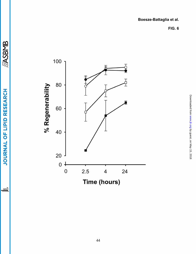
AY9944 treatment alters regenerability of rhodopsin
The changes in the membrane environment that occur at three months are concomitant
with the previously described retinal degeneration in this model (8), which also entails
defective phototransduction. To determine if AY9944-induced alterations in membrane
dynamics are correlated with a parameter pertinent to phototransduction (e.g., rhodopsin
function) we next analyzed the ability of the bleached photopigment (opsin) to recombine with
exogenously supplied 11-cis retinal chromophore to regenerate rhodopsin in vitro, using ROS
membranes from control vs. AY9944-treated rats. As shown in Fig. 6, at one postnatal month,
no significant decrease in rhodopsin regenerability was observed between ROS obtained from
control and AY9944-treated rat retinas (95 ± 2.6% in controls, 92 ± 1.5% in AY9944-treated
retinas). The total extent and rate of rhodopsin regeneration, however, were decreased
significantly by three postnatal months as a function of AY9944 treatment (65 ± 1.5%
regenerability) relative to controls (82 ± 3.0 % regenerability).
21
by guest, on May 15, 2018
ww
w.jlr.org
Dow
nloaded from
DISCUSSION
In the present study, we have demonstrated that perturbation of sterol biosynthesis by
use of the distal pathway inhibitor, AY9944, profoundly alters both ROS membrane sterol
composition as well as fatty acid composition, relative to age-matched control rats, in a time-
dependent manner. Concomitantly, as expected, the physical properties of the membranes
are altered; notably, there is a significant reduction in membrane fluidity by three months, as
measured by use of fluorescent fatty acid probes. Also, the ability of opsin to combine with its
11-cis retinal chromophore to regenerate rhodopsin is compromised by three months of
treatment. Interestingly, although the changes in membrane sterol and fatty acid composition
are substantial even by one month of treatment, there is no retinal degeneration nor profound
electrophysiological deficits at this stage in the treatment time course (7), nor is rhodopsin
regeneration affected. By three months of AY9944 treatment, however, ROS sterol and fatty
acid composition are even more deranged, most notably with the profound (>40%) loss in DHA
content, and rhodopsin regenerability is significantly compromised. This stage of treatment
corresponds to the progression to marked retinal degeneration, particularly rod photoreceptor
degeneration and death, as well as defective rod and cone visual function (8). The question
arises as to whether the observed effects on membrane fluidity and visual pigment
regenerability are due primarily to the altered sterol composition or to the altered fatty acid
composition of the ROS membranes? In order to answer this question, one must consider in
further detail the changes in these two distinct lipid classes.
Since the absolute recovery efficiency of ROS membranes varies from one preparation
to the next, one cannot confidently make direct comparisons of absolute values for sterol mass
between membrane preparations. However, if one normalizes the total sterol mass to total
fatty acid mass on a molar basis, a comparison can be made. Using the empirically
22
by guest, on May 15, 2018
ww
w.jlr.org
Dow
nloaded from

determined values for the total sterol and fatty acid content of ROS membranes from both
AY9944-treated and control rats, we calculate a sterol/fatty acid mole ratio of 0.062 (±12%).
This is in good agreement with published values for both the cholesterol content (∼8-10 mol%
of total ROS lipid) and phospholipid content (∼85-90 mol% of total ROS lipid, and 65-75 moles
of phospholipid per mole of rhodopsin) of ROS membranes, (reviewed in (29)). To a first
approximation (excluding plasmalogens), each glycerophospholipid molecule contains two acyl
chains. Hence, if the Chol/phospholipid mole ratio is approximately 1:9 in ROS membranes,
the Chol/fatty acid mole ratio should be about 1:18, or 0.056. The fact that this ratio is the
same in ROS membranes from both normal control and AY9944-treated rats further reveals
that the total sterol mass in ROS membranes is unaffected by AY9944 treatment (i.e., there is
no net loss of sterol mass in the membranes). Hence, there is a one-for-one molecular
replacement of Chol with 7DHC under the conditions employed. However, the chemical
composition of the membrane sterol pool is dramatically and progressively altered: by one
month of AY9944 treatment, the 7DHC/Chol mole ratio is 3.5, whereas by three months it is
5.6; in contrast, cholesterol is essentially the only sterol present in normal rat ROS membranes
at any age.
The impact of sterol chemistry on the structure and function of biological membranes
has been a subject of extensive investigation over the years (reviewed in (31, 32)). In sum,
there do not appear to be uniformly applicable rules to predict with certainty the consequences
of replacing cholesterol with sterols of different structure, such as 7DHC; rather, the impact of
such changes tends to be a function of the particular sterol and the biological system being
examined. However, recent studies from our lab using model membrane systems suggest that
7DHC has physical properties very similar to those of cholesterol, including packing densities
and molecular volumes in mixed sterol-phospholipid monolayers (Langmuir films) (34, 35).
23
by guest, on May 15, 2018
ww
w.jlr.org
Dow
nloaded from

These findings, in conjunction with the fact that sterols account for <10 mol% of the total lipid in
ROS membranes (29), would tend to support the conclusion that altered sterol composition is
not a major factor in the observed changes in membrane fluidity or rhodopsin regenerability.
Our results obtained with fluorescent probe measurements demonstrate that the
hydrophobic core of the ROS membranes becomes more ordered (less fluid), as has been
previously reported in cases where there is a substantial decrease in the polyunsaturated fatty
acid (e.g., 22:6n3) content of the ROS membrane (30). In a study of the effects of dietary n3
deficiency, a similar increase in the acyl chain order reported by DPH in ROS membranes was
accompanied by a 50% increase in the time required for transducin binding to light-activated
rhodopsin and three-fold decrease in light-stimulated phosphodiesterase activity (30). An
earlier study of the relationship between acyl chain packing, as reported by fv, and formation of
the light-activated conformation metarhodopsin II indicates that the decrease in fv found in the
present study would depress metarhodopsin II by 25-30% (35). Increasing cholesterol in ROS
disk membranes from 12% to 30% produced a reduction of fv comparable to treatment with
AY9944 and this was accompanied by a 20% reduction in metarhodopsin II (36). In all of
these earlier studies, increased acyl chain order in the membrane hydrophobic core was
accompanied by an increase in DPH fluorescence lifetime, indicating increased order in the
head group region. In the present study, the decreased order in the headgroup/interfacial
region reported by both DPH and cPA strongly suggests that treatment with AY9944 produces
a unique set of changes in the physical properties of ROS membranes. In the core of the
membrane, these changes appear to be dominated by the marked reduction in the number of
22:6n3 acyl chains, consistent with the increased order and decreased motion of DPH (Fig. 4
and Fig. 5, respectively). However, the changes reported by cPA (Fig. 3) and DPH
fluorescence lifetime suggest additional significant modifications of membrane architecture.
This simultaneous decrease in fluidity in the membrane core and slight increase in fluidity in
24
by guest, on May 15, 2018
ww
w.jlr.org
Dow
nloaded from
the headgroup/interfacial region may be due to the particular molecular species that lost
22:6n3 acyl chains, or a change in lipid lateral phase organization.
Our fatty acid analyses (Fig. 1) showed that although there were several ROS fatty acyl
species that exhibited statistically significant altered levels, comparing AY9944-treated rats to
age-matched controls, the magnitude of these changes tended to be rather modest for most
species, particularly for saturated and monounsaturated acyl chains. The spectroscopic
results obtained with cPA indicate that these changes were not substantial enough to
appreciably alter membrane fluidity after only one month of AY9944 treatment. However, more
dramatic alterations in ROS membrane fatty acid composition were observed by three months
of AY9944 treatment, relative to age-matched controls. Notably, by normalizing the 22:6n3
content to that of 18:0, an invariant fatty acid (Table 2), there was a ∼41 mol% loss of 22:6n3,
compared to controls. This finding is consistent with the results obtained recently by lipidomic
analysis of whole retinas from AY9944-treated and control rats, where the most notable
alteration in retinal glycerophospholipids (phosphatidylethanolamine, phosphatidylcholine, and
phosphatidylserine) was a profound loss in molecular species containing 22:6n3 with AY9944
treatment (9). Clearly, the magnitude of this change was sufficient to cause appreciable
changes in the physical properties of the ROS membranes (see Figs. 3, 4, and 5).
Increased order in the membrane core and simultaneous decreased order in the
headgroup and interfacial regions is somewhat unique and suggests that other changes in
membrane composition also play a role in altering the membrane. Significantly large
alterations in the protein-to-lipid ratio would result in marked differences in the physical
properties of the membranes, which would be readily detectable by the spectroscopic methods
employed here. In the case of the ROS membrane, the visual pigment, rhodopsin, is
estimated to account for at least 90% of the total integral membrane protein and the lipid-to-
protein mass ratio is approximately 1:1 (see (29)). Rhodopsin packing density also has been
25
by guest, on May 15, 2018
ww
w.jlr.org
Dow
nloaded from
correlated with alterations in membrane function (assessed by rhodopsin activation, which
impacts efficiency of the phototransduction cascade) (37, 38). However, our analyses showed
that there was no significant alteration in the opsin-to-phospholipid ratio of ROS from AY9944-
treated rats, compared to age-matched controls; hence, the protein packing density in ROS
membranes was unaffected by AY9944 treatment. In fact, a reduction in opsin levels, and
concomitant reduction in protein packing density, would be predicted to cause an increase in
membrane fluidity, which clearly was not observed here.
It is important to note that, while no histological degeneration or apparent loss in rod or
cone electrophysiological responsiveness was observed in the AY9944-induced rat model of
SLOS after one month of treatment, there was an appreciable and statistically significant delay
in the timing of those photoresponses (i.e., increased “implicit times”) (7). The implicit time is
related to the efficiency of the phototransduction cascade operative in the photoreceptor cell,
involving both the protein components organized within and at the surface of the ROS disk
membranes as well as the cyclic nucleotide-gated ion channels located in the ROS plasma
membrane (39). Hence, it is apparent that even the relatively modest (in absolute magnitude)
changes in the fatty acid profile of the ROS membranes that occur by one month of AY9944
treatment have some biological impact at the membrane level, although not enough to cause
overt cellular dysfunction or degeneration. The retina requires high levels of DHA for optimal
function, particularly with regard to supporting photoreceptor viability and electrophysiological
competence (see (29)). ROS membranes in which the DHA content has been significantly
reduced compared to normal levels exhibit slower than normal photoresponses, which
correlates with impaired activation of transducin, the cognate G-protein and key player in the
phototransduction cascade (30). It is tempting to speculate that, faced with a metabolic insult
(such as disrupting normal cholesterol biosynthesis), the cell responds by globally remodeling
its membrane lipidome in an effort to preserve cellular integrity and function, including
26
by guest, on May 15, 2018
ww
w.jlr.org
Dow
nloaded from

preservation of membrane fluidity that is critical to the function of a host of membrane-
associated enzymes, receptors, and ion channels (40). However, while the retinal
photoreceptor cells may be able to tolerate this level of stress for at least one month,
eventually they lose physiological competence and cellular integrity, undergoing degeneration
and cell death by three months of exposure to AY9944 (8, 15). At that point, rod and cone
electrophysiology and structure are severely compromised, and this also correlates temporally
with the dramatic losses in the DHA content of ROS membranes documented in this study, as
well as those recently reported under virtually identical conditions for whole retina (16).
Notably, it is at three months of AY9944 treatment that we also observe altered membrane
properties, as assessed by the behavior of fluorescent probes added in small amounts to
isolated ROS membranes, which also are consistent with the more dramatic changes in fatty
acid composition, particularly DHA.
Herein, we show for the first time that one pathologic consequence of decreased
cholesterol biosynthesis in a rat model of SLOS is reduced rhodopsin regenerability. It should
be noted that our in vitro studies of rhodopsin regeneration were performed in the presence of
excess exogenous 11-cis retinal; hence, the decreased regeneration cannot be due to
insufficient retinoid availability. Reduction in the regenerability of rhodoposin also would be
expected to negatively impact the efficiency of phototransduction and, hence, photoreceptor
function, consistent with our prior in vivo observations with the SLOS rat model (7, 8, 15). We
speculate that opsin’s ability to assume a conformational state requisite for optimal
regeneration is impaired as a consequence of alterations in membrane lipid composition and
biophysical properties (e.g., fluidity), such alterations being more extensive by three months
than by one month of exposure of rats to AY9944. This may represent a threshold
phenomenon: beyond some level of deviation from normal membrane composition and
biophysical properties, opsin regenerability is compromised, whereas deviations below that
27
by guest, on May 15, 2018
ww
w.jlr.org
Dow
nloaded from
level (even though not normal) are apparently tolerable with respect to opsin regenerability. A
similar hypothesis (the “molecular-spring model”) was put forward by Dratz and Holte (41) to
explain diminished rhodopsin bleaching kinetics in the presence of altered levels of cholesterol
and DHA. Whether an analogous mechanism involving lipid-protein interactions in rhodopsin
regeneration is at play here is unknown. However, we suggest that alterations in membrane
composition that decrease headgroup/interfacial order and increase hydrocarbon order also
result in decreased efficiency of rhodopsin regeneration, consistent with the observed
electrophysiological defects in the SLOS rat model and in human SLOS patients, and may
contribute to long-term retinal degeneration due to the inability of the retina to adapt to the
continuous exposure to light.
With the exception of our recent studies with the SLOS rat model (9), the implication of
a linkage between disrupted sterol biosynthesis and altered fatty acid metabolism, particularly
in association with SLOS, has never been proposed. We have proposed (9) that cross-talk
between sterol and fatty acid metabolism may explain the results observed with the AY9944-
induced rat model, and that additional metabolic compromise beyond the primary defect in
sterol biosynthesis may contribute to the pathology of SLOS. The exact mechanism by which
AY9944 (which allegedly is a selective inhibitor of DHCR7 in the sterol pathway) perturbs fatty
acid metabolism remains to be elucidated. Interestingly, altered membrane fluidity has been
reported recently by one of us (K.B.B.) in a study of skin fibroblasts obtained from SLOS
patients, using the same spectroscopic techniques as employed in the current study (42).
While this finding had been considered only within the context of altered membrane sterol
composition, our results suggest the possibility that additional changes in the steady-state fatty
acid composition of the fibroblast membranes also may underlie the observed alterations in
membrane fluidity.
28
by guest, on May 15, 2018
ww
w.jlr.org
Dow
nloaded from

ACKNOWLEDGMENTS
This study was supported by U.S.P.H.S. (NIH) grants EY007361 (SJF), EY00871
(REA), EY04149 (REA), EY10420 (KBB), EY12190 (REA), and RR17703 (REA); the
Foundation Fighting Blindness (REA); and unrestricted departmental grants from Research to
Prevent Blindness (SJF and REA). KBB is the recipient of an E. Matilda Ziegler Vision Award.
SJF is the recipient of a Research to Prevent Blindness Senior Scientific Investigator Award.
29
by guest, on May 15, 2018
ww
w.jlr.org
Dow
nloaded from

REFERENCES
1. Kelley, R. I. and G. E. Hermann. 2001. Inborn errors of sterol biosynthesis. Annu. Rev.
Genomics Hum. Genet. 2: 299-341.
2. Porter, F. D. 2003. Human malformation syndromes due to inborn errors of cholesterol
synthesis. Curr. Opin. Pediatr. 15: 607-613.
3. Smith, D. L., L. Lemli, and J. M. Opitz. 1964. A newly recognized syndrome of multiple
congenital anomalies. J. Pediatr. 64: 210-217.
4. Correa-Cerro, L. S. and F. D. Porter. 2005. 3beta-hydroxysterol Delta7-reductase and
the Smith-Lemli-Opitz syndrome. Mol. Genet. Metab. 84: 112-126.
5. Elias, E. R., R. M. Hansen, M. Irons, N. B. Quinn, and A. B. Fulton. 2003. Rod
photoreceptor responses in children with Smith-Lemli-Opitz syndrome. Arch.
Ophthalmol. 121: 1738-1743.
6. Kolf-Klauw, M., F. Chevy, C. Wolf, B. Siliart, D. Citadelle, and C. Roux. 1996.
Inhibition of 7-dehydrocholesterol reductase by the teratogen AY9944: a rat model for
Smith- Lemli-Opitz syndrome. Teratology. 54: 115-125.
7. Fliesler, S. J., M. J. Richards, C. -Y. Miller, and N. S. Peachey. 1999. Marked alteration
of sterol metabolism and composition without compromising retinal development or
function. Invest. Ophthalmol. Vis. Sci. 40: 1792-1801.
8. Fliesler, S. J., N. S. Peachey, M. J. Richards, B. A. Nagel, and D. K. Vaughan. 2004.
Retinal degeneration in a rodent model of Smith-Lemli-Opitz syndrome:
electrophysiologic, biochemical, and morphologic features. Arch. Ophthalmol. 122:
1190-1200.
9. Ford, D. A., J. K. Monda, R. S. Brush, R. E. Anderson, M. J. Richards, and S. J.
Fliesler. 2008. Lipidomic analysis of the retina in a rat model of Smith-Lemli-Opitz
30
by guest, on May 15, 2018
ww
w.jlr.org
Dow
nloaded from

syndrome: Alterations in docosahexaenoic acid content of phospholipid molecular
species. J. Neurochem., in press [Epub ahead of Print 21 Dec. 2007].
10. Lentz, B. R. 1993. Use of fluorescent probes to monitor molecular order and motions
within liposome bilayers. Chem. Phys. Lipids. 64: 99-116.
11. Drummen, G. P. C., J. A. F. Op den Kamp, and J. Post. 1999. Validation of the
peroxidative indicators, cis-parinaric acid and parinaroyl-phospholipids, in a model
system and cultured cardiac myocytes. Biochim. Biophys. Acta. 1436: 370-382.
12. Trevors, J. T. 2003. Fluorescent probes for bacterial cytoplasmic membrane research.
J. Biochem. Biophys. Methods. 57: 87–103.
13. Zannoni, C., A. Argioni, and P. Cavatorta. 1983. Fluorescence depolarization in liquid
crystals and membrane bilayers. Chem. Phys. Lipids. 32: 179-250.
14. van der Meer, B. W., H. Pottel, W. Herreman, M. Ameloot, H. Hendricks, and H.
Schroder. 1984. Effect of orientational order on the decay of the fluorescence
anisotropy in membrane suspensions. Biophys. J. 46: 515-523.
15. Fliesler, S. J., D. K. Vaughan, E. C. Jenewein, M. J. Richards, B. A. Nagel, and N. S.
Peachey. 2007. Partial rescue of retinal function and sterol steady-state in a rat model
of Smith-Lemli-Opitz syndrome. Pediatr. Res. 61: 273-278.
16. Fliesler, S. J., R. Florman, and R. K. Keller. 1995. Isoprenoid lipid metabolism in the
retina: dynamics of squalene and cholesterol incorporation and turnover in frog rod
outer segment membranes. Exp. Eye Res. 60: 57-69.
17. Fliesler, S. J., M. J. Richards, C. Y. Miller, and R. J. Cenedella. 2000. Cholesterol
synthesis in the vertebrate retina: effects of U18666A on rat retinal structure,
photoreceptor membrane assembly, and sterol metabolism and composition. Lipids. 35:
289-296.
31
by guest, on May 15, 2018
ww
w.jlr.org
Dow
nloaded from

18. Cusanovich, M. 1982. Kinetics and mechanism of rhodopsin regeneration with 11-cis-
retinal. Methods Enzymol. 81: 443-447.
19. Boesze-Battaglia, K., H. Song, M. Sokolov, C. Lillo, L. Pankoski-Walker, C. Gretzula, B.
Gallagher, R. A. Rachel, N. A. Jenkins, N. G. Copeland, F. Morris, J. Jacob, P. Yeagle,
D. S. Williams, and M. Damek-Poprawa. 2007. The tetraspanin protein peripherin-2
forms a complex with melanoregulin, a putative membrane fusion regulator.
Biochemistry. 46: 1256-1272.
20. Towbin, H., T. Staehelin, and J. Gordon. 1979. Electrophoretic transfer of proteins from
polyacrylamide gels to nitrocellulose sheets: Procedure and some applications. Proc.
Natl. Acad. Sci. U.S.A. 76: 4350-4354.
21. Bartlett, G. R. 1959. Phosphorus assay in column chromatography. J. Biol. Chem.
234: 466-468.
22. Sklar, L. A., B. S. Hudson, M. Petersen, and J. Diamond. 1977. Conjugated polyene
fatty acids as fluorescent probes: spectroscopic characterization. Biochemistry. 16:
813- 819.
23. Calafut, T., J. Dix, and A. Verkman. 1989. Fluorescence depolarization of cis-and
trans-parinaric acids in artificial and red cell membranes. Biochemistry. 21: 5051-5058.
24. Mitchell, D. C. and B. J. Litman. 1998. Molecular order and dynamics in bilayers
consisting of highly polyunsaturated phospholipids. Biophys. J. 74: 879-891.
25. Straume M. and B. J. Litman. 1987. Equilibrium and dynamic structure of large,
unilamellar, unsaturated acyl chain phosphatidylcholine vesicles. Higher order analysis
of 1,6-diphenyl-1,3,5-hexatriene and 1-[4-(trimethylammonio) phenyl]-6-phenyl-1,3,5-
hexatriene anisotropy decay. Biochemistry. 26: 5113-5120.
26. Heyn, M. P. 1979. Determination of lipid order parameters and rotational correlation
times from fluorescence depolarization experiments. FEBS Lett. 108: 359-364.
32
by guest, on May 15, 2018
ww
w.jlr.org
Dow
nloaded from

27. Bligh, E. J. and W. J. Dyer. 1959. A rapid method of total lipid extraction and
purification. Can. J. Biochem. Physiol. 37: 911–917.
28. Martin, R. E., M. H. Elliott, R. S. Brush, and R. E. Anderson. 2005. Detailed
characterization of the lipid composition of detergent-resistant membranes from
photoreceptor rod outer segment membranes. Invest. Ophthalmol. Vis. Sci. 46: 1147-
1154.
29. Fliesler, S.J. and R. E. Anderson. 1983. Chemistry and metabolism of lipids in the
vertebrate retina. Prog. Lipid Res. 22: 79-131.
30. Niu, S. L., D. C. Mitchell, S. Y. Lim, Z. M. Wen, H. Y. Kim, N. Salem Jr., and B. J.
Litman. 2004. Reduced G protein-coupled signaling efficiency in retinal rod outer
segments in response to n-3 fatty acid deficiency. J. Biol. Chem. 279: 31098-31104.
31. Bloch, K. 1983. Sterol structure and membrane function. Crit. Rev. Biochem. 14: 47-92.
32. Yeagle, P. L. 1993. The biophysics and cell biology of cholesterol: A hypothesis for the
essential role of cholesterol in mammalian cells. In Cholesterol in Membrane Models.
L. Finegold, editor. CRC Press, Boca Raton, FL. 1-12.
33. Serfis, A. B., S. Brancato, and S. J. Fliesler. 2001. Comparative behavior of sterols in
phosphatidylcholine-sterol monolayer films. Biochim. Biophys. Acta. 1511: 341-348.
34. Berring, E. E., K. Borrenpohl, S. J. Fliesler, and A. B. Serfis. 2005. A comparison of the
behavior of cholesterol and selected derivatives in mixed sterol-phospholipid Langmuir
monolayers: a fluorescence microscopy study. Chem. Phys. Lipids. 136: 1-12.
35. Mitchell, D. C., M. Straume. and B. J. Litman. 1992. Role of sn-1-saturated,sn-2-
polyunsaturated phospholipids in control of membrane receptor conformational
equilibrium: effects of cholesterol and acyl chain unsaturation on the metarhodopsin I in
equilibrium with metarhodopsin II equilibrium. Biochemistry. 31: 662-670.
33
by guest, on May 15, 2018
ww
w.jlr.org
Dow
nloaded from
36. Niu, S. L., D. C. Mitchell, and B. J. Litman. 2002. Manipulation of cholesterol levels in
rod disk membranes by methyl-beta-cyclodextrin: effects on receptor activation. J. Biol.
Chem. 277: 20139-20145.
37. Albert, A. D. and K. Boesze-Battaglia. 2005. The role of cholesterol in rod outer
segment membranes. Progr. Lipid Res. 44: 99-124.
38. Niu, S. L. and D. C. Mitchell. 2005. Effect of packing density on rhodopsin stability and
function in polyunsaturated membranes. Biophys. J. 89: 1833-1840.
39. Fulton, A. B., R. M. Hansen, and O. Findl. 1995. The development of the rod
photoresponse from dark-adapted rats. Invest. Ophthalmol. Vis. Sci. 36: 1038-1045.
40. Vigh, L., H. Nakamoto, J. Landry, A. Gomez-Munoz, J. L. Harwood, and I. Horvath.
2007. Membrane regulation of the stress response from prokaryotic models to
mammalian cells. Ann. N. Y. Acad. Sci. 1113: 40-51.
41. Dratz, E.A. and L.L. Holte. 1992. The molecular spring model for the function of
docosahexaenoic acid (22:6w3) in biological membranes. In Essential Fatty Acids and
Eicosanoids: Invited Papers from the Third International Congress. A. Sinclair and R.
Gibson, editors. American Oil Chemists' Society, Champaign, IL. 122-127.
42. Tulenko, T. N., K. Boeze-Battaglia, R. P. Mason, G. S. Tint, R. D. Steiner, W. E.
Connor, and E. F. Labelle. 2006. A membrane defect in the pathogenesis of the Smith-
Lemli- Opitz syndrome. J. Lipid Res. 47: 134-143.
34
by guest, on May 15, 2018
ww
w.jlr.org
Dow
nloaded from

Table 1. Sterol composition and content of serum and ROS membranes from AY9944-
treated and control rats.
___________________________________________________________________________
Treatment Age 7DHC/Chol % of
Tissue Group (mo) (mole ratio) Total Sterols* Control
Serum Control 1 (7) 0 100.5 ± 10.7 100
+AY9944 1 (19) 2.0 ± 0.8 16.5 ± 6.2 16.4
Control 3 (12) 0 95.5 ± 17.5 100
+AY9944 3 (16) 4.6 ± 1.0 20.8 ± 5.6 21.8
ROS Control 1 (4) 0 --- ---
+AY9944 1 (6) 3.5 ± 0.4 --- ---
Control 3 (6) 0 --- ---
+AY9944 3 (4) 5.6 ± 1.6 --- ---
Aliquots (50 µL each) of serum or ROS membranes (resuspended in buffer) were saponified in
methanolic KOH and the nonsaponifiable lipids were extracted with petroleum ether,
evaporated to dryness, redissolved in methanol, and analyzed by reverse-phase HPLC.
Values represent the mean ± S.D., with number of biologically independent samples (N) given
in parentheses, corrected for recovery efficiency using an internal standard of [3H]cholesterol.
7DHC, 7-dehydrocholesterol; Chol, cholesterol.
* Values for total sterols in serum given in mg/dL.
35
by guest, on May 15, 2018
ww
w.jlr.org
Dow
nloaded from

Table 2. Normalized PUFA content of ROS membranes from control and AY9944-treated
rats.
___________________________________________________________________________
1 Month 3 Months
Fatty Normalized Value* Normalized Value*
Acid Control +AY9944 %Change Control +AY9944 %Change
20:4n6 0.286 0.333 +16.4a 0.181 0.155 -14.4a
22:5n6 0.067 0.233 +348a 0.041 0.180 +439a
22:6n3 1.44 1.19 -17.9b 1.39 0.825 -40.6a
Data are average values, obtained by dividing the relative mole % values (see Fig. 1) of the
given fatty acid species by the relative mole percent of stearic acid (18:0).
a Statistically significant (P < 0.05; N=3).
b Not statistically significant (P > 0.05; N=3).
36
by guest, on May 15, 2018
ww
w.jlr.org
Dow
nloaded from
FIGURE LEGENDS
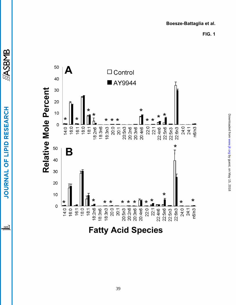
Figure 1. Fatty acid profiles of retinal rod outer segment (ROS) membranes from
AY9944-treated (filled bars) and control (open bars) rats at (A) one and (B) three
postnatal months of age. Values are mean ± S.D. (N=3), expressed as relative mol %.
Statistical significance (P<0.05; multivariant ANOVA with post-hoc Scheffe test) of differences
between treated and control values for a given fatty acid species is noted by an asterisk (*).
Figure 2. Opsin protein levels in control (C) and AY9944-treated (AY) rat ROS
membranes. A. Western blot probed with monoclonal anti-opsin antibodies (mAb 4D2).
Samples were adjusted in order to load an equivalent amount (1 mg total) of lipid phosphorus
per lane. Immunoreactivity was visualized using HRP-conjugated secondary antibodies and
enhanced chemifluorescence (ECF) detection. Migration positions of protein molecular weight
markers (Mr in kDa) are indicated in the left-hand side of figure. B. Quantitative densitometric
analysis of Western blots. Graph represents measurements (mean ± S.D.) obtained from four
independent blots per treatment and time point (one and three months). C. Quantitative RT-
PCR analysis of opsin expression in retinas isolated from control and AY9944-treated rats.
Graph shows values (mean ± S.D., N=4) expressed in arbitrary units.
Figure 3. Steady-state anisotropy measurements of cPA in purified ROS membranes
from one- and three-month old rats. Open ( ) bars, controls; filled ( ) bars, AY9944-
treated. Values are mean ± S.D. (N= 4), measured at 37 oC. Asterisk (*) denotes a
statistically significant difference (P = 0.0068) between treated vs control samples at three
months of age.
37
by guest, on May 15, 2018
ww
w.jlr.org
Dow
nloaded from

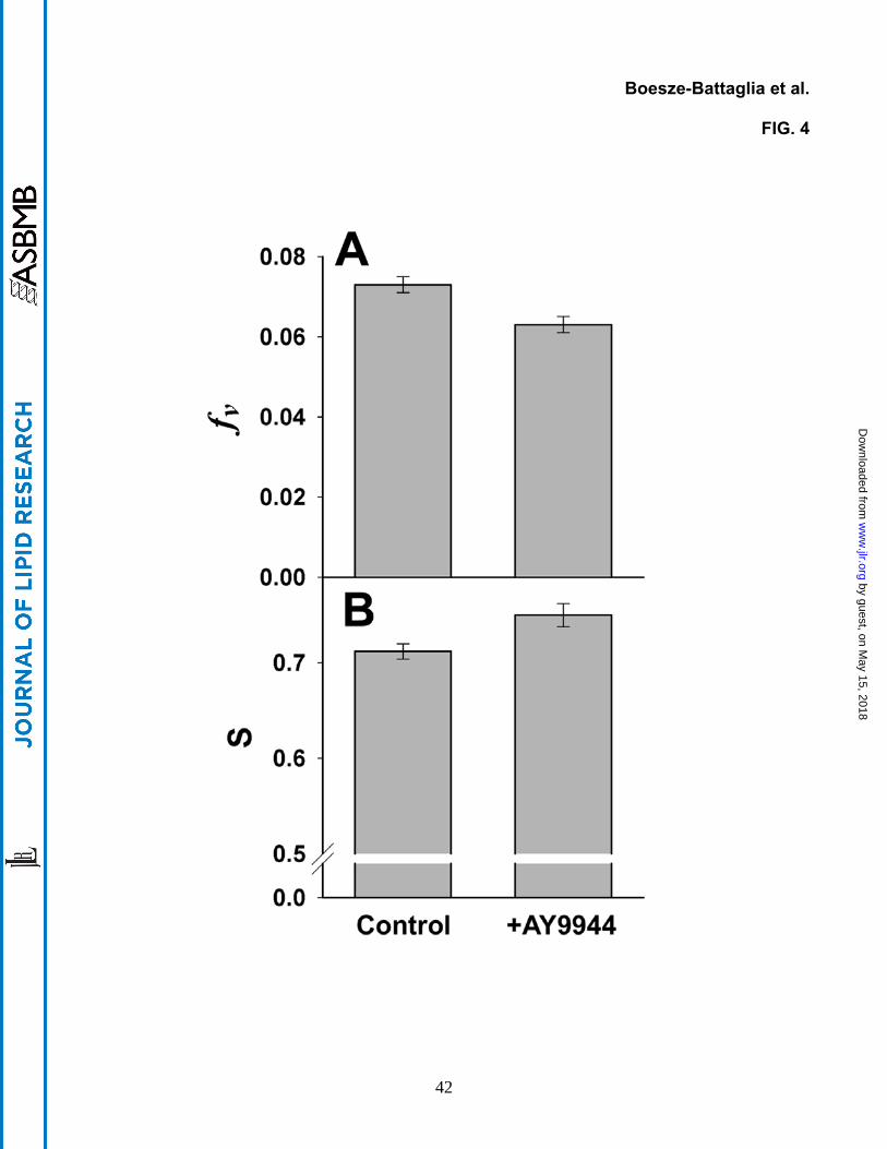
Figure 4. Measures of acyl chain ensemble order (membrane fluidity) obtained from
analysis of DPH fluorescence anisotropy decays. A. The free volume (or disorder
parameter), fv, from analysis via the BRD model. The smaller value for the membranes from
AY9944-treated animals, compared to controls, indicates decreased acyl chain packing
disorder (P < 0.001, N=6). B. The order parameter, S, calculated from the model-independent
analysis of the anisotropy decays in terms of the sum of three exponential decays. The higher
S value for the membranes from AY9944-treated animals, compared to controls, indicates
increased orientational order of DPH (P < 0.001, N=6).
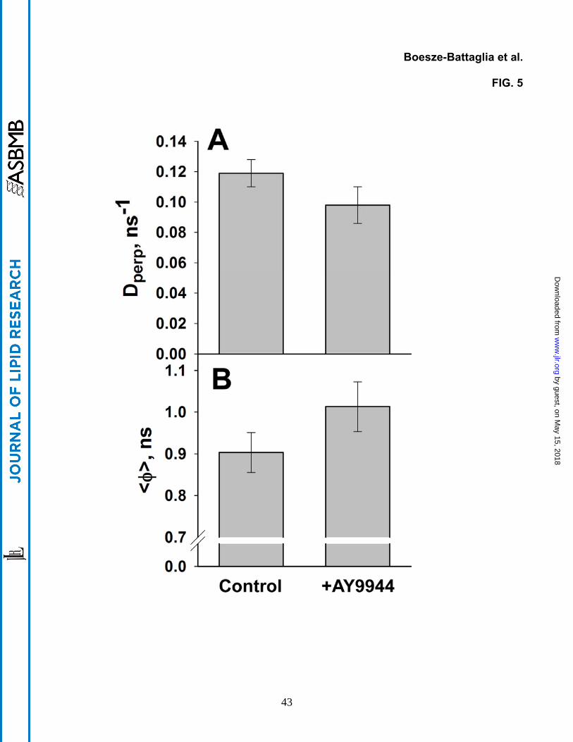
Figure 5. Measures of DPH rotational motion obtained from analysis of DPH
fluorescence anisotropy decays. A. The diffusion constant for DPH rotation about its long
axis, Dperp, from the BRD model analysis. The smaller value in ROS membranes from the
AY9944-treated animals, compared to controls, indicates slower DPH rotation (P= 0.006, N=6).
B. The weighted average of the three empirical rotational time constants, <φ>, from the model-
independent analysis. The higher value for membranes from AY9944-treated animals,
compared to controls, indicates slower DPH rotation (P = 0.006, N=6).
Figure 6. Rhodopsin regeneration in ROS membranes from control and AY9944-treated
rats. Values expressed as percent regeneration, shown as a function of time ROS
membranes were incubated with excess 11-cis retinal at 37 oC. Comparison of values from
one-month old control (○) versus AY9944-treated rats (●) shows no statistically significant
difference (N=3). In contrast, comparison of values from three-month old control ( ) and
AY9944-treated rats ( ) showed a statistically significant difference (P = 0.0006, N=3, at 24 h).
38
by guest, on May 15, 2018
ww
w.jlr.org
Dow
nloaded from
Boesze-Battaglia et al.
FIG. 1
39
by guest, on May 15, 2018
ww
w.jlr.org
Dow
nloaded from

Boesze-Battaglia et al.
FIG. 2
40
by guest, on May 15, 2018
ww
w.jlr.org
Dow
nloaded from

Boesze-Battaglia et al.
FIG. 3
41
by guest, on May 15, 2018
ww
w.jlr.org
Dow
nloaded from
Boesze-Battaglia et al.
FIG. 4
42
by guest, on May 15, 2018
ww
w.jlr.org
Dow
nloaded from
Boesze-Battaglia et al.
FIG. 5
43
by guest, on May 15, 2018
ww
w.jlr.org
Dow
nloaded from
Boesze-Battaglia et al.
FIG. 6
44
by guest, on May 15, 2018
ww
w.jlr.org
Dow
nloaded from