lipid uptake and metabolism in the parasitic protozoan
TRANSCRIPT

University of Texas at El PasoDigitalCommons@UTEP
Open Access Theses & Dissertations
2009-01-01
Lipid Uptake and Metabolism in the ParasiticProtozoan Giardia lamblia.Mayte YichoyUniversity of Texas at El Paso, [email protected]
Follow this and additional works at: https://digitalcommons.utep.edu/open_etdPart of the Molecular Biology Commons, and the Parasitology Commons
This is brought to you for free and open access by DigitalCommons@UTEP. It has been accepted for inclusion in Open Access Theses & Dissertationsby an authorized administrator of DigitalCommons@UTEP. For more information, please contact [email protected].
Recommended CitationYichoy, Mayte, "Lipid Uptake and Metabolism in the Parasitic Protozoan Giardia lamblia." (2009). Open Access Theses & Dissertations.387.https://digitalcommons.utep.edu/open_etd/387

LIPID UPTAKE AND METABOLISM IN THE PARASITIC
PROTOZOAN GIARDIA LAMBLIA
MAYTE YICHOY
Department of Biological Sciences
APPROVED: ___________________________________ Siddhartha Das, Ph.D., Co-Chair ___________________________________ Stephen B. Aley, Ph.D., Co-Chair ___________________________________ Igor C. Almeida, Ph.D. ___________________________________ Sukla Roychowdhury, Ph.D. ____________________________________ James Becvar, Ph.D.
___________________________________ Patricia D. Witherspoon, Ph.D., Dean of the Graduate School

Copyright
by
Mayte Yichoy
2009

Dedication
This dissertation is dedicated the friends, family and colleagues who have supported and
encouraged me through the last five years, but especially to Añin and Aye.
LIPID UPTAKE AND METABOLISM IN THE PARASITIC PROTOZOAN GIARDIA LAMBLIA
by
Mayte Yichoy, B.A.
DISSERTATION
Presented to the Faculty of the Graduate School of
The University of Texas at El Paso
in Partial Fulfillment
of the Requirements
for the Degree of
DOCTOR OF PHILOSOPHY
Department of Biological Sciences
The University of Texas at El Paso
May 2009

v
Acknowledgements
This work would not have been possible without the guidance and assistance of several
people, the most important of these being Dr. Sid Das, who has been not only a mentor, but also
a great friend. His dedication, guidance, support and encouragement are much appreciated.
I would also like to thank the members of my committee, first and foremost, co-chair Dr.
Steve Aley, as well as Drs. Sukla Roychowdhury, Igor C. Almeida, and James Becvar for taking
the time to guide me through this endeavor. Dr. Almeida and Dr. Roychowdhury have been
especially helpful and provided much helpful discussion and guidance.
I would also like to thank Dr. Ernesto Nakayasu. This work would not have been
possible without his great knowledge of mass spectrometry and lipidomics as well as his
friendship.
Drs. Rosa Maldonado and Armando Varela have provided me with much wisdom and
support, which are much appreciated. I would also like to thank past and present members of the
Das laboratory, especially Tavis Mendez, Debarshi Roy and Dr. Suparna Ray who have been
great friends and made not only scholarly contributions, but also infused much humor into my
life. I would also like to thank Dr. Yunuen Hernandez and Trevor Duarte for their intellectual
contributions to this work. I am also grateful to Dr. Judi Ellzey and Marian Viveros of the
Analytical Cytology Core for their assistance with the confocal microscope.
I would also like to credit Drs. Bob Wallace and Elizabeth Walsh. Without them, I would
have never thought this was possible. Dr. Bob has been especially instrumental in guiding me
towards this academic path.

vi
Abstract
Giardia lamblia is a protozoan parasite that causes various intestinal syndromes, and it is
a common cause of water-borne illness worldwide, both in developed and developing countries.
Giardia attaches to the mucosal epithelia of the duodenum below the bile duct, where it is
exposed to bile salts and dietary lipids. G. lamblia is unable to synthesize lipids de novo and
must therefore scavenge necessary lipids from its extracellular environment and remodel them as
needed. However, the current lipidomic analysis (presented in this dissertation) has revealed that
while the Giardia lipidome is rich in phosphatidylcholine (PC), phosphatidylethanolamine (PE),
and phosphatidylglycerol (PG), its growth medium contains only PC, lyso-PC, and some
diacylglycerol. Therefore, it is quite likely that Giardia has more lipid synthesis abilities than
previously reported. The lipid analysis also includes fatty-acid analysis by GC-MS, which has
revealed that Giardia trophozoites, encysting cells, and in vitro-derived cysts contain odd
carbon-chain fatty acids (OCFAs), as well as a number of fatty acids not present in the medium,
indicating that elongases and desaturases are also active in Giardia.
The Giardia Genome Database (giardiaDB.org) was searched for the presence of genes
encoding enzymes for the synthesis of PG and PE—i.e., two newly synthesized lipids in Giardia.
Analyses identified phosphatidylglycerol phosphate synthase (PGPS) and phosphatidylserine
decarboxylase (PSD) genes, which are expressed throughout the Giardia life cycle. Furthermore,
I also searched for the genes linked to the synthesis of phospholipid transporters that could be
involved in importing phospholipids from outside sources. Interestingly, the search indicated that
Giardia has the genes for flippase enzymes, which indicates that this parasite relies on
scavenging lipids from the host. In order to further elucidate the mechanisms of lipid uptake and
incorporation, I labeled cells with [13C]-glycerol isotope to determine whether generation of PG

vii
occurs via the base- or head-group exchange reactions between PC and glycerol. Side by side, I
have also used fluorescent lipids as reporter molecules to understand whether Giardia uses
flippase enzymes to uptake lipid molecules. These studies indicate that PG is indeed generated
via head-group exchange and that uptake of PC and PE is completely dependent upon
internalization through a flippase-like transporter. The current investigation suggests that
Giardia has some capability to synthesize its own phospholipids de novo but that it mostly
depends upon the supplies from outside sources. I speculate that lipid synthesis and transport
systems that are operative in Giardia are interesting and may serve as potential targets for
developing new therapies against this waterborne pathogen that affects millions of children
worldwide, especially in poor countries.

viii
Table of Contents
Acknowledgements ……………………………………………………………………..………..v Abstract ……….……………………………………………………………………………….…vi Table of Contents ……………………………………………………………………………..…vii List of Figures …………………………………………………………...………………………xii Chapter 1: Introduction …………………………………………………………………………...1
1.1. Giardia lamblia as a human pathogen ……………………………………………….1 1.2. The Giardial genome …………………………………………………………………3
1.3. Molecular mechanisms of Giardial differentiation …………………………………..4
1.4. Energy production by Giardia ……………………………………………………….6
1.5. Lipid metabolism in Giardia ………………………………………………………...7
1.6. The goal of my thesis ………………………………………………………………...9
Chapter 2: Lipidomic analysis reveals that Giardia has the ability to synthesize new phospholipids and fatty acids ……………………………………………………………………15 2.1. Materials and Methods ……………………………………………………………...15 2.1.1. Materials ……………………………………………………………….....15 2.1.2. Organisms ………………………………………………………………...16 2.1.3. Digestion of cysts …………………………………………………………16 2.1.4. Lipid extraction …………………………………………………………...17 2.1.5. Phospholipid and sterol purification ……………………………………...17 2.1.6. Phospholipid analysis by ESI-QTOF-MS ………………………………...18 2.1.7. Gas chromatography-mass spectrometry (GC-MS) analysis ……………..18 2.2. Results ………………………………………………………………………………20
ix
2.2.1. Mass spectrometric analysis reveals that phosphatidylglycerol, phosphatidylethanolamine and phosphatidylcholine are newly generated phospholipids in Giardia ………………………………………………………………………….20 2.2.2. GC-MS analysis of fatty acids ……………………………………………24
2.3. Conclusions ………………………………………………………………………...25 Chapter 3: Genomic and transcriptional analyses of putative phospholipid synthesis genes …..41 3.1. Materials and Methods ……………………………………………………………..42 3.2. Results ……………………………………………………………………………...43 3.2.1. Genomic analysis of PG and PE synthesis and Flippase genes ………….43
3.2.2. Transcriptional analysis suggests that gpgps and gpsd genes are transcribed in Giardia …………………………………………………………..44
3.3. Conclusions ………………………………………………………………………...45 Chapter 4: Elucidating the mechanism of phosphatidylglycerol (PG) synthesis ……………….50 4.1. Materials and Methods ……………………………………………………………..50 4.1.1. Materials ………………………………………………………………….50 4.1.2. Labeling with the non-radioactive isotope [13C]-glycerol ………………..51 4.1.3. Phospholipid analysis by linear ion trap mass spectrometry ……………..52 4.1.4. Labeling with a lipid-staining reagent ……………………………………52 4.1.5. Labeling with fluorescent lipid probes …………………………………...53 4.1.6. Analysis of lipid uptake by confocal microscopy ………………………..53 4.2. Results ……………………………………………………………………………...54 4.2.1. Labeling with [13C]-glycerol ……………………………………………..54 4.2.2. Uptake and incorporation of fluorescent lipid probes ……………………55 4.3. Conclusions ………………………………………………………………………...56

x
Chapter 5: Discussion and future directions ……………………………………………………67 5.1. Final conclusion ……………………………………………………………………71 References ………………………………………………………………………………………74 Appendix ………………………………………………………………………………………..81 Curriculum Vita ………………………………………………………………………………...82

xi
List of Tables TABLE 1: Lipid analysis and composition of the major phospholipids from differentiating Giardia lamblia ………………………………………………………………………………….32 TABLE 2: Positive-ion mode MS-MS analysis of phospholipids from bile and serum ………..34 TABLE 3: Fatty acid analysis by GC-MS ……………………………………………………...39 TABLE 4: Predicted open reading frames and Pfam matches of giardial PGPS, PSD, and Flippases ………………………………………………………………………………………...47 TABLE 5: Topology and localization predictions of giardial lipid metabolic enzymes using PsortII ……………………………………………………………………………………………48

xii
List of Figures
FIGURE 1: The life cycle of Giardia lamblia …………………………………………………11 FIGURE 2: Structures of nitroheterocyclic drugs for treatment of Giardiasis …………………11 FIGURE 3: Evolutionary tree showing the evolutionary basal position of Giardia lamblia …..12 FIGURE 4: Phospholipid structures ……………………………………………………………13 FIGURE 5: Phospholipid biosynthesis in Plasmodium infected erythrocytes …………………14 FIGURE 6: Full-scan spectra of lipid analysis by MS of G. lamblia …………………………..27 FIGURE 7: MS-MS spectra of lipid analysis by MS of G. lamblia …………………………....29 FIGURE 8: ESI-QTOF-MS spectra of relative quantitative analysis of giardial phospholipids ……………………………………………………………………………………35 FIGURE 9: GC-MS spectra of fatty acid content ………………………………………………37 FIGURE 10: Sterol analysis by GC-MS ………………………………………………………..40 FIGURE 11: Differential expression of giardial phosphatidylserine decarboxylase (gpsd) and giardial phosphatidylglycerolphosphate synthase (gpgps) genes ……………………………….49 FIGURE 12: Structures of non-radioactive isotopic phospholipid precursors …………………57 FIGURE 13: Product mass spectra for [13C] - glycerol incorporation .………………………...58 FIGURE 14: Full-scan spectra of [13C] – glycerol incorporation ……………………………...59 FIGURE 15: Uptake and incorporation of Nile Red …………………………………………...60 FIGURE 16: Uptake and incorporation of BODIPY-PC ………………………………………62 FIGURE 17: Uptake and incorporation of BODIPY-PE ………………………………………64 FIGURE 18: Uptake and incorporation of NBD-PG …………………………………………..66 FIGURE 19: Model of phospholipid, fatty acid and diacylglycerol remodeling in Giardia …..73

1
Chapter 1: Introduction Giardiasis is a common disease worldwide and occurs in humans as well as in livestock, cats and
dogs. In developed countries, it is common amongst day-care aged children as well as
backpackers and campers. In developing countries, it is widespread primarily because of the
association of the disease with contaminated water supply (CDC 2004) and the World Health
Organization estimates that approximately 200 million people are infected each year. Giardiasis
manifests itself as various intestinal syndromes, including diarrhea, malabsorption, and stomach
cramps (Adam 2001). While the infection clears without treatment in the majority of patients, it
can persist in immunocompromised patients, particularly those deficient in IgA (Langford et al.
2002). In the United States, giardiasis is considered a public health risk and has also become a
concern of food industry, because several cases of food-borne giardiasis (with seven outbreaks)
were reported since 1993 (Rose and Slifko 1999). Giardia is also considered a class B
biodefense agent (Thompson 2000).
1.1. Giardia lamblia as a human pathogen
The trophozoite form of Giardia lamblia colonizes the lumenal surface of the human
small intestine, is non-invasive and multiplies by asexual reproduction (binary fission). Increased
giardiasis can be attributed to dense populations, poor sanitation, and lack of clean drinking
water and/or environmental pollution. Infection begins with ingestion of water-resistant cysts. It
is estimated that only ten cysts or less are needed to contract the infection (Talal and Murray
1994). Once ingested, cysts travel through the gastrointestinal tract to the stomach, where the
stomach acid facilitates the process of excystation through an unknown mechanism. Newly

2
excysted trophozoites travel further downstream in the small intestine and colonize below the
bile duct and causes giardiasis. Trophozoites can also travel to the jejunum, where they encyst
(Figure 1). During their colonization in the small intestine newly-generated cysts are passed in
the feces, thus continuing the cycle. (Adam 2001). While genes coding for toxins have not been
found in the Giardia genome (Morrison et al. 2007), and the cause of symptoms is not well
characterized, though it is thought that the malabsorption and diarrhea are caused by epithelial
barrier dysfunction, as a result of epithelial apoptosis as well as decreased expression of the tight
junction proteins claudin-1, -2, -4, and -7, and occludin (Troeger et al. 2007).
The small intestinal environment is extremely harsh for trophozoites. In a series of
pioneering experiments, Gillin et al. (1988) have demonstrated the process of encystation, by
injecting trophozoites into the stomachs of mice, following the fate of trophozoites and
understanding the process of encystation. Interestingly, the largest numbers of trophozoites and
cysts were found in the jejunum, and fatty acids and bile salts with slightly alkaline pH (7.8)
were found to be stimulators of encystation (Lauwaet et al. 2007). Lujan et al. (1996)
subsequently showed that starvation for cholesterol was necessary and sufficient to induce
encystation. More recently, Hernandez et al. (2008) demonstrated that sphingolipid biosynthesis
is also important for giardial encystation
Giardiasis is commonly treated with nitroheterocyclic compounds such as metronidazole
and tinidazole (Figure 2). However, resistance by the parasite to these chemotherapeutics has
been reported, and patients tend to suffer severe side-effects to the drugs (Upcroft and Upcroft
2001). The severe side-effects of metronidazole are likely due to the mechanism of action of the
drug. Metronidazole targets the glycolytic pathway—more specifically, it is reduced by pyruvate
ferredoxin-oxidoreductase (PFOR) into its active form (Harris et al. 2001). The action of PFOR

3
ultimately leads to the inhibition of DNA segregation and cell cycle arrest. PFOR is only found
in anaerobic organisms (such as anaerobic protozoans such as Giardia, Entamoeba, and
Trichomonas, as well as anaerobic bacteria), and replaces the function of the pyruvate
dehydrogenase found in aerobic organisms (Upcroft and Upcroft 2001). This provides some
degree of specificity, since the drug itself is relatively non-toxic until it is reduced by PFOR
(Harris et al. 2001). However, because PFOR is also found in bacteria, metronidazole is
considered a wide-spectrum drug. It is likely that metronidazole is cytotoxic to the bacterial flora
of the intestinal tract, therefore causing the increase of diarrhea common to this treatment.
1.2. The giardial genome
The giardial genome is distributed through five chromosomes and is approximately
11Mbp is length and 6470 open reading frames (ORFs) have been identified. Of the identified
ORFs, a number of them are similar to bacteria or archaea (Morrison et al. 2007). This can likely
be attributed to lateral gene transfer. Giardia, like Entamoeba and Trichomonas; is exposed to
bacteria throughout its life cycle, including during infection, giving all three protozoan parasites
ample opportunity to exchange genes with bacterial cells . Other intestinal protozoans also have
similar genome sizes—Cryptosporidium parvum, for instance, has a 9Mbp genome spread across
eight chromosomes, but with only 3807 genes, and like Giardia, has very few introns (only 5
percent of genes have introns) (Abrahamsen et al. 2004).
Giardia is a binucleate protozoan, belonging to the family of Diplomonads, and each
nuclei has two complete copies of the genome in a stationary phase trophozoites (Yu et al. 2002;
Keeling 2007). However, it has been proposed that the number of gene copies varies anywhere

4
from 4N to 16N throughout the life cycle, which trophozoites ranging from 4N to 8N, depending
on the cell cycle. Cysts, on the other hand, have 4 nuclei, giving rise to gene copy numbers
ranging from 8N to 16N (Bernander et al. 2001). The binucleated nature of this protozoan, as
well as its high gene copy number, is therefore a limitation in generating gene knockdowns and
other transfections.
1.3. Molecular mechanisms of giardial differentiation
Giardia is an evolutionarily basal eukaryote, having branched off from prokaryotes early
on. However, because of its unique position in the evolutionary tree, it shares similarities with
both eukaryotes and prokaryotes (Figure 3). Like other eukaryotes, it has distinct nuclei and
enclosed organelles, such as an endoplasmic reticulum. However, Giardia does lack some
organelles, such as distinct mitochondria, peroxisomes, and has only a transient Golgi (Adam
2001). This transient Golgi appears only during encystation (also called encystation secretory
vesicles, or ESVs) (Reiner et al. 1990; Gillin et al. 1991; Lanfredi-Rangel et al. 1999) and is
responsible for the transport of cyst wall proteins to the cell membrane (Gillin et al. 1991). While
Giardia lacks a mitochondrion, it has been proposed that this protozoon does possess a
mitosome; which is thought to be a vestigial mitochondrion. It is possible that Giardia acquired a
mitochondrion through and endosymbiotic event, but later its function was lost (Tovar et al.
2003; Regoes et al. 2005). The lack of peroxisomes is logical, given this parasite’s limited lipid
metabolic abilities
Differentiation into cysts begins with the internalization of flagella as well as breakdown
of the ventral disk, causing trophozoites to detach (Lauwaet et al. 2007). In addition, synthesis of

5
cyst wall proteins (CWP) 1, 2, and 3 begins, and these acidic proteins are transported to the
plasma membrane by ESVs (Reiner et al. 1990; Gillin et al. 1991). Once CWPs are deposited
and the cyst wall is formed, the cell is no longer motile and is rounded, rather than pear-shaped
(Adam 2001; Lauwaet et al. 2007). During encystation, a trophozoites in G1 phase also
undergoes two complete DNA replication cycles, without subsequent cytokinesis, resulting in a
cyst with four nuclei and a total gene ploidy of 16N. Thus, a cyst is composed of two
trophozoites (Bernander et al. 2001). The cyst wall is composed of 40% protein, and the
remainder is composed of carbohydrates and lipids, with the primary carbohydrate being N-
acetylgalactosamine (Jarroll et al. 1989; Gerwig et al. 2002). This tough exterior acts to protect
the cyst from the environmental conditions present outside the host, including temperature
changes and water.
While the exact mechanisms that trigger encystation and excystation are not well defined,
it has been previously suggested that exposing cells to conditions that most closely resemble
those within the host are most effective at producing in vitro-derived trophozoites from
excystation (Bingham et al. 1979). In the host, cysts are exposed to highly acidic conditions as
they pass through the stomach, followed by a quick neutralization once in the duodenum. In the
small intestine, because of the site of colonization below the bile duct, trophozoites are exposed
to bile salts and digestive enzymes with detergent activity. In the jejunum, where excystation
likely occurs, Giardia is exposed to lactic acid, a by-product of the metabolic activities of the
bacteria present. Furthermore, it has been shown that exposure to low pH and pancreatic
proteases is crucial in the excystation process (Boucher and Gillin 1990). An excyzoite (ie: a cyst
in the process of excystation) divides twice to produce four trophozoites (Bernander et al. 2001).

6
1.4. Energy production by Giardia
The conversion of glucose to pyruvate is the major source of carbohydrate-derived
energy production in Giardia and occurs via the Embden-Meyerhof-Parnas and hexose
monophosphate shunt pathways (Adam 2001). Carbohydrate metabolism in Giardia is not
compartmentalized and occurs in the cytosol (Lindmark 1980), unlike trypanosomatids, which
carry out glycolysis in the glycosome (Opperdoes and Borst 1977), or Trichomonas vaginalis, in
which glycolytic enzymes are found in the cytosol, but the oxidation of pyruvate occurs in the
hydrogenosome (Johnson et al. 1993). Glucose metabolism in Giardia produces a net two ATPs
and one NADH (Adam 2001).
Aside from carbohydrate metabolism, energy is also produced from amino acid
metabolism. In particular, alanine, arpartate, and arginine (Mendis et al. 1992; Schofield et al.
1992; Schofield et al. 1995). In particular, the conversion of arginine to ornithine is a favorable
reaction for ATP production, because ATP is produced through substrate level phosphorylation,
and therefore, oxygen and redox systems are not necessary. Because Giardia is microaerophilic,
this makes it a favorable reaction. Arginine is rapidly consumed from the media, and the ATP
production from the catabolism of arginine to citrulline is 7-8 higher than from glucose
(Schofield et al. 1992). This reaction is catalyzed by arginine deiminase, which has also has a
role in regulating antigenic variation and has been shown to bind to the 5-amino acid conserved
anchor (CRGKA) of variant surface proteins (VSP) (Touz et al. 2008).

7
1.5. Lipid metabolism in Giardia
Giardia is unable to synthesize lipids de novo, and is therefore is dependent on
exogenous lipids in order maintain and synthesize membranes and generate lipid-based signaling
molecules (Das et al. 2002). Giardia colonizes the duodenum below the bile duct, where it is
exposed to dietary lipids and bile salts from its host (Adam 2001). These bile salts are thought to
be involved in carrier-mediated uptake of lipids (Das et al. 1997). Transport systems for lipid
uptake depend largely on their structure. For instance, ceramide, a sphingolipid, is internalized
by clathrin-coated vesicles (Hernandez et al. 2007), while uptake of phospholipids in Giardia is
likely carried out by flippases. Flippases belong to a family of phospholipid transporters, which
also include floppases and scramblases. Flippases transport phospholipids into the cell, while
floppases are responsible for transporting phospholipids outward, and scramblases are capable of
carrying out transport in both directions (Daleke and Lyles 2000). While the mechanism of
flippase action has not been previously delineated in Giardia, the Giardia genome database
contains sequences for four flippases, annotated in the database as phospholipid-transporting
ATPases (Morrison et al. 2007).
Once transported into the cell, phospholipid (PL) remodeling can occur by either acyl or
head-group exchange. Fatty acyl groups are likely cleaved from the PL by a lysophospholipase
and exchanged for another fatty acid by a lysophosphatidic acid acyl transferase (Chapoy 2005).
It has been shown that trophozoites exposed to [3H]-oleate, -myristate, -palmitate, and –
arachidonate are able to incorporate these fatty acids into PC, PG, and PE (Gibson et al. 1999).
Exchange of the phosphate head-group likely occurs through the activities of various enzymes.
For instance, trophozoites labeled with [3H]-myo-inositol incorporate this base into PI as well as

8
lyso-PI (Subramanian et al. 2000). Also, unpublished data from Das et al shows that when
trophozoites are exposed to [14C]-labeled head-groups, these bases are incorporated into
phospholipids. In particular, choline incorporates into PC and lyso-PC, ethanolamine into lyso-
PE, glycerol into PG, and interestingly, serine incorporates into PE, rather than
phosphatidylserine (PS) (Das 2005). The incorporation of serine into PE indicates that a
phosphatidylserine decarboxylase (PSD) is highly active in Giardia.
The mechanisms of phospholipid head-group remodeling enzymes differ greatly in
Giardia from higher eukaryotes, including mammals and even other protozoans. For instance, in
Plasmodium falciparum-infected erythrocytes, PG, phosphatidic acid (PA), PS, and PI are all
directly formed from CDP-DAG (Vial et al. 2003) (Figure 5). Specifically, PG biosynthesis
occurs by the conversion of CDP-DAG and glycerol-3-phosphate to
phosphatidylglycerolphosphate (PGP) by a PGP synthase. PGP is then dephosphorylated to form
phosphatidylglycerol (PG). Two molecules of PG can then be fused to form its dimer, cardiolipin
(Xu et al. 1999). PC and PE are generated via the Kennedy pathway, in which ethanolamine and
choline are phosphorylated and combined with cytidine triphosphate (CTP) and then DAG to
form PE and PC, respectively. These two PLs can also be generated from PS via base-exchange
reactions (Vial et al. 2003), which likely involves decarboxylation to form PE, and
decarboxylation and methylation to form PC. Infected erythrocytes and Plasmodium have very
similar PL pathways—the only difference being that Plasmodium is not capable of exchanging
the serine base of PS for ethanolamine or choline head-groups (Figure 5) (Vial et al. 2003).
In Saccharomyces cerevisiae, both the Kennedy and base-exchange pathways for PE
biosynthesis are present, though each is catalyzed by a different enzyme. In this yeast, two
isoforms of PSD are present. PSD1 is responsible for the decarboxylation of PS, a reaction which

9
takes place in the inner mitochondrial membrane. PSD2, localized in the Golgi, is responsible for
generation of PE via the Kennedy pathway. PE can also be methylated by PE methyltransferases
to form PC (Birner et al. 2001). However, such de novo synthesis pathways are thought to be
unlikely to occur in Giardia, given its evolutionarily basal position. Interestingly, S. cerevisiae
psd1 and psd2 mutants can be rescued with the addition of choline to the media, which suggests
that the presence of PC is essential for the formation of PE (Birner et al. 2001). Similarly, in
Giardia, trophozoites cultured without serum (the source of PC) have severely reduced
attachment (23%) and growth (52%), indicating that PC is a required PL (Lujan et al. 1994).
1.7. The goal of my thesis
The goal of this work is to answer the following questions: (1) Does Giardia have the
ability to synthesize new lipids, given the previous reports that the lipid profile of this parasite
quite likely resembles that of its growth medium? (2) Is it possible for Giardia to generate
necessary membrane lipids by altering or remodeling phospholipids that have been scavenged
from the small intestinal milieu? Because the enzymes and pathways of remodeling reactions are
unique, efforts should be taken to determine if they could be used as targets for the development
of new therapies against this pathogen.
Giardia is dependent upon exogenous lipids in order to maintain membrane integrity and
generate signaling molecules such as phosphatidylinositol-3–kinase (Hernandez et al. 2007),
which indicates that such mechanisms could be exploited as targets for developing
chemotherapeutic compounds against giardiasis.
In order to be able to obtain the necessary lipids, Giardia must have a lipid transport
system because facilitated diffusion is likely to be a rather slow reaction. The Giardia Genome

10
database (www.giardiadb.org/giardiadb) shows that sequences encoding four such proteins are
present—four copies of a flippase, or a phospholipid ATPase transporter (Morrison et al. 2007).
Once transported into the cell via flippases, phospholipids can either be incorporated into
membranes “as is” or remodeled via base- or acyl-exchange reactions (Das et al. 2001). In an
acyl-exchange reaction, a fatty acid chain is cleaved from the glycerol backbone and replaced
with another chain, of either shorter or longer carbon length. Restructuring phospholipids via
base-exchange involves replacing the phosphate head group of one phospholipid for another—
for instance, replacing the trimethyl amine head group of a PC for a glycerol group to form PG
via the action of a phosphatidylglycerolphosphate synthase (PGPS), or decarboxylating a PS to
form PE in a reaction catalyzed by a phosphatidylserine decarboxylase (PSD). Both of these
enzymes appear to be housekeeping genes because their transcript levels to not vary greatly
throughout the life cycle, indicating that maintenance of lipid composition occurs in both
trophozoites and cysts.
In order to determine how lipids are metabolized, it was necessary to identify the lipid
composition of the cell using mass spectrometry, a technique with high resolving power that was
not available when the lipid composition of Giardia was previously described. Here, I show that
the lipid composition of Giardia is much more diverse than that of its growth medium, which
indicates that remodeling enzymes are highly active and crucial to the survival of the cell and
that Giardia is much more capable of generating new phospholipids than previously thought.
Furthermore, I have identified a flippase for the transport of phospholipids, as well as remodeling
enzymes responsible for the production of new phospholipids. I propose that lipid metabolic
pathways in Giardia are unique and thus should be considered as new targets for developing
potential therapies against Giardia and related intestinal parasites.

Trophozoite
Cyst
Figure 1. The life cycle of Giardia lamblia. Adapted from Hernandez et al. (2007).
N
N
CH3O2N
OH
Metronidazole
NN
CH3
O2N
SCH3 O
O
Tinidazole
Figure 2. Structures of nitroheterocyclic drugs for treatment of Giardiasis.
11

Figure 3. Evolutionary tree showing the evolutionary basal
position of Giardia lamblia (Keeling 2007).
12

13
O
O
O
R1
O
PO
O-O
O
R2
X
A.
CH3N+
CH3
CH3
CH3
Figure 4. Phospholipid structures. A. Structure of a phospholipid, where “X” is a head-group. B. PL head-groups.
OH
NH3+
EthanolamineCholine
OH
OHOH
Glycerol
Inositol
OHOH OH
OHOH
OHOH
NH3+
OH
O
Serine
B.

Figure 5. Phospholipid biosynthesis in Plasmodium infected erythrocytes.CDS: CDP-DAG
synthase, PIP: phosphatidylinositol phosphate, PIP2: PI bis-phosphate, SDn: Serine
decarboxylase, PSS: phosphatidylserine synthase (Vial et al. 2003).
14

15
Chapter 2: Lipidomic analysis reveals that Giardia has the ability to synthesize new
phospholipids and fatty acids
The focus of this chapter is to determine the lipid composition of Giardia trophozoites,
encysting cells, and cysts. Two fundamental questions were asked: (1) Which lipids are present
in the growth medium? (2) Does Giardia have the ability to synthesize new lipids via de novo
pathways, given that previous reports have indicated that the lipid profile of this parasite quite
likely resembles that of its growth and encystation medium (Jarroll et al. 1981; Kaneda and
Goutsu 1988; Mohareb et al. 1991; Ellis et al. 1996), (which contains bovine bile and serum)?
Interestingly, those studies were performed using instruments such as high-performance liquid
chromatography (HPLC) and thin-layer chromatography (TLC). While useful, both HPLC and
TLC have limited lipid resolution capacities, especially in identifying Sn1 and Sn2 fatty acids of
phospholipids, as well as phosphorylated head groups. Using newly acquired Electrospray
Ionization-Quadrupole Time-of-Flight-Mass Spectrometry (ES-QTOF-MS; Micromass Qtof-1,
Waters) equipment, it was possible to overcome these challenges and obtain a better resolution.
Furthermore, with small amounts of cells (~1 x 10⁶), it was possible to quantify the lipid contents
using internal standards.
2.1. Materials and Methods
2.1.1 Materials
All chemicals were purchased from Sigma-Aldrich (St. Louis, MO) and were of the highest
purity available. Serratia marcescens chitinase and α-N-acetyl-galactosaminidase were purchased

16
from Sigma-Aldrich and Genway Biotech, Inc. (San Diego, CA), respectively. Phospholipid and
fatty acid standards were obtained from Avanti Polar Lipids (Alabaster, AL) and Supelco
(Bellefonte, PA), respectively.
2.1.2. Organisms
Giardia lamblia trophozoites (strain WB, ATCC No. 30957) were cultivated in TYI-S-33
medium supplemented with adult bovine serum and bile (Diamond et al. 1978; Keister 1983).
The antibiotic piperacillin (100 μg/ml) was added during routine culture of the parasite (Gillin et
al. 1989), and parasites were detached by chilling in ice and harvested by centrifugation at 1,500
x g for 10 min at 4 °C, followed by repeated washes. Encystation in culture was carried out by
the method of Gillin et al. (1989) by culturing trophozoites in TYI-S-33 medium (pH 7.8),
supplemented with bovine serum (10%, v/v), lactic acid (5 mM), and porcine bile (250 mg/ml)
for various time points, as described in the text and figure legends. In vitro-derived cysts were
generated by culturing trophozoites in high-bile medium (TYI-S-33 medium, supplemented with
10% bovine serum and bile, pH 7.8) as described previously by Kane et al. (1991).
2.1.3. Digestion of cysts
In vitro-derived giardial cysts and 48 hour (h) encysting cells were suspended in 0.1 M
phosphate buffer (pH 7.0) and incubated overnight at 37 oC with chitinase (1.5 mg/ml) and α-N-
acetyl-galactosaminidase (5.3 Units/ml). Digested cell extracts were subjected to lipid extraction
as described below.

17
2.1.4. Lipid extraction
Non-encysting and encysting Giardia trophozoites were grown to confluency and
harvested as described above (section 2.2). Lipids from giardial cell pellets (vegetative and
encysting trophozoites), cysts, bovine serum, and bile were extracted 3 times in 10 volumes of
each of the following chloroform (CHCl3)/methanol (CH3OH) solutions: CHCl3:CH3OH:H2O
(1:2:0.8, v/v/v) and CHCl3:CH3OH (2:1, v/v). After adding each solvent solution, the tube was
vortexed for approximately 1 min, then centrifuged at room temperature for 30 min at 2,000 x g.
Following centrifugation, the organic phase was transferred to a clean glass tube with
Teflon/PTFE inner disks and stored at -20 °C until further use. The sample was dried under
highly pure nitrogen stream after the last extraction in each step (Almeida et al. 2000).
2.1.5. Phospholipid and sterol purification
Glass columns prepared in Pasteur pipettes were packed with fine glass wool and
approximately 100 mg silica gel resin (pore size 60 Å, 200–400 μm mesh, Sigma-Aldrich).
Columns were washed with CH3OH and acetone and equilibrated with CHCl3. Following the
equilibration, the lipid sample dissolved in 1 ml CHCl3 was loaded onto the column. Neutral
lipids (e.g., sterols, triglycerides etc.) and free fatty acids were eluted out with 2–3 ml CHCl3.
This was followed by 2–3 ml acetone to elute glycolipids and ceramides, and 2–3 ml methanol to
elute phospholipids (Pernet et al. 2006). All samples were dried under nitrogen stream and stored
at -20 °C until further use.

18
2.1.6. Phospholipid analysis by ESI-QTOF-MS
MS spectra for fractionated lipids were acquired in an ESI-QTOF-MS (Micromass Qtof1,
Waters). Samples analyzed in positive-ion mode were dissolved in CHCl3:CH3OH (1:1, v/v)
containing 10 mM LiOH. For analysis in negative-ion mode, samples were dissolved in CHCl3:
CH3OH: formic acid: NH4OH (1:1:0.1%:0.1%). Samples were injected by infusion at a flow rate
of 0.5 μl/min, and the capillary voltage was set at 2.5 kV. Full-scan spectra were collected in the
200–2,000 m/z range, and MS/MS spectra were automatically collected for each parent ion with
abundance higher than 20 counts using ramp-collision energy (20–65 eV) according to the mass
range. Argon was used as the collision gas. For quantitative analysis, samples were normalized
to 5000 cells/μl and spiked with an internal standard at a final concentration of 2.5 µM
C11:0/C11:0-PC for positive-ion mode, or 5 µM C12:0/C12:0-PE for negative-ion mode. Peak
height of the standards was normalized to 30% of the total peak height.
2.1.7. Gas chromatography-mass spectrometry (GC-MS) analysis
The analysis of the sterol fraction by GC-MS was carried out as described by Fridberg et
al. (2008). For analysis of fatty acids by GC-MS, total lipids were isolated as described above.
Alkaline hydrolysis of total fatty acids was carried out following a method adapted from
Maldonado et al. (2006). Twenty-five-µl aliquots of the total lipid extracts were dried under
nitrogen stream and re-suspended in 100 µl 13 N ammonium hydroxide: methanol (1:1, v:v),
then incubated for 1 h at 37 °C and dried under nitrogen stream. The samples were then washed
twice with 100 µl of dry methanol, with complete drying under nitrogen stream between each

19
wash. For the methylation, 100 µl 0.5 N methanolic HCl (Supelco, Sigma-Aldrich) were added,
and the reaction mixture was incubated for 1 h at 75 °C. The reaction mixture was allowed to
cool to room temperature and then neutralized with 100 µl 0.5 N NaOH. To remove HCl from
the reaction, samples were washed with 1 ml each deionized water and dichloromethane (DCM).
The aqueous phase was extracted and samples were washed two more times with water. Finally,
the organic phase was transferred to a fresh tube and briefly dried under nitrogen stream
(Maldonado et al. 2006).
For GC-MS analysis, samples were redisolved in 100 µl DCM and 1 µl was used for
analysis in a trace gas chromatographer (Thermo Fisher Scientific, Austin, TX) coupled to a
mass spectrometer (Polaris Q, Thermo Fisher Scientific) (GC-MS). Samples were separated in a
SP-2380 fused silica column (30 m x 250 µm x 0.20 µm, Supelco, Sigma-Aldrich). The injector
was set at 200ºC, and the following gradient was used: 70°C for 5 min, followed by 4°C/min up
to 140°C, 2°C/min up to 185°C, and 185°C for 10 min. Helium was used as the carrier gas, with
a flow rate of 1 ml/min. The molecules were ionized by electron impact at 70 eV and 200 ºC.
The spectra were collected in the 30–400 m/z range, and a chromatogram was generated by
plotting the spectra of diagnostic fragment-ion species for m/z 41, 43, and 55. Fatty-acid species
were identified by comparison with the FAME 37 methylated FA mix standard (Supelco, Sigma-
Aldrich).

20
2.2. Results
2.2.1. Mass spectrometric analysis reveals phosphatidylglycerol (PG),
phosphatidylethanolamine (PE) and phosphatidylcholine are newly generated phospholipids in
Giardia
Several reports suggest that dietary lipids (i.e., PLs, fatty acids, and bile acids) are
important for giardial growth and encystation in the human small intestine (Das et al. 2002).
Reports also indicate that many of these lipids are not synthesized de novo; instead, they are
scavenged from outside sources. To further elucidate whether giardial lipid compositions are
similar to the growth medium or Giardia has the capacity to synthesize new lipids, the total lipid
composition of vegetative and encysting trophozoites, and water-resistant cysts was determined
by ESI-QTOF-MS (Figure 6A, B). The analysis in positive-ion mode identified multiple species
of sphingomyelin (SM) and PC (Fig. 6A, B). Giardial PCs were composed mostly of palmitic
(C16:0), palmitoleic (C16:1), stearic (C18:0), oleic (C18:1), and linoleic (C18:2) acids (Table 1).
A few lipid species had the highest peak heights and were present in both vegetative and
encysting trophozoites, as well as in in vitro-derived cysts — i.e., C16:0/d18:1-SM,
C16:0/C18:1-PC, C18:1/C18:2-PC, C18:1/C18:1-PC, and C18:0/C18:1-PC. 18:1/18:1-PC
appeared to be the major species (Figures 6A and B, 7A and B, Table 1). Supplemental Fig. 1A
shows the fragmentation of the major PC species (C18:1/C18:1-PC) observed at m/z 792.6 (Fig.
6A). Loss of the choline head group is identified by the fragment ion at m/z 603.7, and loss of the
acyl chains by ions at m/z 451.5 and 504.6 (Figure 7A).
Negative-ion mode spectra revealed that PGs are predominant PLs in Giardia not only in
vegetative trophozoites but also in encysting cells and cysts. The phospholipids found include

21
primarily PG but also PE, PC, and two species of phosphatidylinositol (PI) (Figures 6B, 7C, 7D,
7E, Table 1). Interestingly, no phosphatidylserine (PS) could be detected. The fatty acids
covalently linked to these PLs were similar to those found in positive-ion mode, with C16:0,
C16:1, C18:0, and C18:1 fatty acids being the most predominant ones. In both negative- and
positive-ion modes, we found that some of these fatty acids included species with retention time
similar or identical to odd-carbon number fatty acids (OCFAs) (i.e., C15:0, C17:0, C17:1, C19:0)
(Table 1). Since the GC-MS analysis was carried out using internal standards containing only
linear OCFAs and a column that was not appropriate for discriminating between linear and
monomethyl-branched OCFAs, we could not determine here the true nature of giardial OCFAs;
thus, further experiments are required.
Moreover, it is possible sometimes to misidentify PG as lysobisphosphatidic acid (LBPA)
or even other glycerophospholipids, because they share the same dehydrated glycerophosphate
fragment-ion at m/z 153. In the current study, however, some characteristic fragment ions led to
the unambiguous identification of these ion species as PGs (Boettner et al. 2005). For instance,
Figure 7C shows fragmentation of the parent-ion species at m/z 749.7, tentatively assigned as
C18:0/C16:0-diacyl-PG. The phospholipid was unequivocally identified as PG by the presence
of the daughter-ion at m/z 675.5 ([M – glycerol – H]-), which is derived from the neutral loss of
the glycerol moiety linked to phosphate at the headgroup. The resulting fragment represents a
phosphatidic acid with two fatty acids attached and, therefore, it is specific of PG and could not
be generated from a lysobisphosphatidic acid (LBPA) species, because in the latter only one fatty
acid is linked to each glycerol moiety. We also observed a fragment-ion at m/z 689.8, which
most likely resulted from the internal fragmentation of the double-bond formed between C-2 and
C-3 by dehydration of the glycerol backbone at the headgroup. Further fragmentation analysis of

22
m/z 749.7 confirmed the presence of C18:0 (m/z 283.3) and C16:0 (m/z 255.3) at sn-1 and sn-2,
respectively. The position of the sn-1 and sn-2 fatty acids was determined by their relative peak
heights obtained by low collision energy dissociation, as reviewed by Pulfer and Murphy (Pulfer
and Murphy 2003). Other fragments corresponding to the neutral loss of these fatty acids were
also observed at m/z 465.4 ([M – C18:0 – H]-), 483.4 ([M – C18:0 + H2O – H]-), 493.5 ([M –
C16:0 – H]-), and 511.5 ([M – C16:0 + H2O – H]-). The glycerol head group was confirmed by
the formation of daughter-ions at m/z 391.3 and 419.3, which represent the loss of the sn-1 and
sn-2 fatty acids, respectively, plus the glycerol moiety. A similar pattern of fragmentation was
observed for other PG species identified in this study (Tables 1 and 2).
Earlier Ellis et al. (1996) have reported that PC, SM, and PE were the major
phospholipids in Giardia, and that PG comprises only 10% of the total PL content in
trophozoites (Ellis et al. 1996). In this study, however, the peak intensities and the number of PG
species present in negative-ion mode indicate that PG could be a major phospholipid in this
protozoan parasite (Figure 6C). As far as PE is concerned, we have found that this particular
phospholipid is bound to common fatty acids such as oleate, linoleate, and stearate. Furthermore,
the peak heights for PE species are higher than 50% in the full-scan spectra, indicating that these
are a fairly abundant species (Table 1, and Figure 6B).
When a relative quantitative analysis of phospholipids was performed in positive-ion
mode (Figure 8A), the major PC species observed was 18:1/18:1-PC (m/z 792.6), with higher
(~75%) relative abundance observed in non-encysting trophozoites, and lower (~25%) in 12-h
encysting cells. In cysts, nevertheless, the relative abundance of 18:1/18:1-PC returned to levels
comparable to that of trophozoites. Another point of interest is the observation of C16:0/d18:1-
SM at m/z 709.6 (Figure 8A). While the relative abundance of this sphingosine-based

23
phospholipid in trophozoites was <10% or even undetectable in 12-h encysting cells, it increased
to ~25% in cysts, suggesting that SM might be important for cyst wall synthesis (Hernandez et
al. 2008). For negative-ion mode analysis, the most predominant PL species observed was
C18:0/C16:0-PG (m/z 749.5), with a relative abundance of 85%, as compared to 30% for the
C12:0/C12:0-PE internal standard (m/z 578.4). Nevertheless, 18:0/16:0-PG decreased to ~45% in
12-h encysting cells and cysts. Other abundant PLs of interest in negative-ion mode, particularly
in trophozoites, were 16:0/16:0-PG and 18:0/14:0-PG (both at m/z 721.5), with relative
abundance of ~25%, making it clear that while the most commonly bound fatty acids were
C16:0, C18:0, and C18:1, shorter fatty acids such as C14:0 were also present in reasonable
amounts.
Giardia is cultured in TYI-S-33 media supplemented with adult bovine serum as a source
of lipids. Bovine bile is also added to the media and is thought to facilitate the growth of G.
lamblia by a mechanism yet to be elucidated. Studies also suggest that bile acids are taken up by
the parasite through carrier-dependent pathway and most likely involve transporting lipid
molecules by forming mixed micelles (Das et al. 1997). In the current study, an attempt was
made to analyze the PL composition of serum and bile to verify which lipids could be present in
the culture medium, because previous reports suggest that the lipid composition of Giardia
closely resembles that of its growth medium, especially the medium containing high bile content
(Jarroll et al. 1981; Kaneda and Goutsu 1988; Mohareb et al. 1991; Ellis et al. 1996). Our mass
spectrometric data, on the other hand, show that serum and bile are rich in PC, lyso-PC, and
diacylglycerol (DAG) (Table 2), and do not contain other PLs. This difference could be due to
the fact that thin-layer chromatography (TLC) was used to separate PLs (Ellis et al. 1996), rather
than a more sensitive and powerful resolving tool like mass spectrometry. Mass spectrometry has

24
the advantage over TLC and other methodologies of being able to resolve complex lipids with
much higher sensitivity. Because the growth medium lacks PG and PE, the current results
support the idea that at least PG and PE are newly synthesized PLs in Giardia and most likely
generated via remodeling reactions as proposed earlier (Das et al. 2001; Das et al. 2002)
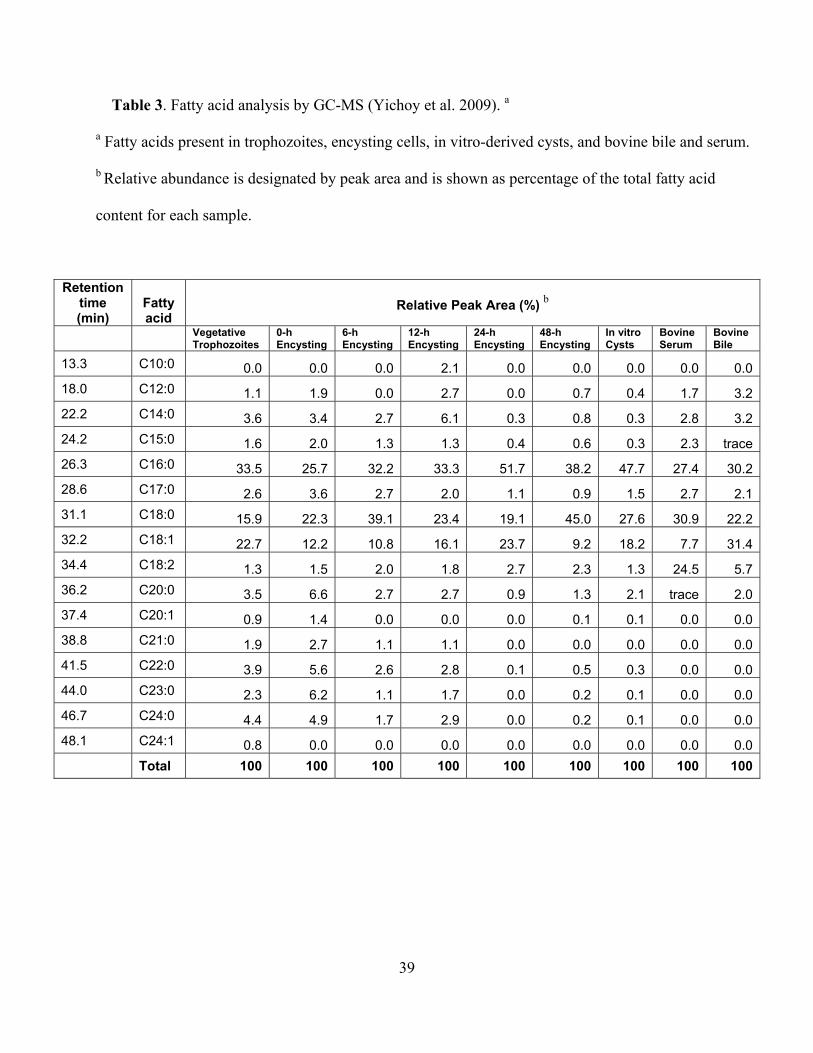
2.2.2. GC-MS analysis of fatty acids
Like PLs, fatty acids are also important for giardial growth and encystation (Das et al.
2001). Although fatty acids trigger encystation in vitro (Gillin et al. 1987), excess fatty acids are
toxic and possess anti-giardial properties. It has been demonstrated that medium-chain fatty
acids, especially dodecanoic acid (C12:0), kill Giardia in vitro with (LD50 ~13.78 µg/ml) (Rayan
et al. 2005). Previous reports (Kaneda and Goutsu 1988; Mohareb et al. 1991; Ellis et al. 1996)
indicate that C16:0 is the major fatty acid in vegetative and encysting Giardia, followed by
C18:0, C18:1, and C18:2. Traces of C14:0, C15:0, C17:0, C18:3, C19:0, C20:0, C22:0, C24:0,
C26:0, and C28:0 have also been detected. Interestingly, no dramatic differences were observed
in fatty-acid compositions between vegetative and encysting trophozoites. In the current study,
the total fatty acid content of trophozoites, encysting cells, and cysts was analyzed by GC-MS,
and the results revealed that C16:0, C18:0, and C18:1 were indeed the major fatty acids in all
giardial lipid species, as evidenced by relative peak areas (Figure 9A). However, less common
acyl groups such as C10:0, C12:0 and C14:0 were also present. The fatty-acid composition
remained unchanged in vegetative trophozoites, encysting cells (0–48 hours), and water-resistant
cysts (Figure 9A, Table 3). In serum and bile, the major fatty acids are C16:0, C18:0, C18:1, and
C18: 2 (Figure 9B, Table 3). Traces of other fatty acids such as C12:0, C14:0, C15:0, C17:0,

25
C20:0, and C22:0, were also identified. Our results also suggest that some long chain fatty acids
(i.e., C24:0, C24:1, C23:0, etc.), which are present in Giardia, could be synthesized de novo by
the action of elongase(s). Although no attempt was made to measure the elongase activity, the
BLAST search of the Giardia Genome Database (http://giardiadb.org/giardiadb/) (Morrison et al.
2007) yielded the presence of fatty acid elongase 1 gene (accession no. XM_00170849.1, E-
value 4e-81), suggesting that fatty acid elongation machinery may be present in this pathogen.
Interestingly, in Dictyostelium discoideum, the same sequence was annotated as fatty acid
elongase 3-ketoacyl-CoA synthase (NCBI accession no. XP_638938). Our GC-MS analysis also
revealed that the sterol fractions purified from trophozoites, encysting cells and cysts contain
only cholesterol (Figure 10). However, a previous report suggests the presence of low levels of
ergosterol along with the more abundant cholesterol in this protozoan parasite (Ellis et al. 1996).
2.3. Conclusions
The current lipidomic analysis generated valuable information regarding the metabolism
and synthesis of phospholipids in Giardia. It was observed that giardial lipid composition was
different than the lipid and fatty-acid contents of the growth medium, and that PG and PE are
newly generated phospholipids, which suggests that this evolutionarily basal organism may use a
sophisticated pathway to carry out lipid synthesis. In addition to PG and PE, I also detected new
fatty acids, which are not present in the medium. Interestingly, the Giardia genome database
(www.GiardiaDB.org) did not reveal the existence of well-known fatty-acid biosynthesis
pathways that are operative in bacteria and mammalian cells, which further indicated its

26
evolutionary position—i.e., deep in the branch leading from prokaryotes to higher eukaryotes
(Sogin et al. 1989). The presence of mono-methylated branched-chain fatty acids (MBCFA)
further supports the primitive metabolic characteristics of this unicellular protozoan because
MBCFAs are ubiquitous in bacteria and archaea but are not present in mammalian cells
(Chattopadhyay and Jagannadham 2003). Thus, the lipid metabolic abilities of this organism are
quite varied, with some being very primitive and others being more evolved than previously
reported.
The analysis presented in the current dissertation also emphasizes the use of sophisticated
instruments, and so analyzing software should be employed to generate more accurate
descriptions of any biological processes. The mass spectrometric analysis of the giardial
lipidome reveals the presence of many PL and fatty-acid species that could not be detected
earlier by TLC and HPLC. Detection of new lipid and fatty-acid species in Giardia encouraged
me to investigate the pathways that allow this parasite to synthesize new phospholipids by
genomic and biochemical analyses described in Chapter 3 and Chapter 4.

Figure 6
27

Figure 6. Lipid analysis by MS of G. lamblia. Total phospholipids were fractionated by silica-
gel 60 and analyzed by ESI-QTOF-MS. (A and B) Positive- and negative-ion mode full-scan
spectra of giardial phospholipids, respectively. Total lipids and phospholipids from vegetative,
encysting and water-resistant cysts were isolated as described in the Materials and Methods.
(Yichoy et al. 2009).
28

Figure 7
(A)
(B)
29

Figure 7 (C)
(D)
30

(E)
Figure 7. Lipid analysis by MS of G. lamblia. Total phospholipids were fractionated by silica-
gel 60 and analyzed by ESI-QTOF-MS. (A and B) Positive-ion mode MS-MS spectra of
C18:1/18:1-PC (m/z 792.7) C16:0/d16:1-SM at m/z 709.9, respectively. (C) MS-MS spectrum of
C18:0/16:0-PG parent-ion at m/z 749.5, ionized in negative-ion mode. “x16” indicates that the
portion of that spectrum was magnified sixteen times to make the peaks more visible. The
number at the top right corner of each spectrum indicates signal strength, measured as ion
intensity at 100% relative abundance. m/z, mass to charge ratio. (D) Negative-ion mode MS-MS
spectra of C18:1/C16:0-PE (m/z 716.7). (E) C16:0/C16:0-PI (m/z 809.7), respectively. The
number at the top right corner of each spectrum indicates signal strength, measured as ion
intensity at 100% relative abundance. m/z, mass to charge ratio (Yichoy et al. 2009).
31

32
Table 1. Lipid analysis and composition of the major phospholipids from differentiating Giardia lamblia (Yichoy et al. 2009). a
a Phospholipid species were identified by MS-MS analysis in positive- and negative-ion modes. b Relative abundance is designated by the peak height: ++++, up to 100%; +++, up to 75%; ++, up to 50%; +, 10% or less. IS, internal standard.
c Relative abundances for each sample were similar and therefore only the peak heights for trophozoites are shown. N/A, not applicable.
PL m/z Ion Species Proposed Structure sn-1/sn-2 b
Relative Abundance c
Positive-Ion Mode
PC 502.4 M + Li lyso-C16:0 +
526.4 M + Li lyso-C18:2 +
528.4 M + Li lyso-C18:1 +
600.5 M + Li C11:0/C11:0 (IS) N/A
740.8 M + Li C16:0/C16:0 +
752.7 M + Li C16:0/C17:1 and/or C18:1/C15:0 +
754.8 M + Li C16:0/C17:1 +
764.8 M + Li C16:1/C18:1 +
766.8 M + Li C16:0/C18:1 ++
768.8 M + Li C16:0/C18:0 +
780.6 M + Li C17:0 /C18:1 +
788.6 M + Li C18:2/C18:2 +
790.7 M + Li C18:1/C18:2 +
792.7 M + Li C18:1/C18:1 ++
794.6 M + Li C18:0/C18:1 ++++
806.8 M + Na C18:1/C18:2 +
808.8 M + Na C18:1/C18:1 ++
814.6 M + Li C20:4/C18:1 ++
816.6 M + Li C20:4/C18:0 +
SM 709.9 M + Li C16:0/d18:1 ++
725.7 M + Na C16:0/d18:1 +
737.8 M + Li C18:0/d18:1 +

33
Table 1. Continued.
PL m/z Ion Species Proposed Structures sn-1/sn-2 b
Relative Abundance c
Negative-Ion Mode
PC 804.6 M + formate C18:1/C16:0 +
806.6 M + formate C18:0/C16:0 +
820.6 M + Cl C18:1/C18:1 +
828.6 M + formate C18:2/C18:1 +
830.6 M + formate C18:1/C18:1 +
832.6 M - H C18:0/C18:1 +
PE 578.4 M - H C12:0/C12:0 (IS) N/A
636.4 M + NaCl - H C12:0/C12:0 (IS) N/A
646.4 M + Na + formate - H C12:0/C12:0 (IS) N/A
714.5 M - H C16:0/C18:2 and/or C16:1/C18:1 ++
716.5 M - H C18:1/C16:0 +++
834.6 M - H C22:6/C22:6 (IS) N/A
PG 609.4 M - H C12:0/C12:0 (IS) N/A
707.5 M - H C16:0/15:0 and/or C14:0/C17:0 +
719.6 M - H C16:0/C16:1 and/or C18:1/C14:0 +
721.5 M - H C16:0/16:0 and/or C18:0/C14:0 +++
733.5 M - H C16:0/C17:1 and/or C18:1/C15:0 +
735.5 M - H C16:0/C17:0 and/or C15:0/C18:0 +
745.5 M - H C18:2/C16:0 and/or C16:1/C18:1 +
747.5 M - H C18:1/C16:0 ++++
749.5 M - H C18:0/C16:0 ++++
761.5 M - H C18:1/C17:0 +
763.6 M - H C18:0/C17:0 and/or C19:0/C16:0 +
771.5 M - H C18:1/C18:2 +
773.5 M - H C18:1/C18:1 +
775.5 M - H C18:0/C18:1 +
777.6 M - H C16:0/20:0 and/or C18:0/C18:0 +
789.5 M + Na + formate – H C16:0/C16:0, C18:0/C14:0, and/or C15:0/C17:0 +
817.5 M + Na + formate – H C18:0/C16:0 +
PI 809.5 M - H C16:0/C16:0 +
835.5 M - H C18:1/C16:0 +

34
Table 2. Positive-ion mode MS-MS analysis of phospholipids from bile and serum (Yichoy et al.
2009).
m/z Ion Species Proposed Structures sn-1/sn-2
526.4 M + Li lyso-C18:2-PC 528.4 M + Li lyso-C18:1-PC 530.5 M + Li lyso-C18:0-PC 552.4 M + Li lyso-C20:3-PC 653.7 M + Li C18:0/C20:3-DAG 764.7 M + Li C16:0/C18:2-PC 766.7 M + Li C16:0/C18:1-PC 790.7 M + Li C18:1/C18:2-PC and/or C18:0/C18:3-PC 792.7 M + Li 18:0/18:2-PC 794.7 M + Li 18:0/18:0-PC 816.7 M + Li 18:0/20:4-PC

Figure 8
35

36
Figure 8. ESI-QTOF-MS spectra of relative quantitative analysis of giardial phospholipids.
Total lipids were extracted and fractionated as described in Materials and Methods. Cell numbers
were adjusted to 5000 cells/µL. (A and B) Positive- and negative-ion mode analysis,
respectively. For positive-ion (ESI+) mode MS analysis, giardial lipid samples were spiked with
2.5 µM 11:0/11:0-PC (m/z 600.4) ([M + Li]+), used as internal standard (IS). For negative-ion
(ESI-) mode MS analysis, 5 µM 12:0/12:0-PE (m/z 578.4) ([M - H]-) was used as the IS. m/z,
mass to charge ratio (Yichoy et al. 2009).

Figure 9
37

38
Figure 9. GC-MS spectra of fatty acid content. Total lipids from vegetative trophozoites,
encysting cells, water-resistant cysts, bile and serum were isolated and processed as described in
Materials and Methods. (A) Giardial fatty acids; (B) Fatty acids from the bile and serum. The
retention times (min) of identified fatty acids and internal standards are indicated (Yichoy et al.
2009).
39
Table 3. Fatty acid analysis by GC-MS (Yichoy et al. 2009). a
a Fatty acids present in trophozoites, encysting cells, in vitro-derived cysts, and bovine bile and serum.
b Relative abundance is designated by peak area and is shown as percentage of the total fatty acid
content for each sample.
Retention time (min)
Fatty acid
Relative Peak Area (%) b
Vegetative Trophozoites
0-h Encysting
6-h Encysting
12-h Encysting
24-h Encysting
48-h Encysting
In vitro Cysts
Bovine Serum
Bovine Bile
13.3 C10:0 0.0 0.0 0.0 2.1 0.0 0.0 0.0 0.0 0.018.0 C12:0 1.1 1.9 0.0 2.7 0.0 0.7 0.4 1.7 3.222.2 C14:0 3.6 3.4 2.7 6.1 0.3 0.8 0.3 2.8 3.224.2 C15:0 1.6 2.0 1.3 1.3 0.4 0.6 0.3 2.3 trace26.3 C16:0 33.5 25.7 32.2 33.3 51.7 38.2 47.7 27.4 30.228.6 C17:0 2.6 3.6 2.7 2.0 1.1 0.9 1.5 2.7 2.131.1 C18:0 15.9 22.3 39.1 23.4 19.1 45.0 27.6 30.9 22.232.2 C18:1 22.7 12.2 10.8 16.1 23.7 9.2 18.2 7.7 31.434.4 C18:2 1.3 1.5 2.0 1.8 2.7 2.3 1.3 24.5 5.736.2 C20:0 3.5 6.6 2.7 2.7 0.9 1.3 2.1 trace 2.037.4 C20:1 0.9 1.4 0.0 0.0 0.0 0.1 0.1 0.0 0.038.8 C21:0 1.9 2.7 1.1 1.1 0.0 0.0 0.0 0.0 0.041.5 C22:0 3.9 5.6 2.6 2.8 0.1 0.5 0.3 0.0 0.044.0 C23:0 2.3 6.2 1.1 1.7 0.0 0.2 0.1 0.0 0.046.7 C24:0 4.4 4.9 1.7 2.9 0.0 0.2 0.1 0.0 0.048.1 C24:1 0.8 0.0 0.0 0.0 0.0 0.0 0.0 0.0 0.0 Total 100 100 100 100 100 100 100 100 100

Figure 10. Sterol analysis by GC-MS. Neutral lipids eluted with choloroform from total lipid extract Cho: cholesterol (Yichoy et al. 2009)
40

41
Chapter 3: Genomic and transcriptional analyses of putative phospholipid metabolic and transporter genes
The goal of this chapter is to identify the genes of lipid metabolic and transporter
enzymes present in Giardia. The data presented in Chapter 2 indicates that PG and PE are most
likely newly generated phospholipids (PLs) and that they are not obtained from the culture media
(Yichoy et al. 2009). Determining whether the Giardia genome contains the open-reading frames
(ORFs) for PL synthesis, metabolism, and transport proteins will provide important information
regarding lipid biochemistry of this protozoan parasite. Because of the fact that the giardial
growth medium contains only PC and lyso-PC (discussed in Chapter 2), and due to the fact that
Giardia has a limited ability to synthesize its own lipids (Jarroll et al. 1981), it is conceivable
that PC/lyso-PC serve as precursors of PG and PE. Most likely the lipids from the growth
medium are internalized by lipid transporters (i.e., flippase) and remodeled by head-group or
base-exchange enzymes such as phosphatidylglycerolphosphate synthase (PGPS) and
phosphatidylserine decarboxylase (PSD). The focus of Chapter 3 is to carry out various
bioinformatic (i.e., Pfam, phylogeny, predicted cellular localizations, topology, etc.) analyses,
along with SYBR-Green assisted transcriptional assays of pgps and psd to comprehend the
nature and functions of these lipid biosynthesis and transport genes in Giardia. Such analyses
should predict considerable information with regard to the lipid metabolomics of this early
divergent eukaryote.

42
3.1. Materials and Methods
Predicted open-reading frames (ORFs) were obtained from the Giardia genome
database (http://giardiadb.org/giardiadb/) and were compared using BLASTP (Morrison et al.
2007). Sequences for PE and PG biosynthesis were identified, including the genes of
phosphatidylserine decarboxylase (psd, accession no. XM_001707858, ORF no. 16495) and
phosphatidylglycerolphosphate synthase (pgps, accession no. XP_769290, ORF no. 7259). PCR
primers were designed using Primer3 software (http://frodo.wi.mit.edu) and were synthesized by
MWG Biotech (High Point, NC) or Sigma Genosys (St. Louis, MO). The sequences of the
primer pairs are as follows.
Following harvest, cysts were re-suspended in 4 ml TRI reagent (Sigma) and freeze-
thawed at -20 °C three times. Following the last freeze-thaw, 0.5-mm glass beads (BioSpec
Products) were added, and the cyst wall was broken by vortexing for approximately 5 min. Total
RNA was extracted from trophozoites and encysting cells using TRI reagent and was reverse
transcribed using the ImProm-II Reverse Transcriptase Kit (Promega). Levels of PGPS
(GenBank accession number XP_769290) and PSD (GenBank accession number
XM_001707858) transcription were quantified by quantitative, real-time PCR. RT2 Real-Time
SYBR Green/Fluorescein PCR Master Mix (SuperArray) was used in the reaction mixture, and
all reactions were performed three times. Primers targeting α-tubulin were used as a control to
normalize the samples. Transcript levels were quantified using the relative standard-curve
method (Hernandez et al. 2007; Morrison et al. 2007). Statistical analysis was performed with
one-way ANOVA with Dunnett’s Post Test using GraphPad Prism version 4.00 for Windows
(GraphPad Software, San Diego, CA).

43
3.2. Results
3.2.1. Genomic analysis of PG and PE synthesis and Flippase genes
Because PG and PE are not present in bile or serum (Table 2), we asked whether they
could be newly synthesized in Giardia. Therefore, we searched the Giardia Genome Database
(http://giardiadb.org/giardiadb) (Morrison et al. 2007) to identify PG- and PE-related
synthesis/remodeling genes. Furthermore, because it has been suggested that the Giardia genome
has limited lipid-remodeling abilities and that many of the traditional de novo lipid synthesis
enzymes are not found in the giardial genome, we asked whether this protozoan has flippases, or
ATP-dependent phospholipid transporters (Daleke and Lyles 2000; Morrison et al. 2007). The
BLASTp search yielded giardial phosphatidylglycerolphosphate synthase (gpgps) and
phosphatidylserine decarboxylase (gpsd) genes, as well as four flippases. Pfam analyses revealed
that gpgps matched well with bacterial and trichomonads pgps (see Table 4). The two closest
matches were Opitutaceae bacterium and Trichomonas vaginalis, with E-values of 1.00 e-32 and
5.00 e-25, respectively. However, the open-reading frame (ORF) annotations
(http://giardiadb.org/giardiadb/) (Morrison et al. 2007) for these matches were CDP-DAG-
glycerol-3-phosphate and 3-phosphatidyltransferase, rather than PGPS, and they belong to the
CDP-alcohol phosphatidyltransferase family of enzymes. On the other hand, gpsd yielded
matches of the eukaryotic phylum, with the closest being Plasmodium vivax (E-value 2.00 e-22).
Other protozoa with psd sequences that closely matched the giardial psd included Toxoplasma
gondii and Plasmodium falciparum. For example, gpsd belongs to the phosphatidylserine
decarboxylase family of enzymes. Unlike gPSD and gPGPS, three of the flippase protein
sequences did not match to bacteria or other protozoans. Instead, these most closely match to

44
Homo sapiens (E-value 5 e-76 and 5 e-26) and Rattus norvegicus (E-value 4 e-94), while the last
matches to Pediculus humanis corporis (2 e-94) as well as to Entamoeba histolytica (2 e-90).
Pfam analysis shows that all four giardial flippases belong to the family of E1 and E2 ATPases
(Table 4).
To retrieve the predicted sub-cellular localization of gPGPS and gPSD, DNA sequences
were translated into protein using the translation tool, available at the Expert Protein Analysis
System (ExPaSy) proteomics server (www.expasy.ch/) (Gasteiger et al. 2003; Morrison et al.
2007). The translated protein sequences were then entered into PSORTII to predict the sub-
cellular localizations. PORTII predicts that gPGPS is an ER-bound protein and that gPSD
localizes in the cytoplasm in Giardia (Horton and Nakai 1997; Nakai and Horton 1999) (Table
5). Because the PSORTII tool only has options for fungal, bacterial, plant and mammalian
sequences, the CSS-Palm tool was employed to verify that membrane localization of the four
flippases. Protein palmitoylation is a post-translational modification that acts as a membrane
target anchor. Therefore, if the giardial flippases are indeed membrane-bound proteins, CSS-
Palm would act as another prediction tool in addition to PSORT (Greaves and Chamberlain
2007; Emmer et al. 2009). The CSS-Palm tool indicates that all four flippases as well as PGPS
and PSD have palmitoylation sites (Table 5).
3.2.2. Transcriptional analysis suggests that gpgps and gpsd genes are transcribed in Giardia
Recent results from our laboratory suggest that many lipid synthesis genes in Giardia are
regulated differentially during encystation (Hernandez et al. 2008). Here, we asked whether gpsd
and gpgps are transcribed and are expressed in trophozoites and cysts. Quantitative, reverse–

45
transcription, polymerase chain-reaction (qRT-PCR) results indicate that both gpsd and gpgps
are expressed in vegetative and encysting trophozoites as well as in water-resistant cysts. It is
clear from Figure 11 that these two genes display differential patterns of expression. For
example, in 6-h and 12-h encysting cells, gpsd transcript levels decrease ~60% and 50%,
respectively, whereas in 24-h encysting cells the transcript level is two-fold higher than in the
vegetative state and remains high (~1.5 fold) in cysts. Transcript levels were found to be
significantly different from vegetative trophozoites by one-way ANOVA with Dunnett’s Post
Test (p < 0.05). In the case of gpgps, the transcript levels decrease by ~30% in 6-, 12-, and 24-h
encysting cells. In 48-h encysting cells and cysts, expression increases ~1.5 fold (Figure 10B).
Interestingly, Dunnett’s Post Test did not show that gpgps transcript levels in encysting
cells or cysts were significantly different from trophozoites (p > 0.05). Although it is not known
whether these genes are translated into products, data indicate that both gPSD and gPGPS could
play significant roles in giardial biology. For example, while gPSDs are required for encystation,
they might play precise roles in forming cysts and during excystation to trophozoites.
3.3. Conclusions
Despite the fact that giardial PSD most closely resembles the protein sequence of
Plasmodium vivax, it is unlikely that Giardia shares many similarities with the phospholipid
metabolism of Plasmodium. While both sets of organisms are capable of generating PE by head-
group exchange, the similarities end there (Vial et al. 2003). Plasmodium has more capabilities
for generating phospholipids de novo, unlike Giardia. Also, the similarities between the giardial
PGPS and bacterial homologues reiterates the position of this protozoan as an evolutionarily

46
basal organism and reinforces the fact that Giardia maintains metabolic similarities with both
other eukaryotes and bacteria and Archaea. Furthermore, given that PG is primarily found in the
plasma membrane of bacteria and mitochondrial membranes of eukaryotes, these similarities to
bacteria are logical. It would be of interest to determine whether giardial PG localizes in the
mitosome. Previously, it has been reported that PG localizes throughout the cytoplasm and in the
perinuclear regions (Das et al. 2001), but it is not known whether these areas of fluorescence
would co-localize with mitosome antibodies.
The Pfam matches for the four gFlip enzymes indicate that flippases are well conserved,
despite the differences in lipid metabolism among protozoans and mammalian systems.
However, these similarities also indicate that despite their more sophisticated lipid synthesis
abilities, mammalian cells also require the uptake of exogenous lipids. It is surprising that none
of the four giardial flippases are predicted to be localized in the ER, given that phospholipids,
including sphingomyelin (Hernandez et al. 2007), are likely synthesized in the ER, thereby
creating a need for transport molecules to move newly synthesized lipids through the ER
membrane and ultimately to their designated location within the cell. Since PGPS is predicted to
localize to the ER as well, it is logical to assume that newly generated PGs and sphingolipids
would require a transport protein in order to pass through the ER membrane. In fact, it has been
shown that flippases in other organisms, such as Saccharomyces cerevisiae, do indeed localize to
the ER and are responsible for the translocation of not only phospholipids but also dolichol-
phosphate-anchored oligosaccharides across the ER membrane (Sanyal et al. 2008). It is
possible, however, that an undescribed lipid transport system exists solely for the purpose of
transport out of organelles.

47
Table 4. Predicted open reading frames and Pfam matches of giardial
PGPS, PSD, and Flippases.a
Designation (gORF no.)
GenBank Accession
Match to Pfam (motif location)
Species with best BLASTP match
(E-value) Pfam family match
Flippase IA 16958 XP_001704967
E-value 3.1e-10 (aa 87-173)
Pediculus humanis corporis (2e-94)
Entamoeba histolytica (2e-90) E1-E2 ATPase
Flippase IA 10019 XP_001710085
E-value 5.3e-11 (aa 97-185)
Homo sapiens (5e-76) E1-E2 ATPase
Flippase IIB 101810 XP_001707954
E-value 3.2e-8 (aa 207-301)
Homo sapiens (5e-26) E1-E2 ATPase
Flippase IIB 38104 XP_001704293.1
E-value 1.2e-13 (aa 100-295)
Rattus norvegicus (4e-94) E1-E2 ATPase
PGPS (7259) XP_769290
2.3E-29 (aa 63-191)
Opitutacaea bacterium (1e-32)
Trichomonas vaginalis (5e-25) CDP-alcohol phosphatidyl transferase
PSD (16495) XP_779868
1.1E-13 (aa 186-409)
Plasmodium vivax (2e-22) Phosphatidylserine decarboxylase
aPutative genes encoding flippases, phosphatidylglycerolphosphate synthase (PGPS) and
phosphatidylserine decarboxylase (PSD) were identified in Giardia using the NCBI and Protein
Family (Pfam) database. Adapted from Yichoy et al (2009).

Table 5. Topology and localization predictions of giardial lipid metabolic enzymes using PsortII (Horton and Nakai 1997; Nakai and
Horton 1999).
Predictions
Designation (gORF)
GenBank Accession
Topology (Transmembrane domains) k-NN
Palmitoylation Sites
Flippase IA (16958) XP_001704967 9 plasma membrane 69.6% 12
Flippase IA (10019) XP_001710085 10 plasma membrane 65.2% 7
Flippase IIB (101810) XP_001707954 10 plasma membrane 78.3% 7
Flippase IIB (38104) XP_001704293.1 10 plasma membrane 69.6% 9
PGPS (7259) XP_769290 4
endoplasmic reticulum 43.5% plasma membrane 21.7% mitochondrial 21.7% 1
PSD (16495) XP_779868 1
mitochondrial 60.9% cytoplasmic 26.1% 1
48

Figure 11. Differential expression of giardial phosphatidylserine decarboxylase (gpsd) and
giardial phosphatidylglycerolphosphate synthase (gpgps) genes, relative to vegetative
trophozoites. Experiments were carried out three times with three replicates each time. The data
are the means and standard deviations of these replicates. An asterisk indicates significant
difference compared to vegetative trophozoites with p<0.05. Panel A indicates the expression of
gpsd, and Panel B denotes gpgps expression (Yichoy et al. 2009). 49

50
Chapter 4: Elucidating the mechanism of phosphatidylglycerol (PG) synthesis
Because phosphatidylglycerol (PG) is one of the two phospholipids newly synthesized by
Giardia and abundant in trophozoites and cysts, it is likely that it’s synthesis important for the
growth and viability of this waterborne pathogen. This statement can be further supported by the
fact that knocking down the gpgps gene (linked to PG synthesis) kills Giardia instantly (not
shown). Genomic analyses (see Chapter 3) indicate that a majority of PG synthesis genes are not
present in Giardia, and therefore that the question of how new PG is synthesized is extremely
important. The goal of Chapter 4, therefore, is to delineate the mechanism of
phosphatidylcholine (PC) internalization (because PC is the major phospholipid in the growth
medium) and its subsequent conversion into PG and other phospholipids through remodeling
reactions. Using fluorescently conjugated phospholipids, I show that the flipasse-like
sulphohydryl (-SH) group containing transport proteins is involved in PC uptake across the
plasma membranes of Giardia. These transporters are also efficient in transporting other amino-
phospholipid (i.e., PE) but not glycerol-containing phospholipids such as PG. I speculate that PC
is the major source of PG and that it most likely occurs by way of a headgroup remodeling step
through an enzyme that is yet to be identified and characterized.
4.1. Materials and Methods
4.1.1. Materials
Stock solutions (100 mM) of N-ethylmalemide (NEM) and Verapamil (Sigma, St. Louis,
MO) were prepared freshly before carrying out the experiments. Stock solutions of BODIPY-PE,

51
BODIPY-PC (Invitrogen, Carlsbad, CA), and NBD-PG (Avanti Polar Lipids, Alabaster, AL)
were prepared by resuspending the lyophilized dyes in 1ml ethanol.
4.1.2. Labeling with the non-radioactive isotope [13C]-glycerol
Media from confluent 8-ml culture tubes were discarded, and tubes were refilled with
phosphate-buffered saline (PBS) supplemented with 2 mM cysteine and 100 µL dialyzed fetal
bovine serum (FBS) per 8-ml tube, pH 7.1. [13C]-glycerol was added directly to the culture tubes
to create a final concentration of 5 mM, which was incubated at 37 ºC for 5 h. N-ethylmalemide
(NEM) and Verapamil (flippase inhibitors) were also added to final concentrations of 200 µM
and 50 µM, respectively (Biagini et al. 2000; Reiner et al. 2003). All labeling reactions were
carried out in duplicate, and cells from duplicate tubes were later combined in order to achieve a
sufficient number of cells to carry out the lipid isolation and analyses. Following the incubation
period, liquid was removed by decanting the medium and refilling the tubes with PBS. Tubes
were kept in ice-cold water for 20–30 min to remove the attached cells, and labeled cells were
then harvested by centrifugation at 3,000 rpm for 7 min at 4°C to collect the cell pellets. The
pellets were washed with PBS and kept at -20oC until lipid extraction. Total lipids were isolated
later by the Folch method, and phospholipids were fractionated using a Silica60 column, as
previously described in Chapter 2.

52
4.1.3. Phospholipid analysis by linear ion trap mass spectrometry
MS spectra for fractionated lipids were acquired in a linear ion trap (LTQ XL, Thermo
Fisher Scientific). Samples were dissolved in CHCl3: CH3OH: formic acid: NH4OH
(1:1:0.1%:0.1%) and were analyzed in negative-ion mode. Samples were injected using a
Triversa Nanomate System (Advion), full-scan spectra were collected in the 550–900 m/z range,
and MS/MS spectra were automatically collected by total-ion mapping using pulsed Q
dissociation. The collision energy was set at 29% and Q = 0.7 for 0.1 ms, and helium was used as
the collision gas. For quantitative analysis, samples were normalized to 5,000 cells/μl and spiked
with 0.5 µM C12:0/C12:0–PG as an internal standard.
4.1.4. Labeling cells with a lipid-staining reagent.
Growth medium was discarded from culture tubes and refilled with PBS. Trophozoites
were pre-incubated with 50 µM Verapamil or 200 µM NEM for 1 h at 37 °C. Following the pre-
incubation, Nile Red, a lipid-staining reagent (Sigma, St. Lous, MO) was added to a final
concentration of 10 µg/ml, incubated for 15 min, washed five times with PBS, and then
harvested by centrifugation as described above. Trophozoites were then transferred to 4-well
chambered slides (Nalgene Nunc, Naperville, IL) and allowed to attach for 30 min at 37 °C.
Slides were then washed once in Coplin jars and trophozoites were fixed with 4%
paraformaldehyde in 1x PBS for 15 min, washed again 3 times with 1x PBS and then allowed to
air dry. Cover slips were mounted using DAKO Fluorescence Mounting Medium (DAKO

53
Corporation, Carpinteria, CA) and allowed to set overnight at 4 °C before analyzing by confocal
microscopy.
4.1.5. Labeling with fluorescent lipid probes
The media in the culture tubes were discarded, and the tubes were refilled with PBS.
Then the flippase inhibitors NEM and Verapamil were added, at the concentrations described
above. Cells were pre-incubated for 1 h for Verapamil-treated cells and 45 min for NEM-treated
cells. After the pre-incubation, the fluorescent lipid probes were added directly to the cultures in
the following concentrations: BODIPY-PE, 200 µM; BODIPY-PC, 200 µM; and NBD-PG, 5
µM. Trophozoites were incubated with the fluorescent probes for 1 h at 37 °C, washed once,
harvested by centrifugation as described above, and re-suspended in 1 ml PBS. Trophozoites
were transferred to 4-well chambered slides (Nalgene Nunc, Naperville, IL) and incubated at
37°C for 30 min for Verapamil-treated cells and 45 min for NEM-treated cells to allow cells to
attach to the slides. After the attachment period, slides were washed, fixed, and mounted as
described above.
4.1.6. Analysis of lipid uptake by confocal microscopy
Images of fluorescent-labeled trophozoites were taken using an LSM5 Pascal laser-
scanning, confocal microscope (Zeiss). For Nile Red BODIPY-PC and NBD-PG uptake
experiments, images were scanned 16 times at a scan speed of 6. For BODIPY-PE, images were
scanned 8 times, and a scan speed of 6 was used. Nine images were taken for each treatment

54
group, and control and fluorescence intensity were analyzed using LSM5 Pascal software,
version 3.2 (Zeiss). To compare differences between treatments and controls in NBD-PG-labeled
cells, one-way ANOVA with Dunnet’s post test was performed. For all other fluorescent-
labeling experiments, an unpaired, two-tailed t-test was performed. All statistical analyses
included a sample size of at least n = 9 and a confidence interval of 95%, and were performed
using GraphPad Prism version 4.00 for Windows® (GraphPad Software, San Diego, CA).
4.2. Results
4.2.1. Labeling with [13C]-glycerol
The previous experiment from our laboratory suggested that [14C]-labeled glycerol
incorporates into PG in Giardia (unpublished). However, these studies were performed using
TLC, making it difficult to identify specifically the species into which the radio-labeled bases
were incorporating. Therefore, these assays were repeated using [13C]-glycerol, a non-radioactive
isotope, which allowed for analysis by mass spectrometry. Because bases with a two-carbon
label were used (Figure 12), a 2-Dalton shift should be evident in the MS/MS spectrum if the
isotopes are incorporated,. Therefore, if a [13C] 1, 3-Glycerol C2 label incorporates into a PG, a 2-
Dalton shift for m/z 153 to m/z 155 (Figure 13) should be seen with the peak corresponding to the
glycerol phosphate moiety of PG. While a few technical problems caused a limited amount of
labeling, these studies reveal that [13C]-glycerol incorporates into the three most abundant PGs
(Yichoy et al. 2009): C16:0/C16:0-PG, C18:0/C14:0-PG, and C18:0/C16:0-PG (Figure 13).
Labeling with non-radioactive glycerol was also performed in the presence of NEM, a flippase
inhibitor, and Verapamil to determine whether remodeling of PLs is dependent upon the uptake

55
of exogenous head groups. Because low levels of incorporation were observed, treatment with
these drugs did not result in significant differences in incorporation levels. Interesting results
were observed in the full-scan spectra, however (Figure 14). In NEM-treated cells, the total
amount of PGs appears to be dramatically decreased. This indicates that Giardia is dependent
upon lipid transport systems in order to generate new PLs. Differences in lipid composition were
not observed between untreated trophozoites and Verapamil-treated cells, which suggests that the
flippase-like transporters present in this protozoan are not Ca2+ dependent.
4.2.2. Uptake and incorporation of fluorescent lipid probes
To further examine whether the distribution and uptake of lipids is dependent upon a PL
transport system, trophozoites were treated with NEM and Verapamil and then labeled with the
lipid tracker Nile Red, as well as with the fluorescent lipid probes BODIPY-PC, BODIPY-PC,
and NBD-PG. Major differences were not observed in overall lipid distribution when cells were
labeled with Nile Red (Figure 15). However, localization of lipids in the nuclear region appeared
to be decreased in both NEM-treated cells, but increases when cells are treated with Verapamil.
This indicates that while uptake of other lipids (ie; cholesterol and sphingolipids), is not
dependent upon a flippase-like enzyme, transport between organelles is indeed affected. Also,
while Verapamil and NEM are considered broad-spectrum inhibitors, this data indicates that
formation of clathrin-coated vesicles, which is responsible for the uptake of ceramide, is not
affected by either of these compounds (Hernandez et al. 2007).
More marked differences were observed in BODIPY-PC and -PE uptake and
incorporation in the presence of flippase inhibitors (Figures 16 and 17). In fact, both BODIPY-

56
PC and -PE uptake are dramatically decreased, and the fluorescence intensity is many times
lower than untreated controls. For Verapamil-treated cells, the only observed difference is
decreased localization of BODIPY-PE in the membranes, but there is no difference for BODIPY-
PC distribution. For cells labeled with NBD-PG (Figure 18), while fluorescence intensity is
decreased with both inhibitors, the difference is not quite as dramatic as with BODIPY-PC and -
PE, indicating that the flippase-like transporter localized in the giardial plasma membrane is
specific to PC and PE and that PG can be internalized via other mechanisms. Although uptake of
NBD-PG is significantly affected, the fact that it is not completely reduced indicates that PG can
be internalized from the host through another transport mechanism.
Conclusions
The [13C]-incorporation and fluorescent uptake assays described in this chapter raised
several possibilities regarding lipid scavenging and PG synthesis by Giardia. The confocal
analysis reveals that Giardia is largely dependent upon PC from the host, and most likely PG can
be generated by the parasite itself. Therefore, it can be concluded that while Giardia has more
lipid-synthesis activities than previously thought, this organism is largely dependent upon
exogenous sources of lipids, particularly PC.

13C 13COH OH
OH
Glycerol-1,3-13C2
13C13C
NH2OH
Ethanolamine-13C2
Figure 12. Structures of non-radioactive isotopic phospholipid precursors.
57

Figure 13. Product mass spectra for [13C]- glycerol incorporation.
A. Product mass m/z 153. B. Product mass m/z 155.
58

Figure 14. Full-scan spectra of [13C] – glycerol incorporation. The internal standard and its adducts are represented by peaks at m/z
609, 667, 677 and 681.
59

Figure 15A.
60

Figure 15B.
Figure 15. Uptake and incorporation of Nile Red. Confocal images showing distribution and
statistical analysis of quantitation of fluorescence intensity/µM. P-values for each experiment are
shown below each graph. A. Nile Red incorporation in the presence of NEM. B. Incorporation in
Verapamil-treated cells.
61

Figure 16A.
62

Figure 16B.
Figure 16. Uptake and incorporation of BODIPY-PC. Confocal images showing distribution and
statistical analysis of quantitation of fluorescence intensity/µM. P-values for each experiment are
shown below each graph. A. NEM-treated cells. B. Verapamil-treated cells.
63

Figure 17A.
64

Figure 17B.
Figure 17. Uptake and incorporation of BODIPY-PE. Confocal images showing distribution and
statistical analysis of quantitation of fluorescence intensity/µM. P-values for each experiment are
shown below each graph. A. NEM-treated cells. B. Verapamil-treated cells.
65

A.
B.
Figure 18. Uptake and incorporation of NBD-PG. A. statistical analysis of quantitation of
fluorescence intensity/µM. P-values for each experiment are shown below each graph. B.
Confocal images showing distribution of NBD-PG.
66

67
Chapter 5: Discussion and Future Directions
Because of its inability to synthesize membrane lipids de novo, it has been proposed that
Giardia has evolved well-orchestrated mechanisms to import and utilize pre-formed lipids and
fatty acids from the cell-exterior (Gibson et al. 1999; Subramanian et al. 2000; Das et al. 2001).
Many of these exogenous lipids/fatty acids undergo remodeling before they are incorporated into
giardial membranes (Subramanian et al. 2000; Das et al. 2002). Although, these reports provide
some insights into the metabolism of exogenously acquired lipids, the detailed mechanisms of
lipid metabolism in Giardia are yet to be delineated.
The current study was undertaken to address two fundamental questions related to the
lipid metabolism by Giardia. First, we asked whether giardial lipid compositions are similar to
that of the culture medium, as proposed by other investigators (Jarroll et al. 1981; Kaneda and
Goutsu 1988; Mohareb et al. 1991; Ellis et al. 1996), and second we asked whether any new
membrane lipids are synthesized by this parasite.
We began our experiments by identifying membrane lipids and fatty-acid constituents
through ESI-QTOF-MS. Lipid samples from trophozoites, encysting cells, and cysts were
analyzed in positive- as well as negative-ion mode to retrieve overall information. Interestingly,
significant differences in the lipid species present in any stage of the life cycle were not detected
(Fig. 5). Furthermore, our results indicate that PG, PE, and PC are major phospholipids in
Giardia and most likely synthesized de novo, since the analysis of phospholipid content in
bovine bile and serum yielded no PG or PE (Tables 1 and 2, and Fig. 7). In another experiment
(not shown), we examined phospholipid compositions of the culture medium (containing
peptone, yeast extract, salts, serum, and bile) by thin-layer chromatography, and again, no PG

68
and PE could be identified. Although PG is abundant in bacteria, trace amounts of this acidic PL
can also be found in mammalian and protozoan cells (Besteiro et al. 2008). On the other hand,
PE is present in both prokaryotic and eukaryotic membranes and is involved in the formation,
fusion, and vesiculation of lipid bilayers (Besteiro et al. 2008). Various biochemical and
structural studies (Besteiro et al. 2008; Zhao et al. 2008) have indicated that PG interacts with
other charged lipids (i.e., PC and PE) and that it acts as a membrane stabilizer in addition to
regulating the lipid-protein interactions in bacterial membranes. PG-PE interactions are also
important for vesicle morphology, membrane permeability, and overall maintenance of the
membrane stability (Garidel and Blume 2000). Based on these studies, and considering the
challenges that Giardia meet in the human gut, it can be proposed that the interactions between
PG and PE as well as PG and PC are important for this protozoan parasite to survive and
complete its life cycle in the hostile environment of the human small intestine. Furthermore, the
abundance of PG in Giardia could also be traced back to its evolutionary origin. It has been
demonstrated that Giardia synthesizes a cryptic mitochondrion or mitosome as part of its
putative mitochondrial ancestry (Tovar et al. 2003; Regoes et al. 2005). A recent report suggests
that cardiolipin (a PG dimer), which is present in bacterial membrane, is also present in
hydrogenosome (i.e., a mitosome-like organelle) in Tritrichomonas foetus (de Andrade Rosa et
al. 2006). Therefore, it can be hypothesized that the abundance PG in Giardia could be a
signature of its endosymbiotic past and that it originated from alphaproteobacteria during
symbiosis (van der Giezen and Tovar 2005). However, more in-depth experiments are required
to support this notion.
It appears that both PG and PE in Giardia are generated by base-exchange reactions
rather than the de novo (CDP-DAG) synthesis. This postulation can further be supported by the

69
fact that when live trophozoites were metabolically labeled with radioactive glucose, threonine,
glycerol, and acetate no radioactivity could be detected in lipid fractions (Jarroll et al. 1981).
Conversely, we found that radiolabeled bases, fatty acids, and other preformed lipids can be
assembled to larger lipid molecules (Das et al. 2002). In a separate experiment, it was noted that
[14C]-glycerol and [14C]-ethanolamine are incorporated into PG and PE, which suggests that
Giardia might synthesize enzymes that allows these bases (i.e., glycerol and ethanolamine) to
incorporate them into respective phospholipids. Intriguingly, we could not detect PS in Giardia,
bile, or serum. This could be due to the reasons that PS is a minor PL that exist in few molecular
species. It is possible that during our analyses, PS was broken down to serine and diacylglycerol.
In fact, Table 2 shows the presence of DAG when phospholipid composition of bile and serum
was determined by MS analysis. Nonetheless, when live trophozoites are cultured in the presence
of [14C]-serine the majority of serine is assimilated into PE, which indicates the presence of
strong PSD activity and may explain why we failed to detect PS in Giardia (Das 2005).
As has previously been demonstrated (Kaneda and Goutsu 1988; Mohareb et al. 1991;
Ellis et al. 1996) and also confirmed by our MS analysis (Table 1, Fig. 6), PC is also abundant in
Giardia and is the only major PL available in the growth medium. In light of the fact that the
medium contains only PC and lyso-PC, the number of phospholipid species (i.e., PG, PE, and PI)
identified in trophozoite and cyst supports the idea that base-exchange enzymes are present in
this organism (Subramanian et al. 2000). However, except for gpsd, no genes for base-exchange
enzymes have so far been revealed from the genome project, and therefore detailed biochemical
analyses are required for further characterization of these enzymes.
Our GC-MS analysis of total fatty acid content reveals that Giardia contains few
additional fatty acids that are not present in the medium. Fatty acids like 21:1, 23:0, 24:0, and

70
24:1 are only present in Giardia, which suggests that these are newly generated fatty acids
produced by elongation and desaturation reactions (Fig. 8, Table 3). The presence of a fatty acid
desaturase enzyme has already been reported by Ellis et al. (1996), and the Giardia Genome
Database has identified the presence of a fatty acid elongase gene; the two could be responsible
for the exchange and elongation of acyl chains, respectively. As to the presence of odd-carbon
fatty acids (OCFAs), in our GC-MS analysis conditions we could not determine whether they
were linear or methyl-branched structures.
Our genomic and in silico analyses further emphasize that lipid metabolism is crucial to
parasite survival. The fact that pgps and psd are continually transcribed throughout the life cycle
suggests that both of these the sequences for both enzymes are housekeeping genes.
Furthermore, unpublished qRT-PCR data generated in our laboratory show that all four flippase
genes are transcribed throughout the life cycle and that, in fact, one is expressed five-fold higher
in pre-encysting cells than in trophozoites (Aguilera et al., 2009, unpublished). This indicates
that lipid uptake continually occurs during the encystation period, especially while the cell is
building energy stores, constructing the cyst wall, and preparing for its semi-dormant state.
In addition to the lipidomic and transcriptional analysis, I have also used various
bioinformatics tools to predict the cellular localization of PGPS, PSD, and the four flippases. It is
surprising that none of the flippases are localized in the ER because lipid assembly and
remodeling likely happen in this organelle. It would be logical for a lipid transporter to localize
in the ER and Golgi, although it is possible that another uncharacterized transporter is present.
Our functional analyses confirm the fact that Giardia is dependent upon exogenous
sources of lipids. The fact that the full-scan spectra from NEM-treated cells show only
background levels of PG and PE emphasizes that, while Giardia is capable of some lipid

71
synthesis activities, this protozoan is still largely dependent upon a lipid source from the
extracellular milieu, which may act as a sort of “template” from which new PLs can be
constructed. Also, since the fluorescent labeling assays show very minimal uptake of BODIPY-
PC and -PE, it can be concluded that the four Giardial flippase-like transporters are PC and PE
specific, which suggests that some other PG-specific internalization system exists. Furthermore,
while the [13C]-glycerol and -ethanolamine labeling experiments require that the culturing
conditions be perfected, the [13C]-glycerol incorporation indicates that PL remodeling through
exchange of phosphate head groups does indeed occur.
5.1. Final Conclusions
Based on biochemical data as well as available genomic and bioinformatics tools, I have
constructed a model of phospholipid, fatty acid, and diacylglycerol DAG remodeling (Figure
19). PC and lyso-PC enter the cell by way of a flippase-like transporter with multiple trans-
membrane domains. PC can be remodeled into PG through multiple though yet-to-be-
characterized steps by PGPS. PG, in turn, can be used to form PI by a PI synthase in a base-
exchange reaction. Conversely, PC may also be demethylated by a methyltransferase to form PE.
If PE is used to generate PS, it is likely converted back to PE because PS was not evident in the
lipidomic analysis. The lyso-PC taken up from the growth medium may also be remodeled into
other PCs through the exchange of acyl groups. Quite likely, these same enzymes,
lysophosphatidic acid acyltransferase and 1-acyl-sn-glycerol-3-phosphate acyl transferase, are
also responsible for exchanging fatty acid chains to form remodeled DAG and triacylglycerols.
Finally, elongation and desaturation of fatty acids also occurs.

72
The work presented here begins to elucidate the mechanisms of lipid uptake and
remodeling in Giardia. In order to continue this endeavor, it would be of interest to confirm that
PG is generated from PC by labeling with [14C]-glycerol and chasing with non-radioactive PC. If
PG is indeed generated from exogenous PC, PG levels should be higher in the samples exposed
to cold PC. It would also be of interest to knock down each individual flippase in order to
examine whether the level of any phospholipids is decreased in order to determine the specificity
of each transporter. Furthermore, while the PSORTII predictions of subcellular localization are
useful, tagging each flippase as well as PGPS and PSD with a GFP to localize these within the
cell would provide more concrete data. Finally, the isolation and characterization of a PG-
remodeling enzyme, as I have proposed in this dissertation, should be unique and useful for
screening small molecules by high-throughput screening as part of an anti-giardial drug-
discovery program currently ongoing in our laboratory.

Figure 19. Model of phospholipid, fatty acid and diacylglycerol remodeling in Giardia.
73

74
References
Abrahamsen, M.S., Templeton, T.J., Enomoto, S., Abrahante, J.E., Zhu, G., Lancto, C.A., Deng, M., Liu, C., Widmer, G., Tzipori, S., Buck, G.A., Xu, P., Bankier, A.T., Dear, P.H., Konfortov, B.A., Spriggs, H.F., Iyer, L., Anantharaman, V., Aravind, L. and Kapur, V. (2004). "Complete genome sequence of the apicomplexan, Cryptosporidium parvum." Science. 304(5669): 441-5.
Adam, R.D. (2001). "Biology of Giardia lamblia." Clinical Microbiology Reviews. 14(3): 447-
475. Almeida, I.C., Camargo, M.M., Procopio, D.O., Silva, L.S., Mehlert, A., Travassos, L.R.,
Gazzinelli, R.T. and Ferguson, M.A. (2000). "Highly purified glycosylphosphatidylinositols from Trypanosoma cruzi are potent proinflammatory agents." Embo Journal. 19(7): 1476-85.
Bernander, R., Palm, J.E. and Svard, S.G. (2001). "Genome ploidy in different stages of the
Giardia lamblia life cycle." Cellular Microbiology. 3(1): 55-62. Besteiro, S., Bertrand-Michel, J., Lebrun, M., Vial, H. and Dubremetz, J.F. (2008). "Lipidomic
analysis of Toxoplasma gondii tachyzoites rhoptries: further insights into the role of cholesterol." Biochemical Journal. 415(1): 87-96.
Biagini, G.A., Lloyd, D., Kirk, K. and Edwards, M.R. (2000). "The membrane potential of
Giardia intestinalis." FEMS Microbiology Letters. 192(1): 153-7. Bingham, A.K., Jarroll, E.L., Jr., Meyer, E.A. and Radulescu, S. (1979). "Giardia sp.: physical
factors of excystation in vitro, and excystation vs eosin exclusion as determinants of viability." Exp Parasitol. 47(2): 284-91.
Birner, R., Burgermeister, M., Schneiter, R. and Daum, G. (2001). "Roles of
phosphatidylethanolamine and of its several biosynthetic pathways in Saccharomyces cerevisiae." Molecular Biology of the Cell. 12(4): 997-1007.
Boettner, D.R., Huston, C.D., Sullivan, J.A. and Petri, W.A., Jr. (2005). "Entamoeba histolytica
and Entamoeba dispar utilize externalized phosphatidylserine for recognition and phagocytosis of erythrocytes." Infection and Immunity. 73(6): 3422-30.
Boucher, S.-E.M. and Gillin, F.D. (1990). "Excystation of in vitro-derived Giardia lamblia
cysts." Infection and Immunity. 58(11): 3516-3522. CDC. (2004). "Giardiasis fact sheet." from
http://www.cdc.gov/ncidod/dpd/parasites/giardiasis/factsht_giardia.htm.

75
Chapoy, J.R. (2005). Identification and characterization of phospholipid metabolic enzymes in Giardia lamblia. Biological Sciences. El Paso, University of Texas at El Paso. Ph.D.: 102.
Chattopadhyay, M.K. and Jagannadham, M.V. (2003). "A branched chain fatty acid promotes
cold adaptation in bacteria." Journal of Biosciences. 28(4): 363-4. Daleke, D.L. and Lyles, J.V. (2000). "Identification and purification of aminophospholipid
flippases." Biochimica et Biophysica Acta. 1486(1): 108-27. Das, S. (2005). Incorporation of free bases into phospholipids. Das, S., Castillo, C. and Stevens, T.L. (2001). "Phospholipid remodeling/generation in Giardia:
the role of the Lands cycle." Trends in Parasitology. 17(7): 316-319. Das, S., Schteingart, C.D., Hofmann, A.F., Reiner, D.S., Aley, S.B. and Gillin, F.D. (1997).
"Giardia lamblia: evidence for carrier-mediated uptake and release of conjugated bile acids." Experimental parasitology. 87(2): 133-141.
Das, S., Stevens, T.L., Castillo, C., Villasenor, A., Arredondo, H. and Reddy, K. (2002). "Lipid
metabolism in mucous-dwelling amitochondriate protozoa." International Journal for Parasitology. 32(6): 655-675.
de Andrade Rosa, I., Einicker-Lamas, M., Roney Bernardo, R., Previatto, L.M., Mohana-Borges,
R., Morgado-Diaz, J.A. and Benchimol, M. (2006). "Cardiolipin in hydrogenosomes: evidence of symbiotic origin." Eukaryotic Cell. 5(4): 784-7.
Diamond, L.S., Harlow, D.R. and Cunnick, C.C. (1978). "A new medium for the axenic
cultivation of Entamoeba histolytica and other Entamoeba." Transactions of the Royal Society of Tropical Medicine and Hygiene. 72(4): 431-2.
Ellis, J.E., Wyder, M.A., Jarroll, E.L. and Kaneshiro, E.S. (1996). "Changes in lipid composition
during in vitro encystation and fatty acid desaturase activity of Giardia lamblia." Molecular and Biochemical Parasitology. 81(1): 13-25.
Emmer, B.T., Souther, C., Toriello, K.M., Olson, C.L., Epting, C.L. and Engman, D.M. (2009).
"Identification of a palmitoyl acyltransferase required for protein sorting to the flagellar membrane." J Cell Sci. 122(Pt 6): 867-74.
Garidel, P. and Blume, A. (2000). "Miscibility of phosphatidylethanolamine-
phosphatidylglycerol mixtures as a function of pH and acyl chain length." European Biophysics Journal. 28(8): 629-38.
Gasteiger, E., Gattiker, A., Hoogland, C., Ivanyi, I., Appel, R.D. and Bairoch, A. (2003).
"ExPASy: The proteomics server for in-depth protein knowledge and analysis." Nucleic Acids Research. 31(13): 3784-8.

76
Gerwig, G.J., van Kuik, J.A., Leeflang, B.R., Kamerling, J.P., Vliegenthart, J.F.G., Karr, C.D.
and Jarroll, E.L. (2002). "The Giardia intestinalis filamentous cyst wall contains a novel b(1-3)-N-acetyl-D-galactosamine polymer: a structural and conformational study." Glycobiology. 12(8): 499-505.
Gibson, G.R., Ramirez, D., Maier, J., Castillo, C. and Das, S. (1999). "Giardia lamblia:
incorporation of free and conjugated fatty acids into glycerol-based phospholipids." Experimental parasitology. 92(1): 1-11.
Gillin, F.D., Boucher, S.E., Rossi, S.S. and Reiner, D.S. (1989). "Giardia lamblia: the roles of
bile, lactic acid and pH in the completion of the life cycle in vitro." Experimental parasitology. 69(2): 164-174.
Gillin, F.D., Reiner, D.S. and Boucher, S.E. (1988). "Small-intestinal factors promote
encystation of Giardia lamblia in vitro." Infect Immun. 56(3): 705-7. Gillin, F.D., Reiner, D.S., Gault, M.J., Douglas, H., Das, S., Wunderlich, A. and Sauch, J.F.
(1987). "Encystation and expression of cyst antigens by Giardia lamblia in vitro." Science. 235(4792): 1040-3.
Gillin, F.D., Reiner, D.S. and McCaffery, M. (1991). "Organelles of protein transport in Giardia
lamblia." Parasitology Today. 7(5): 113-6. Gillin, F.D., Reiner, D.S. and McCaffery, M. (1991). "Organelles of protein transport in Giardia
lamblia." Parasitol Today. 7(5): 113-6. Greaves, J. and Chamberlain, L.H. (2007). "Palmitoylation-dependent protein sorting." J Cell
Biol. 176(3): 249-54. Harris, J.C., Plummer, S. and Lloyd, D. (2001). "Antigiardial drugs." Applied Microbiology and
Biotechnology. 57(5-6): 614-9. Hernandez, Y., Castillo, C., Roychowdhury, S., Hehl, A., Aley, S.B. and Das, S. (2007).
"Clathrin-dependent pathways and the cytoskeleton network are involved in ceramide endocytosis by a parasitic protozoan, Giardia lamblia." International Journal for Parasitology. 37(1): 21-32.
Hernandez, Y., Shpak, M., Duarte, T.T., Mendez, T.L., Maldonado, R.A., Roychowdhury, S.,
Rodrigues, M.L. and Das, S. (2008). "Novel role of sphingolipid synthesis genes in regulating giardial encystation." Infection and Immunity. 76(7): 2939-49.
Hernandez, Y., Zamora, G., Ray, S., Chapoy, J., Chavez, E., Valvarde, R., Williams, E., Aley,
S.B. and Das, S. (2007). "Transcriptional analysis of three major putative phosphatidylinositol kinase genes in a parasitic protozoan, Giardia lamblia." Journal of Eukaryotic Microbiology. 54(1): 29-32.

77
Horton, P. and Nakai, K. (1997). "Better prediction of protein cellular localization sites with the
k nearest neighbors classifier." Proceedings International Conference on Intelligent Systems for Molecular Biology. 5: 147-52.
Jarroll, E.L., Manning, P., Lindmark, D.G., Coggins, J.R. and Erlandsen, S.L. (1989). "Giardia
cyst wall-specific carbohydrate: evidence for the presence of galactosamine." Molecular and Biochemical Parasitology. 32(2-3): 121-31.
Jarroll, E.L., Muller, P.J., Meyer, E.A. and Morse, S.A. (1981). "Lipid and carbohydrate
metabolism in Giardia lamblia." Molecular and Biochemical Parasitology. 2(3-4): 187-196.
Johnson, P.J., Lahti, C.J. and Bradley, P.J. (1993). "Biogenesis of the hydrogenosome in the
anaerobic protist Trichomonas vaginalis." J Parasitol. 79(5): 664-70. Kane, A., Ward, H.D., Keusch, G.T. and Pereira, M.E.A. (1991). "In vitro encystation of Giardia
lamblia: large-scale production of in vitro cysts and strain and clone differences in encystation efficiency." Journal of Parasitology. 77(6): 974-81.
Kaneda, Y. and Goutsu, T. (1988). "Lipid analysis of Giardia lamblia and its culture medium."
Annals of Tropical Medicine and Parasitology. 82(1): 83-90. Keeling, P.J. (2007). "Genomics. Deep questions in the tree of life." Science. 317(5846): 1875-6. Keister, D. (1983). "Axenic culture of Giardia lamblia in TYI-S-33 medium supplemented with
bile." Transactions of the Royal Society of Tropical Medicine and Hygiene. 77(4): 487-88.
Lanfredi-Rangel, A., Kattenbach, W.M., Diniz Jr, J.A. and de Souza, W. (1999). "Trophozoites
of Giardia lamblia may have a Golgi-like structure." FEMS Microbiology Letters. 181(2): 245-251.
Langford, T.D., Housley, M.P., Boes, M., Chen, J., Kagnoff, M.F., Gillin, F.D. and Eckmann, L.
(2002). "Central importance of immunoglobulin A in host defense against Giardia spp." Infection and Immunity. 70(1): 11-8.
Lauwaet, T., Davids, B.J., Reiner, D.S. and Gillin, F.D. (2007). "Encystation of Giardia lamblia:
a model for other parasites." Curr Opin Microbiol. 10(6): 554-9. Lindmark, D.G. (1980). "Energy metabolism of the anaerobic protozoon Giardia lamblia." Mol
Biochem Parasitol. 1(1): 1-12. Lujan, H.D., Byrd, L.G., Mowatt, M.R. and Nash, T.E. (1994). "Serum Cohn fraction IV-1
supports the growth of Giardia lamblia in vitro." Infection and Immunity. 62(10): 4664-4666.

78
Lujan, H.D., Mowatt, M.R., Byrd, L.G. and Nash, T.E. (1996). "Cholesterol starvation induces
differentiation of the intestinal parasite Giardia lamblia." Proceedings of the National Academy of Science. 93(15): 7628-7633.
Maldonado, R.A., Kuniyoshi, R.K., Linss, J.G. and Almeida, I.C. (2006). "Trypanosoma cruzi
oleate desaturase: molecular characterization and comparative analysis in other trypanosomatids." Journal of Parasitology. 92(5): 1064-74.
Mendis, A.H., Thompson, R.C., Reynoldson, J.A., Armson, A., Meloni, B.P. and Gunsberg, S.
(1992). "The uptake and conversion of L-[U14C-] aspartate and L-[U14C-] alanine to 14CO2 by intact trophozoites of Giardia duodenalis." Comp Biochem Physiol B. 102(2): 235-9.
Mohareb, E.W., Rogers, E.J., Weiner, E.J. and Bruce, J.I. (1991). "Giardia lamblia:
phospholipid analysis of human isolates." Annals of Tropical Medicine and Parasitology. 85(6): 591-7.
Morrison, H.G., McArthur, A.G., Gillin, F.D., Aley, S.B., Adam, R.D., Olsen, G., Best, A.,
Cande, W.Z., Chen, F., MJ, C., Davids, B.J., Dawson, S.C., Elmendorf, H.G., Hehl, A., Holder, M.E., Huse, S., Kim, U., Lasek-Nesselquist, E., Manning, G., Niqam, A., Nixon, J., Palm, D., Passamaneck, N.Q.E., Prabhu, A., Reich, C.I., Reiner, D.S., Samuelson, J., Svard, S.G. and Sogin, M.L. (2007). "Genomic minimalism in the early diverging intestinal parasite Giardia lamblia." Science. 317(5846): 1921-6.
Nakai, K. and Horton, P. (1999). "PSORT: a program for detecting sorting signals in proteins
and predicting their subcellular localization." Trends in Biochemical Science. 24(1): 34-6. Opperdoes, F.R. and Borst, P. (1977). "Localization of nine glycolytic enzymes in a microbody-
like organelle in Trypanosoma brucei: the glycosome." FEBS Lett. 80(2): 360-4. Pernet, F., Pelletier, C.J. and Milley, J. (2006). "Comparison of three solid-phase extraction
methods for fatty acid analysis of lipid fractions in tissues of marine bivalves." Journal of Chromatography. A.
Pulfer, M. and Murphy, R.C. (2003). "Electrospray mass spectrometry of phospholipids." Mass
Spectrometry Reviews. 22(5): 332-64. Rayan, P., Stenzel, D. and McDonnell, P.A. (2005). "The effects of saturated fatty acids on
Giardia duodenalis trophozoites in vitro." Parasitol Res. 97(3): 191-200. Regoes, A., Zourmpanou, D., Leon-Avila, G., van der Giezen, M., Tovar, J. and Hehl, A.B.
(2005). "Protein import, replication, and inheritance of a vestigial mitochondrion." Journal of Biological Chemistry. 280(34): 30557-63.

79
Reiner, D.S., Hetsko, M.L., Meszaros, J.G., Sun, C.H., Morrison, H.G., Brunton, L.L. and Gillin, F.D. (2003). "Calcium signaling in excystation of the early diverging eukaryote, Giardia lamblia." Journal of Biological Chemistry. 278(4): 2533-40.
Reiner, D.S., McCaffery, M. and Gillin, F.D. (1990). "Sorting of cyst wall proteins to a regulated
secretory pathway during differentiation of the primitive eukaryote, Giardia lamblia." European Journal of Cell Biology. 53(1): 142-53.
Rose, J.B. and Slifko, T.R. (1999). "Giardia, Cryptosporidium, and Cyclospora and their impact
on foods: a review." J Food Prot. 62(9): 1059-70. Sanyal, S., Frank, C.G. and Menon, A.K. (2008). "Distinct flippases translocate
glycerophospholipids and oligosaccharide diphosphate dolichols across the endoplasmic reticulum." Biochemistry. 47(30): 7937-46.
Schofield, P.J., Edwards, M.R., Grossman, G. and Tutticci, E.A. (1995). "Amino acid exchange
activity of the alanine transporter of Giardia intestinalis." Exp Parasitol. 80(1): 124-32. Schofield, P.J., Edwards, M.R., Matthews, J. and Wilson, J.R. (1992). "The pathway of arginine
catabolism in Giardia intestinalis." Mol Biochem Parasitol. 51(1): 29-36. Sogin, M.L., Gunderson, J.H., Elwood, H.J., Alonso, R.A. and Peattie, D.A. (1989).
"Phylogenetic meaning of the kingdom concept: an unusual ribosomal RNA from Giardia lamblia." Science. 243(4887): 75-7.
Subramanian, A.B., Navarro, S., Carrasco, R.A., Marti, M. and Das, S. (2000). "Role of
exogenous inositol and phosphatidylinositol in glycosylphosphatidylinositol anchor synthesis of GP49 by Giardia lamblia." Biochimica et Biophysica Acta. 1483(1): 69-80.
Talal, A.H. and Murray, J.A. (1994). "Acute and chronic diarrhea. How to keep laboratory
testing to a minimum." Postgrad Med. 96(3): 30-2, 35-8, 43 passim. Thompson, R.C. (2000). "Giardiasis as a re-emerging infectious disease and its zoonotic
potential." International Journal for Parasitology. 30(12-13): 1259-67. Touz, M.C., Ropolo, A.S., Rivero, M.R., Vranych, C.V., Conrad, J.T., Svard, S.G. and Nash,
T.E. (2008). "Arginine deiminase has multiple regulatory roles in the biology of Giardia lamblia." Journal of Cell Science. 121(Pt 17): 2930-8.
Tovar, J., Leon-Avila, G., Sanchez, L.B., Sutak, R., Tachezy, J., van der Giezen, M., Hernandez,
M., Muller, M. and Lucocq, J.M. (2003). "Mitochondrial remnant organelles of Giardia function in iron-sulphur protein maturation." Nature. 426(6963): 172-6.
Troeger, H., Epple, H.J., Schneider, T., Wahnschaffe, U., Ullrich, R., Burchard, G.D., Jelinek,
T., Zeitz, M., Fromm, M. and Schulzke, J.D. (2007). "Effect of chronic Giardia lamblia

80
infection on epithelial transport and barrier function in human duodenum." Gut. 56(3): 328-35.
Upcroft, P. and Upcroft, J.A. (2001). "Drug targets and mechanisms of resistance in the
anaerobic protozoa." Clinical Microbiology Reviews. 14(1): 150-64. van der Giezen, M. and Tovar, J. (2005). "Degenerate mitochondria." EMBO Rep. 6(6): 525-30. Vial, H.J., Eldin, P., Tielens, A.G. and van Hellemond, J.J. (2003). "Phospholipids in parasitic
protozoa." Molecular and Biochemical Parasitology. 126(2): 143-54. Xu, F.Y., Kelly, S.L. and Hatch, G.M. (1999). "N-Acetylsphingosine stimulates
phosphatidylglycerolphosphate synthase activity in H9c2 cardiac cells." Biochemical Journal. 337 ( Pt 3): 483-90.
Yichoy, M., Nakayasu, E.S., Shpak, M., Aguilar, C., Aley, S.B., Almeida, I.C. and Das, S.
(2009). "Lipidomic Analysis Reveals That Phosphatidylglycerol and Phosphatidylethanolamine are Newly Generated Phospholipids in an Early-Divergent Protozoan, Giardia lamblia." Molecular and Biochemical Parasitology. 165(1): 67-78.
Yu, L.Z., Birky, C.W., Jr. and Adam, R.D. (2002). "The two nuclei of Giardia each have
complete copies of the genome and are partitioned equationally at cytokinesis." Eukaryot Cell. 1(2): 191-9.
Zhao, W., Rog, T., Gurtovenko, A.A., Vattulainen, I. and Karttunen, M. (2008). "Role of
phosphatidylglycerols in the stability of bacterial membranes." Biochimie. 90(6): 930-8.

Appendix
81

82
Curriculum Vita
Mayte Yichoy was born on May 12, 1982 in Lima, Peru. The youngest of three children
of Victor M. Yichoy and Irma Cavero, and raised by her grandparents, Victor and Julia Yichoy,
she finished her undergraduate studies at Ripon College in 2004, obtaining the degree of
Bachelor of Arts in Biology with a minor in Chemistry. In the fall of 2004 she enrolled at The
University of Texas at El Paso and will graduate in the spring of 2009. During the five years
spent at UTEP, she has been working under Drs. Siddhartha Das and Steve Aley on lipid uptake
and metabolism in Giardia lamblia. She has presented her work at numerous conferences,
including the Molecular Parasitology Meeting in Woods Hole, MA in 2008 and 2007 as well at
the annual meeting of the Rio Grande Branch of the American Society of Microbiology held in
Albuquerque, NM in 2008. In 2009 she published an article in Molecular and Biochemical
Parasitology titled “Lipidomic analysis reveals that phosphatidylglycerol and
phosphatidylethanolamine are newly generated phospholipids in an early-divergent protozoan,
Giardia lamblia.” She has also taught several courses at UTEP, including General Microbiology,
Pathogenic Microbiology, and Microbial Physiology. In the Spring of 2009 she received the
Dodson Graduate School Dissertation Fellowship from UTEP.
Permanent address: 3341 Lacock Place Fremont, CA 94555 This dissertation was typed by Mayte Yichoy.