lipid droplet and early autophagosomal membrane targeting ... · lipid droplet and phagophore...
TRANSCRIPT

This article is available online at http://www.jlr.org Journal of Lipid Research Volume 55, 2014 1267
Copyright © 2014 by the American Society for Biochemistry and Molecular Biology, Inc.
Cellular autophagy is an evolutionarily highly conserved degradation pathway for both stochastic and selective degradation of cytoplasmic cargo within the lysosomal compartment. As such, this process of self-eating secures cellular homeostasis and survival, while malfunction con-tributes to the initiation and development of many age-related human diseases including diabetes, tumorigenesis, and neurodegeneration ( 1–4 ). Autophagy is constitutively active on a low basal level resulting in constant cytoplasmic turnover and the specifi c elimination of macromolecule aggregates and damaged organelles. Following a great va-riety of cellular insults such as nutrient starvation, autophagy is induced above basal level and produces monomers and energy for subsequent recycling processes.
Autophagy is hallmarked by the formation of double-membrane vesicles called autophagosomes that are gener-ated from initial preautophagosomal membrane precursors or phagophores. Elongation of the phagophore and sub-sequent vesicle closure complete the formation of au-tophagosomes that sequester the cytoplasmic cargo and communicate with the lysosomal compartment to acquire acidic hydrolases for cargo breakdown ( 5–7 ).
Abstract Autophagy is a lysosomal bulk degradation path-way for cytoplasmic cargo, such as long-lived proteins, lip-ids, and organelles. Induced upon nutrient starvation, autophagic degradation is accomplished by the concerted actions of autophagy-related (ATG) proteins. Here we demonstrate that two ATGs, human Atg2A and Atg14L, co-localize at cytoplasmic lipid droplets (LDs) and are func-tionally involved in controlling the number and size of LDs in human tumor cell lines. We show that Atg2A is targeted to cytoplasmic ADRP-positive LDs that migrate bidirec-tionally along microtubules. The LD localization of Atg2A was found to be independent of the autophagic status. Fur-ther, Atg2A colocalized with Atg14L under nutrient-rich conditions when autophagy was not induced. Upon nu-trient starvation and dependent on phosphatidylinositol 3-phosphate [PtdIns(3)P] generation, both Atg2A and Atg14L were also specifically targeted to endoplasmic reticulum-associated early autophagosomal membranes, marked by the PtdIns(3)P effectors double-FYVE contain-ing protein 1 (DFCP1) and WD-repeat protein interacting with phosphoinositides 1 (WIPI-1) , both of which function at the onset of autophagy. These data provide evidence for additional roles of Atg2A and Atg14L in the formation of early autophagosomal membranes and also in lipid metabolism. —Pfi sterer, S. G., D. Bakula, T. Frickey, A. Cezanne, D. Brigger, M. P. Tschan, H. Robenek, and T. Proikas-Cezanne. Lipid droplet and early autophagosomal mem-brane targeting of Atg2A and Atg14L in human tumor cells. J. Lipid Res . 2014. 55: 1267–1278.
Supplementary key words autophagy • autophagosome • Atg2A • Atg14L • double-FYVE containing protein 1 • WIPI-1
This work was supported by grants from the German Research Foundation (DFG, SFB 773), the Federal Ministry for Education and Science (BMBF, Bio-Profi le), and the Landesstiftung and the Forschungsschwerpunktprogramm Baden Wuerttemberg, Germany (T.P-C.). The Landesgraduiertenfoerderung Baden Wuerttemberg, Germany, supported S. G. Pfi sterer with a predoctoral fellowship.
Manuscript received 10 December 2013 and in revised form 16 April 2014.
Published, JLR Papers in Press, April 28, 2014 DOI 10.1194/jlr.M046359
Lipid droplet and early autophagosomal membrane targeting of Atg2A and Atg14L in human tumor cells
Simon G. Pfi sterer , 1, * Daniela Bakula , * ,† Tancred Frickey , § Alice Cezanne , * Daniel Brigger , ** Mario P. Tschan , ** Horst Robenek , †† and Tassula Proikas-Cezanne 2, * ,†
Autophagy Laboratory, Department of Molecular Biology,* Interfaculty Institute for Cell Biology, Eberhard Karls University Tuebingen , Tuebingen, Germany ; International Max Planck Research School ‘From Molecules to Organisms’, † Max Planck Institute for Developmental Biology and Eberhard Karls University Tuebingen , Tuebingen, Germany ; Applied Bioinformatics Laboratory, § University of Konstanz , Konstanz, Germany ; Division of Experimental Pathology, Institute of Pathology,** University of Bern , Bern, Switzerland ; and Leibniz Institute for Arteriosklerosis Research, †† University of Muenster , Muenster, Germany
Abbreviations: ADRP, adipocyte differentiation-related protein; ATG, autophagy related; CM, control medium; DAPI, 4,6-diamidino-2-phenylindole; DFCP1, double-FYVE containing protein 1; EM, electron microscopy; ER, endoplasmic reticulum; FYVE, Fab1 YOTB Vac1p EEA1 domain; GFP, green fl uorescent protein; LD, lipid droplet; LSM, laser scanning microscopy; MOC, Mander’s overlap coeffi cient; NF, nutrient-free; OA, oleic acid; PCC, Pearson’s correlation coefficient; PtdIns(3)P, phosphatidylinositol 3-phosphate; VPS, vacuolar protein sorting; WIPI, WD-repeat protein interacting with phosphoinositides; WM, wortmannin .
1 Present address of S. G. Pfi sterer: Institute of Biomedicine, Univer-sity of Helsinki, Helsinki, Finland.
2 To whom correspondence should be addressed. e-mail: [email protected]
The online version of this article (available at http://www.jlr.org) contains supplementary data in the form of seven fi gures and six videos.
by guest, on April 8, 2019
ww
w.jlr.org
Dow
nloaded from
.html http://www.jlr.org/content/suppl/2014/04/28/jlr.M046359.DC1Supplemental Material can be found at:

1268 Journal of Lipid Research Volume 55, 2014
ER ( 12 ). In this context, it was suggested that LD bio-genesis is connected to phagophore formation, likely involving unknown adaptor proteins that facilitate such functional interactions ( 8 ).
Here we provide evidence that both human Atg2A and Atg14L colocalize at LDs under conditions when au-tophagy is not induced above constitutive basal level. Upon nutrient starvation and dependent on PtdIns(3)P produc-tion, both Atg14L and Atg2A also localize to early au-tophagosomal membranes, decorated with the PtdIns(3)P effectors DFCP1 and WIPI-1. Interestingly, our high-content imaging analysis further showed that the numbers of cel-lular LDs increase upon nutrient starvation. In this con-text, we discuss the function of ATGs in both LD and au-tophagosome biogenesis.
MATERIALS AND METHODS
DNA constructs The human Atg2A cDNA (gi:83404909, IRAKp961E12232Q2,
ImaGenes) was amplifi ed by PCR with the oligonucleotides 5 ′ gagagagaattcttcacgatggctgtggccatg’3 and 5 ′ gagaggatcctcagtcttgg-gcactgtccgt’3 and subcloned ( Bam HI/ Eco RI) into pEGFP.C1 (Clon-tech) to generate green fl uorescent protein (GFP)-Atg2A. To obtain myc -Atg2A, the human Atg2A was isolated from GFP-Atg2A ( Bam HI/ Eco RI), and ends were blunted and subcloned ( Eco RV) into pCMV-Tag3b (Stratagene). To generate Atg2A- myc , the Atg2A cDNA was amplifi ed by PCR using pEGFP.C1-Atg2A as a template along with the oligonucleotides 5 ′ gagagaggatccaccatgtc-acgatggctgtggccatg’3 and 5 ′ ctctctgtcgacgtcttgggcactgtccgagcg’3 and subcloned ( Bam HI/ Sal I) into pCMV-Tag1 (Stratagene). Hu-man Atg14L cDNA (gi:80475011, IRCMp5012D055D, ImaGenes) was amplifi ed by PCR with the oligonucleotides 5 ′ gagagaattctgcg-tctcccagtgggaagggagcc’3 and 5 ′ gagagtcgacttaacggtgtccagtgtaagc-tttaaacc’3 and subcloned ( Eco RI/ Sal I) into i ) pEGFP.C1 (Clontech) to generate GFP-Atg14L or ii ) pCMV-Tag3A (Stratagene) to gen-erate myc -Atg14L. Construct integrities were verified by PCR, restriction digest, and automated DNA sequence analysis. GFP-WIPI-1 ( 34 ) and myc -DFCP1 ( 39 ) were described earlier. Roger Tsien, University of California, San Diego, provided mCherry-tubulin.
Cell culture and DNA transfection U2OS, HeLa, and G361 cells were obtained from ATCC and
cultured in DMEM (Cat. No. 31966, Life Technologies), supple-mented with 10% FCS (Cat. No. A15-101, PAA), 100 U/ml penicillin/100 µg/ml streptomycin (Cat. No. 15140, Life Tech-nologies), and plasmocin (Cat. No. ant-mpp, InvivoGen) at 37°C, 5% CO 2 . For DNA transfections, cells were seeded on cover slips, and according to the manufacturer’s protocol, transfections were conducted with Lipofectamine 2000 (Cat. No. 11668, Life Tech-nologies) or Promofectin (Cat. No. PK-CT-2000-100, PromoCell). U2OS cells stably expressing GFP-Atg2A were selected by using 0.6 mg/ml G418 (Cat. No. 11811, Life Technologies).
siRNA transfection ATG2A siRNA (sc-96345), ATG-14 siRNA (sc-92229), and con-
trol siRNA-A (sc-37007) were purchased from Santa Cruz. HeLa or G361 cells were transfected with 25 nM siRNA using Lipo-fectamine RNAimax (11668, Invitrogen) according to the manu-facturer’s reverse transfection protocol. siRNA was diluted in 20 µl Opti-MEM, and 0.2 µl of the transfection reagent was added.
The interconnection between autophagy and energy metabolism, including lipid metabolism, is of intense in-terest, especially in the context of dysfunctional autophagy in metabolic disorders ( 8, 9 ). Lipophagy, the degradation of lipid droplets (LDs) through autophagy, was recently discovered ( 10, 11 ). LDs are intracellular lipid storage res-ervoirs and major contributors to lipid homeostasis ( 12, 13 ). Lipophagy facilitates the release of fatty acids result-ing from LD degradation and also prevents excessive ac-cumulation of cellular lipids ( 8, 14 ).
Lipids, in particular phosphoinositides, play an impor-tant role in the regulation of autophagy ( 15–17 ). The gen-eration of phosphatidylinositol 3-phosphate [PtdIns(3)P] is evidenced to represent an essential and conserved initia-tion step of autophagy ( 18–22 ). In detail, phagophore for-mation is preceded by localized PtdIns(3)P production upon activation of the lipid kinase phosphatidylinositol 3-kinase class III (PtdIns3KC3) in complex with Beclin 1, autophagy-related protein 14L (Atg14L), and vacuolar protein sorting (Vps) 15 ( 20, 23–28 ). Thereby, the PtdIns-3KC3 complex is targeted via Atg14L to the endoplasmic reticulum (ER) ( 29 ) where PtdIns(3)P effectors permit subsequent membrane rearrangements that lead to phago-phore and autophagosome formation. Different PtdIns(3)P binding proteins involved in early steps of phagophore formation include the FYVE-domain proteins Alfy, involved in selective autophagy of protein aggregates ( 30, 31 ), and DFCP1, which decorates ER-associated omegasome structures formed prior to the phagophore ( 32 ). Human WD-repeat proteins interacting with phosphoinositides (WIPIs), 7-bladed � -propeller proteins ( 33, 34 ), include the PtdIns(3)P effectors WIPI-1 and WIPI-2, both of which are functionally essential for autophagy ( 35 ). WIPI-1 was found to specifi cally localize to both ER and plasma mem-brane (PM) when autophagy is induced by nutrient starva-tion; WIPI-2 also localizes to the PM upon starvation, in addition to membranes situated close to the Golgi cister-nae ( 36 ). Both WIPI-1 and WIPI-2 subsequently become membrane proteins of the forming phagophore ( 34, 37–39 ) and of the inner and outer membrane of autophagosomes, as found by freeze-fracture immuno-electron microscopy (EM) ( 36 ).
Yeast Atg2 was identifi ed through functional screening and shown to play an essential role in both autophagy and the cytoplasm-to-vacuole pathway ( 40, 41 ). Subsequently, it was demonstrated that yeast Atg2 is targeted via Atg18, the ancestral human WIPI-1/-2 protein, to the phagophore ( 42, 43 ). Recently, it was shown that yeast Atg2 can be tar-geted to the phagophore also in the absence of Atg18, but dependent on PtdIns(3)P ( 44 ). Human Atg2 proteins are essential for autophagosome formation ( 45 ), suggesting that WIPI proteins and Atg2 share overlapping functions at the phagophore.
Although the membrane origin of autophagosomes is still unclear, landmark experiments showed that phago-phore formation initiates from discrete ER regions re-ferred to as cradle ( 46, 47 ) where several ATGs colocalize ( 48 ). Interestingly, analogous ER regions have also been postulated to facilitate lipid transfer between LDs and the
by guest, on April 8, 2019
ww
w.jlr.org
Dow
nloaded from
.html http://www.jlr.org/content/suppl/2014/04/28/jlr.M046359.DC1Supplemental Material can be found at:

Lipid droplet and phagophore targeting of Atg2A and Atg14L 1269
subjected to automated image analysis using the In Cell Analyzer 1000 Workstation 3.4 software. The number of LDs per cell was determined by using different parameters for nuclei, cell, and inclusions. The cells were recognized by the nuclei (DAPI chan-nel) and GFP channel. The characteristic cell area was set to 800 µm 2 (G361) or 1,500 µm 2 (HeLa), with a sensitivity of 25. For the detection of inclusions within the recognized cells, only inclu-sions with a size between 0.5 µm and 5 µm were counted [sensitiv-ity 25 (G361) or 40 (HeLa)]. Additionally, HeLa cells with an inclusion intensity of <600 were excluded from the analysis.
Live-cell video microscopy Live-cell imaging was conducted as described earlier ( 50 ), and
media supplemented with 11.4 mM ascorbic acid to reduce photo-toxicity. Movies with fi ve images per second were generated. ImageJ software with the MTrackJ plug-in was used for still image represen-tation and to calculate migration distances of selected structures.
EM For freeze-fracture immune-EM, unfi xed stably transfected
GFP-Atg2A U2OS cells were scraped from the culture vessels, centrifuged to remove excess medium, and recentrifuged briefl y (<30 s) in 30% glycerol. Cells were mounted in 30% glycerol on gold-nickel alloy carriers and immediately rapidly frozen in Freon 22 cooled with liquid nitrogen. The samples were fractured in a BA310 freeze-fracture unit (Balzers AG) at –100°C. Replicas of the fractured cells were immediately made by electron beam evaporation of platinum-carbon and carbon at angles of 38° and 90° and to thicknesses of 2 and 20 nm, respectively. The replicas were incubated overnight in 5% SDS to remove cellular material except for those molecules adhering directly to the replicas ( 51 ). They were then washed in distilled water and incubated briefl y in 5% BSA before immunolabeling. Freeze-fracture replicas of the cells were immunogold labeled with primary rabbit polyclonal antibodies raised against the entire sequence of GFP (ab290, Ab-cam). The secondary antibodies used were goat anti-rabbit anti-bodies coupled to 18 nm colloidal gold (conjugates from Jackson ImmunoResearch) ( 52 ). Control specimens were prepared with-out the primary antibodies. Examination of the immunogold-labeled freeze-fracture replicas was carried out using a Philips 410 transmission electron microscope. Observations on freeze-fracture immunogold replicas were based on examination of >200 cells from three separate experiments.
For standard EM, subconfl uent G361 cells were treated with 500 µM OA for 24 h and fi xed in 4% paraformaldehyde (sc-281692, Santa Cruz) and further in 2% glutaraldehyde and 0.5% osmium tetroxide in 0.1 M PBS. Subsequently, fi xed cells were dehydrated with ethanol, and embedded in Epon as previously described ( 53 ). Thin sections were (ultramicrotome) contrasted with uranyl acetate and lead citrate and examined using an EM410 electron microscope (Philipis) and documented digitally (Ditabis).
Quantitative real-time RT-PCR Total RNA was extracted from 1× 10 5 to 1× 10 6 cells (G361 or
HeLa) using InnuPrep RNA Mini Kit (Cat. No. 845-KS-2040250, Analytik Jena), and 0.2 � g of total RNA was reverse transcribed using pd(N)6 random primers (Roche Diagnostics AG, Rotkreuz, Switzerland). Gene expression assays for ATG2A, ATG2B, and ATG14L used in a 96-well format on the StepOnePlus Sequence detection system were Hs00390076_m1, Hs00216083_m1, and Hs00208732_m1, respectively (Life Technologies, Zug, Switzer-land). Hydroxymethylbilane synthase (HMBS) primers and probes have been described previously ( 54 ), and data analysis was per-formed as described ( 55 ).
The transfection mixture was incubated for 20 min and com-bined with 100 µl DMEM/10% FCS containing 1× 10 4 HeLa cells or 3× 10 4 G361 cells in 96-well plates. Forty-eight hours after transfection, cells were subjected to starvation treatments and high-content LD analysis.
Autophagy assays Autophagy was induced by using nutrient-free (NF) medium
(Earl’s balanced salt solution, Cat. No. E2888, Sigma-Aldrich) or by administration of 330 nM rapamycin (RM; Cat. No. R0395, Sigma-Aldrich) for 3 h (37°C, 5% CO 2 ). Autophagy was inhibited by administration of 233 nM wortmannin (WM; Cat. No. W1628, Sigma-Aldrich) for 3 h (37°C, 5% CO 2 ). GFP-WIPI-1 puncta-formation analysis was conducted as previously described ( 37 ), and likewise applied for GFP-Atg14L. Oleic acid (OA; 400 � M, Cat. No. O3008, Sigma-Aldrich) was used to pretreat the cells for 24 h prior to autophagy-modulating treatments.
Antibodies and fl uorescent dyes The following primary antibodies were used in this study: anti-
tubulin (B-5-1-2) (Cat. No. T5168, Sigma-Aldrich), anti- myc (9E10) (Cat. No. sc-40 or sc-789, Santa Cruz Biotechnology), anti-GFP antibody (Cat. No. 11814460001, Roche), anti-adipocyte differentiation-related protein (ADRP) (Cat. No. 610102, Pro-Gen), anti-LC3 (Cat. No. 0231-100/LC3-5F10, Nano Tools), and anti-GAPDH (Cat. No. ACR001P, Acris). The following second-ary antibodies were used: anti-rabbit IgG Alexa 488 (Cat. No. A11008, Life Technologies), anti-mouse (Cat. No. A11003, Life Technologies) or anti-rabbit (Cat. No. A11010, Life Technolo-gies) IgG Alexa 546, anti-rabbit IgG Alexa 633 (Cat. No. A21070, Life Technologies), and anti-mouse (Cat. No. NA931V, GE Healthcare) or anti-rabbit (Cat. No. NA934V, GE Healthcare) IgG-HRP conjugated antibodies. The following reagents were purchased from Life Technologies: HCS LipidTOX Green neu-tral lipid stain (Cat. No. H34475), HCS LipidTOX Red neutral lipid stain (Cat. No. H34476), and TO-PRO-3 (Cat. No. T3605).
Confocal laser scanning microscopy Immunostaining and confocal laser scanning microscopy
(LSM) were previously described ( 34 ). For the visualization of LDs cells were incubated with HCS LipidTOX Green or HCS LipidTOX Red neutral lipid stain (1:1,000) for 30 min at room temperature . For quantitative colocalization analysis, image pro-jections from confocal LSM sections (in distances of 0.5 µm) were acquired with identical laser intensities and detector gains. Subsequently, images were background subtracted, and ADRP LD signals were thresholded and analyzed for individual cells using the ImageJ colocalization threshold plug-in. Alternatively, images were analyzed by using Image Pro Plus software (Media Cybernetics). Using Volocity 3.1 (Improvision), individual confo-cal LSM sections (in distances of 0.2 µm) were applied for 3D reconstruction and fl y-through movie presentations.
Automated high-throughput fl uorescence image acquisition and analysis
G361, HeLa, or U2OS cells were cultured in 96-well plates sub-jected to starvation treatments, fi xed with 3.7% paraformaldehyde in PBS for 15 min, stained with 4,6-diamidino-2-phenylindole (DAPI; 5 µg/ml in PBS) (Cat. No. 4099, Applichem) for 15 min, incubated with HCS LipidTOX Green (1:1,000 in PBS) for 30 min at room temperature, and subjected to automated image acquisi-tion using an In Cell Analyzer 1000 high content platform (GE Healthcare) as previously described ( 39, 49 ). Twenty DAPI and GFP image fi elds (each containing about 20 cells) were auto-matically acquired per well (Nikon 40× Planfl uor objective) and
by guest, on April 8, 2019
ww
w.jlr.org
Dow
nloaded from
.html http://www.jlr.org/content/suppl/2014/04/28/jlr.M046359.DC1Supplemental Material can be found at:

1270 Journal of Lipid Research Volume 55, 2014
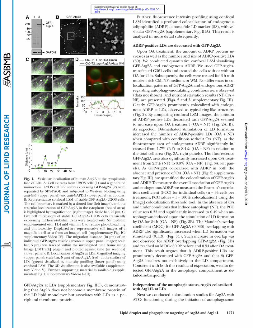
protein extracts from stable GFP-Atg2A/U2OS cells, we detected GFP-Atg2A at � 240 kDa, as expected from the calculated molecular mass for human Atg2A (213 kDa) and GFP (27 kDa) ( Fig. 1A ). In support, myc -tagged Atg2A was detected at � 220 kDa (data not shown).
By confocal LSM of transiently expressed GFP-Atg2A the vesicular localization of Atg2A in the cytoplasm was appar-ent ( Fig. 1B ). This result was verifi ed by live-cell microscopy of GFP-Atg2/U2OS cells (supplementary Fig. IA; supple-mentary Videos I–III) and by confocal LSM of myc -Atg2A (supplementary Fig. IB) or Atg2A- myc (data not shown). Be-cause yeast Atg2 was identifi ed as an essential protein of the autophagic machinery, we conducted treatments that mod-ulate the autophagic activity throughout this study, along with appropriate controls. In general, we used NF medium or RM to induce autophagy above basal level, or WM to inhibit the formation of autophagosomes by inhibiting PtdIns(3)P generation. Independent of the cellular au-tophagic status Atg2A displayed a vesicular localization pat-tern (supplementary Fig. I; supplementary Videos I–III). We quantifi ed the area of vesicular Atg2A structures per cell to assess if the autophagic status infl uences the abundance of vesicular Atg2A structures (supplementary Figs. I and IVA) and found that autophagy induction resulted in a con-sistent increase in vesicular Atg2A compared with control medium (CM) or autophagy inhibition (supplementary Figs. I and IVA). As vesicular GFP-Atg2A structures displayed high mobility (supplementary Videos I–III), indicative for microtubule-assisted movement, we expressed mCherry-tubulin in GFP-Atg2A/U2OS cells followed by live-cell micro-scopy ( Fig. 1C , upper image panels; supplementary Fig. IC and supplementary Video IV). Indeed, GFP-Atg2A vesicles moved distances of � 3–5 µm bidirectionally on microtu-bule tracks ( Fig. 1C , lower panel).
Human Atg2A localizes to cytoplasmic LDs In order to defi ne the identity of vesicular Atg2A struc-
tures, we conducted colocalization studies with a variety of cellular markers (e.g., lysosomes, mitochondria, or FYVE-positive endosomes; data not shown) and found that Atg2A specifi cally localizes to LDs. In detail, by differential inter-ference contrast microscopy we found that LDs colocal-ized with myc -Atg2A vesicles (supplementary Fig. ID). This result was further confi rmed by visualizing transiently ex-pressed myc -Atg2A in U2OS cells treated with LipidTOX Green, which labels neutral lipids (supplementary Fig. IE). We verifi ed that LipidTOX Green specifi cally detects LDs by stimulation of LD formation using OA (supple-mentary Fig. IIA) and EM analysis of OA-loaded cells (sup-plementary Fig. IIB). Clearly, myc -Atg2A was expressed at the surface of LDs as fl uorescence intensity profi ling showed maximal myc -Atg2A signal intensity ( Fig. 1D , in red) at the cytoplasmic face of LDs ( Fig. 1D , in green). Localization of myc -Atg2A at the cytoplasmic face of LDs labeled with LipidTOX Green is further highlighted by 3D reconstruction and fl y-through movie presentations us-ing confocal LSM sections (supplementary Video V). By freeze-fracture immuno-EM of the GFP-Atg2A/U2OS cell line with anti-GFP antibodies, we were unable to detect
Western blotting Cells were rinsed with PBS (37°C) and lysed with preheated
(100°C) 100 mM Tris (pH 6.8), 2.5 mM EDTA (pH 8), 25% glyc-erol, 4% SDS, 100 mM DTT, 5% � -mercaptoethanol, and 0.05% bromophenol blue, followed by chromatin shearing using a 23G needle. Total protein extracts were separated by SDS-PAGE and transferred to polyvinylidene difl uoride membranes (Cat. No. IPVH00010, Millipore). Standard ECL detection was performed with Immobilon Western Chemiluminescent HRP Substrate (Cat. No. WBKLS0100, Millipore).
Sucrose density centrifugation LD purifi cation was performed as described previously ( 56 ). Up
to 1.5× 10 7 OA-treated (500 � M, 24 h) cells were lysed in 3 ml dis-ruption buffer [25 mM Tris (pH 7.4), 100 mM KCl, 1 mM EDTA, 5 mM EGTA, 10 µg/ml leupeptin, 0.7 � g/ml pepstatin A, 0.1 mM PMSF (Cat. No. 04 693 132 001, Roche Complete)] by dounce homogenization. Cell lysates were centrifuged (1,000 g ) for 10 min at 4°C to pellet cell nuclei and debris. The supernatant was mixed with an equal volume of disruption buffer supplemented with 1.08 M sucrose, transferred to 14 ml centrifugation tubes (Cat. No. 331374, Beckman), and overlayed with 2 ml 0.27 M sucrose [25 mM Tris (pH 7.4), 1 mM EDTA, 1 mM EGTA, 0.27 M sucrose], 2 ml 0.135 M sucrose solution, and 2 ml top solution [25 mM Tris (pH 7.4), 1 mM EDTA, 1 mM EGTA]. Samples were centrifuged at 150,500 g (Beckman Coulter OPTIMA XL-100K ultracentrifuge, Beckman SW-40Ti rotor) for 2 h at 4°C and fractionated into lipid-fl oating, intermediate, and cytosolic fractions.
Bioinformatics analyses Cluster analysis: Human Atg2A protein (gi:239047271) was
used for a Basic Local Alignment Search Tool search against the NCBI nonredundant protein database ‘nr’ (version November 2012). All sequences producing High Scoring Segment-Pairs (HSPs) up to E-values of 10 were extracted as full-length se-quences (813 sequences) and analyzed in Cluster Analysis of Se-quences (CLANS) ( 57 ). The sequence similarity groups identifi ed in the CLANS map were used as a basis to identify all sequences of the Atg2 protein family and select the animal as well as suitable outgroup sequences from which to infer a phylogeny.
Phylogenetic inference: All sequences from the animal Atg2 sequence similarity groups, as well as the selected outgroup se-quences, were combined into one fi le and aligned using Mul-tiple Sequence Comparison by Log-Expectation (MUSCLE). Fragment or truncated sequences present in the alignment were manually identifi ed and removed prior to phylogenetic infer-ence. The phylogeny was inferred based on the neighbor-joining approach (1,000 bootstrap replicates) using the ASATURA soft-ware, the JTT substitution matrix, and no mutational saturation cutoff .
RESULTS
Human Atg2A is expressed in vesicular structures that migrate bidirectionally along microtubules
We generated plasmids for transient and stable expres-sion of N-terminal tagged GFP- or myc -Atg2A and C-termi-nal tagged Atg2A- myc in human tumor cell lines (U2OS, G361, and HeLa). In addition, human U2OS cells derived from G418-selected clones that stably express GFP-Atg2A protein at a low level (GFP-Atg2A/U2OS hereafter), were also generated for our studies. By Western blotting of
by guest, on April 8, 2019
ww
w.jlr.org
Dow
nloaded from
.html http://www.jlr.org/content/suppl/2014/04/28/jlr.M046359.DC1Supplemental Material can be found at:
Lipid droplet and phagophore targeting of Atg2A and Atg14L 1271
Further, fl uorescence intensity profi ling using confocal LSM identifi ed a profound colocalization of endogenous adipophilin (ADRP), a bona fi de LD marker ( 58 ), with ve-sicular GFP-Atg2A (supplementary Fig. IIIA). This result is analyzed in more detail subsequently.
ADRP-positive LDs are decorated with GFP-Atg2A Upon OA treatment, the amount of ADRP protein in-
creases as well as the number and size of ADRP-positive LDs ( 59 ). We conducted quantitative confocal LSM visualizing GFP-Atg2A and endogenous ADRP. We used GFP-Atg2A-transfected G361 cells and treated the cells with or without OA for 24 h. Subsequently, the cells were treated for 3 h with nutrient-rich CM, NF medium, or WM. No differences in co-localization patterns of GFP-Atg2A and endogenous ADRP regarding autophagy-modulating conditions were observed (data not shown), and nutrient starvation results (NF, OA + NF) are presented ( Figs. 2 and 3 ; supplementary Fig. III). Clearly, GFP-Atg2A prominently colocalized with endoge-nous ADRP at LDs, observed as typical ring-like structures ( Fig. 2 ). By comparing confocal LSM images, the amount of ADRP-positive LDs decorated with GFP-Atg2A seemed to increase upon OA treatment (OA + NF) ( Fig. 2A, B ). As expected, OA-mediated stimulation of LD formation increased the number of ADRP-positive LDs (OA + NF) when compared with conditions without OA (NF), as the fl uorescence area of endogenous ADRP signifi cantly in-creased from 1.7% (NF) to 8.4% (OA + NF) in relation to the total cell area ( Fig. 3A , right panels). The fl uorescence GFP-Atg2A area also signifi cantly increased upon OA treat-ment from 2.3% (NF) to 8.8% (OA + NF) ( Fig. 3A , left pan-els). As GFP-Atg2A colocalized with ADRP in both the absence and presence of OA (OA + NF) ( Fig. 2 ; supplemen-tary Fig. III), we quantifi ed the colocalization of GFP-Atg2A and ADRP. To measure the overall association of GFP-Atg2A and endogenous ADRP, we measured the Pearson’s correla-tion coeffi cient (PCC) for individual cells (n = 34 cells per treatment; PCC values + 1 = 100% colocalization) using the ImageJ colocalization threshold tool. In the absence of OA and under conditions that induce autophagy (NF), the PCC value was 0.33 and signifi cantly increased to 0.49 when au-tophagy was induced upon the stimulation of LD formation by OA for 24 h (OA + NF) ( Fig. 3B ). The Mander’s overlap coeffi cient (MOC) for GFP-Atg2A (0.036) overlapping with ADRP also signifi cantly increased when LD formation was stimulated (0.119) ( Fig. 3C ). Such increase in overlap was not observed for ADRP overlapping GFP-Atg2A ( Fig. 3D ) and reached an MOC of 0.92 before and 0.94 after OA treat-ment. This result argues that i ) ADRP-positive LDs are prominently decorated with GFP-Atg2A and that ii ) GFP-Atg2A localizes not exclusively to the LD compartment. Consistent with both this result and expectation, we also de-tected GFP-Atg2A in the autophagic compartment as de-tailed subsequently.
Independent of the autophagic status, Atg2A colocalized with Atg14L at LDs
Next we conducted colocalization studies for Atg2A with ATGs functioning during the initiation of autophagosome
GFP-Atg2A at LDs (supplementary Fig. IIC), demonstrat-ing that Atg2A does not become a membrane protein of the LD lipid monolayer but associates with LDs as a pe-ripheral membrane protein.
Fig. 1. Vesicular localization of human Atg2A at the cytoplasmic face of LDs. A: Cell extracts from U2OS cells (1) and a generated monoclonal U2OS cell line stably expressing GFP-Atg2A (2) were separated by SDS-PAGE and subjected to Western blotting using anti-GFP (upper panel) and anti-GAPDH (lower panel) antibodies. B: Representative confocal LSM of stable GFP-Atg2A/U2OS cells. The cell boundary is marked by a dotted line (left image), and the vesicular localization of GFP-Atg2A in the cytoplasm (boxed area) is highlighted by magnifi cation (right image). Scale bar, 20 µm. C: Live cell microscopy of stable GFP-Atg2A/U2OS cells transiently expressing mCherry-tubulin. Cells were treated with NF medium supplemented with 11.4 mM vitamin C to reduce photobleaching and phototoxicity. Displayed are representative still images of a magnifi ed cell area from an imaged cell (supplementary Fig. IC; supplementary Video IV). The migration distance (in µm) of an individual GFP-Atg2A vesicle (arrows in upper panel images; scale bar, 5 µm) was tracked within the investigated time frame using Image J/MTrackJ plug-in and plotted against time (in seconds) (lower panel). D: Localization of Atg2A at LDs. Magnifi ed imaging (upper panel; scale bar, 5 µm) of myc -Atg2A (red) at the surface of LDs (green) visualized by intensity profi ling (lower panel) using confocal LSM. The 3D visualization is also available (supplemen-tary Video V). Further supporting material is available (supple-mentary Fig. I; supplementary Videos I–III).
by guest, on April 8, 2019
ww
w.jlr.org
Dow
nloaded from
.html http://www.jlr.org/content/suppl/2014/04/28/jlr.M046359.DC1Supplemental Material can be found at:

1272 Journal of Lipid Research Volume 55, 2014
however, signifi cantly increased the number of GFP-Atg14L puncta-positive cells to an average of 63% ( Fig. 4B , left panels).
On this basis, we transiently coexpressed GFP-Atg14L and myc -Atg2A in U2OS cells and treated the cells with nu-trient-rich CM, NF medium, or WM. We found that both ATGs colocalized at vesicular structures that were present irrespective of the autophagic status, indicating that these structures represent the colocalization of myc -Atg2A and GFP-Atg14L at LDs ( Fig. 4C ). In support, fl uorescence in-tensity profi ling of GFP-Atg14L-expressing cells incubated with LipidTOX Red demonstrated that in part GFP-Atg14L localizes at the cytoplasmic face of LDs ( Fig. 4D ), and this result was further highlighted by 3D reconstruction and fl y-through movie presentation (supplementary Video VI). Of note, although vesicular GFP-Atg14L ( Fig. 4B ) and myc -Atg2A structures (supplementary Fig. IVA) increased upon autophagy induction, colocalization of GFP-Atg14L and myc -Atg2A was not signifi cantly changed (68% to 77% of GFP-Atg14L colocalized with myc -Atg2A, whereas 52% to 63% of myc -Atg2A colocalized with GFP-Atg14L) (supple-mentary Fig. IVB, C). This indicates that Atg2A and Atg14L localize in part to the autophagic compartment upon au-tophagy induction and in addition to the LD compartment irrespective of the autophagic activity.
formation of Atg14L, WIPI-1, and DFCP1. ( Figs. 4 and 6 ). First, we investigated the localization of myc -Atg2A with re-gard to GFP-Atg14L, a phagophore marker that is partially also present in vesicular structures apart from phago-phores ( 26 ). We expressed GFP-Atg14L in U2OS cells ( Fig. 4A ) followed by treatments with nutrient-rich CM, NF medium, or WM and quantifi ed the number of GFP-Atg14L puncta-positive cells ( Fig. 4B , left panels). In paral-lel, we also analyzed transiently expressed GFP-WIPI-1 ( Fig. 4A ). As expected, nutrient starvation using NF me-dium resulted in a signifi cant increase of GFP-WIPI-1 puncta-positive cells, and WM-mediated inhibition of PtdIns(3)P generation (WM) resulted in a signifi cant de-crease ( Fig. 4B , right panels). This result was also achieved by quantifying GFP-Atg14L puncta-positive cells. However, more control cells already displayed a prominent amount of GFP-Atg14L puncta (an average of 40% puncta-positive cells), and WM administration did not completely abolish the presence of GFP-Atg14L puncta. Nutrient starvation,
Fig. 2. Atg2A localizes to ADRP-positive LDs independent of treatments that modulate autophagy. GFP-Atg2A/U2OS cells were incubated with or without OA for 24 h, followed by treatments with NF medium for 3 h. Subsequently, the cells were immunostained with anti-ADRP/IgG-conjugated Alexa 546 to detect endogenous ADRP and with TO-PRO-3 to mark cell nuclei, followed by confocal LSM (n = 3). A: Representative images from NF-treated cells. B: Representative images from OA + NF-treated cells. The cell bound-aries are marked by a dotted line in the upper panels, and the typi-cal ring-like ADRP-positive LD appearance is highlighted by further magnifi cation in the lower panels. Scale bar, 20 µm. Supporting material is available (supplementary Figs. II and IIIA).
Fig. 3. OA treatment increased Atg2A targeting to ADRP-positive LDs. G361 cells transiently expressing GFP-Atg2A were incubated with or without OA for 24 h, followed by treatments with NF me-dium for 3 h. Cells were immunostained with anti-ADRP/IgG Al-exa 546 antibodies analyzed by confocal LSM (n = 3). A: Image projections of confocal sections were quantifi ed with regard to fl uorescence areas by using Image Pro Plus, providing the percent-age of the total inclusion area (ADRP or GFP-Atg2A vesicles) in relation to the total cell area. NF, n = 21 cells; OA + NF, n = 18 cells (from 3 independent experiments). B: Using the ImageJ colocal-ization threshold tool, the PCC for individual cells (n = 34 cells per treatment) was measured. The MOC for GFP-Atg2A overlapping with ADRP (C) and ADRP overlapping with GFP-Atg2A (D) was also measured using the ImageJ colocalization threshold. Mean ± SEM. P values: n.s. (not signifi cant) P � 0.05, *** P � 0.001. Sup-porting material is available (supplementary Fig. IIIB).
by guest, on April 8, 2019
ww
w.jlr.org
Dow
nloaded from
.html http://www.jlr.org/content/suppl/2014/04/28/jlr.M046359.DC1Supplemental Material can be found at:

Lipid droplet and phagophore targeting of Atg2A and Atg14L 1273
and GFP-Atg14L, but negative for HA-WIPI-1 (red arrow) ( Fig. 6A , left image panels). Fluorescence intensity pro-fi ling underlined this fi nding ( Fig. 6A , right panel). WM administration abolished the triple colocalization of myc -Atg2A, GFP-Atg14L, and HA-WIPI-1, while colocaliza-tion of myc -Atg2A and GFP-Atg14L at LDs was maintained
Detection of GFP-Atg2A and GFP-Atg14L in the ADRP-positive LD fraction separated by density gradient centrifugation
We conducted cell fractionation upon density centrifu-gation prepared from GFP-Atg2A/U2OS cells transiently transfected with myc -Atg14L. As expected, endogenous ADRP was exclusively present in the LD fraction. GFP-Atg2A was also detected in this fraction of fl oating ADRP-positive LDs; however, GFP-Atg2A was more prominently detected in cytoplasmic fractions. To a minor extent, we also detected myc -Atg14L, while the GAPDH control was restricted to cytoplasmic fractions ( Fig. 5 ).
PtdIns(3)P-dependent recruitment of Atg2A to WIPI-1-positive phagophores upon nutrient starvation
Next, we analyzed the localization of Atg2A and Atg14L to the autophagic compartment. We visualized myc -Atg2A, GFP-Atg14L, and HA-WIPI-1 by confocal LSM using U2OS cells ( Fig. 6A, B ; supplementary Fig. VA, B). Upon au-tophagy induction by NF medium treatments of 3 h, myc -Atg2A, GFP-Atg14L, and HA-WIPI-1 colocalized at cy-toplasmic puncta (white arrow, puncta 1 and 2), easily dis-tinguishable from LDs that were positive for myc -Atg2A
Fig. 4. Atg2A colocalizes with Atg14L at LDs inde-pendent of autophagy. A: Protein extracts from U2OS cells transiently expressing GFP-Atg14L or GFP-WIPI-1 prepared at different hours posttransfec-tion were subjected to Western blotting using anti-GFP or anti-tubulin antibodies (n = 3). B: From 400 cells per treatment (n = 4) GFP-Atg14L or GFP-WIPI-1 puncta formation was determined (36 h post-transfection) and expressed as the percentage of puncta-positive cells. Mean ± SD; P value: *** P � 0.001. C: U2OS cells transiently coexpressing GFP-Atg14L (green) and myc -Atg2A (red) for 24 h were treated with nutrient-rich CM, NF medium, or WM for 3 h, followed by indirect immunofl uorescence anal-ysis by confocal LSM using anti- myc /IgG-conjugated Alexa 546 antibodies [CM, NF (n = 4), WM (n = 3)]. The cell boundaries are marked by a dotted line (left panels), and boxed areas highlighted by magnifi ca-tion (right panels). D: U2OS cells expressing GFP-Atg14L for 24 h were incubated with LipidTOX Red and analyzed by confocal LSM (n = 4) followed by intensity profi ling. GFP-Atg14L localization at LDs (1) and cytoplasmic puncta (2) is indicated. The 3D visualization is also available (supplementary Video VI). Scale bars, 20 µm. Further supporting ma-terial is also available (supplementary Fig. IV).
Fig. 5. Density gradient centrifugation reveals the presence of ADRP, GFP-Atg2A, and myc -Atg14L in the fl oating LD fraction. Stable GFP-Atg2A/U2OS cells were transiently transfected with myc -Atg14L and incubated with OA for 24 h. Cell lysates were frac-tionated upon sucrose density centrifugation (n = 2). The fl oating LD fraction (boxed with a dotted red line) and intermediate and fi rst cytoplasmic fractions were analyzed by Western blotting using anti-ADRP, anti-GFP, anti- myc , and anti-GAPDH antibodies.
by guest, on April 8, 2019
ww
w.jlr.org
Dow
nloaded from
.html http://www.jlr.org/content/suppl/2014/04/28/jlr.M046359.DC1Supplemental Material can be found at:

1274 Journal of Lipid Research Volume 55, 2014
induced by nutrient starvation using NF medium, we in-vestigated the effect of NF on the number of LDs by con-ducting automated, high-throughput fl uorescence image acquisition and analysis ( 49 ) of cells stained with Lipid-TOX Greens in 96-well plates ( Fig. 8 ). In HeLa, G361, and U2OS cells treated with NF medium, the number of
( Fig. 6B ). We conducted parallel experiments by using GFP-Atg2A, HA-WIPI-1, and myc -DFCP1 (marking prephago-phore omegasome structures) and found that, apart from the localization of GFP-Atg2A at LDs (red arrow), GFP-Atg2A, myc -DFCP1, and HA-WIPI-1 colocalized at cytoplas-mic puncta (white arrow, puncta 1 and 2) upon the induction of autophagy ( Fig. 6C ; supplementary Fig. VC) which was also abrogated by WM administration (data not shown). Similarly, upon nutrient starvation myc -Atg2A co-localized with endogenous WIPI-1 in cytoplasmic puncta but not with myc -Atg2A at LDs ( Fig. 7A ). WM-mediated au-tophagy inhibition abrogated colocalization of endoge-nous WIPI-1 and myc -Atg2A, whereas LD localization of Atg2A was maintained ( Fig. 7B ).
Nutrient starvation leads to an increase of LDs in the human tumor cell lines G361 and U2OS
The previously discussed results demonstrated that Atg2A and Atg14L colocalize at LDs irrespective of the autophagic status of the cell, but that a subpopulation of both ATGs is targeted to early autophagosomal mem-branes enriched in PtdIns(3)P, and positive for DFCP1 and WIPI-1, upon the induction of autophagy. As we de-tailed our analysis using conditions when autophagy was
Fig. 6. Upon starvation, Atg2A and Atg14L colo-calize at DFCP1- and WIPI-1-positive early autopha-gosomal membranes. A: U2OS cells transiently expressing GFP-Atg14L (green), myc -Atg2A (red), and HA-WIPI-1 (blue) were starved in NF medium for 3 h and immunostained with anti- myc /IgG-conjugated Alexa 546 and anti-WIPI-1/IgG-conjugated Alexa 633 antibodies. Images (left panels) were acquired by confocal LSM followed by intensity pro-fi ling (right panel). Colocalization of GFP-Atg14L, myc -Atg2A, and HA-WIPI-1 (1, 2, white arrow) and additional GFP-Atg14L/ myc -Atg2A LDs (red arrow) are indicated. B: In parallel to A, cells were treated with WM for 3 h. GFP-Atg14L/myc-Atg2A-positive LDs (1, 2) are indicated, whereas HA-WIPI-1 (blue) is distributed throughout the cytoplasm. C: U2OS cells transiently transfected with GFP-Atg2A (green), myc -DFCP-1 (red), and HA-WIPI-1 (blue) were starved in NF medium for 3 h, immunostained with anti- myc /IgG-conjugated Alexa 546 and anti-WIPI-1/IgG-conjugated Alexa 633 antibodies, and analyzed by con-focal LSM (n = 2). The intensity profi le is displayed (right panel), and colocalization of GFP-Atg2A, myc-DFCP1, and HA-WIPI-1 is indicated (1, 2, white ar-row). GFP-Atg2A LDs are also indicated (red arrow). Presented are magnifi ed areas from corresponding full cell images (supplementary Fig. V).
Fig. 7. Myc -tagged Atg2A is recruited to endogenous WIPI-1 upon the induction of autophagy. G361 cells transiently expressing myc -Atg2A were treated with NF medium (A) or WM (B) for 3 h; fi xed; immunostained with anti- myc , anti-WIPI-1, and anti-mouse Alexa 546 antibodies; and analyzed by confocal microscopy. Repre-sentative images of three independent experiments are shown. Scale bar, 10 µm
by guest, on April 8, 2019
ww
w.jlr.org
Dow
nloaded from
.html http://www.jlr.org/content/suppl/2014/04/28/jlr.M046359.DC1Supplemental Material can be found at:

Lipid droplet and phagophore targeting of Atg2A and Atg14L 1275
for prolonged energy shortage at a later stage. This phenomenon might even be evolutionarily conserved as Caenorhabditis elegans accumulates LDs upon dauer forma-tion ( 62, 63 ). Starvation-induced nonspecifi c degradation of proteins and organelles can provide acetyl-CoA and also fatty acids and cholesterol required for neutral lipid syn-thesis. Also, starvation is known to trigger autophagosomal degradation of glycogen stores ( 64 ), which could liberate additional energy for lipid synthesis. Furthermore, our starvation treatments include lipoprotein depletion, which
LDs per cell was found to increase signifi cantly after 3 h (U2OS and G361) and after 24 h (all cell lines) ( Fig. 8A ). To functionally address the role of Atg2A and Atg14L in the biogenesis of LDs, we performed siRNA-mediated depletion of Atg2A or Atg14L in HeLa (supplementary Fig. VIA) and in G361 (supplementary Fig. VIB) cells. We assessed the number of LDs upon treatment with CM or NF medium using high-content image analysis. Atg2A depletion in HeLa cells followed by nutrient starvation using NF medium resulted in a signifi cant increase of LD numbers in individual cells ( Fig. 8B , left panels). Atg14L depletion signifi cantly increased the number of LDs in i ) HeLa cells in CM as well as upon nutrient starvation us-ing NF medium ( Fig. 8B , left panels) and ii ) G361 cells in CM ( Fig. 8B , right panels). In G361 cells, the size of LDs signifi cantly increased upon Atg14L depletion and treat-ments using CM as well as NF medium (supplementary Fig. VID). From this, we conclude that Atg2A and Atg14L have dual functions, acting in the regulation of autopha-gosome formation as well as contributing to LD metabo-lism ( Fig. 8C ).
DISCUSSION
In Saccharomyces cerevisiae , PtdIns(3)P-dependent recruit-ment of Atg2 to autophagosome formation sites has been demonstrated ( 43, 44 ). Similarly, PtdIns(3)P-dependent targeting of human Atg2A to WIPI-1 positive phagophores upon starvation-induced autophagy was also found in this study, and WM treatment abrogated this specifi c recruit-ment of Atg2A. In addition we demonstrate that Atg2A is targeted to the surface of ADRP-positive LDs that move bidirectionally on microtubules, independent of autophagic status. Further, we identifi ed Atg14L as a novel LD-associated protein, colocalizing there with Atg2A irre-spective of autophagic status. Evidence has been provided that Atg14L resides at the ER and regulates localized PtdIns(3)P generation, which is then bound by the WIPIs. Apart from being the site of phagophore formation, LDs are also proposed to form at the ER ( 60 ), and thus sites of autophagosomal membrane biogenesis and LD formation seem to be functionally connected. LDs have further been detected in very close association with the forming phagophore ( 47 ).
So far, three ATGs have been found to be targeted to LDs, LC3 ( 61 ), Atg2A ( 45 ) (this study), and Atg14L (this study), indicating that these ATGs either display dual func-tions in autophagy and LD biogenesis or that they func-tionally connect both pathways. Therefore, by high-content analysis, we quantifi ed the number of LDs in human HeLa, G361, and U2OS tumor cells upon short-term nutrient starvation and detected a prominent increase in the num-ber and size of LDs per individual cell, resembling LD formation. This indicates that autophagy induction and LD formation can simultaneously occur in starved cells. Hence starvation-induced autophagy might provide energy to boost lipid storage, preparing the cell to compensate
Fig. 8. Starvation treatment increases LD formation. HeLa, G361, and U2OS cells were treated with CM or NF medium for 3 h, fi xed, and stained with DAPI (blue) and LipidTOX Green (green). Automated image acquisition and analysis was conducted using a high-content imaging platform (In Cell Analyzer 1000). A: Up to 9,305 cells were quantifi ed (n = 3–5) to assess the num-ber of LDs per individual cell (left panel). Representative images for G361 cells are presented (right panel). Scale bar, 20 µm. B: HeLa and G361 cells were transiently transfected with scrambled control siRNA (siControl), siRNA targeting Atg2A (siAtg2A), or Atg14L (siAtg14L), and downregulation of Atg2A and Atg14L mRNA was verifi ed by quantitative PCR (supplementary Fig. VIA, B). Cells transfected with siRNA were incubated with CM or NF medium for 3 h and fi xed, and LD abundance was quantifi ed by high-content analysis (n = 3–4). Mean ± SD. P values: n.s. (not signifi cant) P � 0.05, * P < 0.05, ** P � 0.01, *** P � 0.001. Sup-porting material is available (supplementary Fig. VIC, D). C: A model for the roles of human Atg2A and Atg14L in LD biogenesis and autophagy.
by guest, on April 8, 2019
ww
w.jlr.org
Dow
nloaded from
.html http://www.jlr.org/content/suppl/2014/04/28/jlr.M046359.DC1Supplemental Material can be found at:

1276 Journal of Lipid Research Volume 55, 2014
Atg2, as yet unidentifi ed, might contribute to particular human pathologies correlated with modulated autophagic activity ( 1 ).
The authors thank Roger Tsien for the mCherry-tubulin encod-ing plasmid. The authors kindly acknowledge Andrei Lupas for discussions on the bioinformatics analysis.
REFERENCES
1 . Mizushima , N. , B. Levine , A. M. Cuervo , and D. J. Klionsky . 2008 . Autophagy fi ghts disease through cellular self-digestion. Nature . 451 : 1069 – 1075 .
2 . Levine , B. , and G. Kroemer . 2008 . Autophagy in the pathogenesis of disease. Cell . 132 : 27 – 42 .
3 . Beau , I. , M. Mehrpour , and P. Codogno . 2011 . Autophagosomes and human diseases. Int. J. Biochem. Cell Biol. 43 : 460 – 464 .
4 . Moscat , J. , and M. T. Diaz-Meco . 2011 . Feedback on fat: p62-mTORC1-autophagy connections. Cell . 147 : 724 – 727 .
5 . Mizushima , N. 2007 . Autophagy: process and function. Genes Dev. 21 : 2861 – 2873 .
6 . Jahreiss , L. , F. M. Menzies , and D. C. Rubinsztein . 2008 . The itiner-ary of autophagosomes: from peripheral formation to kiss-and-run fusion with lysosomes. Traffi c . 9 : 574 – 587 .
7 . Yang , Z. , and D. J. Klionsky . 2010 . Eaten alive: a history of macroau-tophagy. Nat. Cell Biol. 12 : 814 – 822 .
8 . Singh , R. , and A. M. Cuervo . 2011 . Autophagy in the cellular ener-getic balance. Cell Metab. 13 : 495 – 504 .
9 . Mathew , R. , and E. White . 2011 . Autophagy in tumorigenesis and energy metabolism: friend by day, foe by night. Curr. Opin. Genet. Dev. 21 : 113 – 119 .
10 . Singh , R. , S. Kaushik , Y. Wang , Y. Xiang , I. Novak , M. Komatsu , K. Tanaka , A. M. Cuervo , and M. J. Czaja . 2009 . Autophagy regulates lipid metabolism. Nature . 458 : 1131 – 1135 .
11 . Koga , H. , S. Kaushik , and A. M. Cuervo . 2010 . Altered lipid content inhibits autophagic vesicular fusion. FASEB J. 24 : 3052 – 3065 .
12 . Martin , S. , and R. G. Parton . 2006 . Lipid droplets: a unifi ed view of a dynamic organelle. Nat. Rev. Mol. Cell Biol. 7 : 373 – 378 .
13 . Fujimoto , T. , Y. Ohsaki , J. Cheng , M. Suzuki , and Y. Shinohara . 2008 . Lipid droplets: a classic organelle with new outfi ts. Histochem. Cell Biol. 130 : 263 – 279 .
14 . Czaja , M. J. 2010 . Autophagy in health and disease. 2. Regulation of lipid metabolism and storage by autophagy: pathophysiological implications. Am. J. Physiol. Cell Physiol. 298 : C973 – C978 .
15 . Simonsen , A. , A. E. Wurmser , S. D. Emr , and H. Stenmark . 2001 . The role of phosphoinositides in membrane transport. Curr. Opin. Cell Biol. 13 : 485 – 492 .
16 . Noda , T. , K. Matsunaga , N. Taguchi-Atarashi , and T. Yoshimori . 2010 . Regulation of membrane biogenesis in autophagy via PI3P dynamics. Semin. Cell Dev. Biol. 21 : 671 – 676 .
17 . Obara , K. , and Y. Ohsumi . 2011 . PtdIns 3-kinase orchestrates au-tophagosome formation in yeast. J. Lipids . 2011 : 498768 .
18 . Seglen , P. O. , and P. B. Gordon . 1982 . 3-Methyladenine: specifi c inhibitor of autophagic/lysosomal protein degradation in isolated rat hepatocytes. Proc. Natl. Acad. Sci. USA . 79 : 1889 – 1892 .
19 . Blommaart , E. F. , U. Krause , J. P. Schellens , H. Vreeling-Sindelarova , and A. J. Meijer . 1997 . The phosphatidylinositol 3-kinase inhibitors wortmannin and LY294002 inhibit autophagy in isolated rat hepa-tocytes. Eur. J. Biochem. 243 : 240 – 246 .
20 . Petiot , A. , E. Ogier-Denis , E. F. Blommaart , A. J. Meijer , and P. Codogno . 2000 . Distinct classes of phosphatidylinositol 3 ′ -kinases are involved in signaling pathways that control macroautophagy in HT-29 cells. J. Biol. Chem. 275 : 992 – 998 .
21 . Kihara , A. , T. Noda , N. Ishihara , and Y. Ohsumi . 2001 . Two dis-tinct Vps34 phosphatidylinositol 3-kinase complexes function in autophagy and carboxypeptidase Y sorting in Saccharomyces cer-evisiae. J. Cell Biol. 152 : 519 – 530 .
22 . Obara , K. , and Y. Ohsumi . 2008 . Dynamics and function of PtdIns(3)P in autophagy. Autophagy . 4 : 952 – 954 .
23 . Codogno , P. , and A. J. Meijer . 2005 . Autophagy and signaling: their role in cell survival and cell death. Cell Death Differ. 12 ( Suppl. 2 ): 1509 – 1518 .
could decrease ER cholesterol levels resulting in sterol regulatory element binding protein (SREBP)-mediated stim-ulation of lipid synthesis ( 65 ). However, autophagy was also found to degrade LDs, thereby contributing to the regulation of lipid metabolism in liver cells ( 10 , 61 ) and adipocyte differentiation ( 66 ), suggesting that the func-tion of autophagosomal proteins at LDs and in adipogen-esis is complex.
Whereas Atg2A and Atg14L are targeted to LDs inde-pendent of the autophagic status, both proteins localize to DFCP1 and WIPI-1-positive omegasome and phago-phore membranes, distinct to LD structures, upon au-tophagy stimulation. In addition, we could demonstrate that Atg2A and Atg14L depletion resulted in increased LD abundance upon treatment with CM (Atg14L deple-tion) or NF medium (Atg2A depletion and Atg14L deple-tion). Therefore, our study provides evidence that Atg2A as well as Atg14L fulfi ll at least two functions in human cells: one exerted as a peripheral membrane protein at the surface of ADRP-positive LDs and the other involving the regulation of autophagy, dependent on PtdIns(3)P generation ( Fig. 8C ). It is possible that LD-localized Atg2A and Atg14L impair excessive lipid storage inside LDs and ensure effi cient energy use (e.g., generated by autophagy induction). Another function of Atg2A and Atg14L at the LD surface might involve modulation of the fatty acid and cholesterol esterifi cation/hydrolysis cycle, contributing to the regulation of lipid storage. Our results are in line with recent work on mammalian Atg2, shown to play an essential role in autophagosome forma-tion and to also regulate LD size and distribution ( 45 ). However, it is also possible that ATGs, such as Atg2A and Atg14L, fulfi ll distinct roles at LDs and autophagosomal membranes, but that they also mediate a functional inter-connection during simultaneous formation of LDs and autophagsomes, maybe by mediating intermembrane lipid traffi c.
Because Atg2 shares protein sequence homologies with Vps13 (supplementary Fig. VII), a protein shown to promote endosome/trans-Golgi network (TGN) mem-brane protein cycling ( 67 ), it might be plausible that Vps13 and Atg2 proteins generally function in mem-brane formation and morphology, and that distinct mem-bers provide specifi city for particular vesicle subsets. In this context, it has been proposed that the yeast Atg18/Atg2 complex is required to generate negative curvature at the forming autophagosome or fulfi lls an essential function at the elongating tips of the phago-phore ( 43 ). Likewise it was recently found that Vps13 regulates membrane bending and promotes the expan-sion of the prospore membrane in yeast ( 68 ). From this, one would assume that malfunctions of either of the hu-man Atg2/Vps13 proteins should be correlated to dis-tinct human pathologies with defects in intracellular membrane biogenesis. Indeed, hereditary mutations of human Vps13 orthologs cause the diseases chorea acan-thocytosis and Cohen syndrome ( 69, 70 ). As Atg2 func-tions in LD and autophagosome biogenesis, it can be assumed that functional or genetic alterations of human
by guest, on April 8, 2019
ww
w.jlr.org
Dow
nloaded from
.html http://www.jlr.org/content/suppl/2014/04/28/jlr.M046359.DC1Supplemental Material can be found at:

Lipid droplet and phagophore targeting of Atg2A and Atg14L 1277
45 . Velikkakath , A. K. , T. Nishimura , E. Oita , N. Ishihara , and N. Mizushima . 2012 . Mammalian Atg2 proteins are essential for au-tophagosome formation and important for regulation of size and distribution of lipid droplets. Mol. Biol. Cell . 23 : 896 – 909 .
46 . Hayashi-Nishino , M. , N. Fujita , T. Noda , A. Yamaguchi , T. Yoshimori , and A. Yamamoto . 2009 . A subdomain of the endoplasmic reticu-lum forms a cradle for autophagosome formation. Nat. Cell Biol. 11 : 1433 – 1437 .
47 . Ylä-Anttila , P. , H. Vihinen , E. Jokitalo , and E. L. Eskelinen . 2009 . 3D tomography reveals connections between the phagophore and endoplasmic reticulum. Autophagy . 5 : 1180 – 1185 .
48 . Tooze , S. A. , and T. Yoshimori . 2010 . The origin of the autophago-somal membrane. Nat. Cell Biol. 12 : 831 – 835 .
49 . Pfi sterer , S. G. , M. Mauthe , P. Codogno , and T. Proikas-Cezanne . 2011 . Ca 2+ /calmodulin-dependent kinase (CaMK) signaling via CaMKI and AMP-activated protein kinase contributes to the regulation of WIPI-1 at the onset of autophagy. Mol. Pharmacol. 80 : 1066 – 1075 .
50 . Proikas-Cezanne , T. , and S. G. Pfi sterer . 2009 . Assessing mammalian autophagy by WIPI-1/Atg18 puncta formation. Methods Enzymol. 452 : 247 – 260 .
51 . Robenek , H. , and N. J. Severs . 2008 . Recent advances in freeze-frac-ture electron microscopy: the replica immunolabeling technique. Biol. Proced. Online . 10 : 9 – 19 .
52 . Robenek , H. , I. Buers , O. Hofnagel , S. Lorkowski , and N. J. Severs . 2009 . GFP-tagged proteins visualized by freeze-fracture immuno-electron microscopy: a new tool in cellular and molecular medi-cine. J. Cell. Mol. Med. 13 : 1381 – 1390 .
53 . Robenek , H. , M. J. Robenek , I. Buers , S. Lorkowski , O. Hofnagel , D. Troyer , and N. J. Severs . 2005 . Lipid droplets gain PAT fam-ily proteins by interaction with specialized plasma membrane do-mains. J. Biol. Chem. 280 : 26330 – 26338 .
54 . Britschgi , C. , M. Rizzi , T. J. Grob , M. P. Tschan , B. Hugli , V. A. Reddy , A. C. Andres , B. E. Torbett , A. Tobler , and M. F. Fey . 2006 . Identifi cation of the p53 family-responsive element in the promoter region of the tumor suppressor gene hypermethylated in cancer 1. Oncogene . 25 : 2030 – 2039 .
55 . Tschan , M. P. , D. Shan , J. Laedrach , M. Eyholzer , E. O. Leibundgut , G. M. Baerlocher , A. Tobler , D. Stroka , and M. F. Fey . 2010 . NDRG1/2 expression is inhibited in primary acute myeloid leuke-mia. Leuk. Res. 34 : 393 – 398 .
56 . Yu , W. , J. Cassara , and P. F. Weller . 2000 . Phosphatidylinositide 3-kinase localizes to cytoplasmic lipid bodies in human polymor-phonuclear leukocytes and other myeloid-derived cells. Blood . 95 : 1078 – 1085 .
57 . Frickey , T. , and A. Lupas . 2004 . CLANS: a Java application for visu-alizing protein families based on pairwise similarity. Bioinformatics . 20 : 3702 – 3704 .
58 . Brasaemle , D. L. , T. Barber , N. E. Wolins , G. Serrero , E. J. Blanchette-Mackie , and C. Londos . 1997 . Adipose differentiation-related protein is an ubiquitously expressed lipid storage droplet-associated protein. J. Lipid Res. 38 : 2249 – 2263 .
59 . Xu , G. , C. Sztalryd , X. Lu , J. T. Tansey , J. Gan , H. Dorward , A. R. Kimmel , and C. Londos . 2005 . Post-translational regulation of adi-pose differentiation-related protein by the ubiquitin/proteasome pathway. J. Biol. Chem. 280 : 42841 – 42847 .
60 . Farese , R. V. , Jr ., and T. C. Walther . 2009 . Lipid droplets fi nally get a little R-E-S-P-E-C-T. Cell . 139 : 855 – 860 .
61 . Shibata , M. , K. Yoshimura , N. Furuya , M. Koike , T. Ueno , M. Komatsu , H. Arai , K. Tanaka , E. Kominami , and Y. Uchiyama . 2009 . The MAP1–LC3 conjugation system is involved in lipid droplet for-mation. Biochem. Biophys. Res. Commun. 382 : 419 – 423 .
62 . Hellerer , T. , C. Axang , C. Brackmann , P. Hillertz , M. Pilon , and A. Enejder . 2007 . Monitoring of lipid storage in Caenorhabditis elegans using coherent anti-Stokes Raman scattering (CARS) mi-croscopy. Proc. Natl. Acad. Sci. USA . 104 : 14658 – 14663 .
63 . Riddle , D. L. , and P. S. Albert . 1997 . Genetic and environmental regu-lation of dauer larva development. In C. elegans II. 2nd edition. D. L. Riddle, T. Blumenthal, B. J. Meyer, and J. R. Priess, editors. Cold Spring Harbor Laboratory Press, Cold Spring Harbor, NY. Chapter 26.
64 . Kotoulas , O. B. , S. A. Kalamidas , and D. J. Kondomerkos . 2004 . Glycogen autophagy. Microsc. Res. Tech. 64 : 10 – 20 .
65 . Goldstein , J. L. , R. A. DeBose-Boyd , and M. S. Brown . 2006 . Protein sensors for membrane sterols. Cell . 124 : 35 – 46 .
66 . Singh , R. , Y. Xiang , Y. Wang , K. Baikati , A. M. Cuervo , Y. K. Luu , Y. Tang , J. E. Pessin , G. J. Schwartz , and M. J. Czaja . 2009 . Autophagy regulates adipose mass and differentiation in mice. J. Clin. Invest. 119 : 3329 – 3339 .
24 . Abeliovich , H. , W. A. Dunn , Jr ., J. Kim , and D. J. Klionsky . 2000 . Dissection of autophagosome biogenesis into distinct nucleation and expansion steps. J. Cell Biol. 151 : 1025 – 1034 .
25 . Itakura , E. , C. Kishi , K. Inoue , and N. Mizushima . 2008 . Beclin 1 forms two distinct phosphatidylinositol 3-kinase complexes with mammalian Atg14L and UVRAG. Mol. Biol. Cell . 19 : 5360 – 5372 .
26 . Matsunaga , K. , T. Saitoh , K. Tabata , H. Omori , T. Satoh , N. Kurotori , I. Maejima , K. Shirahama-Noda , T. Ichimura , T. Isobe , et al . 2009 . Two Beclin 1-binding proteins, Atg14L and Rubicon, reciprocally regulate autophagy at different stages. Nat. Cell Biol. 11 : 385 – 396 .
27 . Zhong , Y. , Q. J. Wang , X. Li , Y. Yan , J. M. Backer , B. T. Chait , N. Heintz , and Z. Yue . 2009 . Distinct regulation of autophagic activity by Atg14L and Rubicon associated with Beclin 1-phosphatidylinosi-tol-3-kinase complex. Nat. Cell Biol. 11 : 468 – 476 .
28 . Pattingre , S. , A. Tassa , X. Qu , R. Garuti , X. H. Liang , N. Mizushima , M. Packer , M. D. Schneider , and B. Levine . 2005 . Bcl-2 antiapoptotic proteins inhibit Beclin 1-dependent autophagy. Cell . 122 : 927 – 939 .
29 . Matsunaga , K. , E. Morita , T. Saitoh , S. Akira , N. T. Ktistakis , T. Izumi , T. Noda , and T. Yoshimori . 2010 . Autophagy requires endo-plasmic reticulum targeting of the PI3-kinase complex via Atg14L. J. Cell Biol. 190 : 511 – 521 .
30 . Filimonenko , M. , P. Isakson , K. D. Finley , M. Anderson , H. Jeong , T. J. Melia , B. J. Bartlett , K. M. Myers , H. C. Birkeland , T. Lamark , et al . 2010 . The selective macroautophagic degradation of aggre-gated proteins requires the PI3P-binding protein Alfy. Mol. Cell . 38 : 265 – 279 .
31 . Simonsen , A. , H. C. Birkeland , D. J. Gillooly , N. Mizushima , A. Kuma , T. Yoshimori , T. Slagsvold , A. Brech , and H. Stenmark . 2004 . Alfy, a novel FYVE-domain-containing protein associated with protein gran-ules and autophagic membranes. J. Cell Sci. 117 : 4239 – 4251 .
32 . Axe , E. L. , S. A. Walker , M. Manifava , P. Chandra , H. L. Roderick , A. Habermann , G. Griffi ths , and N. T. Ktistakis . 2008 . Autophagosome formation from membrane compartments enriched in phosphati-dylinositol 3-phosphate and dynamically connected to the endo-plasmic reticulum. J. Cell Biol. 182 : 685 – 701 .
33 . Jeffries , T. R. , S. K. Dove , R. H. Michell , and P. J. Parker . 2004 . PtdIns-specifi c MPR pathway association of a novel WD40 repeat protein, WIPI49. Mol. Biol. Cell . 15 : 2652 – 2663 .
34 . Proikas-Cezanne , T. , S. Waddell , A. Gaugel , T. Frickey , A. Lupas , and A. Nordheim . 2004 . WIPI-1alpha (WIPI49), a member of the novel 7-bladed WIPI protein family, is aberrantly expressed in human cancer and is linked to starvation-induced autophagy. Oncogene . 23 : 9314 – 9325 .
35 . Codogno , P. , M. Mehrpour , and T. Proikas-Cezanne . 2012 . Canonical and non-canonical autophagy: variations on a common theme of self-eating? Nat. Rev. Mol. Cell Biol. 13 : 7 – 12 .
36 . Proikas-Cezanne , T. , and H. Robenek . 2011 . Freeze-fracture replica immunolabelling reveals human WIPI-1 and WIPI-2 as membrane proteins of autophagosomes. J. Cell. Mol. Med. 15 : 2007 – 2010 .
37 . Proikas-Cezanne , T. , S. Ruckerbauer , Y. D. Stierhof , C. Berg , and A. Nordheim . 2007 . Human WIPI-1 puncta-formation: a novel assay to assess mammalian autophagy. FEBS Lett. 581 : 3396 – 3404 .
38 . Polson , H. E. , J. de Lartigue , D. J. Rigden , M. Reedijk , S. Urbé , M. J. Clague , and S. A. Tooze . 2010 . Mammalian Atg18 (WIPI2) local-izes to omegasome-anchored phagophores and positively regulates LC3 lipidation. Autophagy . 6 : 506 – 522 .
39 . Mauthe , M. , A. Jacob , S. Freiberger , K. Hentschel , Y. D. Stierhof , P. Codogno , and T. Proikas-Cezanne . 2011 . Resveratrol-mediated autophagy requires WIPI-1 regulated LC3 lipidation in the absence of induced phagophore formation. Autophagy . 7 : 1448 – 1461 .
40 . Wang , C. W. , J. Kim , W. P. Huang , H. Abeliovich , P. E. Stromhaug , W. A. Dunn , Jr ., and D. J. Klionsky . 2001 . Apg2 is a novel protein required for the cytoplasm to vacuole targeting, autophagy, and pexophagy pathways. J. Biol. Chem. 276 : 30442 – 30451 .
41 . Shintani , T. , K. Suzuki , Y. Kamada , T. Noda , and Y. Ohsumi . 2001 . Apg2p functions in autophagosome formation on the perivacuolar structure. J. Biol. Chem. 276 : 30452 – 30460 .
42 . Reggiori , F. , K. A. Tucker , P. E. Stromhaug , and D. J. Klionsky . 2004 . The Atg1-Atg13 complex regulates Atg9 and Atg23 retrieval trans-port from the pre-autophagosomal structure. Dev. Cell . 6 : 79 – 90 .
43 . Obara , K. , T. Sekito , K. Niimi , and Y. Ohsumi . 2008 . The ATG18–ATG2 complex is recruited to autophagic membranes via PtdIns(3)P and exerts an essential function . J. Biol. Chem .
44 . Kobayashi , T. , K. Suzuki , and Y. Ohsumi . 2012 . Autophagosome formation can be achieved in the absence of Atg18 by expressing engineered PAS-targeted Atg2. FEBS Lett. 586 : 2473 – 2478 .
by guest, on April 8, 2019
ww
w.jlr.org
Dow
nloaded from
.html http://www.jlr.org/content/suppl/2014/04/28/jlr.M046359.DC1Supplemental Material can be found at:

1278 Journal of Lipid Research Volume 55, 2014
67 . Brickner , J. H. , and R. S. Fuller . 1997 . SOI1 encodes a novel, con-served protein that promotes TGN-endosomal cycling of Kex2p and other membrane proteins by modulating the function of two TGN localization signals. J. Cell Biol. 139 : 23 – 36 .
68 . Park , J. S. , and A. M. Neiman . 2012 . VPS13 regulates membrane morphogenesis during sporulation in Saccharomyces cerevisiae. J. Cell Sci. 125 : 3004 – 3011 .
69 . Rampoldi , L. , C. Dobson-Stone , J. P. Rubio , A. Danek , R. M. Chalmers , N. W. Wood , C. Verellen , X. Ferrer , A. Malandrini , G.
M. Fabrizi , et al . 2001 . A conserved sorting-associated protein is mu-tant in chorea-acanthocytosis. Nat. Genet. 28 : 119 – 120 .
70 . Kolehmainen , J. , G. C. Black , A. Saarinen , K. Chandler , J. Clayton-Smith , A. L. Traskelin , R. Perveen , S. Kivitie-Kallio , R. Norio , M. Warburg , et al . 2003 . Cohen syndrome is caused by mutations in a novel gene, COH1, encoding a transmem-brane protein with a presumed role in vesicle-mediated sort-ing and intracellular protein transport. Am. J. Hum. Genet. 72 : 1359 – 1369 .
by guest, on April 8, 2019
ww
w.jlr.org
Dow
nloaded from
.html http://www.jlr.org/content/suppl/2014/04/28/jlr.M046359.DC1Supplemental Material can be found at: