lipid-cell interactions in human monocytes investigated by ... filelipid-cell interactions in human...
TRANSCRIPT
Journal of Biomedical Optics 16(2), 021117 (February 2011)
Lipid-cell interactions in human monocytes investigatedby doubly-resonant coherent anti-Stokes Ramanscattering microscopy
Tyler Weeks,a Iwan Schie,a Laura J. den Hartigh,b John C. Rutledge,a,b and Thomas Husera,baUniversity of California, Davis, NSF Center for Biophotonics Science and Technology, Sacramento, California 95817bUniversity of California, Davis, Department of Internal Medicine, Sacramento, California 95817
Abstract. We demonstrate that doubly-resonant coherent anti-Stokes Raman scattering can provide enhanced andhighly specific contrast for molecules containing unique Raman-active small molecular groups. This combinationprovides contrast for molecules that can otherwise be difficult to discriminate by Raman spectroscopy. Here,human monocytes were incubated with either deuterated oleic acid or 17-octadecynoic acid (a fatty acid with anend terminal acetylene group). The carbon-deuterium stretching vibration of the deuterated fatty acid, as well asthe unique alkyne stretching vibration of the alkyne-containing fatty acid, were used to provide contrast for theseexogenous free fatty acids. The combination of these unique modes with the common aliphatic carbon-hydrogenstretching vibration inherent to all fatty acid allowed for doubly-resonant detection of these unique molecules andenabled us to detect the presence of these lipids in areas within a cell where each molecular resonance by itselfdid not generate sufficient signal. C©2011 Society of Photo-Optical Instrumentation Engineers (SPIE). [DOI: 10.1117/1.3544585]
Keywords: Raman scattering; coherent Raman scattering; nonlinear microscopy; lipotoxicity.
Paper 10314RR received Jun. 9, 2010; revised manuscript received Dec. 31, 2010; accepted for publication Jan. 3, 2011; publishedonline Feb. 14, 2011.
1 IntroductionIt is well known that the interaction between cells and dietaryand blood lipids plays a key role in the pathogenesis of manydiseases. In fact, it has been shown that a number of eukary-otic cells,1 such as adipocytes,2 hepatocytes,3 and pancreatic β-cells,4 develop lipid accumulations in the form of lipid dropletswhen incubated with fatty acids. Such fundamental lipid-cellinteractions have been identified as the main cause of metabolicdiseases, such as nonalcoholic fatty liver disease.5–7 More re-cently, monocytes have also been observed to develop lipiddroplets in response to direct exposure to a combination oftriglyceride-rich lipoprotein (TGRL) lipolysis products consist-ing of lipoprotein remnant particles, diglycerides, monoglyc-erides, phospholipids, and free fatty acids.8–11 Production ofTGRL lipolysis products in high physiological to pathophys-iological concentrations of TGRL lipolysis products has beenlinked to vascular inflammation and atherosclerosis.6, 8, 10, 11 Ob-taining a better understanding of the biogenesis of lipid dropletsand the steps leading to related cellular lipid accumulations invitro and in vivo will ultimately allow us to devise therapies forthe avoidance and/or treatment of many lipid-related disordersand diseases.
Traditionally, hematoxylin and eosin (H&E) staining hasbeen used to identify lipid inclusions by histopathology. Here,lipid droplets simply appear as circular cavities in the stainedtissue section. Alternatively, lipid droplets can be better visual-ized by incubating samples with hydrophobic fluorescent dyes,such as Oil-Red O, because these dyes preferentially integrateinto hydrophobic regions such as lipid droplets. While these are
Address all correspondence to: Thomas Huser, University of California, Davis,NSF Center for Biophotonics Science and Technology, Sacramento, California95817. E-mail: [email protected].
very useful and widely utilized techniques they lack the abil-ity to determine the chemical composition of lipid droplets andtherefore may not provide sufficient information for studyinglipid-cell interactions. Label-free microscopies based on vibra-tional molecular spectroscopy, such as Raman scattering, onthe other hand, can provide very high spatial resolution andhigh spectral resolution which enables the chemically specificimaging and discrimination of different classes of lipids, e.g.,saturated versus unsaturated fatty acids,12–15 in vitro and in vivo.Coherent anti-Stokes Raman scattering (CARS) microscopy hasbeen shown to be particularly useful for imaging and analyzinglipid droplets in cells, especially when coupled with traditionalRaman spectroscopy.3, 15 Unfortunately, despite the high spectralresolution offered by Raman spectroscopy, its ability to distin-guish different fatty acids in vitro is limited since many lipidspectra differ only in relative peak intensities. These similaritiesmake the analysis of complex lipid droplets composed of manydifferent types of lipids rather difficult. This ability to discrim-inate lipids is all the more important in the case of intracellularlipid droplets formed after lipid-cell interactions, where propermapping of the distribution of specific exogenous lipids and ananalysis of their biochemical effects within the cell may be crit-ical. The use of modified lipids that provide specific and uniqueRaman-active vibrational signatures, e.g., deuterated lipids, oralkyne-modified lipids,15, 16 can provide the needed specificity.The inclusion of such unique molecular bonds is also often re-ferred to as “Raman labeling.”
Raman labeling describes minor chemical modificationsthat result in a biologically unique Raman signature but willotherwise not significantly change the biological or chemical
1083-3668/2011/16(2)/021117/5/$25.00 C© 2011 SPIE
Journal of Biomedical Optics February 2011 � Vol. 16(2)021117-1
Downloaded From: https://www.spiedigitallibrary.org/journals/Journal-of-Biomedical-Optics on 11 May 2019Terms of Use: https://www.spiedigitallibrary.org/terms-of-use
Weeks et al.: Lipid-cell interactions in human monocytes investigated by doubly-resonant ...
nature of the labeled molecule. This is particularly important forsmall molecules, such as fatty acids, sugars, and peptides, which,when labeled with fluorescent moieties, will be significantly al-tered and may no longer exhibit their particular biochemicalfunction. Also, fluorescence is a rather short-lived process, lim-ited by permanent photobleaching, which only allows for ratherlimited windows of observation of biological samples. Modifi-cations that alter the Raman signature of a molecule can includeisotope substitutions as well as bond substitutions. The resultsof these modifications typically present themselves as new vi-brational peaks or peak shifts in the resulting Raman spectra.Ideally, such modifications will lead to new peaks in the 1800 to2700 cm− 1 region, which is a spectral region with few, if any, vi-brational modes for most biomolecules, thus providing excellentspecificity for the labeled molecule. Here, we demonstrate thatRaman labels, detected and amplified by doubly-resonant CARS(DR-CARS),16, 17 can provide specific contrast for exogenouslipids. Specifically, we demonstrate this effect on human mono-cytes, which, when activated by exposure to triglyceride-richlipoprotein lipolysis products, can injure monocytes in bloodand increase monocyte binding to vascular endothelial cells andplatelets, two of the cardinal interactions involved in the devel-opment of atherosclerotic cardiovascular disease.11 Our demon-stration suggests that the combination of DR-CARS with Ramanlabels may be a particularly suitable tool for studying lipid-cellinteractions based on the higher sensitivity and specificity af-forded by this technique.
2 Materials and Methods2.1 Preparation of Human MonocytesTHP-1 human monocytes were purchased from AmericanType Culture Collection and maintained in suspension between5×104 and 8×10⁵ cells/ml in RPMI 1640 medium supplementedwith 2 mM L-glutamine and containing 10 mM HEPES [4-(2-hydroxyethyl)-1-piperazineethanesulfonic acid], 1 mM sodiumpyruvate, 4.5 g/L glucose, 1.5 g/L bicarbonate, 10% fetal bovineserum, and 0.05 mM 2-mercaptoethanol. Monocytes were in-cubated in 5% CO2 and 95% O2 at 37◦C during growth andtreatment. THP-1 monocyte treatments were conducted at a celldensity of 1×10⁶ cells/ml for the times indicated. Monocyteviability was monitored after all treatments using trypan blueexclusion, and remained above 90% for all treatments. THP-1 cells were treated with the synthetic fatty acids deuteratedoleic acid (Cambrige Isotope Labs) or 17-octadecynoic acid(Sigma-Aldrich), at 150 μM each, for 3 h. This concentrationis representative of the physiological fatty acid concentration inblood during the post-prandial phase, approximately 3 h afterconsumption of a moderately high-fat meal. These free fattyacids were first dissolved in phosphate buffered saline solu-tion, then added to the cell’s growth medium. The existence oflipid droplets was independently confirmed by differential in-terference contrast microscopy using a 40× objective or phasecontrast microscopy using a 60× objective.
2.2 ImagingTwo different Raman labels were used to provide contrast forexogenous lipids. The first was isotope substitution by deuter-
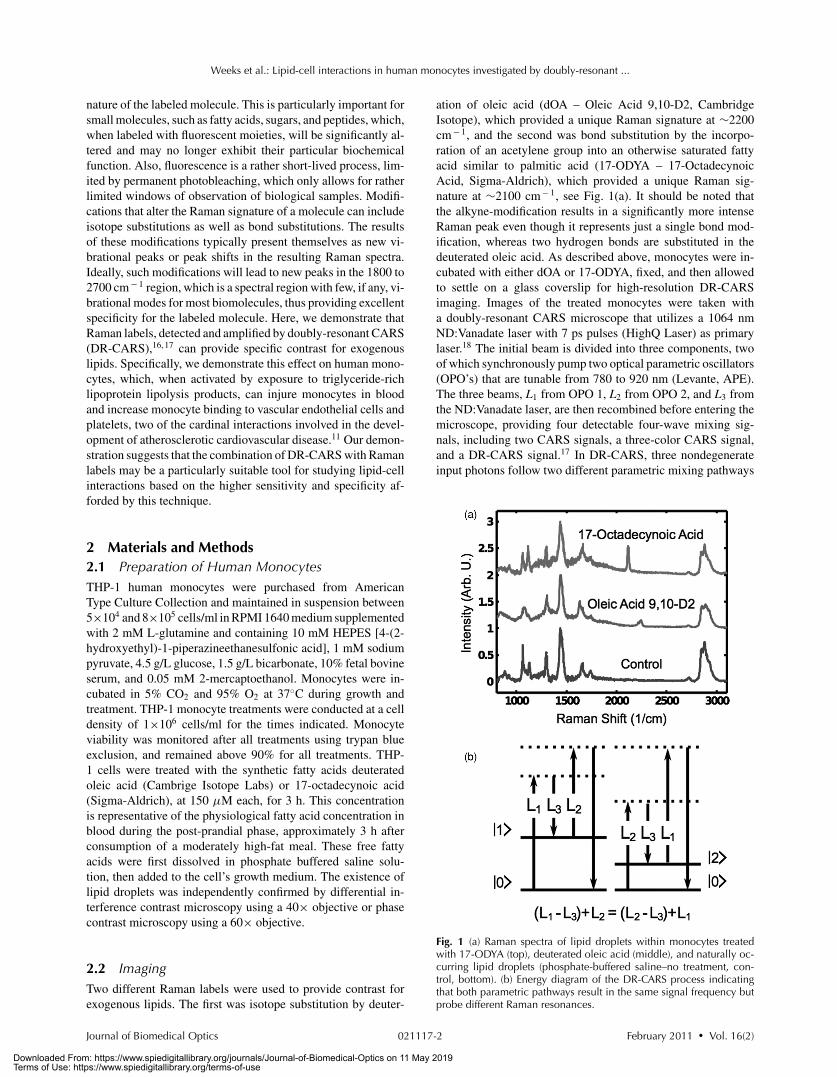
ation of oleic acid (dOA – Oleic Acid 9,10-D2, CambridgeIsotope), which provided a unique Raman signature at ∼2200cm− 1, and the second was bond substitution by the incorpo-ration of an acetylene group into an otherwise saturated fattyacid similar to palmitic acid (17-ODYA – 17-OctadecynoicAcid, Sigma-Aldrich), which provided a unique Raman sig-nature at ∼2100 cm− 1, see Fig. 1(a). It should be noted thatthe alkyne-modification results in a significantly more intenseRaman peak even though it represents just a single bond mod-ification, whereas two hydrogen bonds are substituted in thedeuterated oleic acid. As described above, monocytes were in-cubated with either dOA or 17-ODYA, fixed, and then allowedto settle on a glass coverslip for high-resolution DR-CARSimaging. Images of the treated monocytes were taken witha doubly-resonant CARS microscope that utilizes a 1064 nmND:Vanadate laser with 7 ps pulses (HighQ Laser) as primarylaser.18 The initial beam is divided into three components, twoof which synchronously pump two optical parametric oscillators(OPO’s) that are tunable from 780 to 920 nm (Levante, APE).The three beams, L1 from OPO 1, L2 from OPO 2, and L3 fromthe ND:Vanadate laser, are then recombined before entering themicroscope, providing four detectable four-wave mixing sig-nals, including two CARS signals, a three-color CARS signal,and a DR-CARS signal.17 In DR-CARS, three nondegenerateinput photons follow two different parametric mixing pathways
Fig. 1 (a) Raman spectra of lipid droplets within monocytes treatedwith 17-ODYA (top), deuterated oleic acid (middle), and naturally oc-curring lipid droplets (phosphate-buffered saline–no treatment, con-trol, bottom). (b) Energy diagram of the DR-CARS process indicatingthat both parametric pathways result in the same signal frequency butprobe different Raman resonances.
Journal of Biomedical Optics February 2011 � Vol. 16(2)021117-2
Downloaded From: https://www.spiedigitallibrary.org/journals/Journal-of-Biomedical-Optics on 11 May 2019Terms of Use: https://www.spiedigitallibrary.org/terms-of-use
Weeks et al.: Lipid-cell interactions in human monocytes investigated by doubly-resonant ...
Fig. 2 (a) DR-CARS image of THP-1 monocytes incubated with deuter-ated oleic acid, resonant with both the ∼2200 cm− 1 CD stretch andthe 2845 cm− 1 CH2 stretch. (b) CARS image of the same monocytes,resonant with just the 2845 cm− 1 CH2 stretch. (c) Difference imagebetween (a) and (b) highlighting contrast for the CD stretch resonance.(d) DR-CARS image of monocytes incubated with 17-Octadecynoicacid, resonant with both the ∼2100 cm− 1 alkyne stretch and the2845 cm− 1 CH2 stretch. (e) CARS image of the same monocytes,resonant with just the 2845 cm− 1 CH2 stretch. (f) Difference between(d) and (e) highlighting the contrast for the alkyne stretch resonance.Note the intensity scales for (c) and (f) have been adjusted to highlightcontrast for the lipids.
that probe distinct Raman resonances while resulting in the samesignal photon, see Fig. 1(b). The OPO’s were tuned such that thecombination of L1 and L3 probed the 2845 cm− 1 CH2 stretchand the combination of L2 and L3 probed either the ∼2100 cm− 1
alkyne stretch or the ∼2200 cm− 1 CD stretch. This tuning re-sulted in CARS signals that were resonant with each vibrationindividually and a DR-CARS signal that was resonant with both.For DR-CARS imaging of monocytes the laser powers are ad-justed to 40 mW (OPO1, pump 1), 11 mW (OPO2, pump 2), and12 mW (Nd-Vanadate laser, probe). Initially, images are scannedat an image size of 80 μm × 80 μm at 256×256 pixels withtypical pixel dwell times of 1 ms to identify interesting parts of asample. The zoomed parts of the initial image are then rescannedwith 256×256 pixels and image sizes according to the scale barsshown in each figure, respectively. Spontaneous Raman spectraare acquired with an integration time of 10 s/spectrum, whileDR-CARS signal spectra are acqiured at 0.5 s/spectrum. By tak-ing the difference between the DR-CARS (resonant with Ramanlabel and 2845 cm− 1 CH2 stretch) and CARS (resonant with2845 cm− 1 CH2 stretch) images, signal contributions from theRaman labels were extracted and highlighted.16, 17
3 Results and DiscussionImages of monocytes incubated with 17-ODYA and monocytesincubated with dOA both exhibit a large number of lipid dropletsthat appear to reside in the cytoplasm of the cells, see Figs. 2(a),2(b), 2(d), and 2(e). DR-CARS images [Figs. 2(a) and 2(d)]provide contrast similar to CARS images [Figs. 2(b) and 2(e)]because the labeled fatty acids provide both the Raman label anda contribution to the 2845 cm− 1 CH2 resonance. However, thedifference images, obtained by subtracting a normalized CH2
CARS image from the corresponding DR-CARS image, revealenhanced contrast for the Raman label for both groups of cells,
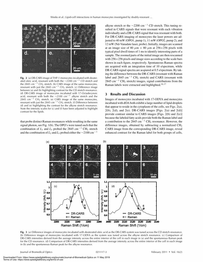
Fig. 3 (a) Difference images of monocytes incubated with deuterated oleic acid as the DR-CARS system was tuned across the CD stretch resonance.(b) Difference images of monocytes incubated with 17-ODYA as the system was tuned across the alkyne stretch resonance. (c) Comparison ofDR-CARS intensities derived from the average intensity across the entire interior of the cell in each image in (a) and the spontaneous Raman peakfor the CD resonance. (d) Comparison of DR-CARS intensities derived from the average intensity across the entire interior of the cell in each imagein (b) and the spontaneous Raman peak for the alkyne resonance.
Journal of Biomedical Optics February 2011 � Vol. 16(2)021117-3
Downloaded From: https://www.spiedigitallibrary.org/journals/Journal-of-Biomedical-Optics on 11 May 2019Terms of Use: https://www.spiedigitallibrary.org/terms-of-use

Weeks et al.: Lipid-cell interactions in human monocytes investigated by doubly-resonant ...
see Figs. 2(c) and 2(f). Interestingly, although the deuteratedoleic acid is present in the intracellular droplets, the bright ringsaround each cell indicate that much of this exogenous lipid maybe located in the cytoplasm near the plasma membrane, seeFig. 2(c). Alternatively, while the monocytes treated with17-ODYA also appear to develop a limited number of lipiddroplets, only a small number of cells seem to incorporate theexogenous saturated free-fatty acid into the intracellular lipiddroplets. Here we do not observe any notable lipid concentra-tion near the plasma membrane, see Fig. 2(f). The “shadows”surrounding each cell are due to the interference of resonant andnonresonant signals near the edge of each cell.
To further validate that the measured difference images inFigs. 2(c) and 2(f) represent contrast for the respective Ramanlabels the system was tuned across each resonance, respectively,and compared with the spontaneous Raman signal of either thealkyne resonance or the CD resonance, see Figs. 3(a) and 3(b). Inboth cases, contrast for the Raman label was minimized as thesystem was tuned away from the resonance. When comparedwith the spontaneous Raman peaks, the peak intensity of thedifference images corresponded to the expected maximum, seeFigs. 3(c) and 3(d). The dependence of the contrast on the tuningof the system relative to either the alkyne or the CD stretchvibrational resonance suggests that it is indeed a result of probingthe respective Raman labels.
Differences in the distribution of the Raman label betweenthe two groups of cells may also serve to validate the observedcontrast. Known lipid-cell interactions suggest that saturatedfatty acids may be more toxic to cells than unsaturated fattyacids.9, 19, 20 A natural response of monocytes exposed to ex-ogenous molecules is cell activation.8–11 Here the result is thegeneration of intracellular lipid droplets, which may incorpo-rate both endogenous and exogenous lipids. In particular, un-saturated lipids, such as oleic acid, are thought to enter cellsby penetrating the plasma membrane through a flip-flop mech-anism, which might explain the apparent high accumulation ofdeuterated oleic acid near the plasma membrane. However, ex-posure to more saturated fatty acids, such as 17-ODYA (exceptfor the terminal group), leads to an overall higher lipotoxicityand apoptosis of cells, which we assume accounts for the per-turbed morphology and relative low density of the cells observedafter incubation with 17-ODYA, as seen in Fig. 2(d). The appar-ent rough shape of the cell shown in Fig. 3(b) may be due to amechanism known as blebbing, which occurs in the late stagesof apoptosis. While the monocytes treated with deuterated oleicacid appear to contain lipid droplets, they maintain their roundshape and an intact plasma membrane typical of healthy humanmonocytes. Close inspection of Fig. 3(a) suggests that diffusionof unsaturated fatty acids across the plasma membrane leadsto a locally higher oleic acid concentration near the plasmamembrane, but lipid droplets are ultimately formed in the cy-toplasm, likely by the endoplasmic reticulum as is the case inmany other mammalian cells. The different entry pathway of un-saturated fatty acids might partially explain their overall lowerlipotoxicity.
4 ConclusionsWe have shown that Raman labels can provide a valuable con-trast for small molecules such as lipids as they interact with
cells. This contrast is generally difficult to achieve with tra-ditional fluorescent labels without perturbing the interaction.Although Raman-based microscopies are much less sensitivethan fluorescence-based microscopies, they do provide signalsthat are much more spectrally narrow which enable better chem-ical analysis of particular molecules and could also be exploitedin terms of broad spectral multiplexing. Furthermore, the useof doubly-resonant CARS can serve to enhance sensitivity forthe Raman label without the use of complicated heterodyningor lock-in schemes. Difference imaging, as demonstrated here,however, cannot completely remove background contributions(e.g., nonresonant background or laser fluctuations) from DR-CARS images. We expect the combination of DR-CARS with amodulation and lock-in detection scheme to lead to completelybackground-free detection. Although the central motivation inthe development of Raman microscopy has traditionally beenits use as a label-free technique, we anticipate that Raman labelsmay become a powerful, if not necessary, complement to label-free sources of contrast since they can provide true chemicalspecificity.
AcknowledgmentsTyler Weeks acknowledges support by the Lawrence ScholarProgram from Lawrence Livermore National Laboratory. Thiswork was supported in part by funding from the AmericanHeart Association through a Grant-in-Aid to T.H., and by theNational Science Foundation. The Center for Biophotonics, anNSF Science and Technology Center, is managed by the Uni-versity of California, Davis, under Cooperative Agreement No.PHY 0120999. Support by the National Institutes of Health,National Cancer Institute under Grant No. 1U54CA136465–01and the National Heart, Lung, and Blood Institute under GrantNo. NHLBI HL055667 is acknowledged. Support is further ac-knowledged from the Richard A. and Nora Eccles Harrison En-dowed Chair in Diabetes Research as well as from the ClinicalTranslational Science Center under Grant No. UL1 RR024146from the National Center for Research Resources (NCRR), acomponent of the National Institutes of Health (NIH), and theNIH Roadmap for Medical Research.
References1. S. Martin, and R. G. Parton, “Lipid droplets: a unified view of a dynamic
organelle,” Nat. Rev. Mol. Cell Biol. 7(5), 373–378 (2006).2. J. P. Kampf. and A. M. Kleinfeld, “Is Membrane Transport of
FFA Mediated by Lipid, Protein, or Both?,” Physiology 22(1), 7–14(2007).
3. Y. Akazawa, “Palmitoleate attenuates palmitate-induced Bim andPUMA up-regulation and hepatocyte lipoapoptosis,” J. Hepatol. 42(4),586–593 (2010).
4. H. J. Welters, M. Tadayyon, J. H. B. Scarpello, S. A. Smith, andN. G. Morgan, “Mono-unsaturated fatty acids protect against [beta]-cell apoptosis induced by saturated fatty acids, serum withdrawal orcytokine exposure,” FEBS Lett. 560(1–3), 103–108 (2004).
5. E. Roldan-Valadez, R. Favila, M. Martınez-Lopez, M. Uribe, andN. Mendez-Sanchez, “Imaging techniques for assessing hepatic fatcontent in nonalcoholic fatty liver disease,” Ann. Hepatol. 7(3), 212–20(2008).
6. P. D. Lang and W. Insull, “Lipid Droplets in Atherosclerotic FattyStreaks of Human Aorta,” J. Clin. Invest. 49(8), 1479–1488 (1970).
7. I. R. Corbin, E. E. Furth, S. Pickup, E. S. Siegelman, and E.J. Delikatny, “In vivo assessment of hepatic triglycerides in murine
Journal of Biomedical Optics February 2011 � Vol. 16(2)021117-4
Downloaded From: https://www.spiedigitallibrary.org/journals/Journal-of-Biomedical-Optics on 11 May 2019Terms of Use: https://www.spiedigitallibrary.org/terms-of-use

Weeks et al.: Lipid-cell interactions in human monocytes investigated by doubly-resonant ...
non-alcoholic fatty liver disease using magnetic resonance spec-troscopy,” Biochim. Biophys. Acta 1791, 757–763 (2009).
8. J. L. Kelley, M. M. Rozek, C. A. Suenram, and C. J. Schwartz,“Activation of human peripheral blood monocytes by lipoproteins,”Am. J. Pathol. 130, 223–231 (1988).
9. M. F. Cury-Boaventura, R. Gorjao, T. M. de Lima, P. Newsholme,and R. Curi, “Comparative toxicity of oleic and linoleic acid on humanlymphocytes,” Life Sci. 78(13), 1448–1456 (2006).
10. A. Alipour, A. J. van Oostrom, A. Izraeljan, C. Verseyden, J. M.Collins, K. N. Frayn, T. W. Plokker, J. W. Elte, and M. Castro Cabezas,“Leukocyte activation by triglyceride-rich lipoproteins,” Arterioscler.Thromb. Vasc. Biol. 28, 792–797 (2008).
11. L. J. den Hartigh, J. E. Connolly-Rohrbach, S. Fore, T. R. Huser, and J.C. Rutledge, “Fatty acids from very low-density lipoprotein Llpolysisproducts induce lipid droplet accumulation in human monocytes,” J.Immunol., 184(7), 3927–3936 (2010).
12. J. W. Chan, D. Motton, J. C. Rutledge, N. L. Keim, and T. Huser,“Raman spectroscopic analysis of biochemical changes in individualtriglyceride-rich lipoproteins in the pre-and postprandial state,” Anal.Chem. 77, 5870–5876 (2005).
13. J. R. Beattie, S. E. J. Bell, and B. W. Moss, “A critical evaluation ofRaman spectroscopy for the analysis of lipids: Fatty acid methyl esters,”Lipids 39(5), 407–419 (2004).
14. C. Krafft, L. Neudert, T. Simat, and R. Salzer, “Near infrared Raman
spectra of human brain lipids,” Spectrochim. Acta, Part A 61(7), 1529–1535 (2005).
15. M. N. Slipchenko, T. T. Le, H. T. Chen, and J. X. Cheng, “High-speedvibrational imaging and spectral analysis of lipid bodies by compoundRaman microscopy,” J. Phys. Chem. B 113(21) 7681–7686 (2009).
16. T. Weeks, S. Wachsmann-Hogiu, and T. Huser, “Raman microscopybased on doubly-resonant four-wave mixing (DR-FWM),” Opt. Express17(19), 17044–17051 (2009).
17. T. Weeks, I. W. Schie, S. Wachsmann-Hogiu, and T. Huser, “Signalgeneration and Raman-resonant imaging by non-degenerate four-wavemixing under tight focusing conditions,” J. Biophoton. 3(3), 169–175(2010).
18. I. W. Schie, T. Weeks, G. P. McNerny, S. Fore, J. K. Sampson,S. Wachsmann-Hogiu, J. C. Rutledge, and T. Huser, “Simultaneousforward and epi-CARS microscopy with a single detector by time-correlated single photon counting,” Opt. Express 16(3), 2168–2175(2008).
19. J. E. de Vries, M. M. Vork, T. H. Roemen, Y. F. de Jong, J. P.Cleutjens, G. J. van der Vusse, and M. van Bilsen, “Saturated but notmono-unsaturated fatty acids induce apoptotic cell death in neonatal ratventricular myocytes,” J. Lipid Res. 38(7), 1384–94 (1997).
20. G. B. Gordon, “Saturated free fatty acid toxicity. II. Lipid accumulation,ultrastructural alterations, and toxicity in mammalian cells in culture,”Exp. Mol. Pathol. 27(2), 262–276 (1977).
Journal of Biomedical Optics February 2011 � Vol. 16(2)021117-5
Downloaded From: https://www.spiedigitallibrary.org/journals/Journal-of-Biomedical-Optics on 11 May 2019Terms of Use: https://www.spiedigitallibrary.org/terms-of-use