light regulation of metabolic pathways in fungi - springer · rhythmicity, both processes have to...
TRANSCRIPT

MINI-REVIEW
Light regulation of metabolic pathways in fungi
Doris Tisch & Monika Schmoll
Received: 20 August 2009 /Revised: 14 October 2009 /Accepted: 14 October 2009 /Published online: 14 November 2009# The Author(s) 2009. This article is published with open access at Springerlink.com
Abstract Light represents a major carrier of information innature. The molecular machineries translating its electro-magnetic energy (photons) into the chemical language ofcells transmit vital signals for adjustment of virtually everyliving organism to its habitat. Fungi react to illumination invarious ways, and we found that they initiate considerableadaptations in their metabolic pathways upon growthin light or after perception of a light pulse. Alterations inresponse to light have predominantly been observed incarotenoid metabolism, polysaccharide and carbohydratemetabolism, fatty acid metabolism, nucleotide and nucleo-side metabolism, and in regulation of production ofsecondary metabolites. Transcription of genes is initiatedwithin minutes, abundance and activity of metabolicenzymes are adjusted, and subsequently, levels of metabo-lites are altered to cope with the harmful effects of light orto prepare for reproduction, which is dependent on light inmany cases. This review aims to give an overview onmetabolic pathways impacted by light and to illustrate thephysiological significance of light for fungi. We provide abasis for assessment whether a given metabolic pathwaymight be subject to regulation by light and how theseproperties can be exploited for improvement of biotechno-logical processes.
Keywords Light response .Metabolic pathways .
Strain improvement . Biotechnological processes
Introduction
Light is a very important signal for every living cell, andsince optimal adaptation to both the beneficial and harmfuleffects of light significantly enhances fitness of anorganism, it can be considered crucial for successfulcompetition and survival in nature. The ability to senselight is essential to recognize and anticipate conditionsunfavorable for vegetative growth, such as loss of waterand deprivation of nutrient on the soil surface. On the otherhand, the same signal is needed for appropriate timing ofproduction and dissemination of conidia, which happens onthe surface, in light. Commonly observed effects of light onfungi range from induction or inhibition of sexual devel-opment and conidiation to circadian clock resetting andsuppression of spore release (Corrochano 2007). However,also phenomena such as hyperpolarization of the cellmembrane (Gresik et al. 1991; Potapova et al. 1984),alterations in intracellular levels of ATP and cyclicadenosine monophosphate (cAMP) as well as an increasedrate of oxygen consumption and glycogen breakdown havebeen observed (Farkas et al. 1990).
In contrast to plants, fungi use light as a source ofinformation but not as a source of energy. During decadesof studies on fungi, at least 100 fungal species, representingall phyla, have been found to react to light (Marsh et al.1959). They have perception mechanisms for blue, nearUV, green, and red light (Herrera-Estrella and Horwitz2007; Purschwitz et al. 2006). However, the influence oflight on metabolic processes has not been investigated inthat much detail in most cases, and therefore, the responses
D. Tisch :M. Schmoll (*)Research Area Gene Technology and Applied Biochemistry,Institute of Chemical Engineering,Vienna University of Technology,Getreidemarkt 9/1665,1060 Vienna, Austriae-mail: [email protected]
D. Tische-mail: [email protected]
Appl Microbiol Biotechnol (2010) 85:1259–1277DOI 10.1007/s00253-009-2320-1

to light described in this review will predominantly refer towhite light or daylight effects.
Although light-related signaling phenomena have mainlybeen studied in Neurospora crassa, influences on variousmetabolic processes and regulators have been elucidated inother fungi. Based on the genes involved in the lightsignaling cascade and their impact on metabolic outputpathways in several fungi, hypothetical connections be-tween these pathways and light signaling can be drawn,which may be worth exploring in the future. Yet, also directmetabolic responses to light have been discovered in fungi,in many cases already decades ago.
In the following, we will give an overview on therelevance of light for fungi as well as how fungi canperceive and transmit the light signal. We will then discussprocesses directly influenced by light as well as suchimpacted by components of the light perception machineryand its downstream signaling cascade. Thereby, we intendto describe the network of factors targeted by light andsubject to light response in fungi. Despite often similaroutput effects of illumination, differences in regulatorymechanisms of light response must be assumed betweendifferent fungal species. Nevertheless, the data summarizedbelow indicate that the influence of light pulses on a certainmetabolic pathway—both with respect to the enzymesconstituting it and the signaling proteins and transcriptionfactors regulating it—cannot be excluded without experi-mental prove. Although this network provided here will bea heterogeneous one, composed of pits and pieces ofinformation from various fungi, it can help to comprehendhow fungi adjust their metabolic pathways in order tosurvive and succeed in competition upon sunrise.
Conidiation as one suitable natural cause for precise timingwith the aid of light
Nothing in biology makes sense unless it is in the light ofreproduction (Dobzhansky 1973; Maheshwari 2007). In thisrespect, it could be speculated that every adaptation ofmetabolism to changing environmental cues is ultimatelyjust meant to provide cellular energy needed for conidiationand/or sexual development in an appropriate amount at theideal time and hence to ensure reproduction and survival ofa species. Conidiation is the most obvious output regulatedby the so-called circadian clock (which ensures idealtiming) and is initiated after dusk in Neurospora. Lightpulses administered during subjective night perturb thistiming and cause resetting of the clock and altered phasesof conidiation (Devlin 2002; Brunner and Kaldi 2008). Inmany fungi, conidiation requires a light pulse to beinitiated. The processes relevant for conidiation and itsregulation are diverse. At several stages in the cascadesleading to initiation of conidiation, mechanisms triggering
the production of enzymes and chemicals the fungusapplies for successful competition in nature are at work.In the physiological sense, the transition from vegetativegrowth (favored inside the substrate, i.e., in darkness) to thereproductive state (which mostly happens on the surface ofthe substrate—in light) can be anticipated to require drasticchanges in metabolism. Still, considering the variations infavorable conditions for sexual or asexual development indifferent fungi, regulation of these changes may differaccordingly and caution is advisable when comparing orextrapolating data from one fungal species to another.Consequently, these often light-responsive adaptationmechanisms of a given organism can be used to gain abetter understanding on biotechnological processes and toimprove their efficiency.
The interrelationship between light and circadian rhythms
Circadian rhythms are apparently ubiquitous amongeukaryotic organisms (Bünning 1973). Constant tempera-ture and darkness are conditions imposed on the organismin the laboratory in order to facilitate and standardizeexperimental work. However, in nature, adaptation of anorganism to changing temperature and light conditions iscrucial for survival. Hence, a complex machinery foranticipation of dusk and dawn, adaptation to the harmfuleffects of light and desiccation during the day, and efficienttiming of conidiation has evolved. The circadian rhythms—internal timekeepers which generate a daily rhythmicity—are present in light-sensitive organisms from cyanobacteriato humans. They have been studied in detail not only inN. crassa, which has been suggested as a model forunderstanding photoperiodism (Tan et al. 2004), but alsoin Aspergillus spp. and other organisms (Brunner and Kaldi2008; Devlin 2002; Dunlap et al. 2007; Greene et al. 2003;Heintzen and Liu 2007). This circadian system is capable ofsensing and interpreting changes in temperature and lightconditions and has a major function in adaptation byadjusting its periodicity and phase (Heintzen and Liu 2007).Even light pulses in the range of minutes can reset thisclock system. The photoperiod—comparable with thelength of a day—can also have an influence on physiologyof fungi, as was shown for example for infectivity of theinsect pathogen Paecilomyces fumosoroseus (Avery et al.2004). In Fusarium oxysporum, a role for the wc-1homolog (a photoreceptor) in pathogenicity with mammalshas been observed (Ruiz-Roldan et al. 2008). Thesefindings are only special examples, which highlight thesignificance of reception of the light signal for fungi. Themechanisms dealing with response and adaptation to lighton one hand and circadian rhythmicity on the other handshare several crucial regulatory components. Because ofthis interdependence of light response and circadian
1260 Appl Microbiol Biotechnol (2010) 85:1259–1277
rhythmicity, both processes have to be considered if theeffect of light on an organism—in particular underlaboratory conditions—is to be assessed. The interrelation-ship between the clock and metabolic processes has beenstudied systematically in N. crassa (Correa et al. 2003) andgenes with products involved in DNA metabolism, ribo-some biogenesis in RNA metabolism, cell cycle, proteinmetabolism, carbon metabolism, nitrogen metabolism, andisoprenoid (including carotenoid) biosynthesis have beenfound to be under clock control (Dong et al. 2008).Thereby, the circadian clock does not only influencemetabolism, which would make it just an output pathway,but metabolism is also involved in control of circadianoscillations (Roenneberg and Merrow 1999), thus consti-tuting a complex and interdependent regulatory machinery.However, besides N. crassa and Aspergillus, detailedreports on circadian rhythms and their regulation are scarce,and in most fungi, the presence of circadian rhythmicity hasnot yet been shown. Nevertheless, it can be expected thatevery organism sensitive to light needs to deal with thechanging environment during day and night. Hence,processes triggered by a circadian rhythm are likely to besubject to certain regulation by light pulses which wouldreset the clock.
How transmission of the light signal works
The perception and transmission of the light signal isorganized by a complex regulatory network, which isinterconnected with circadian rhythmicity by sharingcrucial components (for reviews, see Brunner and Kaldi2008; Dunlap and Loros 2004; Dunlap et al. 2007) inN. crassa. With respect to light response, the majorregulatory proteins in this fungus are the PAS-domaincontaining photoreceptors WHITE COLLAR-1 (WC-1) andWHITE COLLAR-2 (WC-2) which act together as atranscription factor complex (white collar complex,WCC). All blue light responses in N. crassa described todate are mediated by the white collar complex. Thesephotoreceptors are also key components of the circadianclock (Ballario et al. 1996; Crosthwaite et al. 1997; Lindenand Macino 1997; Talora et al. 1999). Also, the WCCactivates a number of clock-controlled genes, which serveas output of the clock mechanism.
Perception of the light signal is achieved by alteredconformation and interaction: The LOV-type PAS domainof WC-1 is responsible for the function of this protein as aphotoreceptor and contains a signature motif for flavinbinding, and between a conserved cysteine residue in theLOV domain and the flavin (FAD or FMN), a blue light-dependent adduct is formed (Crosson and Moffat 2002;Crosson et al. 2003). A photoinduced conformationalchange eventually leads to altered interaction properties
with downstream signal transduction components andinitiates transmission of the light signal (Crosson andMoffat 2002; Crosson et al. 2003; Harper et al. 2003).
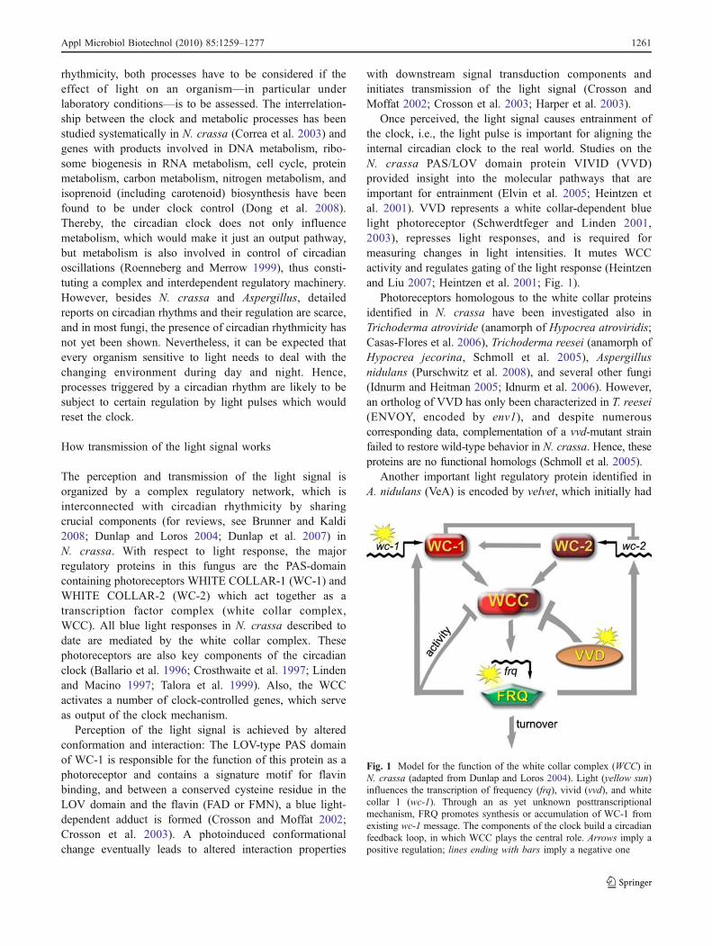
Once perceived, the light signal causes entrainment ofthe clock, i.e., the light pulse is important for aligning theinternal circadian clock to the real world. Studies on theN. crassa PAS/LOV domain protein VIVID (VVD)provided insight into the molecular pathways that areimportant for entrainment (Elvin et al. 2005; Heintzen etal. 2001). VVD represents a white collar-dependent bluelight photoreceptor (Schwerdtfeger and Linden 2001,2003), represses light responses, and is required formeasuring changes in light intensities. It mutes WCCactivity and regulates gating of the light response (Heintzenand Liu 2007; Heintzen et al. 2001; Fig. 1).
Photoreceptors homologous to the white collar proteinsidentified in N. crassa have been investigated also inTrichoderma atroviride (anamorph of Hypocrea atroviridis;Casas-Flores et al. 2006), Trichoderma reesei (anamorph ofHypocrea jecorina, Schmoll et al. 2005), Aspergillusnidulans (Purschwitz et al. 2008), and several other fungi(Idnurm and Heitman 2005; Idnurm et al. 2006). However,an ortholog of VVD has only been characterized in T. reesei(ENVOY, encoded by env1), and despite numerouscorresponding data, complementation of a vvd-mutant strainfailed to restore wild-type behavior in N. crassa. Hence, theseproteins are no functional homologs (Schmoll et al. 2005).
Another important light regulatory protein identified inA. nidulans (VeA) is encoded by velvet, which initially had
Fig. 1 Model for the function of the white collar complex (WCC) inN. crassa (adapted from Dunlap and Loros 2004). Light (yellow sun)influences the transcription of frequency (frq), vivid (vvd), and whitecollar 1 (wc-1). Through an as yet unknown posttranscriptionalmechanism, FRQ promotes synthesis or accumulation of WC-1 fromexisting wc-1 message. The components of the clock build a circadianfeedback loop, in which WCC plays the central role. Arrows imply apositive regulation; lines ending with bars imply a negative one
Appl Microbiol Biotechnol (2010) 85:1259–1277 1261

been implicated regulation of conidiation (Mooney andYager 1990). VeA is involved in activation of sexualdevelopment and inhibition of asexual development andadditionally functions as a key global metabolic regulator inthe biosynthesis of many secondary metabolites (for areview, see Calvo 2008). Identified already in 1965 (Kafer1965), the mechanism of its involvement in light responsehas only recently become subject to detailed studies. Aconnection between fruiting body formation, secondarymetabolism, and light has been shown (Busch et al. 2003;Kato et al. 2003; Kim et al. 2002), and velvet, which isprimarily expressed in the dark (Bayram et al. 2008b), maybe one factor on the crossroads of the underlying pathways(Dreyer et al. 2007; Krappmann et al. 2005; Li et al. 2006).Transport of VeA into the nucleus is inhibited by light(Fig. 2), and it acts as a negative regulator of asexualdevelopment (Mooney and Yager 1990) and antibioticproduction (Sprote and Brakhage 2007). A complexconsisting of VeA, the velvet-like protein VelB, and LaeA,a master regulator of secondary metabolism in Aspergilli,connects light-responding developmental regulation andcontrol of secondary metabolism (Bayram et al. 2008b).On the other hand, the bridge to light response isconstituted by an interaction of VeA with thephytochrome-like red light receptor FphA, thus forming acomplex which also comprises LreA and LreB (Fig. 3),the A. nidulans orthologs of the N. crassa photoreceptorsWC-1 and WC-2 (Purschwitz et al. 2008, 2009).
Blue light is also perceived by cryptochromes, which arebelieved to have evolved from the photolyase proteinfamily. These photoreceptors regulate entrainment by lightof the circadian clock in animals and plants (Lin and Todo2005). In fungi, cryptochrome/photolyase-like proteinshave been described from T. atroviride (PHR1; Berrocal-Tito et al. 2007), A. nidulans (CryA; Bayram et al. 2008a),
Cercospora zeae-maydis (PHL1; Bluhm and Dunkle 2008),and the CRY-DASH-type photolyase/cryptochrome fromSclerotinia sclerotiorum (Veluchamy and Rollins 2008).The output effects of these photoreceptors include regula-tion of the veA gene, UV protection, and functions indevelopment.
Besides the rather direct transmitters of the signal, theregulatory mechanism further includes light-dependentgene regulation by protein kinase C (PKC) in N. crassa(Arpaia et al. 1999). Upon activation of this kinase, itinteracts with WC-1 in a light-dependent manner, phos-phorylates it, and leads to decreased WC-1 protein levels(Franchi et al. 2005; Loros 2005). Processes regulated byPKC consequently are likely to be also influenced by lightbecause once activated, PKC not only acts on the respectiveprocess but apparently also interferes with light response.This finding suggests that the signal transmitted via PKChas a light-dependent relevance. In this respect, it isinteresting that inositol metabolites play an indirect role inthe light input pathway in N. crassa. In this fungus, inositoldepletion increases light sensitivity by several orders ofmagnitude. This effect is suggested to be caused by a lack
Fig. 2 Model for the functionand localization of VeA (adaptedfrom Bayram et al. 2008b). a Inlight, VeA is predominantlylocalized in the cytoplasmand VelB supports asexualsporulation. LaeA shows lowactivity. b In darkness, anincreased amount of VeA isimported into the nucleus byKapA
Fig. 3 Model for the VeAprotein complex (adapted fromCalvo 2008). VeA interacts withthe phytochrome-like red lightsensor FphA, where thechromophore-binding region inthis protein is essential formaintenance of this interaction.FphA in turn interacts with thephotoreceptor homolog LreBand hence connects VeA to theA. nidulans equivalent of theN. crassa WCC
1262 Appl Microbiol Biotechnol (2010) 85:1259–1277
of inositol leading to increased levels of diacylglycerol andto consequently enhanced protein kinase C activity (Lakin-Thomas 1992).
Also, another kinase is essential for clock function inN. crassa: cAMP-dependent protein kinase A (PKA), thefunction of which has been well studied with severalmetabolic processes. This kinase inhibits WC activity byserving as a priming kinase for the subsequently actingcasein kinases (Huang et al. 2007). For both kinases, afunction in conidiation has been shown (Banno et al. 2005;Herrmann et al. 2006), which might be the physiologicalreason for their function in light response and regulation ofmetabolic processes. Additionally, also regulatory cyclesinvolving the clock component FREQUENCY (FRQ),which activates or deactivates the WCC (Fig. 1), arecontrolled by phosphorylation events (Heintzen and Liu2007), hence emphasizing that phosphorylation is a crucialevent in triggering circadian timing and light response.
What would happen if we let the sun rise?
An early study on blue light-induced genes in 1989(Sommer et al. 1989) started the endeavor to answer thequestion: How many genes are regulated by blue light, inwhat way, and how do they influence the organism? Thisanalysis revealed that as early as 2 min after the blue lightsignal, transcripts are detectable and the authors estimatedthat within 30 min, 60–80 genes are regulated in N. crassa.It can, however, be assumed that initiation of transcriptionafter illumination already starts much earlier, since forexample, only 2 s of light is sufficient to induce sporulationin Trichoderma viride (Betina and Zajacova 1978). Thus,transcription of genes involved in this process must havebeen triggered by this 2 s of light. Later on, a similar studydealing with light regulation of expression of the toxincercosporin by Cercospora kikuchii led to the identificationof six light-enhanced cDNA clones, transcript accumulationof which paralleled cercosporin production (Ehrenshaft andUpchurch 1991). Also, the role of the photoreceptor WC-1in light response was investigated on a broad scale inN. crassa by microarray analysis (Lewis et al. 2002) andfunctions of homologs of WC-1, WC-2, and VVD wereelucidated on a genome-wide scale in Trichoderma spp.:T. atroviride was shown to regulate gene expression notonly in response to blue light but also to red light and thatthe respective signaling pathways are interconnected.Moreover, this fungus exhibits blue light regulator (BLR;orthologs to the white collar proteins)-independent photo-responses (Rosales-Saavedra et al. 2006). For T. reesei,different growth patterns in light and darkness have beendetected on various carbon sources, and these patterns werealtered in a strain lacking the light regulatory proteinENVOY (Schmoll et al. 2005; Schuster et al. 2007). In
agreement with this report, crosstalk between carbonmetabolism, blue light receptors, and reaction to oxidativestress was shown by investigation of growth of T. atrovirideon 96 carbon sources (Friedl et al. 2008a). On a similarlybroad scale, it was shown that conidiation is primarilycarbon source dependent and illumination plays a catalyticrole in T. atroviride. Also, the study revealed cross talkof the photoreceptors BLR-1 and BLR-2 (homologs ofN. crassa WC-1 and WC-2) with regulation of cyclic AMPlevels (Friedl et al. 2008b).
Recent comprehensive genome-wide approaches providea broad picture on the transcriptional response to light. InN. crassa, more than 5% of the total detectable transcriptsare responsive to light. Also, a differentiation between earlylight-responsive genes (ELRGs), which peak between 15and 45 min after onset of light, and late light-responsivegenes (LLRGs), which peak later until 90 min after lightson, is possible (Chen et al. 2009). While ELRGs essentiallyrepresent the attempt to rapidly adapt to the harmful effectsof light, such as genes involved in synthesis of photo-protective pigments, secondary metabolism, and signaling,LLRGs rather reflect adjustment of metabolic functions todaylight condition, as they comprise genes involved incarbohydrate metabolism, oxidation of fatty acids, anddetoxification. In summary, this study revealed a tightlyscheduled sequence of cellular events in response to thedaily light stimulus in N. crassa (Chen et al. 2009).
Aiming to provide insights into the systems biology ofthe clock in Neurospora, a recent study revealed thatribosomal RNA processing and assembly rather thantranscription seems to be under clock control, suggestingan unexpected mechanism of posttranscriptional regulationof clock-controlled genes (Dong et al. 2008). In support ofthis finding, light-dependent splicing of the T. viridehydrophobin gene tvsrh1 was reported (Vargovic et al.2006). In F. oxysporum, also differential splicing of ahydrophobin was observed. In this case, this phenomenonwas dependent on the presence of the WC-1 homolog(Ruiz-Roldan et al. 2008). Consequently, a role of thisphotoreceptor in light-dependent differential splicing—notonly of hydrophobin genes—could explain discrepanciesbetween transcript abundance and protein accumulation orfunctionality in light.
Metabolic pathways as output pathways of lightsignaling
Literature shows that the fundamental adaptations fungiapply to survive and succeed in the daily changes broughtabout by the rotation of earth are largely underestimated.While numerous studies deal with light regulation ofcarotenoid metabolism, the implications of early studies
Appl Microbiol Biotechnol (2010) 85:1259–1277 1263

suggesting an influence of light on other metabolicprocesses (for example, Hill 1976; Nambudiri et al. 1973;Ram et al. 1984) were often not considered with laterstudies on transcription and expression of metabolic genes.At the same time, an increasing number of reports showthat a significant portion of the transcribed genes of anorganisms responds to light (Chen et al. 2009; Sommer etal. 1989), often within minutes. Additionally, it can beassumed that this response is influenced by the carbonsource the fungus grows on (Friedl et al. 2008a, b; Schusteret al. 2007). Quite early on it was recognized that the natureof the medium on which the fungus is cultivated candetermine whether light stimulates or decreases the growthrate in many fungi (Carlile 1965). This phenomenonbecomes very clear in recent studies investigating growthand conidiation on 96 carbon sources in wild-type andmutant strains of T. atroviride (Friedl et al. 2008a, b).Substantial metabolic differences have been detectedbetween cultures growing in light and darkness. Lightseems to have a different effect in dependence on whetherthe nutritional conditions are optimal or not. Hence, underoptimal conditions, light and dark metabolism should beapproximately equally effective in supporting growth, butalterations in conditions would affect the processes differ-ently (Carlile 1965).
In the following, we will summarize both quite early andvery recent studies dealing with light-responsive regulationof transcription of genes involved in metabolic pathways,activation of enzymes, alterations of nutrient uptake, andproduction of metabolites. The genes and enzymes describedwere assigned to a chapter according to the classification ofthe MIPS Functional Catalogue (FunCatDB; Ruepp et al.2004). Nevertheless, in some cases, overlaps were unavoid-able in order to maintain clarity.
Carotenoid metabolism
Carotenoids are soluble pigments produced in bacteria,fungi, and plants (Britton et al. 1998). They belong to thegroup of terpenoids and comprise over 600 organicpigments, split into two classes: xanthophylls (whichcontain oxygen) and carotenes (contain no oxygen). Thesepigments can act as antioxidants and generally absorb bluelight, which is crucial for their function in numerousorganisms from bacteria to plants. Besides their natural,mainly protective function in the organisms producingcarotenoids, these molecules also have beneficial effectson animals and humans (Stahl and Sies 2005), who are notable to produce carotenoids. This is also the main reasonwhy they are produced on a large scale both chemically aswell as biotechnologically (Avalos and Cerda-Olmedo2004). Investigations of carotenoid production in fungi(Fig. 4) have already been done more than 100 years ago
(Kohl 1902) and elaborate studies with Phycomycesblakesleeanus followed in the 1950s (Bergman et al. 1969;Garton et al. 1951; Meissner and Delbruck 1968). Amongthe many fungi-producing carotenoids, the zygomyceteBlakeslea trispora and the Mucorales P. blakesleeanusand Mucor circinelloides are of industrial interest, butseveral other fungi also deserve closer evaluation withrespect to their carotenoid production. While the respectivebiosynthetic pathways are quite conserved, regulation ofcarotenoid production (Almeida and Cerda-Olmedo 2008;Cerda-Olmedo 2001) is more variable among species.Nevertheless, blue light is a common signal triggeringupregulation of the pathway (Avalos et al. 1993; Avalos andCerda-Olmedo 2004; Carlile 1965; Jayaram et al. 1979).P. blakesleeanus is well studied with respect to its responseto light and its production of carotenoids. Besides sexualstimulation, also light has an enhancing effect on carotenoidproduction in this fungus (Cerda-Olmedo 2001). Bluelight causes enhanced accumulation of carRA and carBtranscripts (Blasco et al. 2001; Rodriguez-Romero andCorrochano 2004; Ruiz-Hidalgo et al. 1997), encoding
Fig. 4 Biosynthetic pathway for β-carotene in the Mucorales andneurosporaxanthin in Neurospora. Conversion of β-carotene toabscisic acid (empty arrows) has only been shown in Cercosporarosicola, but in no other fungus (Assante et al. 1977)
1264 Appl Microbiol Biotechnol (2010) 85:1259–1277

enzymes of the carotenoid biosynthetic pathway, whichare crucial for light response and carotenogenesis inP. blakesleeanus (Bejarano et al. 1991). A similar effecthas been observed for the homologs of these genes inM. circinelloides (Velayos et al. 2000a, b), B. trispora(Quiles-Rosillo et al. 2005), and N. crassa (Nelson et al.1989; Schmidhauser et al. 1990, 1994). The so-called madmutants of Phycomyces (denominated after Nobel laurateMax Delbrück), which are defective in phototropism, showa corresponding effect: Deletion of madA and madB leadsto defective photocarotenogenesis (Cerda-Olmedo 2001;Idnurm et al. 2006). While both brief light pulses as well asprolonged illumination increase carotene accumulation inPhycomyces (Bejarano et al. 1991), continuous illuminationresults in even decreased carotene production in Blakeslea(Sutter 1970). Yet it shows clear enhancement afterillumination of dark-grown mycelia in this fungus (Quiles-Rosillo et al. 2005). With respect to regulation, it isinteresting that in Phycomyces, a heterotrimeric G-proteinapparently plays a role photocarotenogenesis (Tsolakis etal. 2004).
Besides these industrially important fungi, also Gibberellafujikuroi (Fusarium moniliforme) produces considerableamounts of carotenoids (predominantly neurosporaxanthinand minor amounts of intermediate carotenes), and hence,when grown in light, mycelia of this fungus are orange(Avalos and Schrott 1990). In darkness, mycelia are whiteand have a low carotenoid content, albeit several mutants,called pale-orange, contain an increased amount of carote-noids even in darkness (Avalos and Cerdà-Olmedo 1987). Incontrast to Neurospora, which already responds to a fewseconds of illumination with carotenoid production (Schrott1980), G. fujikuroi needs at least 8 min of light for adetectable response (Avalos and Schrott 1990). Moreover,G. fujikuroi still considerably responds to illumination bycarotenoid production upon deletion of the photoreceptorhomolog wc-1, albeit to a lesser extent than the wild type(Estrada and Avalos 2008; Ruiz-Roldan et al. 2008). Studieson the interrelationship of the positive effect of nitrogenstarvation on carotenogenesis and the effect of light revealedthat these effects are additive (Rodriguez-Ortiz et al. 2009).
In N. crassa, carotenoid metabolism and its regulationby light and the circadian clock has been studied exten-sively predominantly because it is a convenient outputpathway of these regulatory circuits. Also in this fungus,carotenoid biosynthesis is being studied already for decades(Harding et al. 1969), and carotenoids have been implicatedin protection of respiratory systems in this fungus (Ramadan-Talib and Prebble 1978). As might be expected from theimportance of the light signal for carotenogenesis, thephotoreceptors WC-1 and WC-2 both are important regu-lators of this process, and mutants in the respective genes aresignificantly impaired in its regulation (Nelson et al. 1989;
Perkins et al. 1982). On the other hand, the photoreceptorprotein VVD has a negative effect on carotenogenesis, andvvd mutants show a vivid orange color when grown in light,reflecting overproduction of carotenoids, which also gave thisgene its name (Heintzen et al. 2001; Shrode et al. 2001).Biosynthetic enzymes of the carotenoid pathway such asAL-1 (Schmidhauser et al. 1990), the phytoene synthaseAL-2 (Schmidhauser et al. 1994), the prenyl transferase AL-3(Sandmann et al. 1993), and CAO-2 are regulated by light ina WC-1 and WC-2 dependent manner (Saelices et al. 2007).Exposure of mycelia to open air is crucial for photocaroteno-genesis, and reactive oxygen species (ROS) play a major rolein this process (Iigusa et al. 2005). Also, a function of theputative green light opsin photoreceptor NOP-1 in caroteno-genesis has been suggested (Bieszke et al. 2007).
The clear light regulation of carotene biosynthesis infungi suggested a similar effect in regulation of a closelyrelated compound, ubiquinone, which shares the majorityof biosynthetic reactions with carotene. It has been shownthat carotenoid protects ubiquinone from photodestructionin mitochondria (Ramadan-Talib and Prebble 1978).Ubiquinone is an essential cellular component, which isused as an ingredient in cosmetics and for medicalapplications (Kuzina and Cerda-Olmedo 2007 and refer-ences therein). A comparable regulation of its production tocarotenoid biosynthesis would greatly facilitate strainimprovement. Interestingly, studies with B. trispora andP. blakesleeanus revealed that light indeed had a positiveeffect on the former (albeit a moderate one), but no effecton the latter (Kuzina and Cerda-Olmedo 2007). Hence,carotenoid and ubiquinone biosynthesis are not coregulated,and also, their regulation is not conserved between species.
Another compound derived from carotenoids (ABA) isabscisic acid, an important plant hormone (Gubler et al.2005). This secondary metabolite is produced not only inplants but also in the fungus Cercospora rosicola, and aswould be expected for a carotenoid derivative, it is alsosignificantly regulated by light (Norman et al. 1981).
Polysaccharide/carbohydrate metabolism
The finding that deviation from optimal conditions causesdifferent effects of light and darkness on growth of fungi(Carlile 1965) as do different carbon sources (Friedl et al.2008a, b) indicates that carbon source availability andquality are closely interlinked with light response. It seemsthat not only utilization but also uptake of nutrients issubject to regulation by light (Hill 1976), which in turnimpacts metabolism due to the lack of the respectivecompound in the cell. Inhibition of growth could therebybe caused by limited uptake of an essential nutrient. InAspergillus ornatus, glucose uptake (but not lysine uptake)is significantly decreased upon cultivation in light. This
Appl Microbiol Biotechnol (2010) 85:1259–1277 1265

could be interpreted in a way that the rate of glucose uptakeis decreased prior to production of conidia. It has beensuggested that this effect is due to biosynthesis of adialyzable inhibitor of glucose uptake in light (Hill 1976),which is supported by data on Neurospora: In this fungus,it was shown that altered banding in a mutant strain is dueto glucose transport deficiency (Halaban 1975). Theinterrelationship of this effect to conidiation is furtheremphasized by the finding that high glucose concentrationsinhibit conidiation in T. viride, although they have noinfluence on growth (Betina and Zajacova 1978). Conse-quently, the limitation of glucose uptake in light may serveas a precondition for induction of conidiation. In support ofthese data, a study aimed at identification of early light-regulated genes in T. reesei revealed two putative trans-porters to be regulated by light and by the light regulatoryprotein ENVOY (Schuster et al. 2007).
Light also controls several metabolic processes ofAspergillus giganteus. In this fungus, occurrence of certainpolysaccharides (Fiema 1983; Fiema and Golbiewska 1981)as well as the amount of glucans (Fiema et al. 1991) orglycogen content (Zurzycka 1991) depends on lightintensity and at least in part on glucose concentration inthe nutrient medium. Accordingly, a decrease in endoge-nous reserves, as reflected by a lower glycogen contentupon cultivation in light, has also been observed in T. reesei(Farkas et al. 1990).
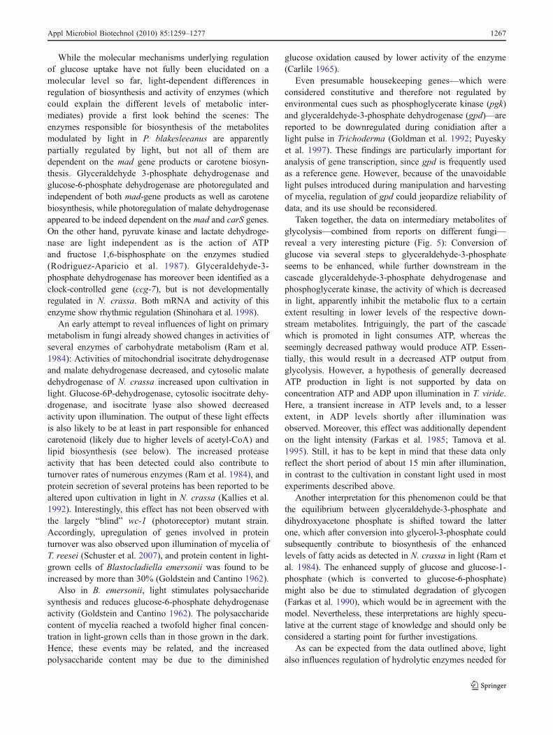
However, not only uptake but also intracellular synthesisand/or degradation of carbohydrates is altered in responseto illumination, as are certain steps of glycolysis, thepentose phosphate pathway, or the citric acid cycle. Theeffects of light on intermediates of glycolysis reported fromseveral different fungi as outlined in the following aresummarized in Fig. 5.
In Penicillium isariiforme, polysaccharide synthesis isstimulated by light (Graafmans 1976). Blue light alsocauses increased excretion of citric acid in this fungusbecause of the lower supply of pyruvate in light, which iscarboxylated to form citric acid via oxaloacetate. In light,higher levels of intermediates such as glucose-6-phosphate,glucose-1-phosphate, and fructose-6-phosphate were ob-served (Graafmans 1977). In this fungus, light stimulatesthe pentose phosphate pathway and blocks the supply ofpyruvate for citric acid synthesis via the Embden–Meyerhof–Parnas (EMP) pathway. Since uptake of sucrose is not alteredin light in P. isariiforme (Graafmans 1974), this indicates thatless endogenous substrate is channeled via the glycolyticEMP pathway upon growth in light (Graafmans 1977).
Influences of light on carbohydrate metabolism are alsopresent in P. blakesleeanus (Rua et al. 1987). Themetabolites fructose 6-phosphate, glyceraldehyde 3-phosphate, dihydroxyacetone phosphate, and lactate werepresent in higher concentrations in light, whereas glucose 6-
phosphate, 2-phosphoglycerate, phosphoenolpyruvate, andpyruvate accumulated in cultures grown in darkness. Theconcentration of fructose 1,6-bisphosphate appeared to belight independent. With respect to pyruvate metabolism,levels of acetyl coenzyme A and citrate were higher inlight, and L-alanine and L-malate were higher in darkness(Rua et al. 1987).
Fig. 5 Schematic representation of the influence of light onglycolysis. Green diamonds indicate increased levels of the respectivemetabolite upon cultivation in light, and red diamonds reflect lowerlevels. For fructose-1,6-bisphosphate (yellow diamond), no alterationwas found, and in case of white diamonds, no data on the levels ofthese metabolites in light were available. Red balloons indicate lowerenzymatic activities of the respective enzyme
1266 Appl Microbiol Biotechnol (2010) 85:1259–1277
While the molecular mechanisms underlying regulationof glucose uptake have not fully been elucidated on amolecular level so far, light-dependent differences inregulation of biosynthesis and activity of enzymes (whichcould explain the different levels of metabolic inter-mediates) provide a first look behind the scenes: Theenzymes responsible for biosynthesis of the metabolitesmodulated by light in P. blakesleeanus are apparentlypartially regulated by light, but not all of them aredependent on the mad gene products or carotene biosyn-thesis. Glyceraldehyde 3-phosphate dehydrogenase andglucose-6-phosphate dehydrogenase are photoregulated andindependent of both mad-gene products as well as carotenebiosynthesis, while photoregulation of malate dehydrogenaseappeared to be indeed dependent on the mad and carS genes.On the other hand, pyruvate kinase and lactate dehydroge-nase are light independent as is the action of ATPand fructose 1,6-bisphosphate on the enzymes studied(Rodriguez-Aparicio et al. 1987). Glyceraldehyde-3-phosphate dehydrogenase has moreover been identified as aclock-controlled gene (ccg-7), but is not developmentallyregulated in N. crassa. Both mRNA and activity of thisenzyme show rhythmic regulation (Shinohara et al. 1998).
An early attempt to reveal influences of light on primarymetabolism in fungi already showed changes in activities ofseveral enzymes of carbohydrate metabolism (Ram et al.1984): Activities of mitochondrial isocitrate dehydrogenaseand malate dehydrogenase decreased, and cytosolic malatedehydrogenase of N. crassa increased upon cultivation inlight. Glucose-6P-dehydrogenase, cytosolic isocitrate dehy-drogenase, and isocitrate lyase also showed decreasedactivity upon illumination. The output of these light effectsis also likely to be at least in part responsible for enhancedcarotenoid (likely due to higher levels of acetyl-CoA) andlipid biosynthesis (see below). The increased proteaseactivity that has been detected could also contribute toturnover rates of numerous enzymes (Ram et al. 1984), andprotein secretion of several proteins has been reported to bealtered upon cultivation in light in N. crassa (Kallies et al.1992). Interestingly, this effect has not been observed withthe largely “blind” wc-1 (photoreceptor) mutant strain.Accordingly, upregulation of genes involved in proteinturnover was also observed upon illumination of mycelia ofT. reesei (Schuster et al. 2007), and protein content in light-grown cells of Blastocladiella emersonii was found to beincreased by more than 30% (Goldstein and Cantino 1962).
Also in B. emersonii, light stimulates polysaccharidesynthesis and reduces glucose-6-phosphate dehydrogenaseactivity (Goldstein and Cantino 1962). The polysaccharidecontent of mycelia reached a twofold higher final concen-tration in light-grown cells than in those grown in the dark.Hence, these events may be related, and the increasedpolysaccharide content may be due to the diminished
glucose oxidation caused by lower activity of the enzyme(Carlile 1965).
Even presumable housekeeping genes—which wereconsidered constitutive and therefore not regulated byenvironmental cues such as phosphoglycerate kinase (pgk)and glyceraldehyde-3-phosphate dehydrogenase (gpd)—arereported to be downregulated during conidiation after alight pulse in Trichoderma (Goldman et al. 1992; Puyeskyet al. 1997). These findings are particularly important foranalysis of gene transcription, since gpd is frequently usedas a reference gene. However, because of the unavoidablelight pulses introduced during manipulation and harvestingof mycelia, regulation of gpd could jeopardize reliability ofdata, and its use should be reconsidered.
Taken together, the data on intermediary metabolites ofglycolysis—combined from reports on different fungi—reveal a very interesting picture (Fig. 5): Conversion ofglucose via several steps to glyceraldehyde-3-phosphateseems to be enhanced, while further downstream in thecascade glyceraldehyde-3-phosphate dehydrogenase andphosphoglycerate kinase, the activity of which is decreasedin light, apparently inhibit the metabolic flux to a certainextent resulting in lower levels of the respective down-stream metabolites. Intriguingly, the part of the cascadewhich is promoted in light consumes ATP, whereas theseemingly decreased pathway would produce ATP. Essen-tially, this would result in a decreased ATP output fromglycolysis. However, a hypothesis of generally decreasedATP production in light is not supported by data onconcentration ATP and ADP upon illumination in T. viride.Here, a transient increase in ATP levels and, to a lesserextent, in ADP levels shortly after illumination wasobserved. Moreover, this effect was additionally dependenton the light intensity (Farkas et al. 1985; Tamova et al.1995). Still, it has to be kept in mind that these data onlyreflect the short period of about 15 min after illumination,in contrast to the cultivation in constant light used in mostexperiments described above.
Another interpretation for this phenomenon could be thatthe equilibrium between glyceraldehyde-3-phosphate anddihydroxyacetone phosphate is shifted toward the latterone, which after conversion into glycerol-3-phosphate couldsubsequently contribute to biosynthesis of the enhancedlevels of fatty acids as detected in N. crassa in light (Ram etal. 1984). The enhanced supply of glucose and glucose-1-phosphate (which is converted to glucose-6-phosphate)might also be due to stimulated degradation of glycogen(Farkas et al. 1990), which would be in agreement with themodel. Nevertheless, these interpretations are highly specu-lative at the current stage of knowledge and should only beconsidered a starting point for further investigations.
As can be expected from the data outlined above, lightalso influences regulation of hydrolytic enzymes needed for
Appl Microbiol Biotechnol (2010) 85:1259–1277 1267

degradation of extracellular substrates. It is difficult todetermine at which physiological process the light signalinterferes: Diminished uptake of substrate in response tolight could necessitate slower or altered metabolism. On theother hand, if intracellular processes involved in utilizationof the substrate would be working at different levels inlight, also uptake of a carbon source could be adjustedaccordingly. Together, regulation of these processes in turncould lead to adjustment of expression of extracellularenzymes in light—which would in turn influence theabundance of their degradation products to be taken up bythe fungal cell. In this respect, the latter part of the circuitonly applies if the organism is forced to degrade anextracellular, insoluble compound such as the naturalsubstrates cellulose or lignin. However, also this extracel-lular section of the circuit has been shown to be responsiveto light in that enzymes directly responsible for degradationof nutrients outside the fungal cell along with someintracellular degradative enzymes show light-dependentvariation in activity or abundance:
Glucoamylase activity of blue light-exposed mycelia ofAspergillus niger is increased more than 2.5-fold ascompared to mycelia grown in darkness (Zhu and Wang2005), and light-grown cultures of P. blakesleeanus exhibitlower specific activities for alcohol dehydrogenase thandark-grown cultures. Since this effect has also beenobserved in mutants with altered phototropism, thisadaptation is not mediated by the mad photosystem(Garce's and Medina 1985). Recent studies dealt with theregulatory mechanisms involved in light-responsive pro-cesses in carbohydrate metabolism. In A. nidulans, a rolefor VeA (besides its well-studied function in secondarymetabolism as discussed below) in regulation of a fructosylamino acid oxidase (Jeong et al. 2002) and a mannoprotein(Jeong et al. 2003) has been detected.
A screening for genes involved in cellulase signaling inthe cellulolytic fungus T. reesei led to the identification ofan ortholog of the N. crassa photoreceptor VIVID (Schmollet al. 2004). In the following, cellulase gene expression wasshown to be modulated by light. The identified protein,ENVOY (Schmoll et al. 2005), was not only shown tostrongly respond to light but also to significantly influencecellulase gene expression in T. reesei. Subsequently, furtherparts of the signaling cascade were shown to have a light-dependent effect on cellulase gene expression. The G-alphasubunit GNA3 positively regulates this process only in light(Schmoll et al. 2009), while deletion of another G-alphasubunit, GNA1 (Seibel et al. 2009), leads to abolishedcellulase transcription in light and strongly increasedtranscript levels in darkness. In both cases, constitutiveactivation of the G-alpha subunit did not lead to inducerindependent cellulase expression. Hence, it can be concludedthat these two G-alpha subunits transmit signals important
for adjustment of cellulase gene expression to environmentalconditions, the relevance of which depends on whether thefungus is growing in light or darkness. These signals areobviously not related to the presence of cellulose, andconsequently, other so far undetermined environmental cuesare highly relevant for regulation of cellulase expression andpossibly other extracellular enzymes.
Chitin and other cell wall components
Upon illumination of fungal mycelia, the cell wall is the firstsite of impact of photons. Given the harmful effects of light, itis not surprising that several studies revealed an alteredcomposition of the cell wall in response to light. Transientalterations in cell wall structure were observed in P.blakesleeanus (Herrera-Estrella and Ruiz-Herrera 1983),and the content of chitin in the cell wall of A. giganteus isdoubled if the mycelium is cultivated in light as compared todarkness (Fiema 1983). In this fungus, the levels of S- andR-glucans in the mycelium show alterations in light. The S-glucan proportion is directly proportional to light intensityupon growth in high glucose concentrations. Also therelation between S- and R-glucans is altered. Therefore,both quality and quantity of glucans in the mycelium ofA. giganteus are controlled by light and glucose concentra-tion (Fiema 1993). In agreement with these findings, alsoillumination of Trichoderma harzianum affects enzymesinvolved in biosynthesis of cell wall components: Alreadyafter 10 min of illumination, specific activity of beta-1,3-glucan synthase increases by about 130%, and specificactivity of chitin synthase decreases by 50% compared todark controls. Correspondingly, while the chitin contentremains essentially constant, the beta-1,3-glucan content inilluminated mycelia is elevated by up to 50% (Nemčovič andFarkaš 2001).
In P. blakesleeanus, studies on the fascinating phenom-ena of phototropism, gravitropism, and avoidance led to theidentification of ten genes involved in these processes, theso-called mad genes. All of them show an altered responseto light. The corresponding gene products have beensuggested to be involved in regulation of growth of thecell wall in the growing zone, which may be due to light-regulated interplay of chitinases and new chitin synthesis(Cerda-Olmedo 2001). In this process, chitin synthaseactivity is increased by illumination and two light-sensitive calcium and calcium/calmodulin systems contrib-ute to this activation (Herrera-Estrella and Ruiz-Herrera1983; Ruiz-Herrera et al. 1990).
Fatty acid metabolism
Considering the global picture of gene regulation providedby the detailed microarray analysis of Chen and coworkers,
1268 Appl Microbiol Biotechnol (2010) 85:1259–1277

which revealed upregulation of genes assigned to lipidmetabolism and fatty acid oxidation (Chen et al. 2009),earlier studies on this topic now get additional support. Ithas been suggested that 18:2 polyunsaturated fatty acids(i.e., linoleic acid) have a role in fungal development,particularly in sporulation (Roeder et al. 1982). InNeurospora, oleic acid is the predominant fatty acid indeveloping asci and ascospores, while linoleic acid is themajor fatty acid in asexual mycelia in this fungus (Goodrich-Tanrikulu et al. 1998). The concentration of lipids inN. crassa was found to be higher in mycelia grown in lightas compared to cultivation in darkness (Ram et al. 1984).Similar results were obtained for Alternaria alternata(Haggblom and Unestam 1979). Provision of reducingpower (which could be met by glucose-6P-dehydrogenase)and availability of acetyl CoA (which can be provided bycleavage of excess citrate in the cytosol) both can increaselipid as well as carotenoid levels. If light influences thesepreconditions, the respective regulation would explain howthe influence of light is mediated. However, glucose-6P-dehydrogenase activity is not increased in Neurospora, onthe contrary, but cytosolic malate dehydrogenase showedhigher activity in light and may thus provide reducing power.Also for the active catabolism of citrate, a high level ofisocitrate dehydrogenase is required to remove the substrateisocitrate because it would otherwise be converted to citrateby the reversible aconitase reaction. Hence, the decreasedactivity of isocitrate dehydrogenase in light should causecitrate to accumulate and consequently provide the buildingblocks for lipid and carotenogensis (Ram et al. 1984).
Indirect hints as to a light-dependent regulation ofmembrane fatty acid composition come from severalreports on circadian regulation (which can be phase-shifted by light) in N. crassa (Cote and Brody 1987).Further, effects of supplementation of cultivation mediawith saturated (Mattern and Brody 1979) or unsaturated(Brody and Martins 1979) fatty acids on period length inN. crassa have been reported. However, also the levelsof linoleic and linolenic acid in N. crassa myceliaoscillate, which could be caused by cyclic (de)activationor biosynthesis/degradation of a desaturase. The phase ofthese circadian oscillations can be light-set in a similarmanner as it is the case for conidiation. Nevertheless, theoscillations in fatty acid composition are largely indepen-dent of the developmental events leading to conidiation(Roeder et al. 1982). In A. nidulans, a desaturase, encodedby odeA, has been deleted, and clear effects on fatty acidcomposition and sporulation have been observed. Addi-tionally, it has been shown that also in this fungus, fattyacid composition is influenced by light as well as by velvet(veA) (Calvo 2008; Calvo et al. 2001).
In T. viride, lipid accumulation after photoinduction hasbeen investigated, and significant differences were found in
sporulating, photoinduced, and nonsporulating, noninducedmycelia. However, in this case, it is not possible to decidewhether this effect is due to the light pulse or connected tosporulation (Betina and Koman 1980).
Nucleotide and nucleoside metabolism
Despite the importance of nucleotides and nucleosides for thefungal cell in energy metabolism and adjustment of redoxpotential, knowledge on their regulation in response to light infungi is limited. NAD+ kinase is one of the key enzymes inregulation of relative levels of anabolic and catabolic path-ways in the cell. In N. crassa, specific activity of NAD+kinase increases only minutes after illumination up totwofold (Afanasieva et al. 1982). This activation can beviewed as one step of a coordinated regulatory mechanismfor the activity of enzymes involved in formation andreduction of NADP and consequently in regulation of thepentose phosphate shunt (Afanasieva et al. 1982). Moreover,both activities of NADH reductase and NADH oxidase (aswell as several other enzymes involved in respiration) arenegatively influenced by light not only in wild-type but alsoin albino strains (Ramadan-Talib and Prebble 1978) ofN. crassa. Yet, a generally negative effect of light onrespiration is contradicted by the finding of an “oxidativeburst”, lasting about 10 min after onset of illumination,during which an increase in the rate of oxygen consumptionhas been observed in T. reesei (Farkas et al. 1990).
In B. emersonii, growth in light causes the nucleic acidcontent of the cells to increase more rapidly than indarkness and to reach a 28% higher final quantity ofnucleic acids than dark-grown cells (Goldstein and Cantino1962). Interestingly, also circadian rhythms of nucleic acidcontent and biosynthesis occur in N. crassa with a period ofabout 24 h and also in many other organisms (Martens andSargent 1974). Since this reflects a daily rhythm whichnormally can be reset by light pulses, the detection of acircadian rhythm supports the finding of an influence oflight on nucleotide metabolism.
cAMP
Cyclic adenosine monophosphate (cAMP) is a secondarymessenger in eukaryotes. This compound is produced inresponse to several extracellular stimuli and regulates avariety of physiological processes. Two enzymes are themajor regulators of cAMP levels in fungi: the biosyntheticenzyme adenylyl cyclase and the degradative phosphodies-terase (D'souza and Heitman 2001). Light pulses signifi-cantly impact cAMP levels in T. viride (Farkas et al. 1985;Gresik et al. 1988). Thereby, illumination causes rapid buttransient increase in intracellular concentrations of ATP andcAMP, which might contribute to phosphorylation of
Appl Microbiol Biotechnol (2010) 85:1259–1277 1269

proteins in response to light. With respect to phosphoryla-tion, the effect of light can be substituted by addition of3 mM cAMP (Gresik et al. 1989). These cAMP levelscould be adjusted by the membrane associated adenylylcyclase, which is activated by light—in contrast to 3′5′cyclic AMP phosphodiesterase, for which this is not thecase (Kolarova et al. 1992). In N. crassa in contrast, noalterations of cAMP levels were detected in response tolight (Shaw and Harding 1987). For A. nidulans, however,photoregulation of phosphodiesterase has been reported(Gradisnik-Grapulin and Legisa 1997). In T. atroviride, aconnection between the cAMP-dependent protein kinase A(PKA) and blue light perception has been drawn. PKAactivity increases not only in illuminated cell extracts of thewild-type strain but also in mutants of the photoreceptorsBLR-1 and BLR-2 (Casas-Flores et al. 2006). Since thesestrains show severely perturbed light response, this phe-nomenon could be due to adenylyl cyclase activateddirectly (or at least without participation of these photo-receptors) by light. A connection between cAMP levels andaccumulation of carotenoids has been suggested in N.crassa in that a transient decrease of cAMP levels occurredafter photoinduction of carotenoid syntheses during its lagperiod (Kritsky et al. 1982).
Interestingly, indications for an interconnection betweencAMP levels and expression of extracellular enzymes havebeen detected: In Cryptococcus albidus, xylanase produc-tion can be increased by addition of exogenous cAMP(Morosoli et al. 1989). A connection of cAMP metabolismto carbon metabolism is also obvious in a more prominentcellulase producer: Prior to the onset of cellulase biosyn-thesis, a rise in intracellular cAMP and at the same time adecrease in the intracellular concentration of glucose-6-phosphate can be observed in T. reesei (Farkas et al. 1987,1990). In agreement with these data, cellulase formationcan be enhanced by addition of cAMP, but on the otherhand, enhanced levels of cAMP were not sufficient toinduce cellulase gene expression in the absence of aninducer (Sestak and Farkas 1993).
This interrelationship between cAMP metabolism andexpression of extracellular hydrolytic enzymes may be onemechanism contributing to light-modulated cellulase genetranscription in T. reesei (Schmoll et al. 2005). With thisfungus, several steps of the signaling cascade targetingcellulase gene expression have already been elucidated.With respect to cAMP metabolism, the heterotrimeric G-protein GNA3, a G-alpha subunit (Schmoll et al. 2009),plays an important role. Transcript abundance of thissignaling factor is increased in response to light andnegatively regulated by ENVOY (Schmoll et al. 2005).GNA3 significantly influences cAMP levels and stronglyenhances cellulase gene expression, but only in thepresence of an inducer (which is also in agreement with
data on cAMP; Sestak and Farkas 1993). Intriguingly, anyeffects on cellulase gene expression exerted by GNA3 wereonly detected in light, and the activity of this G-proteinseems to be modulated by an RGS protein. Also, whilecAMP seems to play an important role in this regulation,additional regulatory processes are likely to be involved(Schmoll et al. 2009). Due to the numerous pathwaysimpacted by cAMP as well as heterotrimeric G-proteinsignaling, a widespread and light-dependent regulatoryfunction can also be assumed for further pathways notstudied yet.
Amino acid metabolism
Being the building blocks of proteins, for which significantalterations in both quality and quantity have been shown inresponse to light, also the abundance of amino acids islikely to be adjusted to the different requirements forprotein production as well as other functions uponillumination. As is the case for glucose, in A. ornatus, alsothe uptake of many amino acids was clearly decreased uponcultivation in light, but the uptake of lysine on the otherhand even increased (Hill 1976). In P. blakesleeanus,illumination of dark-adapted cultures decreased ornithinedecarboxylase activity (Lapointe and Cohen 1983). In thebasidiomycete Polyporus hispidus, the cinnamate pathwayleading from phenylalanine to hispidin is regulated by light:Nonoxidative deamination of phenylalanine to cinnamicacid is considerably higher in light-grown mycelia than inthe dark. In contrast to phenylalanine ammonia lyase,tyrosine ammonia-lyase activity was increased upon growthin the dark (Nambudiri et al. 1973).
Activity of glutamic acid decarboxylase, which catalysesalpha-decarboxylation of L-glutamic acid forming gamma-amino butyric acid (GABA), is stimulated by light(Pokorny et al. 2005; Strigacova et al. 2001) in T. viride.This reaction belongs to the metabolic pathway calledGABA shunt (Schmit and Brody 1975) and providesenergy for conidia needed to survive and germinate. Insupport of these findings, GABA content in mycelia ofMonascus pilosus is also remarkably enhanced afterillumination (Miyake et al. 2005).
Nitrogen and sulfur metabolism
For nitrogen metabolism, only a few hints as to aninfluence of light are available. However, the fact thatnitrogen limitation leads to the differentiation of hyphaeinto female organs, the protoperithecia (Ricci et al. 1991;Sommer et al. 1987), and that once induced by nitrogendeprivation, the formation of protoperithecia is stronglyenhanced by blue light (Innocenti et al. 1983; Sommer et al.1987), suggesting that a connection between light response
1270 Appl Microbiol Biotechnol (2010) 85:1259–1277
and nitrogen metabolism may exist. Also, it has beenreported that several blue light-regulated genes are alsoregulated by nitrogen limitation (Sokolovsky et al. 1992)and that among the genes controlled by the circadian clock,there are several genes involved in nitrogen metabolism(Correa et al. 2003). A further indication of an involvementof a light signal could be that the oscillations of nitratereductase activity are phase-advanced in light as comparedto darkness (Christensen et al. 2004). Additionally, inN. crassa, nitrate reductase activity decreases, and theactivity of its small subunit increases upon blue lightinduction (Klemm and Ninnemann 1979). Taken together,these reports arouse suspicion that light could alsoinfluence nitrogen metabolism, but there is clearly moreexperimental support needed to evaluate this hypothesis.
With respect to light response, also sulfur metabolismstill requires elucidation. Although this metabolic pathwayis well studied in fungi (Marzluf 1997), only one studydealt with an influence of light on sulfur metabolism. ForT. reesei, a connection between sulfur metabolism andcellulase gene expression was detected, which is dependenton illumination. Also, uptake of sulfate by this fungusseems to be modulated in response to light (Gremel et al.2008). A further interesting issue of this study is that thereare obviously differences in light response between growthin the presence of the sulfur sources sulfate or methionine,which suggest that the presence of methionine in themedium is not only of relevance as sulfur source but alsorepresents a signal of significant light-dependent influenceon cellulase gene expression. Consequently, this interrela-tionship of sulfur metabolism with carbon metabolism aswell as light response opens a new complex regulatorynetwork which warrants further investigations.
Secondary metabolism
The evolutionary success of fungi on our planet is not onlydue to their versatile metabolism and their ability to adapt todiverse environmental conditions but also to their efficiencyin chemical warfare. Fungi secrete compounds that helpthem compete with other microorganisms, which are notonly beneficial for the fungus but, for example as antibioticsor mycotoxins, are also of crucial importance for the society(Fischer 2008; Yu and Keller 2005). Regulation of thesesecondary metabolites is often not only interconnected withsexual development but also with light response.
Gibberellic acid, which is the most abundant gibberellinthat G. fujikuroi synthesizes, and carotenoids (which aresignificantly regulated by light) are both biosynthesized bythe isoprenoid pathway from hydroxymethylglutaryl coen-zyme A via mevalonic acid, isopentenyl diphosphate, geranyldiphosphate, farnesyl dephosphate, and geranylgeranyl di-phosphate (GGDP). The pathway branches from the general
terpene biosynthetic pathway at GGDP, which is a precursorof both compounds (Tudzynski 1999). There is a relationshipbetween the biosynthesis of gibberellins and carotenoids.Increases in carotenoid production have been found to beaccompanied by decreases in gibberellin production inmutants defective for carotenogenesis (Candau et al. 1991).Studying carotenogenesis in G. fujikuroi—also a water-soluble red pigment, presumably a bikaverin—was detected(Linnemannstons et al. 2002). Instead of being accumulatedwithin the cell, it is secreted into the culture medium.Biosynthesis of this pigment parallels carotenoid biosynthe-sis and is also light inducible (Garbayo et al. 2003).
Production of aflatoxin in Aspergillus flavus has beenshown to be negatively influenced by light already 40 yearsago (Joffe and Lisker 1969). For A. alternata, an inhibitoryeffect of light on production of the mycotoxins alternarioland alternariol monomethyl ether has been found(Haggblom and Unestam 1979; Soderhall et al. 1978). Onthe contrary, another study reports that aflatoxin B1 andochratoxin 1 production by A. flavus or Aspergillusochraceus, respectively, is enhanced upon cultivation inlight (Aziz and Moussa 1997), and except for the influenceof enhanced growth, the photoperiod has no effect onspecific ochratoxin production under field conditions (Belliet al. 2006). In addition to aflatoxin, production of averufinand versicolorin A and C is regulated in response to light inAspergillus parasiticus as well. Interestingly, this regulationis dependent on the temperature. All these compounds aresynthesized by a polyketide pathway, and therefore, animpact of light on the whole pathway could be assumed(Bennett et al. 1981). However, 25 genes involved inaflatoxin biosynthesis and regulation are clustered (Yu et al.2004), and those responsible for integration of the lightsignal into this cascade remain to be determined. Elucida-tion of the potential roles of these genes in light response(and possibly response to other interfering environmentalcues) of aflatoxin production may also help to clarify howconflicting results for light regulation of aflatoxin produc-tion could have occurred.
Although the physiological meaning of light dependentregulation of secondary metabolism remains to be deter-mined, considerable progress has also been made inelucidation of the underlying mechanisms (Bayram et al.2008b, 2009; Fox and Howlett 2008; Purschwitz et al.2009). Interestingly, intracellular cAMP levels mediate aPKA-dependent regulatory influence on aflatoxin synthesisin Aspergillus spp. and FadA (a G-protein alpha subunit)/PKA regulate toxin synthesis (Roze et al. 2004). Althougha connection of this regulatory mechanism to light responsehas not yet been shown, data on light regulation of cAMPlevels, G-protein signaling, and PKA-dependent regulationin other fungi (see above) suggest that this pathway plays arole in light-modulated aflatoxin biosynthesis.
Appl Microbiol Biotechnol (2010) 85:1259–1277 1271

While aflatoxin is one of the most prominent mycotoxins,also for toxins of other fungi, a regulatory influence of lighthas been detected. The polyketide phytotoxin cercosporin isproduced by many Cercospora spp. and generates reactiveoxygen species. Light is an essential inducer for cercosporinbiosynthesis and absolutely necessary for cercosporintoxicity (Daub et al. 2005). Signaling and subsequentregulation of cercosporin production in response to numer-ous environmental cues are suggested to involve Ca2+/calmodulin signaling, mitogen-activated protein kinase sig-naling, and G-protein signaling (You et al. 2008 andreferences therein). Similarly, an interrelationship with G-protein and PKA signaling has been observed for thepeptaibol atroviridin produced by H. atroviridis and wasshown to be dependent on the function of the blue lightregulators BLR1 and BLR2, as could be expected from theinfluence of light on its biosynthesis (Komon-Zelazowska etal. 2007; Kubicek et al. 2007). Also, a role of VeA inmycotoxin production has been shown in Fusariumverticillioides (Myung et al. 2009), A. nidulans (Kato et al.2003), A. parasiticus (Calvo et al. 2004), and A. flavus(Duran et al. 2007).
In contrast to the production of mycotoxins by fungi,many other secondary metabolites are of high industrialvalue because of their applications as antibiotics or foodproduction. One fungus which is used for production oftraditional oriental food is Monascus. This fungus producesboth beneficial secondary metabolites and the polyketidemycotoxin citrinin (Blanc et al. 1995). Interestingly, thestimulatory effect of red light on production of thesecompounds is more pronounced than that of blue light,which only stimulates production of GABA, but not that ofthe other metabolites (Miyake et al. 2005).
With respect to pharmaceutical use, penicillin is one ofthe most important secondary metabolites to date. Inaccordance with several other secondary metabolites dis-cussed above, also the biosynthesis of penicillin is inhibitedin light. One crucial factor regulating penicillin productionin Aspergillus is VeA, which acts on the respective pathwayvia regulation of the acvA gene (Sprote and Brakhage2007). A homolog of another important signaling compo-nent shown to be connected to the light response pathway,protein kinase C (Franchi et al. 2005), has been implicatedin regulation of localization of AnBH1, a repressor ofpenicillin biosynthesis in A. nidulans (Herrmann et al.2006), albeit light-related phenomena have not beeninvestigated in the respective study.
Conclusions and outlook
In summary, the significance of the daily environmentalchanges brought about by the rotation of earth is clearly
reflected in the metabolic reactions of fungi to theseconditions. Literally, every important pathway is adjustedto the requirements of protection from light and/or thepreparations for reproduction, the regulation of which isclosely connected to light signaling. Although the metabol-ic effects outlined in this review represent a patchwork ofdata obtained with numerous fungi, the bottom line is stillvalid: In many cases, the output of a light effect is similar indifferent species; nevertheless, the regulatory mechanismscausing the respective effect are much less conserved.Therefore, a light effect detected in one species can neitherbe taken for granted not be ruled out in another withoutexperimental confirmation.
The obvious adaptations to day and night imply on theone hand that a more detailed understanding of theunderlying regulatory mechanism not only will increaseour knowledge of physiology of fungi but can also help toenhance biotechnological processes. Manipulation of keygenes (once identified) could abolish negative regulation ofa given mechanism or enhance expression of certain geneswithout the need of a light stimulus.
On the other hand, the broad regulatory impact of light onvirtually all important metabolic pathways suggests that therandom light pulses, which are unavoidable with conventionalcultivations and subsequent analysis of fungi, may seriouslyjeopardize reproducibility and validity of data. Therefore,caution is advisable with results obtained under uncontrolledlight regimes. Especially regulatory genes and genes involvedin signal transduction often show a significant transcriptionalresponse to light within minutes. Although particularly thosefungi applied in biotechnology have been cultivated andmaintained under laboratory conditions for decades in somecases, their evolutionary-acquired features persist and shouldbe considered with experimental design in order to make surethat the data obtained indeed reflect the physiologicalcondition of the organism.
Acknowledgments MS is recipient of an APART fellowship (No.11212) of the Austrian Academy of Sciences at the Institute of ChemicalEngineering, TU Vienna. DT is recipient of a DOC fFORTE fellowship(No. 22348) of the Austrian Academy of Sciences at the Institute ofChemical Engineering, TU Vienna. This work was supported by a grantof the Austria Science Fund (FWF; P21072) to MS.
Open Access This article is distributed under the terms of theCreative Commons Attribution Noncommercial License which per-mits any noncommercial use, distribution, and reproduction in anymedium, provided the original author(s) and source are credited.
References
Afanasieva TP, Filippovich S, Sokolovsky V, Kritsky MS (1982)Developmental regulation of NAD+ kinase in Neurosporacrassa. Arch Microbiol 133:307–311
1272 Appl Microbiol Biotechnol (2010) 85:1259–1277

Almeida ER, Cerda-Olmedo E (2008) Gene expression in theregulation of carotene biosynthesis in Phycomyces. Curr Genet53:129–137
Arpaia G, Cerri F, Baima S, Macino G (1999) Involvement of proteinkinase C in the response of Neurospora crassa to blue light. MolGen Genet 262:314–322
Assante G, Merlini L, Nasini G (1977) (+)-Abscisic acid, a metaboliteof the fungus Cercospora rosicola. Cell Mol Life Sci 33:1556–1557
Avalos J, Cerdà-Olmedo E (1987) Carotenoid mutants of Gibberellafujikuroi. Curr Genet 11:505–511
Avalos J, Cerda-Olmedo E (2004) Fungal carotenoid production. In:Arora DK, Bridge PD, Bhatnagar D (eds) Handbook of fungalbiotechnology. Marcel Dekker, New York, pp 367–378
Avalos J, Schrott EL (1990) Photoinduction of carotenoid biosynthesisin Gibberella fujikuroi. FEMS Microbiol Lett 66:295–298
Avalos J, Bejarano ER, Cerda-Olmedo E (1993) Photoinduction ofcarotenoid biosynthesis. Methods Enzymol 214:283–294
Avery PB, Faull J, Simmonds MS (2004) Effect of different photo-periods on the growth, infectivity and colonization of Trinidadianstrains of Paecilomyces fumosoroseus on the greenhouse white-fly, Trialeurodes vaporariorum, using a glass slide bioassay. JInsect Sci 4:38
Aziz NH, Moussa LA (1997) Influence of white light, near-UVirradiation and other environmental conditions on production ofaflatoxin B1 by Aspergillus flavus and ochratoxin A byAspergillus ochraceus. Nahrung 41:150–154
Ballario P, Vittorioso P, Magrelli A, Talora C, Cabibbo A, Macino G(1996) White collar-1, a central regulator of blue light responsesin Neurospora, is a zinc finger protein. EMBO J 15:1650–1657
Banno S, Ochiai N, Noguchi R, Kimura M, Yamaguchi I, Kanzaki S,Murayama T, Fujimura M (2005) A catalytic subunit of cyclicAMP-dependent protein kinase, PKAC-1, regulates asexualdifferentiation in Neurospora crassa. Genes Genet Syst 80:25–34
Bayram O, Biesemann C, Krappmann S, Galland P, Braus GH (2008a)More than a repair enzyme: Aspergillus nidulans photolyase-likeCryA is a regulator of sexual development. Mol Biol Cell19:3254–3262
Bayram O, Krappmann S, Ni M, Bok JW, Helmstaedt K, Valerius O,Braus-Stromeyer S, Kwon NJ, Keller NP, Yu JH, Braus GH (2008b)VelB/VeA/LaeA complex coordinates light signal with fungaldevelopment and secondary metabolism. Science 320:1504–1506
Bayram O, Sari F, Braus GH, Irniger S (2009) The protein kinase ImeBis required for light-mediated inhibition of sexual development andfor mycotoxin production in Aspergillus nidulans. Mol Microbiol71:1278–1295
Bejarano ER, Avalos J, Lipson ED, Cerdá-Olmedo E (1991) Photo-induced accumulation of carotene in Phycomyces. Planta 183:1–9
Belli N, Ramos AJ, Sanchis V, Marin S (2006) Effect of photoperiodand day-night temperatures simulating field conditions on growthand ochratoxin A production of Aspergillus carbonarius strainsisolated from grapes. Food Microbiol 23:622–627
Bennett JW, Dunn JJ, Goldsman CI (1981) Influence of white light onproduction of aflatoxins and anthraquinones in Aspergillusparasiticus. Appl Environ Microbiol 41:488–491
Bergman K, Burke PV, Cerda-Olmedo E, David CN, Delbruck M,Foster KW, Goodell EW, Heisenberg M, Meissner G, Zalokar M,Dennison DS, Shropshire W Jr (1969) Phycomyces. MicrobiolMol Biol Rev 33:99–157
Berrocal-Tito GM, Esquivel-Naranjo EU, Horwitz BA, Herrera-Estrella A (2007) Trichoderma atroviride PHR1, a fungalphotolyase responsible for DNA repair, autoregulates its ownphotoinduction. Eukaryot Cell 6:1682–1692
Betina V, Koman V (1980) Changes in the lipid composition duringthe photo-induced conidiation of Trichoderma viride. FoliaMicrobiol (Praha) 25:295–300
Betina V, Zajacova J (1978) Regulation of periodicity and intensity ofphoto-induced conidiation of Trichoderma viride. Folia Microbiol(Praha) 23:453–459
Bieszke JA, Li L, Borkovich KA (2007) The fungal opsin gene nop-1is negatively-regulated by a component of the blue light sensingpathway and influences conidiation-specific gene expression inNeurospora crassa. Curr Genet 52:149–157
Blanc PJ, Laussac JP, Le Bars J, Le Bars P, Loret MO, Pareilleux A,Prome D, Prome JC, Santerre AL, Goma G (1995) Character-ization of monascidin A from Monascus as citrinin. Int J FoodMicrobiol 27:201–213
Blasco JL, Roeßink D, Iturriaga EA, Eslava AP, Galland P (2001)Photocarotenogenesis in Phycomyces: expression of the carBgene encoding phytoene dehydrogenase. J Plant Res 114:25–31
Bluhm BH, Dunkle LD (2008) PHL1 of Cercospora zeae-maydisencodes a member of the photolyase/cryptochrome familyinvolved in UV protection and fungal development. FungalGenet Biol 45:1364–1372
Britton G, Liaaen-Jensen S, Pfander H (1998) Carotenoids. Birkhäuser,Basel
Brody S, Martins SA (1979) Circadian rhythms in Neurospora crassa:effects of unsaturated fatty acids. J Bacteriol 137:912–915
Brunner M, Kaldi K (2008) Interlocked feedback loops of the circadianclock of Neurospora crassa. Mol Microbiol 68:255–262
Bünning E (1973) The physiological clock, revised third edition.Springer, New York
Busch S, Eckert SE, Krappmann S, Braus GH (2003) The COP9signalosome is an essential regulator of development in thefilamentous fungus Aspergillus nidulans. Mol Microbiol 49:717–730
Calvo AM (2008) The VeA regulatory system and its role inmorphological and chemical development in fungi. Fungal GenetBiol 45:1053–1061
Calvo AM, Gardner HW, Keller NP (2001) Genetic connectionbetween fatty acid metabolism and sporulation in Aspergillusnidulans. J Biol Chem 276:25766–25774
Calvo AM, Bok J, Brooks W, Keller NP (2004) veA is required fortoxin and sclerotial production in Aspergillus parasiticus. ApplEnviron Microbiol 70:4733–4739
Candau R, Avalos J, Cerda-Olmedo E (1991) Gibberellins andcarotenoids in the wild type and mutants of Gibberella fujikuroi.Appl Environ Microbiol 57:3378–3382
Carlile MJ (1965) The photobiology of fungi. Annu Rev Plant Physiol16:175–202
Casas-Flores S, Rios-Momberg M, Rosales-Saavedra T, Martinez-Hernandez P, Olmedo-Monfil V, Herrera-Estrella A (2006) Crosstalk between a fungal blue-light perception system and the cyclicAMP signaling pathway. Eukaryot Cell 5:499–506
Cerda-Olmedo E (2001) Phycomyces and the biology of light andcolor. FEMS Microbiol Rev 25:503–512
Chen CH, Ringelberg CS, Gross RH, Dunlap JC, Loros JJ (2009)Genome-wide analysis of light-inducible responses revealshierarchical light signalling in Neurospora. EMBO J 28:1029–1042
Christensen MK, Falkeid G, Loros JJ, Dunlap JC, Lillo C, Ruoff P(2004) A nitrate-induced frq-less oscillator in Neurospora crassa.J Biol Rhythms 19:280–286
Correa A, Lewis ZA, Greene AV, March IJ, Gomer RH, Bell-PedersenD (2003) Multiple oscillators regulate circadian gene expressionin Neurospora. Proc Natl Acad Sci USA 100:13597–13602
Corrochano LM (2007) Fungal photoreceptors: sensory molecules forfungal development and behaviour. Photochem Photobiol Sci6:725–736
Cote GG, Brody S (1987) Circadian rhythms in Neurospora crassa: aclock mutant, prd-1, is altered in membrane fatty acid composi-tion. Biochim Biophys Acta 904:131–139
Appl Microbiol Biotechnol (2010) 85:1259–1277 1273
Crosson S, Moffat K (2002) Photoexcited structure of a plantphotoreceptor domain reveals a light-driven molecular switch.Plant Cell 14:1067–1075
Crosson S, Rajagopal S, Moffat K (2003) The LOV domain family:photoresponsive signaling modules coupled to diverse outputdomains. Biochemistry 42:2–10
Crosthwaite SK, Dunlap JC, Loros JJ (1997) Neurospora wc-1 andwc-2: transcription, photoresponses, and the origins of circadianrhythmicity. Science 276:763–769
DaubME, Herrero S, Chung KR (2005) Photoactivated perylenequinonetoxins in fungal pathogenesis of plants. FEMS Microbiol Lett252:197–206
Devlin PF (2002) Signs of the time: environmental input to thecircadian clock. J Exp Bot 53:1535–1550
Dobzhansky T (1973) Nothing in biology makes sense except in thelight of evolution. Am Biol Teach 35:125–129
Dong W, Tang X, Yu Y, Nilsen R, Kim R, Griffith J, Arnold J,Schuttler HB (2008) Systems biology of the clock in Neurosporacrassa. PLoS ONE 3:e3105
Dreyer J, Eichhorn H, Friedlin E, Kurnsteiner H, Kuck U (2007) Ahomologue of the Aspergillus velvet gene regulates bothcephalosporin C biosynthesis and hyphal fragmentation inAcremonium chrysogenum. Appl EnvironMicrobiol 73:3412–3422
D'Souza CA, Heitman J (2001) Conserved cAMP signaling cascadesregulate fungal development and virulence. FEMS Microbiol Rev25:349–364
Dunlap JC, Loros JJ (2004) The Neurospora circadian system. J BiolRhythms 19:414–424
Dunlap JC, Loros JJ, Colot HV, Mehra A, Belden WJ, Shi M, HongCI, Larrondo LF, Baker CL, Chen CH, Schwerdtfeger C, CollopyPD, Gamsby JJ, Lambreghts R (2007) A circadian clock inNeurospora: how genes and proteins cooperate to produce asustained, entrainable, and compensated biological oscillator witha period of about a day. Cold Spring Harb Symp Quant Biol72:57–68
Duran RM, Cary JW, Calvo AM (2007) Production of cyclopiazonicacid, aflatrem, and aflatoxin by Aspergillus flavus is regulated byveA, a gene necessary for sclerotial formation. Appl MicrobiolBiotechnol 73:1158–1168
Ehrenshaft M, Upchurch RG (1991) Isolation of light-enhancedcDNAs of Cercospora kikuchii. Appl Environ Microbiol57:2671–2676
Elvin M, Loros JJ, Dunlap JC, Heintzen C (2005) The PAS/LOVprotein VIVID supports a rapidly dampened daytime oscillatorthat facilitates entrainment of the Neurospora circadian clock.Genes Dev 19:2593–2605
Estrada AF, Avalos J (2008) The White Collar protein WcoA ofFusarium fujikuroi is not essential for photocarotenogenesis, butis involved in the regulation of secondary metabolism andconidiation. Fungal Genet Biol 45:705–718
Farkas V, Sulova Z, Lehotsky J (1985) Effect of light on theconcentration of adenine nucleotides in Trichoderma viride. JGen Microbiol 131:317–320
Farkas V, Sesták S, Gresik M, Kolarova N, Labudová I, Bauer S(1987) Induction of cellulase in Trichoderma reesei grown onlactose. Acta Biotechnol 7:425–429
Farkas V, Gresik M, Kolarova N, Sulova Z, Sestak S (1990)Biochemical and physiological changes during photoinducedconidiation and derepression of cellulase synthesis inTrichoderma. In: Kubicek CP, Eveleigh DE, Esterbauer H,Steiner W, Kubicek-Pranz EM (eds) Trichoderma reesei cellulase:biochemistry, genetics, physiology and application. Graham,Cambridge, pp 139–155
Fiema J (1983) Some aspects of nitrogen metabolism in Aspergillusgiganteus mut. alba. I. Chitin content in the cell walls. ActaPhysiol Plant 5:113–121
Fiema J (1993) The effect of light and glucose concentration on thecontent of S- and R-glucans in the mycelium of Aspergillusgiganteus mut. alba. J Basic Microbiol 33:3–8
Fiema J, Golbiewska T (1981) Chitin synthesis during growth ofAspergillus giganteus mut alba in light and darkness. Acta BiolCrac 23:1–6
Fiema J, Zurzycka A, Bruneteau M (1991) Glucans in the myceliumof Aspergillus giganteus mut. alba: alkali-soluble glucans. JBasic Microbiol 31:37–42
Fischer R (2008) Developmental biology. Sex and poison in the dark.Science 320:1430–1431
Fox EM, Howlett BJ (2008) Secondary metabolism: regulation androle in fungal biology. Curr Opin Microbiol 11:481–487
Franchi L, Fulci V, Macino G (2005) Protein kinase C modulates lightresponses in Neurospora by regulating the blue light photore-ceptor WC-1. Mol Microbiol 56:334–345
Friedl MA, Kubicek CP, Druzhinina IS (2008a) Carbon sourcedependence and photostimulation of conidiation in Hypocreaatroviridis. Appl Environ Microbiol 74:245–250
Friedl MA, Schmoll M, Kubicek CP, Druzhinina IS (2008b) Photo-stimulation of Hypocrea atroviridis growth occurs due to a cross-talk of carbon metabolism, blue light receptors and response tooxidative stress. Microbiology 154:1229–1241
Garbayo I, Vílchez C, Nava-Saucedo JE, Barbotin JN (2003)Nitrogen, carbon and light-mediated regulation studies ofcarotenoid biosynthesis in immobilized mycelia of Gibberellafujikuroi. Enzyme Microb Technol 33:629–634
Garce's R, Medina JR (1985) Light-dependent decrease in alcoholdehydrogenase activity of Phycomyces. Exp Mycol 9:94–98
Garton GA, Goodwin TW, Lijinsky W (1951) Studies in caroteno-genesis; general conditions governing beta-carotene synthesis bythe fungus Phycomyces blakesleeanus Burgeff. Biochem J48:154–163
Goldman GH, Geremia RA, Caplan AB, Vila SB, Villarroel R, VanMontagu M, Herrera-Estrella A (1992) Molecular characteriza-tion and regulation of the phosphoglycerate kinase gene fromTrichoderma viride. Mol Microbiol 6:1231–1242
Goldstein A, Cantino EC (1962) Light-stimulated polysaccharideand protein synthesis by synchronized, single generations ofBlastocladiella emersonii. J Gen Microbiol 28:689–699
Goodrich-Tanrikulu M, Howe K, Stafford A, Nelson MA (1998)Changes in fatty acid composition of Neurospora crassaaccompany sexual development and ascospore germination.Microbiology 144(Pt 7):1713–1720
Graafmans WDJ (1974) Metabolism in Penicillium isariiforme onexposure to light, with special reference to citric acid synthesis. JGen Microbiol 82:247–252
Graafmans WDJ (1976) Enige biochemische aspecten van demorfogenese bij Penicillium isariiforme. Ph.D. thesis, Universityof Amsterdam
Graafmans WDJ (1977) Effect of blue light on metabolism inPenicillium isariiforme. J Gen Microbiol 101:157–161
Gradisnik-Grapulin M, Legisa M (1997) A spontaneous change in theintracellular cyclic AMP level in Aspergillus niger is influencedby the sucrose concentration in the medium and by light. ApplEnviron Microbiol 63:2844–2849
Greene AV, Keller N, Haas H, Bell-Pedersen D (2003) A circadianoscillator in Aspergillus spp. regulates daily development andgene expression. Eukaryot Cell 2:231–237
Gremel G, Dorrer M, Schmoll M (2008) Sulphur metabolism andcellulase gene expression are connected processes in thefilamentous fungus Hypocrea jecorina (anamorph Trichodermareesei). BMC Microbiol 8:174
Gresik M, Kolarova N, Farkas V (1988) Membrane potential, ATP,and cyclic AMP changes induced by light in Trichoderma viride.Exp Mycol 12:295–301
1274 Appl Microbiol Biotechnol (2010) 85:1259–1277

Gresik M, Kolarova N, Farkas V (1989) Light-stimulated phos-phorylation of proteins in cell-free extracts from Trichodermaviride. FEBS Lett 248:185–187
Gresik M, Kolarova N, Farkas V (1991) Hyperpolarization andintracellular acidification in Trichoderma viride as a response toillumination. J Gen Microbiol 137:2605–2609
Gubler F, Millar AA, Jacobsen JV (2005) Dormancy release, ABAand pre-harvest sprouting. Curr Opin Plant Biol 8:183–187
Haggblom P, Unestam T (1979) Blue light inhibits mycotoxinproduction and increases total lipids and pigmentation inAlternaria alternata. Appl Environ Microbiol 38:1074–1077
Halaban R (1975) Glucose transport-deficient mutant of Neurosporacrassa with an unusual rhythmic growth pattern. J Bacteriol121:1056–1063
Harding RW, Huang PC, Mitchell HK (1969) Photochemical studiesof the carotenoid biosynthetic pathway in Neurospora crassa.Arch Biochem Biophys 129:696–707
Harper SM, Neil LC, Gardner KH (2003) Structural basis of aphototropin light switch. Science 301:1541–1544
Heintzen C, Liu Y (2007) The Neurospora crassa circadian clock.Adv Genet 58:25–66
Heintzen C, Loros JJ, Dunlap JC (2001) The PAS protein VIVIDdefines a clock-associated feedback loop that represses light input,modulates gating, and regulates clock resetting. Cell 104:453–464
Herrera-Estrella A, Horwitz BA (2007) Looking through the eyes offungi: molecular genetics of photoreception. Mol Microbiol64:5–15
Herrera-Estrella L, Ruiz-Herrera J (1983) Light response in Phycomycesblakesleeanus: evidence for roles of chitin biosynthesis andbreakdown. Exp Mycol 7:362–369
Herrmann M, Sprote P, Brakhage AA (2006) Protein kinase C (PkcA)of Aspergillus nidulans is involved in penicillin production. ApplEnviron Microbiol 72:2957–2970
Hill EP (1976) Effect of light on growth and sporulation ofAspergillus ornatus. J Gen Microbiol 95:39–44
Huang G, Chen S, Li S, Cha J, Long C, Li L, He Q, Liu Y (2007)Protein kinase A and casein kinases mediate sequential phos-phorylation events in the circadian negative feedback loop.Genes Dev 21:3283–3295
Idnurm A, Heitman J (2005) Light controls growth and developmentvia a conserved pathway in the fungal kingdom. PLoS Biol 3:e95
Idnurm A, Rodriguez-Romero J, Corrochano LM, Sanz C, IturriagaEA, Eslava AP, Heitman J (2006) The Phycomyces madA geneencodes a blue-light photoreceptor for phototropism and otherlight responses. Proc Natl Acad Sci USA 103:4546–4551
Iigusa H, Yoshida Y, Hasunuma K (2005) Oxygen and hydrogenperoxide enhance light-induced carotenoid synthesis in Neurosporacrassa. FEBS Lett 579:4012–4016
Innocenti FD, Pohl U, Russo VE (1983) Photoinduction of proto-perithecia in Neurospora crassa by blue light. PhotochemPhotobiol 37:49–51
Jayaram M, Presti D, Delbrück M (1979) Light-induced carotenesynthesis in Phycomyces. Exp Mycol 3:42–52
Jeong HY, Song MH, Back JH, Han DM, Wu X, Monnier V, JahngKY, Chae KS (2002) The veA gene is necessary for the inducibleexpression by fructosyl amines of the Aspergillus nidulans faoAgene encoding fructosyl amino acid oxidase (amadoriase, EC1.5.3). Arch Microbiol 178:344–350
Jeong HY, Kim H, Han DM, Jahng KY, Chae KS (2003) Expressionof the mnpA gene that encodes the mannoprotein of Aspergillusnidulans is dependent on fadA and flbA as well as veA. FungalGenet Biol 38:228–236
Joffe AZ, Lisker N (1969) Effects of light, temperature, and pH valueon aflatoxin production in vitro. Appl Microbiol 18:517–518
Kafer E (1965) Origins of translocations in Aspergillus nidulans.Genetics 52:217–232
Kallies A, Mohsenzadeh S, Rensing L (1992) Effects of light onprotein secretion in Neurospora crassa. Arch Microbiol157:104–106
Kato N, Brooks W, Calvo AM (2003) The expression of sterigmato-cystin and penicillin genes in Aspergillus nidulans is controlledby veA, a gene required for sexual development. Eukaryot Cell2:1178–1186
Kim H, Han K, Kim K, Han D, Jahng K, Chae K (2002) The veA geneactivates sexual development in Aspergillus nidulans. FungalGenet Biol 37:72–80
Klemm E, Ninnemann H (1979) Nitrate reductase—a key enzyme inblue light-promoted conidiation and absorbance change ofNeurospora. Photochem Photobiol 29:629–632
Kohl FG (1902) Untersuchungen über das Carotin und seinephysiologische Bedeutung in der Pflanze. Borntraeger, Leipzig
Kolarova N, Haplova J, Gresik M (1992) Light-activated adenyl cyclasefrom Trichoderma viride. FEMS Microbiol Lett 72:275–278
Komon-Zelazowska M, Neuhof T, Dieckmann R, von Dohren H,Herrera-Estrella A, Kubicek CP, Druzhinina IS (2007) Formationof atroviridin by Hypocrea atroviridis is conidiation associatedand positively regulated by blue light and the G protein GNA3.Eukaryot Cell 6:2332–2342
Krappmann S, Bayram O, Braus GH (2005) Deletion and allelicexchange of the Aspergillus fumigatus veA locus via a novelrecyclable marker module. Eukaryot Cell 4:1298–1307
Kritsky MS, Sokolovsky VY, Belozerskaya TA, Chernysheva EK(1982) Relationship between cyclic AMP level and accumulationof carotenoid pigments in Neurospora crassa. Arch Microbiol133:206–208
Kubicek CP, Komon-Zelazowska M, Sandor E, Druzhinina IS (2007)Facts and challenges in the understanding of the biosynthesis ofpeptaibols by Trichoderma. Chem Biodivers 4:1068–1082
Kuzina V, Cerda-Olmedo E (2007) Ubiquinone and carotene produc-tion in theMucorales Blakeslea and Phycomyces. Appl MicrobiolBiotechnol 76:991–999
Lakin-Thomas PL (1992) Phase resetting of the Neurospora crassacircadian oscillator: effects of inositol depletion on sensitivity tolight. J Biol Rhythms 7:227–239
Lapointe DS, Cohen RJ (1983) Ornithine decarboxylase in Phycomyces: invitro and in vivo properties. Arch Biochem Biophys 224:515–525
Lewis ZA, Correa A, Schwerdtfeger C, Link KL, Xie X, Gomer RH,Thomas T, Ebbole DJ, Bell-Pedersen D (2002) Overexpression ofWhite Collar-1 (WC-1) activates circadian clock-associatedgenes, but is not sufficient to induce most light-regulated geneexpression in Neurospora crassa. Mol Microbiol 45:917–931
Li S, Myung K, Guse D, Donkin B, Proctor RH, Grayburn WS, CalvoAM (2006) FvVE1 regulates filamentous growth, the ratio ofmicroconidia to macroconidia and cell wall formation inFusarium verticillioides. Mol Microbiol 62:1418–1432
Lin C, Todo T (2005) The cryptochromes. Genome Biol 6:220Linden H, Macino G (1997) White collar 2, a partner in blue-light
signal transduction, controlling expression of light-regulatedgenes in Neurospora crassa. EMBO J 16:98–109
Linnemannstons P, Schulte J, del Mar Prado M, Proctor RH, Avalos J,Tudzynski B (2002) The polyketide synthase gene pks4 fromGibberella fujikuroi encodes a key enzyme in the biosynthesis ofthe red pigment bikaverin. Fungal Genet Biol 37:134–148
Loros J (2005) A kinase for light and time. Mol Microbiol 56:299–302Maheshwari R (2007) Circadian rhythm in the pink-orange bread
mould Neurospora crassa: for what? J Biosci 32:1053–1058Marsh PB, Taylor EE, Bassler LM (1959) A guide to the literature on
certain effects of light on fungi: reproduction, morphology,pigmentation, and phototropic phenomena. Plant Disease ReptrSuppl 261:251–312
Martens CL, Sargent ML (1974) Circadian rhythms of nucleic acidmetabolism in Neurospora crassa. J Bacteriol 117:1210–1215
Appl Microbiol Biotechnol (2010) 85:1259–1277 1275

Marzluf GA (1997) Molecular genetics of sulfur assimilation infilamentous fungi and yeast. Annu Rev Microbiol 51:73–96
Mattern D, Brody S (1979) Circadian rhythms in Neurospora crassa:effects of saturated fatty acids. J Bacteriol 139:977–983
Meissner G, Delbruck M (1968) Carotenes and retinal in Phycomycesmutants. Plant Physiol 43:1279–1283
Miyake T, Mori A, Kii T, Okuno T, Usui Y, Sato F, Sammoto H,Watanabe A, Kariyama M (2005) Light effects on cell developmentand secondary metabolism in Monascus. J Ind MicrobiolBiotechnol 32:103–108
Mooney JL, Yager LN (1990) Light is required for conidiation inAspergillus nidulans. Genes Dev 4:1473–1482
Morosoli R, Durand S, Boucher F (1989) Stimulation of xylanasesynthesis in Cryptococcus albidus by cyclic AMP. FEMSMicrobiol Lett 57:57–60
Myung K, Li S, Butchko RA, Busman M, Proctor RH, Abbas HK,Calvo AM (2009) FvVE1 regulates biosynthesis of the mycotoxinsfumonisins and fusarins in Fusarium verticillioides. J Agric FoodChem 57:5089–5094
Nambudiri AM, Vance CP, Towers GH (1973) Effect of light onenzymes of phenylpropanoid metabolism and hispidin biosyn-thesis in Polyporus hispidus. Biochem J 134:891–897
Nelson MA, Morelli G, Carattoli A, Romano N, Macino G (1989)Molecular cloning of a Neurospora crassa carotenoid biosyn-thetic gene (albino-3) regulated by blue light and the products ofthe white collar genes. Mol Cell Biol 9:1271–1276
Nemčovič M, Farkaš V (2001) Cell-wall composition and polysac-charide synthase activity changes following photoinduction inTrichoderma viride. Acta Biol Hung 52:281–288
Norman SM, Maier VP, Echols LC (1981) Influence of nitrogen source,thiamine, and light on biosynthesis of abscisic acid by Cercosporarosicola Passerini. Appl Environ Microbiol 41:981–985
Perkins DD, Radford A, Newmeyer D, Bjorkman M (1982)Chromosomal loci of Neurospora crassa. Microbiol Rev 46:426–570
Pokorny R, Vargovic P, Holker U, Janssen M, Bend J, Hudecova D,Varecka L (2005) Developmental changes in Trichoderma virideenzymes abundant in conidia and the light-induced conidiationsignalling pathway. J Basic Microbiol 45:219–229
Potapova TV, Levina NN, Belozerskaya TA, Kritsky MS, ChailakhianLM (1984) Investigation of electrophysiological responses ofNeurospora crassa to blue light. Arch Microbiol 137:262–265
Purschwitz J, Muller S, Kastner C, Fischer R (2006) Seeing therainbow: light sensing in fungi. Curr Opin Microbiol 9:566–571
Purschwitz J, Muller S, Kastner C, Schoser M, Haas H, Espeso EA,Atoui A, Calvo AM, Fischer R (2008) Functional and physicalinteraction of blue- and red-light sensors in Aspergillus nidulans.Curr Biol 18:255–259
Purschwitz J, Muller S, Fischer R (2009) Mapping the interaction sitesof Aspergillus nidulans phytochrome FphA with the globalregulator VeA and the White Collar protein LreB. Mol GenetGenomics 281:35–42
Puyesky M, Ponce-Noyola P, Horwitz BA, Herrera-Estrella A (1997)Glyceraldehyde-3-phosphate dehydrogenase expression inTrichoderma harzianum is repressed during conidiation andmycoparasitism. Microbiology 143(Pt 10):3157–3164
Quiles-Rosillo MD, Ruiz-Vazquez RM, Torres-Martinez S, Garre V(2005) Light induction of the carotenoid biosynthesis pathway inBlakeslea trispora. Fungal Genet Biol 42:141–153
Ram S, Nair BG, Chhatpar HS (1984) Photoregulation of someenzymes from Neurospora crassa. Experientia 40:1382–1384
Ramadan-Talib Z, Prebble J (1978) Photosensitivity of respiration inNeurospora mitochondria. A protective role for carotenoid.Biochem J 176:767–775
Ricci M, Krappmann D, Russo VEA (1991) Nitrogen and carbonstarvation regulate conidia and protoperithecia formation of
Neurospora crassa grown on solid media. Fungal Genet Newsl38:87–88
Rodriguez-Aparicio LB, Rua J, De Arriaga D, Soler J (1987) Light-induced effects of several enzymes of carbohydrate metabolismin Phycomyces blakesleeanus. Int J Biochem 19:1211–1215
Rodriguez-Ortiz R, Limon MC, Avalos J (2009) Regulation ofcarotenogenesis and secondary metabolism by nitrogen in wild-type Fusarium fujikuroi and carotenoid-overproducing mutants.Appl Environ Microbiol 75:405–413
Rodriguez-Romero J, Corrochano LM (2004) The gene for the heat-shock protein HSP100 is induced by blue light and heat-shock inthe fungus Phycomyces blakesleeanus. Curr Genet 46:295–303
Roeder PE, Sargent ML, Brody S (1982) Circadian rhythms inNeurospora crassa: oscillations in fatty acids. Biochemistry21:4909–4916
Roenneberg T, Merrow M (1999) Circadian systems and metabolism.J Biol Rhythms 14:449–459
Rosales-Saavedra T, Esquivel-Naranjo EU, Casas-Flores S, Martinez-Hernandez P, Ibarra-Laclette E, Cortes-Penagos C, Herrera-Estrella A (2006) Novel light-regulated genes in Trichodermaatroviride: a dissection by cDNA microarrays. Microbiology152:3305–3317
Roze LV, Beaudry RM, Keller NP, Linz JE (2004) Regulation ofaflatoxin synthesis by FadA/cAMP/protein kinase A signaling inAspergillus parasiticus. Mycopathologia 158:219–232
Rua J, Rodriguez-Aparicio LB, Busto F, Soler J (1987) Effect of lighton several metabolites of carbohydrate metabolism in Phycomycesblakesleeanus. J Bacteriol 169:904–907
Ruepp A, Zollner A, Maier D, Albermann K, Hani J, Mokrejs M,Tetko I, Guldener U, Mannhaupt G, Munsterkotter M, MewesHW (2004) The FunCat, a functional annotation scheme forsystematic classification of proteins from whole genomes.Nucleic Acids Res 32:5539–5545
Ruiz-Herrera J, Martinez-Cadena G, Valenzuela C, Reyna G (1990)Possible roles of calcium and calmodulin in the light-stimulationof wall biosynthesis in Phycomyces. Photochem Photobiol52:217–221
Ruiz-Hidalgo MJ, Benito EP, Sandmann G, Eslava AP (1997) Thephytoene dehydrogenase gene of Phycomyces: regulation of itsexpression by blue light and vitamin A. Mol Gen Genet253:734–744
Ruiz-Roldan MC, Garre V, Guarro J, Marine M, Roncero MI (2008)Role of the white collar 1 photoreceptor in carotenogenesis, UVresistance, hydrophobicity, and virulence of Fusarium oxysporum.Eukaryot Cell 7:1227–1230
Saelices L, Youssar L, Holdermann I, Al-Babili S, Avalos J (2007)Identification of the gene responsible for torulene cleavage in theNeurospora carotenoid pathway. Mol Genet Genomics 278:527–537
Sandmann G, Misawa N, Wiedemann M, Vittorioso P, Carattoli A,Morelli G, Macino G (1993) Functional identification of al-3from Neurospora crassa as the gene for geranylgeranyl pyro-phosphate synthase by complementation with crt genes, in vitrocharacterization of the gene product and mutant analysis. JPhotochem Photobiol B 18:245–251
Schmidhauser TJ, Lauter FR, Russo VE, Yanofsky C (1990) Cloning,sequence, and photoregulation of al-1, a carotenoid biosyntheticgene of Neurospora crassa. Mol Cell Biol 10:5064–5070
Schmidhauser TJ, Lauter FR, Schumacher M, Zhou W, Russo VE,Yanofsky C (1994) Characterization of al-2, the phytoenesynthase gene of Neurospora crassa. Cloning, sequence analysis,and photoregulation. J Biol Chem 269:12060–12066
Schmit JC, Brody S (1975) Neurospora crassa conidial germination:role of endogenous amino acid pools. J Bacteriol 124:232–242
Schmoll M, Zeilinger S, Mach RL, Kubicek CP (2004) Cloning ofgenes expressed early during cellulase induction in Hypocrea
1276 Appl Microbiol Biotechnol (2010) 85:1259–1277

jecorina by a rapid subtraction hybridization approach. FungalGenet Biol 41:877–887
Schmoll M, Franchi L, Kubicek CP (2005) Envoy, a PAS/LOVdomain protein of Hypocrea jecorina (Anamorph Trichodermareesei), modulates cellulase gene transcription in response tolight. Eukaryot Cell 4:1998–2007
Schmoll M, Schuster A, Silva Rdo N, Kubicek CP (2009) The G-alphaprotein GNA3 of Hypocrea jecorina (Anamorph Trichodermareesei) regulates cellulase gene expression in the presence of light.Eukaryot Cell 8:410–420
Schrott EL (1980) Fluence response relationship of carotenogenesis inNeurospora crassa. Planta 150:174–179
Schuster A, Kubicek CP, Friedl MA, Druzhinina IS, Schmoll M(2007) Impact of light on Hypocrea jecorina and the multiplecellular roles of ENVOY in this process. BMC Genomics 8:449
Schwerdtfeger C, Linden H (2001) Blue light adaptation anddesensitization of light signal transduction in Neurospora crassa.Mol Microbiol 39:1080–1087
Schwerdtfeger C, Linden H (2003) VIVID is a flavoprotein and servesas a fungal blue light photoreceptor for photoadaptation. EMBOJ 22:4846–4855
Seibel C, Gremel G, do Nascimento Silva R, Schuster A, Kubicek CP,Schmoll M (2009) Light dependent roles of the G-alpha subunit GNA1ofHypocrea jecorina (anamorph Trichoderma reesei). BMC Biol 7:58
Sestak S, Farkas V (1993) Metabolic regulation of endoglucanasesynthesis in Trichoderma reesei: participation of cyclic AMP andglucose-6-phosphate. Can J Microbiol 39:342–347
Shaw NM, Harding RW (1987) Intracellular and extracellular cyclicnucleotides in wild-type and white collar mutant strains ofNeurospora crassa: temperature dependent efflux of cyclic AMPfrom mycelia. Plant Physiol 83:377–383
Shinohara ML, Loros JJ, Dunlap JC (1998) Glyceraldehyde-3-phosphate dehydrogenase is regulated on a daily basis by thecircadian clock. J Biol Chem 273:446–452
Shrode LB, Lewis ZA, White LD, Bell-Pedersen D, Ebbole DJ (2001)vvd is required for light adaptation of conidiation-specific genesof Neurospora crassa, but not circadian conidiation. FungalGenet Biol 32:169–181
Soderhall K, Svensson E, Unestam T (1978) Light inhibits theproduction of alternariol and alternariol monomethyl ether inAlternaria alternata. Appl Environ Microbiol 36:655–657
Sokolovsky VY, Lauter F-R, Muller-rober B, Ricci M, SchmidhauserTJ, Russo VEA (1992) Nitrogen regulation of blue light-induciblegenes in Neurospora crassa. J Gen Microbiol 138:2045–2049
Sommer T, Degli-Innocenti F, Russo VEA (1987) Role of nitrogen inthe photoinduction of protoperithecia and carotenoids in Neurosporacrassa. Planta 170:205–208
Sommer T, Chambers JA, Eberle J, Lauter FR, Russo VE (1989) Fastlight-regulated genes of Neurospora crassa. Nucleic Acids Res17:5713–5723
Sprote P, Brakhage AA (2007) The light-dependent regulator velvet Aof Aspergillus nidulans acts as a repressor of the penicillinbiosynthesis. Arch Microbiol 188:69–79
Stahl W, Sies H (2005) Bioactivity and protective effects of naturalcarotenoids. Biochim Biophys Acta 1740:101–107
Strigacova J, Chovanec P, Liptaj T, Hudecova D, Tursky T, SimkovicM, Varecka L (2001) Glutamate decarboxylase activity inTrichoderma viride conidia and developing mycelia. ArchMicrobiol 175:32–40
Sutter RP (1970) Effect of light on beta-carotene accumulation inBlakeslea trispora. J Gen Microbiol 64:215–221
Talora C, Franchi L, Linden H, Ballario P, Macino G (1999) Role of awhite collar-1-white collar-2 complex in blue-light signaltransduction. EMBO J 18:4961–4968
Tamova G, Farkas V, Betina V (1995) Relationship between lightintensity and the changes in the intracellular level of ATP duringphotoinduction in Trichoderma viride. Folia Microbiol 40(5):516–518
Tan Y, Merrow M, Roenneberg T (2004) Photoperiodism inNeurospora crassa. J Biol Rhythms 19:135–143
Tsolakis G, Moschonas NK, Galland P, Kotzabasis K (2004)Involvement of G proteins in the mycelial photoresponses ofPhycomyces. Photochem Photobiol 79:360–370
Tudzynski B (1999) Biosynthesis of gibberellins in Gibberellafujikuroi: biomolecular aspects. Appl Microbiol Biotechnol52:298–310
Vargovic P, Pokorny R, Holker U, Hofer M, Varecka L (2006) Lightaccelerates the splicing of srh1 homologue gene transcripts inaerial mycelia of Trichoderma viride. FEMS Microbiol Lett254:240–244
Velayos A, Blasco JL, Alvarez MI, Iturriaga EA, Eslava AP (2000a)Blue-light regulation of phytoene dehydrogenase (carB) geneexpression in Mucor circinelloides. Planta 210:938–946
Velayos A, Eslava AP, Iturriaga EA (2000b) A bifunctional enzymewith lycopene cyclase and phytoene synthase activities isencoded by the carRP gene of Mucor circinelloides. Eur JBiochem 267:5509–5519
Veluchamy S, Rollins JA (2008) A CRY-DASH-type photolyase/cryptochrome from Sclerotinia sclerotiorum mediates minor UV-A-specific effects on development. Fungal Genet Biol 45:1265–1276
You BJ, Lee MH, Chung KR (2008) Production of cercosporin toxinby the phytopathogenic Cercospora fungi is affected by diverseenvironmental signals. Can J Microbiol 54:259–269
Yu JH, Keller N (2005) Regulation of secondary metabolism infilamentous fungi. Annu Rev Phytopathol 43:437–458
Yu J, Chang PK, Ehrlich KC, Cary JW, Bhatnagar D, Cleveland TE,Payne GA, Linz JE, Woloshuk CP, Bennett JW (2004) Clusteredpathway genes in aflatoxin biosynthesis. Appl Environ Microbiol70:1253–1262
Zhu JC, Wang XJ (2005) Effect of blue light on conidiationdevelopment and glucoamylase enhancement in Aspergillusniger. Wei Sheng Wu Xue Bao 45:275–278
Zurzycka A (1991) The effect of light intensity and glucoseconcentration on the development of Aspergillus giganteusmutant alba. Mycol Res 95:1197–1200
Appl Microbiol Biotechnol (2010) 85:1259–1277 1277