light reactions of photosynthesis 4
TRANSCRIPT

Bacteria evolved the basic photochemical pathwaysfound in plants todayPhotosynthesis has been pivotal in the development of complex life forms onthis planet. This process converts light energy into chemical energy that isused to support cellular processes and to provide the basic raw materialsfrom which cell structures are made. In addition, photosynthesis releasesoxygen from water as a by-product. The oxygen has accumulated in theatmosphere, transforming the early anoxic conditions on our planet andenabling the evolution of terrestrial life forms. The metabolism of eukaryotesis invariably aerobic. This chapter will concentrate on the photochemical
Light Reactions ofPhotosynthesis 4
Key concepts
● Early cells evolved chlorophyll photosystems capable of capturing lightenergy to supply the energy needs of the cell.
● Charge separation occurs at a chlorophyll molecule in the light reactioncenter of the photosystems, with the reduction of an acceptor molecule,leaving the chlorophyll in an oxidized state.
● Light harvesting chlorophyll proteins extend the antenna area for collectingphotons and channeling energy to the photosystems.
● Two photosystems evolved: one uses water to supply electrons that reducethe oxidized chlorophyll (with the loss of oxygen), the other uses electronsderived from reduced plastocyanin supplied from the first center to reduceNADP+ to NADPH.
● Electron flow between the two centers occurs via a chain of intermediatesassisted by a cytochrome protein complex.
● The splitting of water releases protons into the lumen of the photosyntheticlamellae, and more protons are pumped in during the successive reductionand oxidation steps of the intermediates, with the hydrogen atom carrierplastoquinone playing a crucial part.
● The high proton concentration gradient across the lamella membrane isused to drive a molecular turbine, ATP synthase, which generates ATP.
● Mechanisms exist that balance light energy input into the two photosystems and protect the cell from the consequences of the capture ofexcess light energy.
processes involved in photosynthesis, while Chapter 5 will deal with the sub-sequent chemical events involved in carbon fixation from atmospheric car-bon dioxide. First, the basic features of the photochemical processes found inplants and their evolutionary origins will be reviewed. The key to the successof photosynthesis lies in the rapid separation of electrically charged productsafter an initial light reaction and the subsequent efficient formation of stableproducts that can be used to drive anabolic reactions (molecule-buildingreactions) in the cell. The section on plastids in Chapter 3 should be con-sulted before reading this chapter.
In the initial evolution of life and cellular entities, sources of chemical energywere exploited to facilitate the synthesis of more complex molecules.Initially, relatively simple chemical molecules that were available in theanoxic environment of that time served this purpose. Analogous activitiesare found today in organisms living in the vicinity of marine hydrothermalvents and other geothermal environments. As with all biological systems, rel-atively simple single chemical steps evolved, and were combined to givesequential reactions (pathways) that conferred a selective advantage in theevolutionary struggle for resources, and hence survival. It is probable thatthese cells evolved a high level of capability, providing the forerunners oftoday’s organisms.
Over time (hundreds of millions of years), the chemical energy sourcesbecame depleted, providing a strong selective advantage for the evolution oforganisms capable of using alternative energy sources. One pervasive energysource is sunlight. Light energy at more energetic, shorter, ultraviolet (UV)wavelengths probably promoted the synthesis of carbon and nitrogen com-pounds from simple precursors, such as carbon dioxide, methane, water, andammonia. These compounds would then have been utilized by living organ-isms. Although the high energy quanta of UV light are favorable for generat-ing small molecules, they are far too energetic to be used by cells for this pur-pose. Instead cells arose containing pigment molecules that could safelyabsorb and utilize light energy in the slightly longer wavelength, visible partof the spectrum; this gave them a survival advantage over their competitors.
Specifically, bacteria evolved that could use the energy trapped from light toform reduced compounds and high energy intermediates, which were thenutilized in anabolic processes. The earliest such organisms used chemicalsources of reducing power, for example purple photosynthetic bacteria usedferrous ions (Fe2+), which were oxidized to insoluble ferric (Fe3+) salts. Thesewere deposited as iron bands in sediments. Green sulfur bacteria used hydro-gen sulfide (H2S) as a source of electrons. Elemental sulfur was discarded as awaste product leading to the creation of the large deposits of sulfur that aremined today. These organisms probably evolved early in the history of theevolution of life, about 3¥109 years ago. They left recognizable signals in thegeological record consistent with carbon fixation (reduction of atmosphericcarbon dioxide to organic molecules).
These early photosynthetic systems were not sustainable on a planet-widescale, because of limitations in the local availability of appropriate chemicals,such as H2S. Evolution of variant forms that could utilize other electrondonors was therefore favored. About 3.5¥109 years ago cyanobacteria evolvedthat were able to utilize water (H2O). Water is chemically similar to H2S, and,as a source of electrons, it is clearly far more readily available on this planet.Chlorophyll evolved as the light-absorbing pigment, and the energy trappedwas used to power ATP synthesis, while NADPH was made as the stablereduced end product. Oxygen was released as a waste product and started toaccumulate in the atmosphere about 2.7¥109 years ago.
66 Plant Biochemistry

The appearance of free oxygen, a very reactive molecule, led to dramaticchemical changes on this planet. Soluble ferrous iron salts were oxidized toinsoluble ferric compounds that were laid down in vast beds of iron ore,mined today for the steel industry. Accumulation of molecular oxygen in theatmosphere shielded living organisms from the damaging effects of UVC. Inaddition, some of this oxygen was photochemically converted to ozone form-ing a layer in the upper atmosphere that provided protection against UVBradiation for the emerging life forms below. Loss of UV radiation at the sur-face of the planet meant that abiotic photochemical production of organiccompounds was greatly reduced; therefore, the conditions that had led to theinitial evolution of life were irreversibly changed.
Organisms unable to cope with the presence of oxygen, a strong oxidant, wereforced to live in anaerobic environments, such as deep muds, where similarforms can be found today. Some, the aerobic organisms, evolved mechanismsto take advantage of the presence of oxygen to power oxidation processes thatreleased all the available chemical energy in organic compounds. These wereoxidized to water and carbon dioxide in a process that we call cellular respi-ration (Chapter 6).
How does photosynthesis fundamentally work? Trapping of photons bychlorophyll molecules raises some of their electrons to a higher energy level.These would naturally return to their ground state condition within nanosec-onds (10–9 s), releasing the energy as heat and photons emitted as fluores-cence at a longer (lower energy level) wavelength. The trick evolved by pho-tosynthetic organisms is to move this high energy electron to an acceptormolecule on a much faster, picosecond (10–12 s), time scale. The acceptormolecule is thus reduced and the chlorophyll is left in a photo-oxidized state.
Two sets of reactions and components (two photosystems) evolved incyanobacteria to take advantage of this momentary charge separation. Inone photosystem, electrons had to be supplied to the oxidized chlorophyll,to replenish (reduce) the pigment molecule for the next photon excitationevent, and to minimize the chance of a backflow from the acceptor mole-cule. This was achieved by the evolution of a water-splitting, oxygen-evolv-ing, center that sequentially provided electrons to a pathway culminating atthe chlorophyll molecule. Then the acceptor molecule had to rapidly pass onan electron, again to minimize the risk of backflow, and to enable the accep-tor to receive the next electron from chlorophyll. These two sets of reactionswere kept physically separated, to avoid short-circuiting the whole system,by arranging them in a linear sequence across the width of a lipid bilayermembrane.
This electron flow from chlorophyll to acceptor resulted in the formation of astable reduced form of a copper-containing protein called plastocyanin.Plastocyanin is a relatively large molecule, whose reducing power is inacces-sible for cellular metabolism. However, its utility lies in the relative ease withwhich the reduced form can donate electrons (much easier than water). Thisallows a second light-trapping chlorophyll photosystem, also located across amembrane, to pump these electrons along a carrier chain to eventually reachand reduce NADPH. NADPH is a relatively small molecule that can readilypass on its reducing power in cellular metabolism. Two systems are requiredbecause a single light reaction does not yield enough energy to extract elec-trons from water and move them through the entire pathway to the formationof NADPH.
Crucially, the photosynthetic membranes enclose a lumen, an internal spaceseparated from the stroma outside. Both of these light reactions, and some of
Chapter 4: Light Reactions of Photosynthesis 67
the intermediate steps, result in the accumulation of protons (H+) within thelumen. The concentration gradient of protons established across this mem-brane is then used to drive a molecular turbine, located in these membranes,which synthesizes ATP.
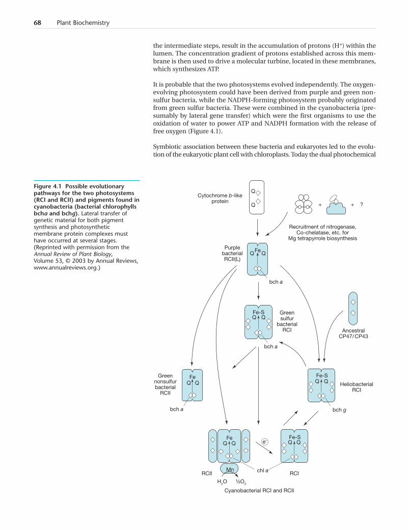
It is probable that the two photosystems evolved independently. The oxygen-evolving photosystem could have been derived from purple and green non-sulfur bacteria, while the NADPH-forming photosystem probably originatedfrom green sulfur bacteria. These were combined in the cyanobacteria (pre-sumably by lateral gene transfer) which were the first organisms to use theoxidation of water to power ATP and NADPH formation with the release offree oxygen (Figure 4.1).
Symbiotic association between these bacteria and eukaryotes led to the evolu-tion of the eukaryotic plant cell with chloroplasts. Today the dual photochemical
68 Plant Biochemistry
+ + ?
Cytochrome b-likeprotein
PurplebacterialRCII(L)
bch a
Recruitment of nitrogenase,Co-chelatase, etc. for
Mg tetrapyrrole biosynthesis
Greensulfur
bacterialRCI Ancestral
CP47/ CP43
HeliobacterialRCI
Greennonsulfurbacterial
RCII
bch gbch a
RCII RCI
e–
bch a
Cyanobacterial RCI and RCII
H2O ½O2
Fe Fe-S
Mn
Fe Fe-S
Fe-S
Fe
Q
Q
Q Q
Q Q
Q Q Q Q
Q Q Q Q
chl a
Figure 4.1 Possible evolutionarypathways for the two photosystems(RCI and RCII) and pigments found incyanobacteria (bacterial chlorophyllsbcha and bchg). Lateral transfer ofgenetic material for both pigmentsynthesis and photosyntheticmembrane protein complexes musthave occurred at several stages.(Reprinted with permission from theAnnual Review of Plant Biology,Volume 53, © 2003 by Annual Reviews,www.annualreviews.org.)
system of terrestrial, aquatic, and marine plants traps a significant propor-tion of the solar energy striking the planet, using it to form stable chemicalproducts that have supported the existence and evolution of most of the lifeforms found on Earth today.
Remarkably, comparison of the structures of the two photosystems fromhigher plants with those from cyanobacteria show that over one thousandmillion years of evolution has resulted in very few changes. Clearly, the sup-posed primitive ancestors of today’s plants had, in fact, evolved a truly effi-cient process that has stood the test of time. This account concentrates on thestructure and function of higher plant photosystems as far as they are knownat present. The major advance that has occurred during evolution is thedevelopment of light harvesting chlorophyll (LHC) proteins that greatlyincrease the area of the receiving antenna for each photosystem. This devel-opment has characterized the few changes that are found in the photosys-tems of modern land plants. Some polypeptides have been lost to make wayfor the effective docking of these new chlorophyll proteins, and others haveevolved to assist the docking process. Across the range of land plants there isa remarkably small level of variation in the structure of their photosystemsand the amino acid sequences of their constituent proteins. In addition tochlorophylls, other pigments, i.e. carotenoids, have been added to these pro-teins, which extend the range of light wavelengths that can be trapped forphotosynthesis. These developments have ensured that plant leaves are ableto intercept and utilize a high proportion of the visible light energy that isincident on the leaf surface.
In summary, photosynthesis starts with two linked photochemical systemsthat trap light energy. Water is split to release oxygen and electrons, which areultimately used to make the reduced form of NADPH. Along the way protons(H+) are accumulated in the grana lumen, creating a charge separation acrossthe photosynthetic membrane that drives the ATP synthesis system.
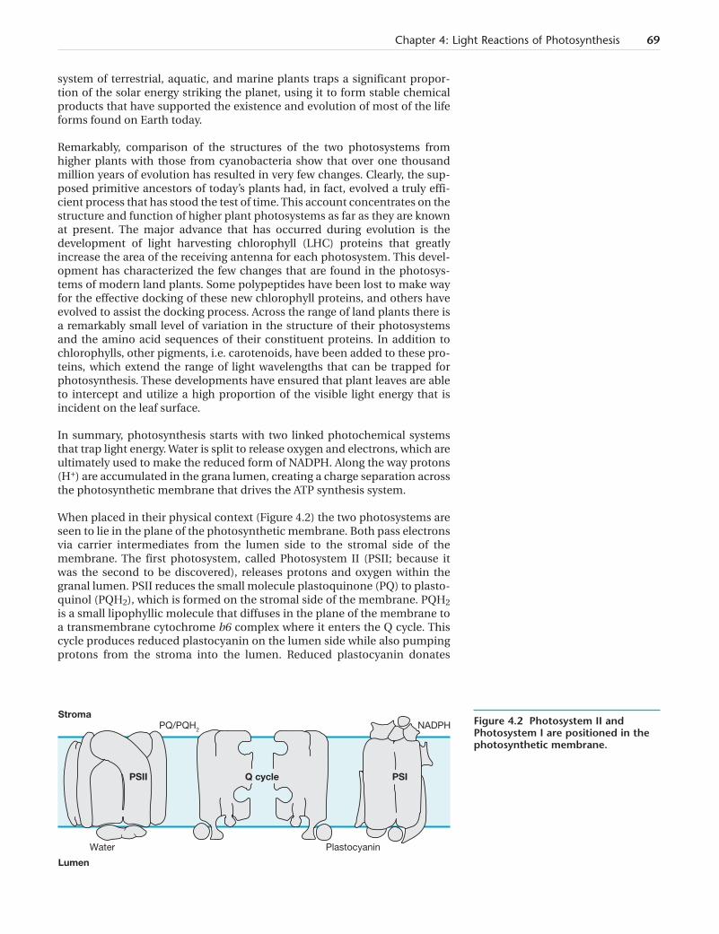
When placed in their physical context (Figure 4.2) the two photosystems areseen to lie in the plane of the photosynthetic membrane. Both pass electronsvia carrier intermediates from the lumen side to the stromal side of themembrane. The first photosystem, called Photosystem II (PSII; because itwas the second to be discovered), releases protons and oxygen within thegranal lumen. PSII reduces the small molecule plastoquinone (PQ) to plasto-quinol (PQH2), which is formed on the stromal side of the membrane. PQH2is a small lipophyllic molecule that diffuses in the plane of the membrane toa transmembrane cytochrome b6 complex where it enters the Q cycle. Thiscycle produces reduced plastocyanin on the lumen side while also pumpingprotons from the stroma into the lumen. Reduced plastocyanin donates
Chapter 4: Light Reactions of Photosynthesis 69
StromaPQ/PQH2
Water Plastocyanin
NADPH
PSII Q cycle PSI
Lumen
Figure 4.2 Photosystem II andPhotosystem I are positioned in thephotosynthetic membrane.
electrons to the second photosystem, Photosystem I (PSI; discovered first),which ultimately leads to the formation of reduced NADPH.
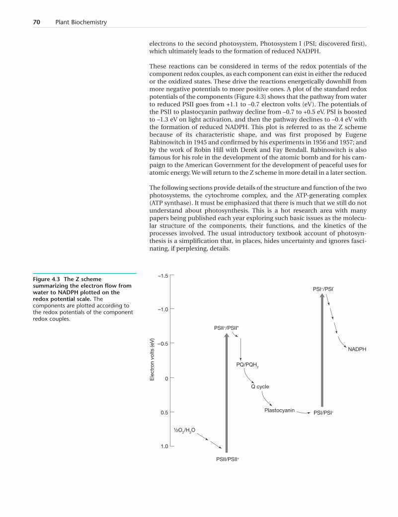
These reactions can be considered in terms of the redox potentials of thecomponent redox couples, as each component can exist in either the reducedor the oxidized states. These drive the reactions energetically downhill frommore negative potentials to more positive ones. A plot of the standard redoxpotentials of the components (Figure 4.3) shows that the pathway from waterto reduced PSII goes from +1.1 to –0.7 electron volts (eV). The potentials ofthe PSII to plastocyanin pathway decline from –0.7 to +0.5 eV. PSI is boostedto –1.3 eV on light activation, and then the pathway declines to –0.4 eV withthe formation of reduced NADPH. This plot is referred to as the Z schemebecause of its characteristic shape, and was first proposed by EugeneRabinowitch in 1945 and confirmed by his experiments in 1956 and 1957; andby the work of Robin Hill with Derek and Fay Bendall. Rabinowitch is alsofamous for his role in the development of the atomic bomb and for his cam-paign to the American Government for the development of peaceful uses foratomic energy. We will return to the Z scheme in more detail in a later section.
The following sections provide details of the structure and function of the twophotosystems, the cytochrome complex, and the ATP-generating complex(ATP synthase). It must be emphasized that there is much that we still do notunderstand about photosynthesis. This is a hot research area with manypapers being published each year exploring such basic issues as the molecu-lar structure of the components, their functions, and the kinetics of theprocesses involved. The usual introductory textbook account of photosyn-thesis is a simplification that, in places, hides uncertainty and ignores fasci-nating, if perplexing, details.
70 Plant Biochemistry
–1.5
–1.0
Ele
ctro
n vo
lts (e
V)
– 0.5
0
0.5
1.0
PQ/PQH2
Q cycle
Plastocyanin PSI/PSI+
PSI+/PSI*
NADPH
½O2/H2O
PSII/PSII+
PSII+/PSII*
Figure 4.3 The Z schemesummarizing the electron flow fromwater to NADPH plotted on theredox potential scale. Thecomponents are plotted according tothe redox potentials of the componentredox couples.
Chlorophyll captures light energy and converts it toa flow of electronsChlorophyll pigments have absorption maxima in the blue and red bands ofthe visible light spectrum and thus reflect and transmit green light.Engelmann and Sachs first discovered the dependence of photosynthesis onchlorophyll and Emerson and Arnold (in 1932) deduced that several hundredmolecules of chlorophyll (a photosynthetic unit) are required for the produc-tion of one molecule of oxygen. Later, 1939, Robin Hill, working with isolatedchloroplasts, demonstrated the direct connection between the light reactionsteps and the release of molecular oxygen. This Hill reaction was extensivelystudied in both isolated chloroplasts and in whole algal cells by Otto Warburg.Eventually (1957) Emerson’s work revealed that two photosystems, bearingchlorophylls of slightly different absorption properties, are involved in thephotosynthetic process.
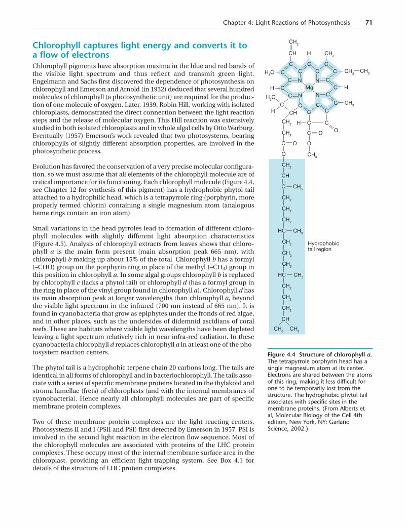
Evolution has favored the conservation of a very precise molecular configura-tion, so we must assume that all elements of the chlorophyll molecule are ofcritical importance for its functioning. Each chlorophyll molecule (Figure 4.4,see Chapter 12 for synthesis of this pigment) has a hydrophobic phytol tailattached to a hydrophilic head, which is a tetrapyrrole ring (porphyrin, moreproperly termed chlorin) containing a single magnesium atom (analogousheme rings contain an iron atom).
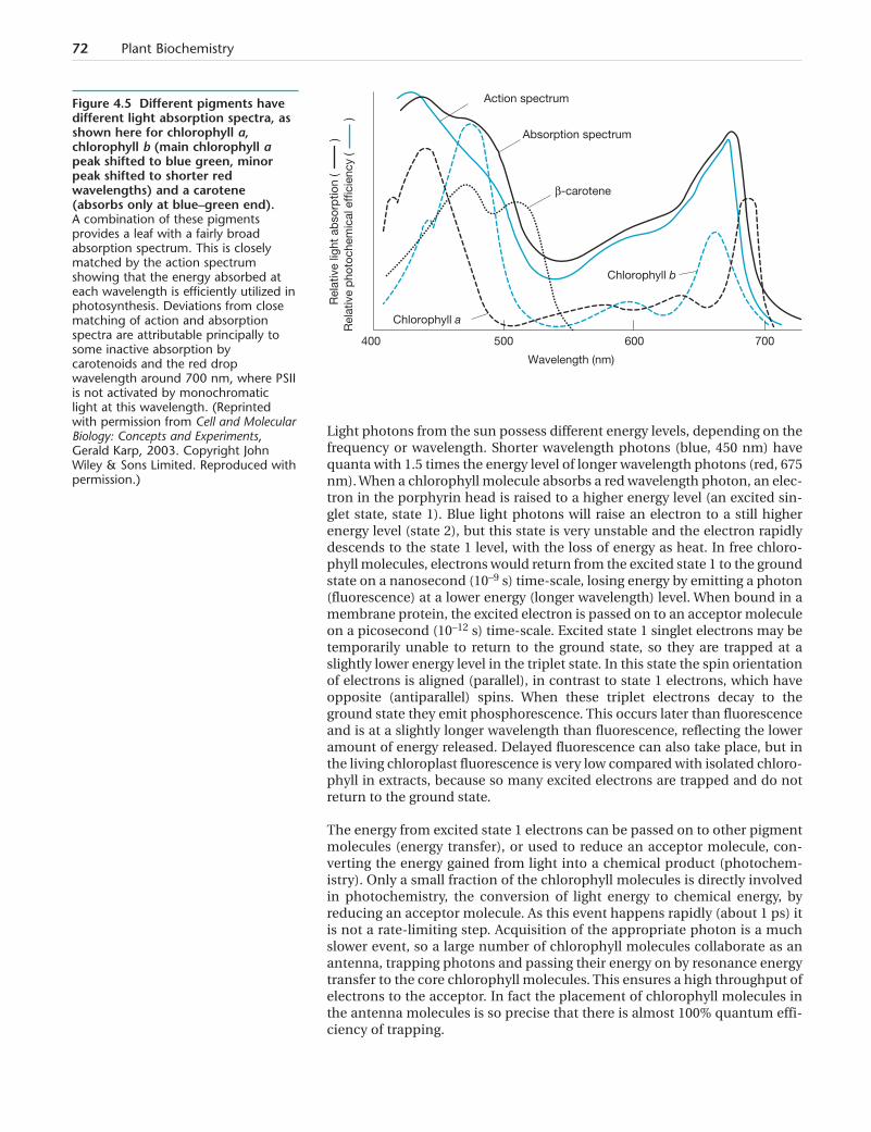
Small variations in the head pyrroles lead to formation of different chloro-phyll molecules with slightly different light absorption characteristics(Figure 4.5). Analysis of chlorophyll extracts from leaves shows that chloro-phyll a is the main form present (main absorption peak 665 nm), withchlorophyll b making up about 15% of the total. Chlorophyll b has a formyl(–CHO) group on the porphyrin ring in place of the methyl (–CH3) group inthis position in chlorophyll a. In some algal groups chlorophyll b is replacedby chlorophyll c (lacks a phytol tail) or chlorophyll d (has a formyl group inthe ring in place of the vinyl group found in chlorophyll a). Chlorophyll d hasits main absorption peak at longer wavelengths than chlorophyll a, beyondthe visible light spectrum in the infrared (700 nm instead of 665 nm). It isfound in cyanobacteria that grow as epiphytes under the fronds of red algae,and in other places, such as the undersides of didemnid ascidians of coralreefs. These are habitats where visible light wavelengths have been depletedleaving a light spectrum relatively rich in near infra-red radiation. In thesecyanobacteria chlorophyll d replaces chlorophyll a in at least one of the pho-tosystem reaction centers.
The phytol tail is a hydrophobic terpene chain 20 carbons long. The tails areidentical in all forms of chlorophyll and in bacteriochlorophyll. The tails asso-ciate with a series of specific membrane proteins located in the thylakoid andstroma lamellae (frets) of chloroplasts (and with the internal membranes ofcyanobacteria). Hence nearly all chlorophyll molecules are part of specificmembrane protein complexes.
Two of these membrane protein complexes are the light reacting centers,Photosystems II and I (PSII and PSI) first detected by Emerson in 1957. PSI isinvolved in the second light reaction in the electron flow sequence. Most ofthe chlorophyll molecules are associated with proteins of the LHC proteincomplexes. These occupy most of the internal membrane surface area in thechloroplast, providing an efficient light-trapping system. See Box 4.1 fordetails of the structure of LHC protein complexes.
Chapter 4: Light Reactions of Photosynthesis 71
CH2
CH H CH3
H3C
CC
CC
CC
C
CH2CH3C
C N N
N NMgH C
C
C CC
C
CC
C
H
H CH
CH3
H3C
CH2
CH2
CH3
CH2
H C C
C
C
CH
O
O O
O O
C
CH3
CH3
CH3
C
HC
CH2
CH2
CH2
CH2
CH2
CH2
CH2
CH2
HC
CH
CH2
CH3 CH3
Hydrophobictail region
Figure 4.4 Structure of chlorophyll a.The tetrapyrrole porphyrin head has asingle magnesium atom at its center.Electrons are shared between the atomsof this ring, making it less difficult forone to be temporarily lost from thestructure. The hydrophobic phytol tailassociates with specific sites in themembrane proteins. (From Alberts etal, Molecular Biology of the Cell 4thedition, New York, NY: GarlandScience, 2002.)
Light photons from the sun possess different energy levels, depending on thefrequency or wavelength. Shorter wavelength photons (blue, 450 nm) havequanta with 1.5 times the energy level of longer wavelength photons (red, 675nm). When a chlorophyll molecule absorbs a red wavelength photon, an elec-tron in the porphyrin head is raised to a higher energy level (an excited sin-glet state, state 1). Blue light photons will raise an electron to a still higherenergy level (state 2), but this state is very unstable and the electron rapidlydescends to the state 1 level, with the loss of energy as heat. In free chloro-phyll molecules, electrons would return from the excited state 1 to the groundstate on a nanosecond (10–9 s) time-scale, losing energy by emitting a photon(fluorescence) at a lower energy (longer wavelength) level. When bound in amembrane protein, the excited electron is passed on to an acceptor moleculeon a picosecond (10–12 s) time-scale. Excited state 1 singlet electrons may betemporarily unable to return to the ground state, so they are trapped at aslightly lower energy level in the triplet state. In this state the spin orientationof electrons is aligned (parallel), in contrast to state 1 electrons, which haveopposite (antiparallel) spins. When these triplet electrons decay to theground state they emit phosphorescence. This occurs later than fluorescenceand is at a slightly longer wavelength than fluorescence, reflecting the loweramount of energy released. Delayed fluorescence can also take place, but inthe living chloroplast fluorescence is very low compared with isolated chloro-phyll in extracts, because so many excited electrons are trapped and do notreturn to the ground state.
The energy from excited state 1 electrons can be passed on to other pigmentmolecules (energy transfer), or used to reduce an acceptor molecule, con-verting the energy gained from light into a chemical product (photochem-istry). Only a small fraction of the chlorophyll molecules is directly involvedin photochemistry, the conversion of light energy to chemical energy, byreducing an acceptor molecule. As this event happens rapidly (about 1 ps) itis not a rate-limiting step. Acquisition of the appropriate photon is a muchslower event, so a large number of chlorophyll molecules collaborate as anantenna, trapping photons and passing their energy on by resonance energytransfer to the core chlorophyll molecules. This ensures a high throughput ofelectrons to the acceptor. In fact the placement of chlorophyll molecules inthe antenna molecules is so precise that there is almost 100% quantum effi-ciency of trapping.
72 Plant Biochemistry
Rel
ativ
e p
hoto
chem
ical
eff
icie
ncy
(
)
Chlorophyll a
Action spectrum
Absorption spectrum
β-carotene
Chlorophyll b
400 500 600 700
Wavelength (nm)
Rel
ativ
e lig
ht a
bso
rptio
n (
)
Figure 4.5 Different pigments havedifferent light absorption spectra, asshown here for chlorophyll a,chlorophyll b (main chlorophyll apeak shifted to blue green, minorpeak shifted to shorter redwavelengths) and a carotene(absorbs only at blue–green end).A combination of these pigmentsprovides a leaf with a fairly broadabsorption spectrum. This is closelymatched by the action spectrumshowing that the energy absorbed ateach wavelength is efficiently utilized inphotosynthesis. Deviations from closematching of action and absorptionspectra are attributable principally tosome inactive absorption bycarotenoids and the red dropwavelength around 700 nm, where PSIIis not activated by monochromaticlight at this wavelength. (Reprintedwith permission from Cell and MolecularBiology: Concepts and Experiments,Gerald Karp, 2003. Copyright JohnWiley & Sons Limited. Reproduced withpermission.)
Chapter 4: Light Reactions of Photosynthesis 73
Most of the chlorophyll molecules in a plant are associatedwith light-harvesting chlorophyll protein complexes(LHC). These are divided into two main classes, those asso-ciated with Photosystem I (PSI), which are known as LHCI,and those predominantly associated with Photosystem II(PSII), termed LHCII. These proteins are very similar toeach other. Both have four helices (A, B, C, and D); with Aand B being closely associated transmembrane helices, Calso lying perpendicular and D lying parallel to the mem-brane. LHCI chlorophyll pigments have absorption max-ima at longer wavelengths than those of LHCII.
LHCI protein molecules contain polypeptides from a fam-ily of four 25 kDa proteins, Lhca 1–4. These bind a total of56 chlorophylls with an a–b ratio of 3.5:1. LHC1 680 con-sists of homodimers of Lhca 2 and 3, while LHC1 730 con-sists of heterodimers of Lhca 1 and 4. Light of up to 750 nmcan be used to oxidize the P700 reaction center. This highconcentration of pigment molecules leads to red-shiftingof their absorption spectra, enabling light harvesting tooccur in dense vegetation canopies where the remaininglight is richer in longer wavelengths (above 680 nm).
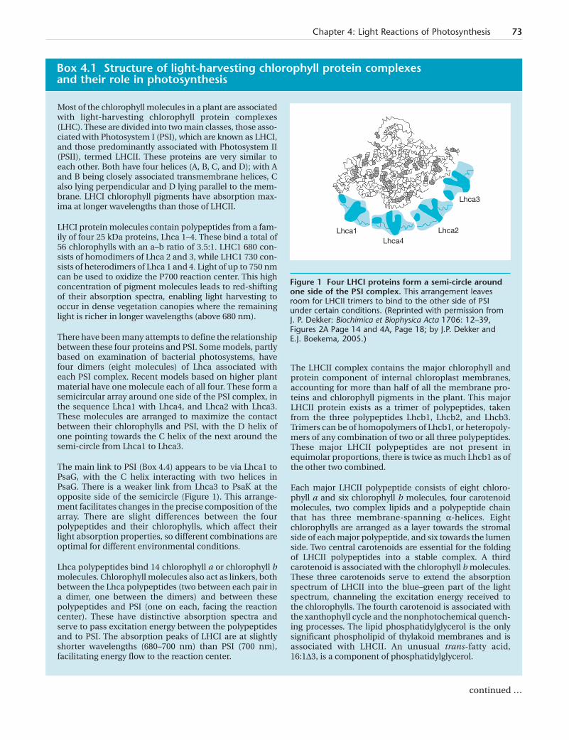
There have been many attempts to define the relationshipbetween these four proteins and PSI. Some models, partlybased on examination of bacterial photosystems, havefour dimers (eight molecules) of Lhca associated witheach PSI complex. Recent models based on higher plantmaterial have one molecule each of all four. These form asemicircular array around one side of the PSI complex, inthe sequence Lhca1 with Lhca4, and Lhca2 with Lhca3.These molecules are arranged to maximize the contactbetween their chlorophylls and PSI, with the D helix ofone pointing towards the C helix of the next around thesemi-circle from Lhca1 to Lhca3.
The main link to PSI (Box 4.4) appears to be via Lhca1 toPsaG, with the C helix interacting with two helices inPsaG. There is a weaker link from Lhca3 to PsaK at theopposite side of the semicircle (Figure 1). This arrange-ment facilitates changes in the precise composition of thearray. There are slight differences between the fourpolypeptides and their chlorophylls, which affect theirlight absorption properties, so different combinations areoptimal for different environmental conditions.
Lhca polypeptides bind 14 chlorophyll a or chlorophyll bmolecules. Chlorophyll molecules also act as linkers, bothbetween the Lhca polypeptides (two between each pair ina dimer, one between the dimers) and between thesepolypeptides and PSI (one on each, facing the reactioncenter). These have distinctive absorption spectra andserve to pass excitation energy between the polypeptidesand to PSI. The absorption peaks of LHCI are at slightlyshorter wavelengths (680–700 nm) than PSI (700 nm),facilitating energy flow to the reaction center.
The LHCII complex contains the major chlorophyll andprotein component of internal chloroplast membranes,accounting for more than half of all the membrane pro-teins and chlorophyll pigments in the plant. This majorLHCII protein exists as a trimer of polypeptides, takenfrom the three polypeptides Lhcb1, Lhcb2, and Lhcb3.Trimers can be of homopolymers of Lhcb1, or heteropoly-mers of any combination of two or all three polypeptides.These major LHCII polypeptides are not present inequimolar proportions, there is twice as much Lhcb1 as ofthe other two combined.
Each major LHCII polypeptide consists of eight chloro-phyll a and six chlorophyll b molecules, four carotenoidmolecules, two complex lipids and a polypeptide chainthat has three membrane-spanning a-helices. Eightchlorophylls are arranged as a layer towards the stromalside of each major polypeptide, and six towards the lumenside. Two central carotenoids are essential for the foldingof LHCII polypeptides into a stable complex. A thirdcarotenoid is associated with the chlorophyll b molecules.These three carotenoids serve to extend the absorptionspectrum of LHCII into the blue–green part of the lightspectrum, channeling the excitation energy received tothe chlorophylls. The fourth carotenoid is associated withthe xanthophyll cycle and the nonphotochemical quench-ing processes. The lipid phosphatidylglycerol is the onlysignificant phospholipid of thylakoid membranes and isassociated with LHCII. An unusual trans-fatty acid,16:1D3, is a component of phosphatidylglycerol.
Box 4.1 Structure of light-harvesting chlorophyll protein complexes and their role in photosynthesis
Lhca1Lhca4
Lhca2
Lhca3
continued …
Figure 1 Four LHCI proteins form a semi-circle aroundone side of the PSI complex. This arrangement leavesroom for LHCII trimers to bind to the other side of PSIunder certain conditions. (Reprinted with permission fromJ. P. Dekker: Biochimica et Biophysica Acta 1706: 12–39,Figures 2A Page 14 and 4A, Page 18; by J.P. Dekker andE.J. Boekema, 2005.)

Carotenoids extend the spectral range of light thatcan be utilized in photosynthesisCarotenoids are long chain pigments that absorb blue and green light (Figure4.5), leaving yellow, orange, and red colors seen in such plant tissues as car-rot roots (for details of carotenoid synthesis see Chapter 12). There are twotypes of carotenoids, carotenes, and xanthophylls. Each is a chain of 40 car-bon atoms with alternating single and double bonds. Carotenes have onlyhydrogen atoms attached to this carbon backbone, but xanthophylls haveone atom of oxygen at each end of the molecule. For example, zeaxanthin isthe same as b carotene but with a hydroxyl group at each end. Carotenoidsare incorporated into the chlorophyll/protein complexes where they performtwo functions. They extend the range of wavelength energies (Figure 4.5) thatcan contribute to photosynthesis by passing on absorbed energy to neighbor-ing chlorophylls. The xanthophyll fucoxanthin in brown seaweeds anddiatoms is especially efficient in harvesting blue and green light and passingenergy to chlorophylls. Also they provide protection for the reaction centers,dissipating excess energized electrons as heat and preventing the formationof damaging reactive oxygen species (see section Nonphotochemicalquenching and the xanthophyll cycle).
74 Plant Biochemistry
In the trimer the three sets of eight chlorophylls on the stro-mal side form two concentric rings. The inner six are chloro-phyll a, acting to transfer energy between monomers, whilethe outer 18 have alternating three chlorophyll a and threechlorophyll b around the circle. These serve to collectenergy from a broad spectrum and pass it on to the P680reaction center. Chlorophylls on the lumen side pass theircollected energy on to these rings on the stromal side.
There are also three minor LHCII proteins, Lhcb4, Lhcb5,and Lhcb6, which do not polymerize with each other, butform bridges between PSII and the major LHCII compo-nents. Lhcb4 and Lhcb6 lie side by side, binding to PSII onone side and to a LHCII trimer on the other (Figure 2).This trimer (M) is less strongly bound to the PSII–LHCIIsupercomplex than the adjacent trimer (S), which bindsdirectly to the PSII surface beside Lhcb4. Lhcb5 is associ-ated with this strongly bound trimer and the PSII surface.Each supercomplex consists of two PSII complexes (thePSII dimer) and four LHCII trimers, two strongly boundand two more weakly bound. Overall analysis of the totalchlorophyll protein complexes shows a ratio of eighttrimers per PSII dimer, so it is assumed that only half thetrimers are bound specifically to the supercomplexes andthe remainder are loosely associated in the surroundingmembrane. As PSII is located in the appressed granalmembranes, it is thought that supercomplexes in adja-cent membranes lie one above the other, permitting exci-tation energy to flow between LHCII proteins on adjacentmembranes of neighboring thylakoids.
Light harvesting by LHCII is assisted by the carotenoids,absorbing at 450–500 nm, passing energy to the chloro-phylls, which absorb at 660–680 nm. This absorption
range is at shorter wavelengths than the PSII reactioncenter (680 nm), so creating a downhill energy flow fromthe LHC to the reaction center (shorter wavelength light ismore energetic).
Box 4.1 Structure of light-harvesting chlorophyll protein complexes and their role in photosynthesis (continued)
L
CP24
M
CP29
S
CP26
X
Figure 2 Major LHCII trimers (M and S) and minorproteins Lhcb4 (CP29), Lhcb5 (CP26), Lhcb6 (CP24)bound to a PSII dimer. The S trimer is strongly bound,while the M trimer shows medium binding. L indicates apossible position for a weakly bound trimer. (Reprinted withpermission from J.P. Dekker: Biochimica et Biophysica Acta1706: 12–39, by J.P. Dekker and E.J. Boekema, 2005.)
Photosystem II splits water to form protons andoxygen, and reduces plastoquinonePSII is located in the membranes of thylakoids making up the granal stacks.It consists of a core dimer of chlorophyll protein molecules that spans themembrane bilayer (Box 4.2). PSII uses light energy to remove electrons fromwater releasing protons and oxygen. The electrons are then used to reducePQ to PQH2. The core chlorophyll a in PSII has a maximum light absorptionpeak at 680 nm, and so is called P680 (pigment 680). This passes an electronon to an acceptor chlorophyll-like molecule (pheophytin, lacks magne-sium), called A0. The first photochemical reaction thus leaves the P680 mol-ecule in an oxidized state (P680+) and the acceptor in a reduced state (A0
–).Two sets of reactions rapidly regenerate the P680/A0 pair in time for the nextlight reaction.
All these electron transfer steps in photosynthesis share a common feature.They involve loss of an electron from one component, leaving it in an oxi-dized state indicated by a plus charge sign; and the gain of the electron byanother component, leaving it in a reduced state, indicated by a minus sign.These components lie close to each other in a chain, and each has a redoxpotential that is slightly higher (more positive) than the previous member ofthe chain. This dictates the direction of flow of the charge along the chain.Typically, chain members are small molecules or atoms of metallic elementsthat can exist in a number of valency states. Iron would be a common exam-ple, existing in either the reduced state Fe2+, or the oxidized state Fe3+. Weshall also meet others, such as copper and manganese. These atoms areheld, either singly or in defined clusters, in an organic matrix created bypolypeptides. Using iron as an example again, it is often held in place bycross-linking to sulfur atoms of cysteine residues as in Rieske proteins(2Fe–2S) and other non-heme iron proteins that carry electrons. Thisarrangement allows the gain or loss of an electron to be shared between thecoordinately linked atoms.
In one set of reactions, P680+ is reduced by the movement of an electron froman adjacent tyrosine molecule (TyrZ) in the polypeptide chain of the D1 pro-tein of the PSII complex (Box 4.2). The oxidized tyrosine is in turn reduced bythe provision of electrons from the oxygen-evolving center.
Electrons are donated from water to replenish those lost by the tyrosine mol-ecule. Two water molecules are split simultaneously to yield one oxygen atom(O2), four protons (H+), and four electrons. However, tyrosine can only acceptone electron at a time. So the oxygen-evolving center has evolved as a chargeaccumulation device, consisting of four manganese atoms held in a proteinmatrix with one atom each of calcium and chlorine (Box 4.2). This centersequentially provides single electrons to the tyrosine molecule, until fourelectrons have been donated, one from each manganese atom. Then twomolecules of water are split simultaneously to replenish the four electronsdonated to tyrosine. The protons are released into the lumen of the thy-lakoid/stroma-lamellar system. Later we will see how these electrons are uti-lized in ATP synthesis (see section ATP synthase utilizes the proton motiveforce to generate ATP).
2H2O –––Æ 4e– + 4H+ + O2
In the dark, the ground state of the oxygen-evolving center is always (+), sothat, on illumination, only three photochemical events are needed to bringabout the evolution of the first oxygen molecule. Thereafter one is formed onevery fourth event. This was first observed by P. Joliot and coworkers in 1969,who illuminated a suspension of algal cells that had been kept in the dark
Chapter 4: Light Reactions of Photosynthesis 75
Author queryDo you need to add anything to linkthis paragraph to the 2nd paragraphon page 78? They are separated bytwo pages of Box 4.2
76 Plant Biochemistry
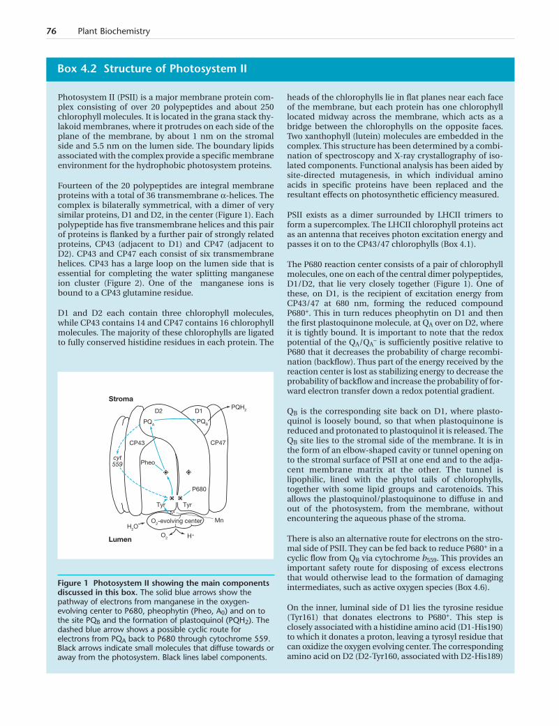
Photosystem II (PSII) is a major membrane protein com-plex consisting of over 20 polypeptides and about 250chlorophyll molecules. It is located in the grana stack thy-lakoid membranes, where it protrudes on each side of theplane of the membrane, by about 1 nm on the stromalside and 5.5 nm on the lumen side. The boundary lipidsassociated with the complex provide a specific membraneenvironment for the hydrophobic photosystem proteins.
Fourteen of the 20 polypeptides are integral membraneproteins with a total of 36 transmembrane a-helices. Thecomplex is bilaterally symmetrical, with a dimer of verysimilar proteins, D1 and D2, in the center (Figure 1). Eachpolypeptide has five transmembrane helices and this pairof proteins is flanked by a further pair of strongly relatedproteins, CP43 (adjacent to D1) and CP47 (adjacent toD2). CP43 and CP47 each consist of six transmembranehelices. CP43 has a large loop on the lumen side that isessential for completing the water splitting manganeseion cluster (Figure 2). One of the manganese ions isbound to a CP43 glutamine residue.
D1 and D2 each contain three chlorophyll molecules,while CP43 contains 14 and CP47 contains 16 chlorophyllmolecules. The majority of these chlorophylls are ligatedto fully conserved histidine residues in each protein. The
heads of the chlorophylls lie in flat planes near each faceof the membrane, but each protein has one chlorophylllocated midway across the membrane, which acts as abridge between the chlorophylls on the opposite faces.Two xanthophyll (lutein) molecules are embedded in thecomplex. This structure has been determined by a combi-nation of spectroscopy and X-ray crystallography of iso-lated components. Functional analysis has been aided bysite-directed mutagenesis, in which individual aminoacids in specific proteins have been replaced and theresultant effects on photosynthetic efficiency measured.
PSII exists as a dimer surrounded by LHCII trimers toform a supercomplex. The LHCII chlorophyll proteins actas an antenna that receives photon excitation energy andpasses it on to the CP43/47 chlorophylls (Box 4.1).
The P680 reaction center consists of a pair of chlorophyllmolecules, one on each of the central dimer polypeptides,D1/D2, that lie very closely together (Figure 1). One ofthese, on D1, is the recipient of excitation energy fromCP43/47 at 680 nm, forming the reduced compoundP680+. This in turn reduces pheophytin on D1 and thenthe first plastoquinone molecule, at QA over on D2, whereit is tightly bound. It is important to note that the redoxpotential of the QA/QA
– is sufficiently positive relative toP680 that it decreases the probability of charge recombi-nation (backflow). Thus part of the energy received by thereaction center is lost as stabilizing energy to decrease theprobability of backflow and increase the probability of for-ward electron transfer down a redox potential gradient.
QB is the corresponding site back on D1, where plasto-quinol is loosely bound, so that when plastoquinone isreduced and protonated to plastoquinol it is released. TheQB site lies to the stromal side of the membrane. It is inthe form of an elbow-shaped cavity or tunnel opening onto the stromal surface of PSII at one end and to the adja-cent membrane matrix at the other. The tunnel islipophilic, lined with the phytol tails of chlorophylls,together with some lipid groups and carotenoids. Thisallows the plastoquinol/plastoquinone to diffuse in andout of the photosystem, from the membrane, withoutencountering the aqueous phase of the stroma.
There is also an alternative route for electrons on the stro-mal side of PSII. They can be fed back to reduce P680+ in acyclic flow from QB via cytochrome b559. This provides animportant safety route for disposing of excess electronsthat would otherwise lead to the formation of damagingintermediates, such as active oxygen species (Box 4.6).
On the inner, luminal side of D1 lies the tyrosine residue(Tyr161) that donates electrons to P680+. This step isclosely associated with a histidine amino acid (D1-His190)to which it donates a proton, leaving a tyrosyl residue thatcan oxidize the oxygen evolving center. The correspondingamino acid on D2 (D2-Tyr160, associated with D2-His189)
Box 4.2 Structure of Photosystem II
cyt559
PQA
PQH2
H2O
H +
Stroma
Lumen
PQB
D2 D1
CP43 CP47
Pheo
P680
Tyr Tyr
MnO2-evolving center
O2
Figure 1 Photosystem II showing the main componentsdiscussed in this box. The solid blue arrows show thepathway of electrons from manganese in the oxygen-evolving center to P680, pheophytin (Pheo, A0) and on tothe site PQB and the formation of plastoquinol (PQH2). Thedashed blue arrow shows a possible cyclic route forelectrons from PQA back to P680 through cytochrome 559.Black arrows indicate small molecules that diffuse towards oraway from the photosystem. Black lines label components.
Chapter 4: Light Reactions of Photosynthesis 77
is oxidized by P680, but is not involved in water oxidation.This oxidation seems to bias electron transfer to the D1side of the reaction center.
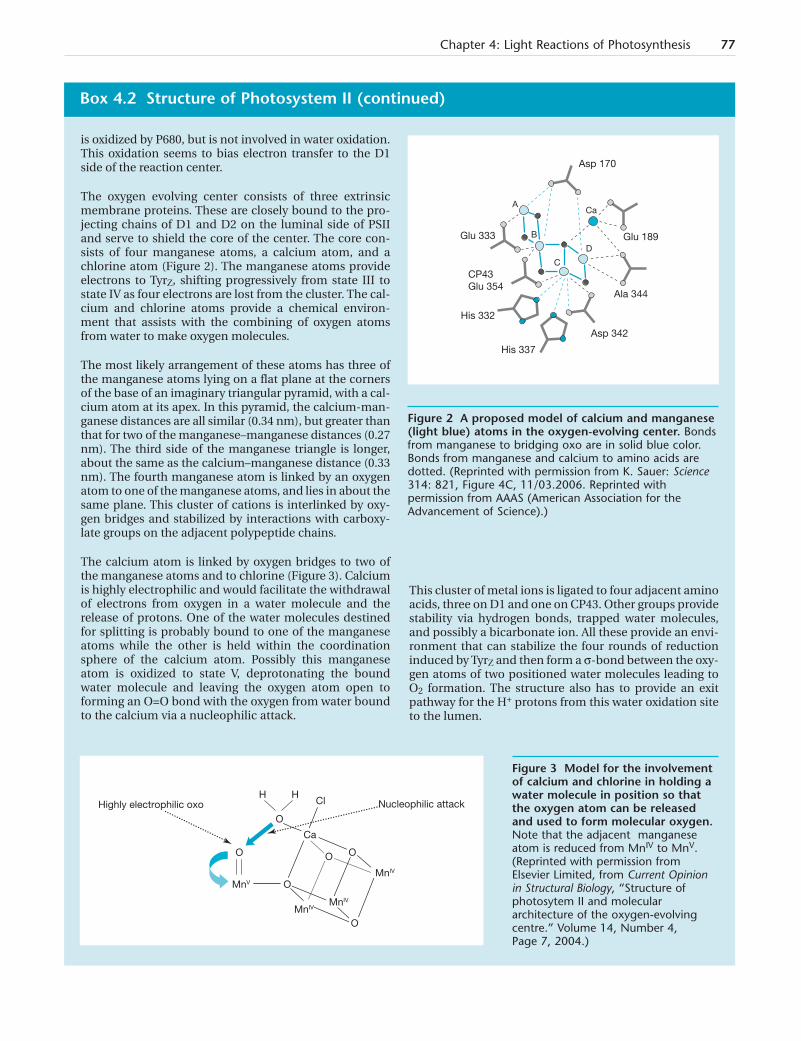
The oxygen evolving center consists of three extrinsicmembrane proteins. These are closely bound to the pro-jecting chains of D1 and D2 on the luminal side of PSIIand serve to shield the core of the center. The core con-sists of four manganese atoms, a calcium atom, and achlorine atom (Figure 2). The manganese atoms provideelectrons to TyrZ, shifting progressively from state III tostate IV as four electrons are lost from the cluster. The cal-cium and chlorine atoms provide a chemical environ-ment that assists with the combining of oxygen atomsfrom water to make oxygen molecules.
The most likely arrangement of these atoms has three ofthe manganese atoms lying on a flat plane at the cornersof the base of an imaginary triangular pyramid, with a cal-cium atom at its apex. In this pyramid, the calcium-man-ganese distances are all similar (0.34 nm), but greater thanthat for two of the manganese–manganese distances (0.27nm). The third side of the manganese triangle is longer,about the same as the calcium–manganese distance (0.33nm). The fourth manganese atom is linked by an oxygenatom to one of the manganese atoms, and lies in about thesame plane. This cluster of cations is interlinked by oxy-gen bridges and stabilized by interactions with carboxy-late groups on the adjacent polypeptide chains.
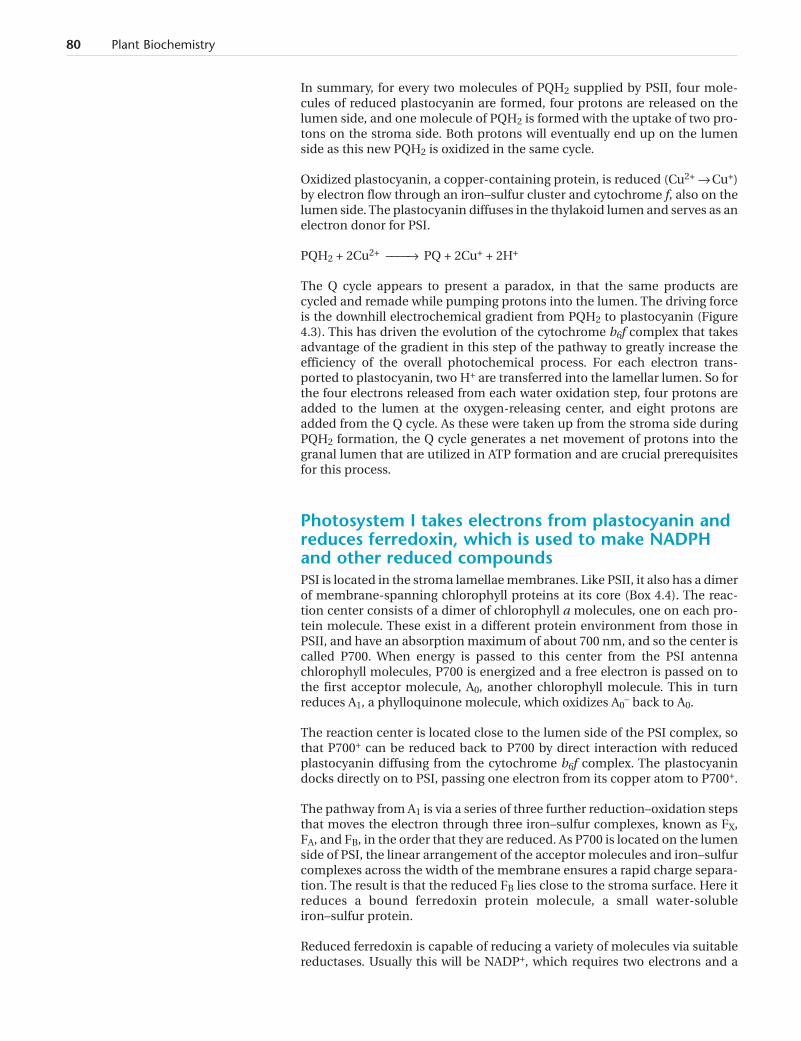
The calcium atom is linked by oxygen bridges to two ofthe manganese atoms and to chlorine (Figure 3). Calciumis highly electrophilic and would facilitate the withdrawalof electrons from oxygen in a water molecule and therelease of protons. One of the water molecules destinedfor splitting is probably bound to one of the manganeseatoms while the other is held within the coordinationsphere of the calcium atom. Possibly this manganeseatom is oxidized to state V, deprotonating the boundwater molecule and leaving the oxygen atom open toforming an O=O bond with the oxygen from water boundto the calcium via a nucleophilic attack.
This cluster of metal ions is ligated to four adjacent aminoacids, three on D1 and one on CP43. Other groups providestability via hydrogen bonds, trapped water molecules,and possibly a bicarbonate ion. All these provide an envi-ronment that can stabilize the four rounds of reductioninduced by TyrZ and then form a s-bond between the oxy-gen atoms of two positioned water molecules leading toO2 formation. The structure also has to provide an exitpathway for the H+ protons from this water oxidation siteto the lumen.
Box 4.2 Structure of Photosystem II (continued)
Highly electrophilic oxo Nucleophilic attackH H
Cl
O
O
MnV
Ca
O
MnIVMnIV
O
MnIV
OO
Glu 333
CP43Glu 354
His 332
His 337
Asp 342
Ala 344
Glu 189
Asp 170
A
B
C
D
Ca
Figure 2 A proposed model of calcium and manganese(light blue) atoms in the oxygen-evolving center. Bondsfrom manganese to bridging oxo are in solid blue color.Bonds from manganese and calcium to amino acids aredotted. (Reprinted with permission from K. Sauer: Science314: 821, Figure 4C, 11/03.2006. Reprinted withpermission from AAAS (American Association for theAdvancement of Science).)
Figure 3 Model for the involvementof calcium and chlorine in holding awater molecule in position so thatthe oxygen atom can be releasedand used to form molecular oxygen.Note that the adjacent manganeseatom is reduced from MnIV to MnV.(Reprinted with permission fromElsevier Limited, from Current Opinionin Structural Biology, “Structure ofphotosytem II and moleculararchitecture of the oxygen-evolvingcentre.” Volume 14, Number 4,Page 7, 2004.)

with 20 ms flashes of light with 0.3-s dark intervals. This was interpreted byB Kok and coworkers (1970) as a cycle of successive oxidations, called theS-cycle. The ground (dark) state is S1, and the center passes to S2, S3, andfinally S4 states when two water molecules are split (Box 4.2).
In the other set of reactions, A0– pheophytin is rapidly (picoseconds) oxidized
to A0 by passing an electron on to the first of two PQ molecules (at site QA, Box4.2) and then, via an iron atom to the next PQ at site QB. PQ requires two elec-trons to become fully reduced. At QA, PQ is tightly bound and only undergoessingle reduction events to the semiquinone state before being reoxidized bythe PQ at QB. Each QB molecule undergoes two successive reduction events tobecome fully reduced. It then takes up two protons from the stromal side ofthe membrane to form PQH2, which is released from the site and leaves thePSII complex. It is replaced by a new PQ molecule binding to the QB site. ThePQH2 diffuses in the lipid bilayer, acting as a mobile carrier of hydrogen atoms.
PQ + 2e– + 2H+ ––––Æ PQH2
The P680+/A0– pair regeneration steps are relatively slow (millisecond time-
scale), so that a second light reaction cannot effectively occur inside 1 ms.However, the first fairly stable end product, PQ at QA, is formed in less than ananosecond, so the electron flow from the excited state of P680 is maintained.
The Q cycle uses plastoquinol to reduce plastocyaninand transport protons into the lumenThe PQH2 formed by PSII is the substrate for the Q cycle on the integral trans-membrane protein complex, cytochrome b6f. The cycle regenerates PQ for PSII,reduces a protein electron carrier, plastocyanin, and pumps more protons fromthe stroma into the lumen of the grana and stroma membrane systems.
As each plastocyanin reduction requires only a single electron, the Q cycleensures that removal of two electrons from PQH2 is achieved safely in twosteps, with one electron going to reduce plastocyanin and the other beingrecycled to reduce a further PQ molecule (Box 4.3).
The cytochrome b6f complex contains two cytochromes, cytochrome b6 andcytochrome f, linked by an iron–sulfur Rieske protein. There are two Q(quinol) binding sites on cytochrome b6. Electrons taken from PQH2 at thebinding site Qp (also called Qo) on the luminal side of the cytochrome aremoved via two iron heme groups to reduce PQ at the other binding site Qn(also called Qi) on the stromal side, to form more PQH2.
PQH2 is oxidized in two steps to PQ (Box 4.3, Figure 1) at the Qp site. The firststep forms plastosemiquinone, with the release of an electron that reducesplastocyanin.
In the second step, plastosemiquinone is oxidized to PQ with the release of anelectron that is passed via a heme molecule to the first step of reducing a fur-ther PQ molecule at the Qn site. Another PQH2 molecule is oxidized in thesame two steps at the Qp site. This forms a further reduced plastocyanin mol-ecule and completes the reduction of plastosemiquinone to PQH2 at the Qnsite, which is fed back into the cycle.
Proton pumping is a very important feature of the Q cycle. Oxidation of thePQH2 releases protons taken up from the stroma, during PQH2 formation onPSII, into the thylakoid lumen. PQH2 synthesis at Qn also takes up protons
78 Plant Biochemistry
Author queryDo you need to add anything to linkthis paragraph to the 3rd paragraphon page 75? They are separated bytwo pages of Box 4.2
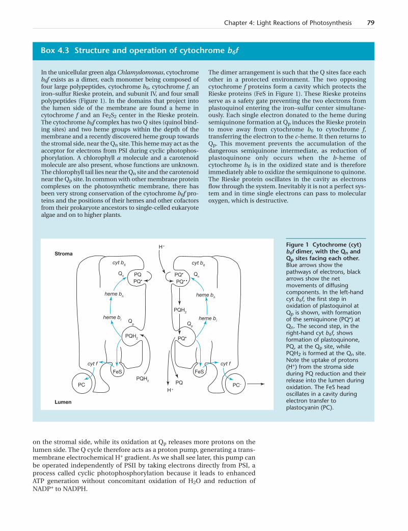
on the stromal side, while its oxidation at Qp releases more protons on thelumen side. The Q cycle therefore acts as a proton pump, generating a trans-membrane electrochemical H+ gradient. As we shall see later, this pump canbe operated independently of PSII by taking electrons directly from PSI, aprocess called cyclic photophosphorylation because it leads to enhancedATP generation without concomitant oxidation of H2O and reduction ofNADP+ to NADPH.
Chapter 4: Light Reactions of Photosynthesis 79
In the unicellular green alga Chlamydomonas, cytochromeb6f exists as a dimer, each monomer being composed offour large polypeptides, cytochrome b6, cytochrome f, aniron–sulfur Rieske protein, and subunit IV, and four smallpolypeptides (Figure 1). In the domains that project intothe lumen side of the membrane are found a heme incytochrome f and an Fe2S2 center in the Rieske protein.The cytochrome b6f complex has two Q sites (quinol bind-ing sites) and two heme groups within the depth of themembrane and a recently discovered heme group towardsthe stromal side, near the Qn site. This heme may act as theacceptor for electrons from PSI during cyclic photophos-phorylation. A chlorophyll a molecule and a carotenoidmolecule are also present, whose functions are unknown.The chlorophyll tail lies near the Qn site and the carotenoidnear the Qp site. In common with other membrane proteincomplexes on the photosynthetic membrane, there hasbeen very strong conservation of the cytochrome b6f pro-teins and the positions of their hemes and other cofactorsfrom their prokaryote ancestors to single-celled eukaryotealgae and on to higher plants.
The dimer arrangement is such that the Q sites face eachother in a protected environment. The two opposingcytochrome f proteins form a cavity which protects theRieske proteins (FeS in Figure 1). These Rieske proteinsserve as a safety gate preventing the two electrons fromplastoquinol entering the iron–sulfur center simultane-ously. Each single electron donated to the heme duringsemiquinone formation at Qp induces the Rieske proteinto move away from cytochrome b6 to cytochrome f,transferring the electron to the c-heme. It then returns toQp. This movement prevents the accumulation of thedangerous semiquinone intermediate, as reduction ofplastoquinone only occurs when the b-heme ofcytochrome b6 is in the oxidized state and is thereforeimmediately able to oxidize the semiquinone to quinone.The Rieske protein oscillates in the cavity as electronsflow through the system. Inevitably it is not a perfect sys-tem and in time single electrons can pass to molecularoxygen, which is destructive.
Box 4.3 Structure and operation of cytochrome b6f
H +
cyt b6 cyt b6
Qn PQ PQ•
PQ• PQ• •
heme bh heme bh
PQH2
Qp Qp
PQ•PQH2
cyt f
Qn
PQH2
FeS
PC
FeS
PQ
cyt f
H +PC–
Stroma
Lumen
heme bl heme bl
Figure 1 Cytochrome (cyt)b6f dimer, with the Qn andQp sites facing each other.Blue arrows show thepathways of electrons, blackarrows show the netmovements of diffusingcomponents. In the left-handcyt b6f, the first step inoxidation of plastoquinol atQp is shown, with formationof the semiquinone (PQ•) atQn. The second step, in theright-hand cyt b6f, showsformation of plastoquinone,PQ, at the Qp site, whilePQH2 is formed at the Qn site.Note the uptake of protons(H+) from the stroma sideduring PQ reduction and theirrelease into the lumen duringoxidation. The FeS headoscillates in a cavity duringelectron transfer toplastocyanin (PC).
In summary, for every two molecules of PQH2 supplied by PSII, four mole-cules of reduced plastocyanin are formed, four protons are released on thelumen side, and one molecule of PQH2 is formed with the uptake of two pro-tons on the stroma side. Both protons will eventually end up on the lumenside as this new PQH2 is oxidized in the same cycle.
Oxidized plastocyanin, a copper-containing protein, is reduced (Cu2+ Æ Cu+)by electron flow through an iron–sulfur cluster and cytochrome f, also on thelumen side. The plastocyanin diffuses in the thylakoid lumen and serves as anelectron donor for PSI.
PQH2 + 2Cu2+ ––––Æ PQ + 2Cu+ + 2H+
The Q cycle appears to present a paradox, in that the same products arecycled and remade while pumping protons into the lumen. The driving forceis the downhill electrochemical gradient from PQH2 to plastocyanin (Figure4.3). This has driven the evolution of the cytochrome b6f complex that takesadvantage of the gradient in this step of the pathway to greatly increase theefficiency of the overall photochemical process. For each electron trans-ported to plastocyanin, two H+ are transferred into the lamellar lumen. So forthe four electrons released from each water oxidation step, four protons areadded to the lumen at the oxygen-releasing center, and eight protons areadded from the Q cycle. As these were taken up from the stroma side duringPQH2 formation, the Q cycle generates a net movement of protons into thegranal lumen that are utilized in ATP formation and are crucial prerequisitesfor this process.
Photosystem I takes electrons from plastocyanin andreduces ferredoxin, which is used to make NADPHand other reduced compoundsPSI is located in the stroma lamellae membranes. Like PSII, it also has a dimerof membrane-spanning chlorophyll proteins at its core (Box 4.4). The reac-tion center consists of a dimer of chlorophyll a molecules, one on each pro-tein molecule. These exist in a different protein environment from those inPSII, and have an absorption maximum of about 700 nm, and so the center iscalled P700. When energy is passed to this center from the PSI antennachlorophyll molecules, P700 is energized and a free electron is passed on tothe first acceptor molecule, A0, another chlorophyll molecule. This in turnreduces A1, a phylloquinone molecule, which oxidizes A0
– back to A0.
The reaction center is located close to the lumen side of the PSI complex, sothat P700+ can be reduced back to P700 by direct interaction with reducedplastocyanin diffusing from the cytochrome b6f complex. The plastocyanindocks directly on to PSI, passing one electron from its copper atom to P700+.
The pathway from A1 is via a series of three further reduction–oxidation stepsthat moves the electron through three iron–sulfur complexes, known as FX,FA, and FB, in the order that they are reduced. As P700 is located on the lumenside of PSI, the linear arrangement of the acceptor molecules and iron–sulfurcomplexes across the width of the membrane ensures a rapid charge separa-tion. The result is that the reduced FB lies close to the stroma surface. Here itreduces a bound ferredoxin protein molecule, a small water-solubleiron–sulfur protein.
Reduced ferredoxin is capable of reducing a variety of molecules via suitablereductases. Usually this will be NADP+, which requires two electrons and a
80 Plant Biochemistry
Chapter 4: Light Reactions of Photosynthesis 81
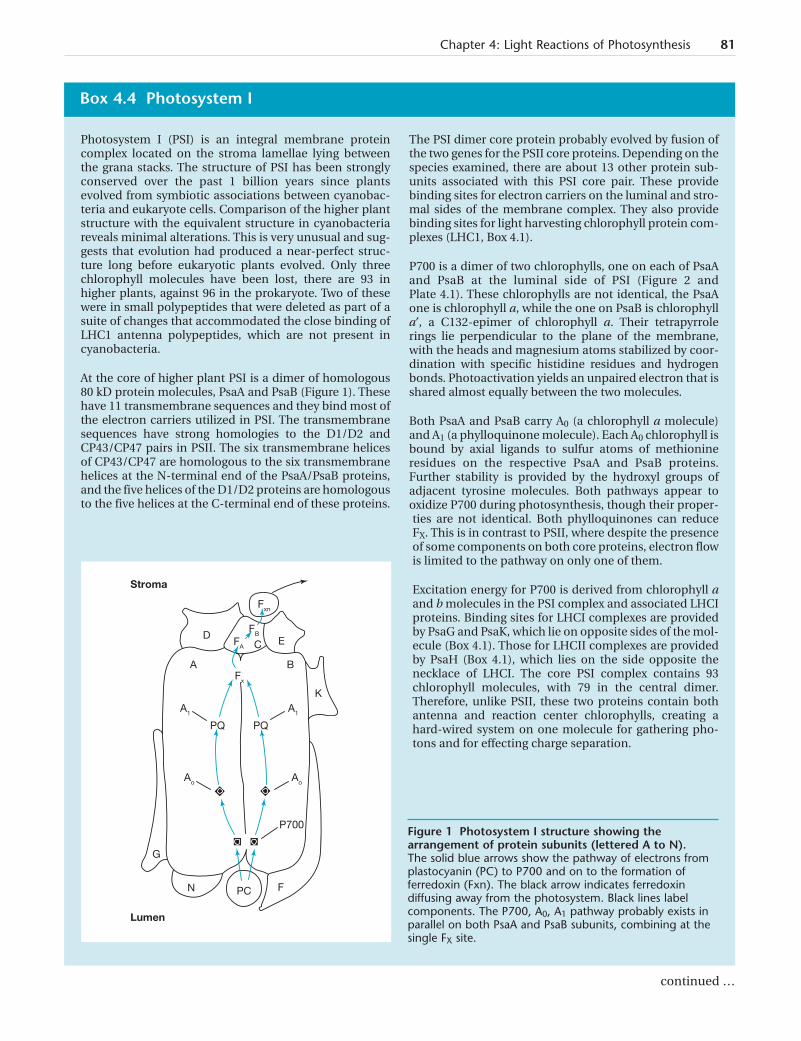
Photosystem I (PSI) is an integral membrane proteincomplex located on the stroma lamellae lying betweenthe grana stacks. The structure of PSI has been stronglyconserved over the past 1 billion years since plantsevolved from symbiotic associations between cyanobac-teria and eukaryote cells. Comparison of the higher plantstructure with the equivalent structure in cyanobacteriareveals minimal alterations. This is very unusual and sug-gests that evolution had produced a near-perfect struc-ture long before eukaryotic plants evolved. Only threechlorophyll molecules have been lost, there are 93 inhigher plants, against 96 in the prokaryote. Two of thesewere in small polypeptides that were deleted as part of asuite of changes that accommodated the close binding ofLHC1 antenna polypeptides, which are not present incyanobacteria.
At the core of higher plant PSI is a dimer of homologous80 kD protein molecules, PsaA and PsaB (Figure 1). Thesehave 11 transmembrane sequences and they bind most ofthe electron carriers utilized in PSI. The transmembranesequences have strong homologies to the D1/D2 andCP43/CP47 pairs in PSII. The six transmembrane helicesof CP43/CP47 are homologous to the six transmembranehelices at the N-terminal end of the PsaA/PsaB proteins,and the five helices of the D1/D2 proteins are homologousto the five helices at the C-terminal end of these proteins.
The PSI dimer core protein probably evolved by fusion ofthe two genes for the PSII core proteins. Depending on thespecies examined, there are about 13 other protein sub-units associated with this PSI core pair. These providebinding sites for electron carriers on the luminal and stro-mal sides of the membrane complex. They also providebinding sites for light harvesting chlorophyll protein com-plexes (LHC1, Box 4.1).
P700 is a dimer of two chlorophylls, one on each of PsaAand PsaB at the luminal side of PSI (Figure 2 andPlate 4.1). These chlorophylls are not identical, the PsaAone is chlorophyll a, while the one on PsaB is chlorophylla¢, a C132-epimer of chlorophyll a. Their tetrapyrrolerings lie perpendicular to the plane of the membrane,with the heads and magnesium atoms stabilized by coor-dination with specific histidine residues and hydrogenbonds. Photoactivation yields an unpaired electron that isshared almost equally between the two molecules.
Both PsaA and PsaB carry A0 (a chlorophyll a molecule)and A1 (a phylloquinone molecule). Each A0 chlorophyll isbound by axial ligands to sulfur atoms of methionineresidues on the respective PsaA and PsaB proteins.Further stability is provided by the hydroxyl groups ofadjacent tyrosine molecules. Both pathways appear tooxidize P700 during photosynthesis, though their proper-ties are not identical. Both phylloquinones can reduceFX. This is in contrast to PSII, where despite the presenceof some components on both core proteins, electron flowis limited to the pathway on only one of them.
Excitation energy for P700 is derived from chlorophyll aand b molecules in the PSI complex and associated LHCIproteins. Binding sites for LHCI complexes are providedby PsaG and PsaK, which lie on opposite sides of the mol-ecule (Box 4.1). Those for LHCII complexes are providedby PsaH (Box 4.1), which lies on the side opposite thenecklace of LHCI. The core PSI complex contains 93chlorophyll molecules, with 79 in the central dimer.Therefore, unlike PSII, these two proteins contain bothantenna and reaction center chlorophylls, creating ahard-wired system on one molecule for gathering pho-tons and for effecting charge separation.
Box 4.4 Photosystem I
Stroma
Lumen
FA
A
FB
Fxn
ED
B
K
G
Fx
A1
C
PQ PQ
Ao
A1
Ao
P700
N PC F
Figure 1 Photosystem I structure showing thearrangement of protein subunits (lettered A to N). The solid blue arrows show the pathway of electrons fromplastocyanin (PC) to P700 and on to the formation offerredoxin (Fxn). The black arrow indicates ferredoxindiffusing away from the photosystem. Black lines labelcomponents. The P700, A0, A1 pathway probably exists inparallel on both PsaA and PsaB subunits, combining at thesingle FX site.
continued …
proton to yield NADPH, a process first discovered by Daniel Arnon in 1962.Arnon had previously (1951) noted that isolated chloroplasts could reduceNADP to NADPH and later (1954) found that ATP is produced at the sametime. The reduction of NADP by reduced ferredoxin is mediated by ferre-doxin-NADP+ reductase, a flavoprotein enzyme.
2Fdreduced + H+ + NADP+ ––––Æ 2Fdoxidized + NADPH
Note that the proton is removed from the stromal side of the membrane,enhancing the transmembrane proton gradient. Two electrons are neededfor each NADPH molecule formed, so the splitting of two molecules of waterat PSII provides enough electrons to form two molecules of NADPH. Thisstoichiometry is not exact, because other molecules can be reduced byferredoxin, as it is not a closed system. The ferredoxin can diffuse to otherreductases and reduce other molecules in the appropriate redox potentialrange. These include nitrite, which is used to produce amino acids (Chapter8), and sulfate, which is reduced to sulfydryl in cysteine (Chapter 8). It canalso donate electrons to the Q cycle (see section above, The Q cycle usesplastoquinol to reduce plastocyanin and transport protons into the lumen).
Thus P700 is capable of pumping electrons from plastocyanin to achievereduction of NADP+ (Figure 4.6). The path from A0 even involves a slightuphill segment from FA to FB, though this is overcome in the overall gradient(Figure 4.7).
82 Plant Biochemistry
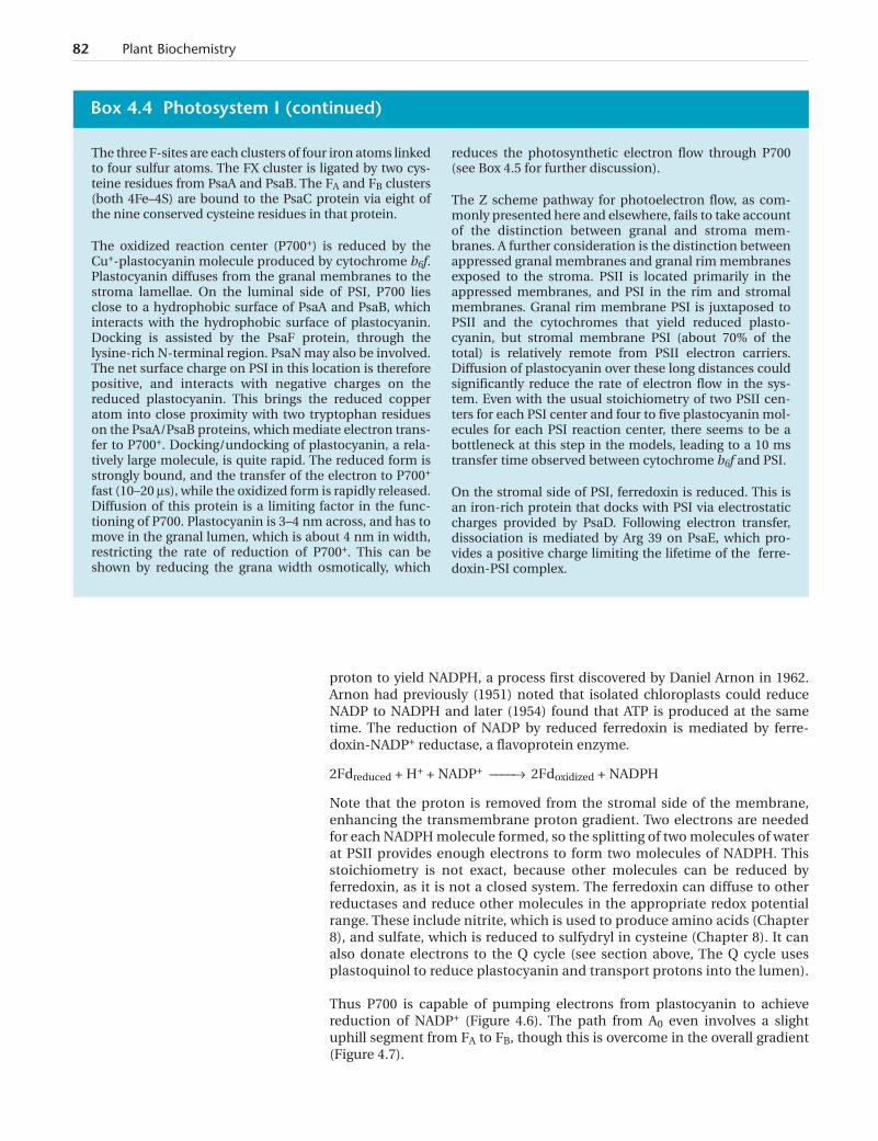
The three F-sites are each clusters of four iron atoms linkedto four sulfur atoms. The FX cluster is ligated by two cys-teine residues from PsaA and PsaB. The FA and FB clusters(both 4Fe–4S) are bound to the PsaC protein via eight ofthe nine conserved cysteine residues in that protein.
The oxidized reaction center (P700+) is reduced by theCu+-plastocyanin molecule produced by cytochrome b6f.Plastocyanin diffuses from the granal membranes to thestroma lamellae. On the luminal side of PSI, P700 liesclose to a hydrophobic surface of PsaA and PsaB, whichinteracts with the hydrophobic surface of plastocyanin.Docking is assisted by the PsaF protein, through thelysine-rich N-terminal region. PsaN may also be involved.The net surface charge on PSI in this location is thereforepositive, and interacts with negative charges on thereduced plastocyanin. This brings the reduced copperatom into close proximity with two tryptophan residueson the PsaA/PsaB proteins, which mediate electron trans-fer to P700+. Docking/undocking of plastocyanin, a rela-tively large molecule, is quite rapid. The reduced form isstrongly bound, and the transfer of the electron to P700+
fast (10–20 ms), while the oxidized form is rapidly released.Diffusion of this protein is a limiting factor in the func-tioning of P700. Plastocyanin is 3–4 nm across, and has tomove in the granal lumen, which is about 4 nm in width,restricting the rate of reduction of P700+. This can beshown by reducing the grana width osmotically, which
reduces the photosynthetic electron flow through P700(see Box 4.5 for further discussion).
The Z scheme pathway for photoelectron flow, as com-monly presented here and elsewhere, fails to take accountof the distinction between granal and stroma mem-branes. A further consideration is the distinction betweenappressed granal membranes and granal rim membranesexposed to the stroma. PSII is located primarily in theappressed membranes, and PSI in the rim and stromalmembranes. Granal rim membrane PSI is juxtaposed toPSII and the cytochromes that yield reduced plasto-cyanin, but stromal membrane PSI (about 70% of thetotal) is relatively remote from PSII electron carriers.Diffusion of plastocyanin over these long distances couldsignificantly reduce the rate of electron flow in the sys-tem. Even with the usual stoichiometry of two PSII cen-ters for each PSI center and four to five plastocyanin mol-ecules for each PSI reaction center, there seems to be abottleneck at this step in the models, leading to a 10 mstransfer time observed between cytochrome b6f and PSI.
On the stromal side of PSI, ferredoxin is reduced. This isan iron-rich protein that docks with PSI via electrostaticcharges provided by PsaD. Following electron transfer,dissociation is mediated by Arg 39 on PsaE, which pro-vides a positive charge limiting the lifetime of the ferre-doxin-PSI complex.
Box 4.4 Photosystem I (continued)
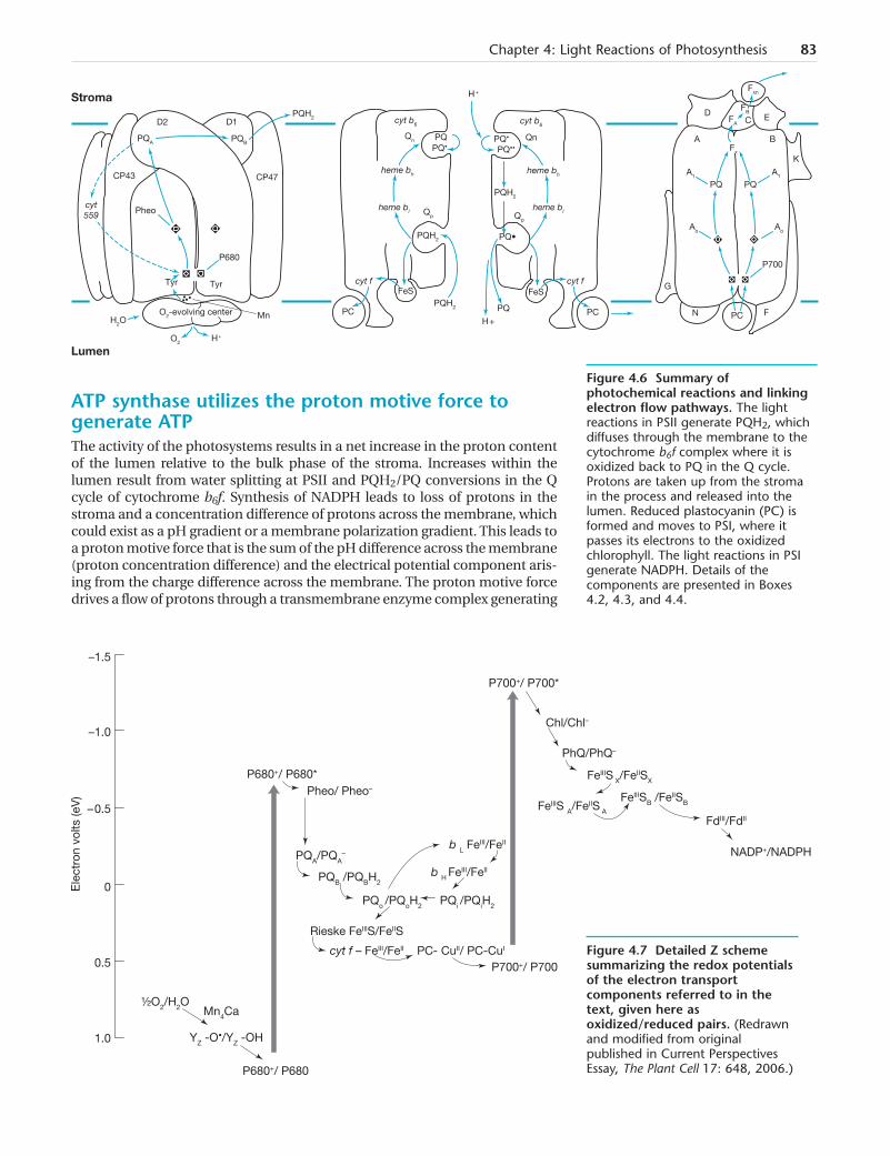
ATP synthase utilizes the proton motive force togenerate ATPThe activity of the photosystems results in a net increase in the proton contentof the lumen relative to the bulk phase of the stroma. Increases within thelumen result from water splitting at PSII and PQH2/PQ conversions in the Qcycle of cytochrome b6f. Synthesis of NADPH leads to loss of protons in thestroma and a concentration difference of protons across the membrane, whichcould exist as a pH gradient or a membrane polarization gradient. This leads toa proton motive force that is the sum of the pH difference across the membrane(proton concentration difference) and the electrical potential component aris-ing from the charge difference across the membrane. The proton motive forcedrives a flow of protons through a transmembrane enzyme complex generating
Chapter 4: Light Reactions of Photosynthesis 83
–1.5
–1.0
Ele
ctro
n vo
lts (e
V)
– 0.5
0
0.5
1.0
P680+/ P680*Pheo/ Pheo–
PQA/PQA–
PQB /PQBH2
PQo /PQoH2 PQi /PQiH2
b L FeIII/FeII
b H FeIII/FeII
Rieske FeIIIS/FeIIS
cyt f – FeIII/FeII
P700+/ P700
P700+/ P700*
P680+/ P680
PC- CuII/ PC-CuI
FeIIIS A/FeIIS A
FeIIIS X/FeIISX
FeIIISB /FeIISB
FdIII/FdII
NADP+/NADPH
Chl/Chl–
PhQ/PhQ–
½O2/H2O Mn4Ca
YZ -O•/YZ -OH
Figure 4.7 Detailed Z schemesummarizing the redox potentialsof the electron transportcomponents referred to in thetext, given here asoxidized/reduced pairs. (Redrawnand modified from originalpublished in Current PerspectivesEssay, The Plant Cell 17: 648, 2006.)
Stroma
Lumen
cyt559
PQA
PQH2
H2O
H +
PQB
D2 D1
CP43 CP47
Pheo
P680
Tyr Tyr
MnO2-evolving center
O2
FA
A
FB
Fxn
ED
B
K
G
Fx
A1
C
PQ PQ
Ao
A1
Ao
P700
N PC F
H +
cyt b6 cyt b6
Qn PQ PQ•
PQ• PQ••
heme bh heme bh
PQH2
Qp
PQ•PQH2
cyt f
Qn
PQH2
FeS
PC
FeS
PQ
cyt f
H +PC
heme bl heme blQp
Figure 4.6 Summary ofphotochemical reactions and linkingelectron flow pathways. The lightreactions in PSII generate PQH2, whichdiffuses through the membrane to thecytochrome b6f complex where it isoxidized back to PQ in the Q cycle.Protons are taken up from the stromain the process and released into thelumen. Reduced plastocyanin (PC) isformed and moves to PSI, where itpasses its electrons to the oxidizedchlorophyll. The light reactions in PSIgenerate NADPH. Details of thecomponents are presented in Boxes4.2, 4.3, and 4.4.
84 Plant Biochemistry
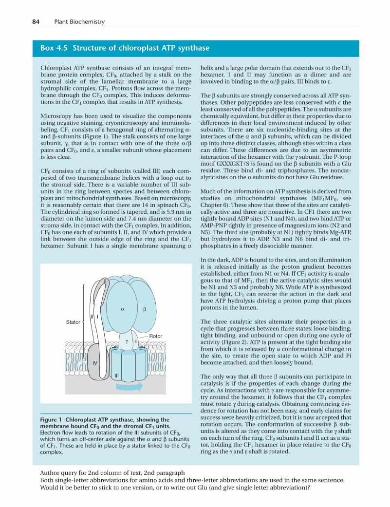
Chloroplast ATP synthase consists of an integral mem-brane protein complex, CF0, attached by a stalk on thestromal side of the lamellar membrane to a largehydrophilic complex, CF1. Protons flow across the mem-brane through the CF0 complex. This induces deforma-tions in the CF1 complex that results in ATP synthesis.
Microscopy has been used to visualize the componentsusing negative staining, cryomicroscopy and immunola-beling. CF1 consists of a hexagonal ring of alternating a-and b-subunits (Figure 1). The stalk consists of one largesubunit, g, that is in contact with one of the three a/bpairs and CF0, and e, a smaller subunit whose placementis less clear.
CF0 consists of a ring of subunits (called III) each com-posed of two transmembrane helices with a loop out tothe stromal side. There is a variable number of III sub-units in the ring between species and between chloro-plast and mitochondrial synthases. Based on microscopy,it is reasonably certain that there are 14 in spinach CF0.The cylindrical ring so formed is tapered, and is 5.9 nm indiameter on the lumen side and 7.4 nm diameter on thestroma side, in contact with the CF1 complex. In addition,CF0 has one each of subunits I, II, and IV which provide alink between the outside edge of the ring and the CF1hexamer. Subunit I has a single membrane spanning a
helix and a large polar domain that extends out to the CF1hexamer. I and II may function as a dimer and areinvolved in binding to the a/b pairs, III binds to e.
The b subunits are strongly conserved across all ATP syn-thases. Other polypeptides are less conserved with e theleast conserved of all the polypeptides. The a subunits arechemically equivalent, but differ in their properties due todifferences in their local environment induced by othersubunits. There are six nucleotide-binding sites at theinterfaces of the a and b subunits, which can be dividedup into three distinct classes, although sites within a classcan differ. These differences are due to an asymmetricinteraction of the hexamer with the g subunit. The P-loopmotif GXXXGKT/S is found on the b subunits with a Gluresidue. These bind di- and triphosphates. The noncat-alytic sites on the a subunits do not have Glu residues.
Much of the information on ATP synthesis is derived fromstudies on mitochondrial synthases (MF1MF0, seeChapter 6). These show that three of the sites are catalyti-cally active and three are nonactive. In CF1 there are twotightly bound ADP sites (N1 and N4), and two bind ATP orAMP-PNP tightly in presence of magnesium ions (N2 andN5). The third site (probably at N1) tightly binds Mg-ATP,but hydrolyzes it to ADP. N3 and N6 bind di- and tri-phosphates in a freely dissociable manner.
In the dark, ADP is bound to the sites, and on illuminationit is released initially as the proton gradient becomesestablished, either from N1 or N4. If CF1 activity is analo-gous to that of MF1, then the active catalytic sites wouldbe N1 and N3 and probably N6. While ATP is synthesizedin the light, CF1 can reverse the action in the dark andhave ATP hydrolysis driving a proton pump that placesprotons in the lumen.
The three catalytic sites alternate their properties in acycle that progresses between three states: loose binding,tight binding, and unbound or open during one cycle ofactivity (Figure 2). ATP is present at the tight binding sitefrom which it is released by a conformational change inthe site, to create the open state to which ADP and Pibecome attached, and then loosely bound.
The only way that all three b subunits can participate incatalysis is if the properties of each change during thecycle. As interactions with g are responsible for asymme-try around the hexamer, it follows that the CF1 complexmust rotate g during catalysis. Obtaining convincing evi-dence for rotation has not been easy, and early claims forsuccess were heavily criticized, but it is now accepted thatrotation occurs. The conformation of successive b sub-units is altered as they come into contact with the g shafton each turn of the ring. CF0 subunits I and II act as a sta-tor, holding the CF1 hexamer in place relative to the CF0ring as the g and e shaft is rotated.
Box 4.5 Structure of chloroplast ATP synthase
Stator
Rotor
IV
III
III
δ
βα
γ ε
Figure 1 Chloroplast ATP synthase, showing themembrane bound CF0 and the stromal CF1 units.Electron flow leads to rotation of the III subunits of CF0,which turns an off-center axle against the a and b subunitsof CF1. These are held in place by a stator linked to the CF0complex.
Author query for 2nd column of text, 2nd paragraphBoth single-letter abbreviations for amino acids and three-letter abbreviations are used in the same sentence.Would it be better to stick to one version, or to write out Glu (and give single letter abbreviation)?
ATP from ADP on the stromal side of the membrane. This enzyme was calledcoupling factor in the earlier literature, but is now more usefully labeled ATPsynthase (Box 4.5). Chloroplast ATP synthase is structurally and functionallysimilar to mitochondrial and bacterial ATP synthases (Chapter 6).
The flow of protons is not linked directly to the synthesis of ATP from ADP andPi. The synthase consists of a series of binding sites for ADP and Pi, and for ATP(Box 4.5). The proton gradient brings about conformational changes in thesubunits binding the newly formed ATP molecule that release it from theenzyme complex. The precise number of protons required depends on theexact structure of the synthase. Using atomic force microscopy, it has beenfound that one molecule of ATP requires 4.67 protons for its production.
Cyclic photophosphorylation generates ATPindependently of water oxidation and NADPHformationThe oxidation of two water molecules at PSII provides sufficient reductants,linked through PSI, to reduce two molecules of NADP+ to NADPH and release12 protons into the lumen. Therefore, the Z pathway alone produces slightlyless than 1.5 ATP per NADPH. However, the carbon dioxide fixation cyclerequires 1.5 moles of ATP for each mole of NADPH (Chapter 5). For this andother reasons additional ATP synthesis is required. The ATP deficit is made upby PSI complexes that are able to function without the involvement of PSII.Electrons from ferredoxin are diverted through the Q cycle on cytochrome b6fto reduce plastocyanin and P700+ (Figure 4.8). This is known as cyclic pho-tophosphorylation, and it operates at the expense of NADPH synthesis. It
Chapter 4: Light Reactions of Photosynthesis 85
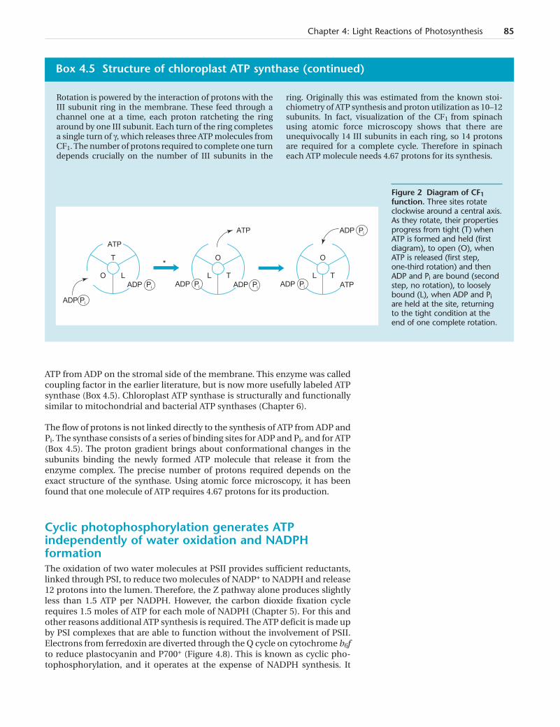
Rotation is powered by the interaction of protons with theIII subunit ring in the membrane. These feed through achannel one at a time, each proton ratcheting the ringaround by one III subunit. Each turn of the ring completesa single turn of g, which releases three ATP molecules fromCF1. The number of protons required to complete one turndepends crucially on the number of III subunits in the
ring. Originally this was estimated from the known stoi-chiometry of ATP synthesis and proton utilization as 10–12subunits. In fact, visualization of the CF1 from spinachusing atomic force microscopy shows that there areunequivocally 14 III subunits in each ring, so 14 protonsare required for a complete cycle. Therefore in spinacheach ATP molecule needs 4.67 protons for its synthesis.
Box 4.5 Structure of chloroplast ATP synthase (continued)
ATP
T
O L
O
L T
O
L T
ADP Pi
ATP ADP Pi
*
ADP Pi ADP Pi ATPADP Pi ADP Pi
Figure 2 Diagram of CF1function. Three sites rotateclockwise around a central axis.As they rotate, their propertiesprogress from tight (T) whenATP is formed and held (firstdiagram), to open (O), whenATP is released (first step,one-third rotation) and thenADP and Pi are bound (secondstep, no rotation), to looselybound (L), when ADP and Piare held at the site, returningto the tight condition at theend of one complete rotation.
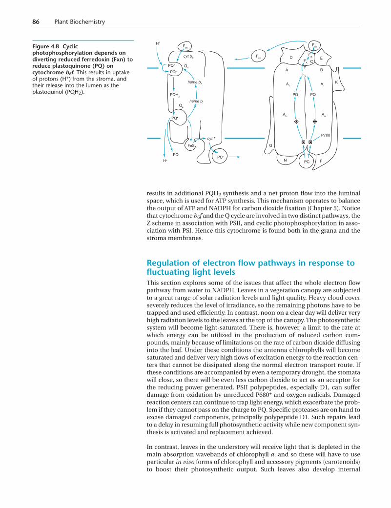
results in additional PQH2 synthesis and a net proton flow into the luminalspace, which is used for ATP synthesis. This mechanism operates to balancethe output of ATP and NADPH for carbon dioxide fixation (Chapter 5). Noticethat cytochrome b6f and the Q cycle are involved in two distinct pathways, theZ scheme in association with PSII, and cyclic photophosphorylation in asso-ciation with PSI. Hence this cytochrome is found both in the grana and thestroma membranes.
Regulation of electron flow pathways in response tofluctuating light levelsThis section explores some of the issues that affect the whole electron flowpathway from water to NADPH. Leaves in a vegetation canopy are subjectedto a great range of solar radiation levels and light quality. Heavy cloud coverseverely reduces the level of irradiance, so the remaining photons have to betrapped and used efficiently. In contrast, noon on a clear day will deliver veryhigh radiation levels to the leaves at the top of the canopy. The photosyntheticsystem will become light-saturated. There is, however, a limit to the rate atwhich energy can be utilized in the production of reduced carbon com-pounds, mainly because of limitations on the rate of carbon dioxide diffusinginto the leaf. Under these conditions the antenna chlorophylls will becomesaturated and deliver very high flows of excitation energy to the reaction cen-ters that cannot be dissipated along the normal electron transport route. Ifthese conditions are accompanied by even a temporary drought, the stomatawill close, so there will be even less carbon dioxide to act as an acceptor forthe reducing power generated. PSII polypeptides, especially D1, can sufferdamage from oxidation by unreduced P680+ and oxygen radicals. Damagedreaction centers can continue to trap light energy, which exacerbate the prob-lem if they cannot pass on the charge to PQ. Specific proteases are on hand toexcise damaged components, principally polypeptide D1. Such repairs leadto a delay in resuming full photosynthetic activity while new component syn-thesis is activated and replacement achieved.
In contrast, leaves in the understory will receive light that is depleted in themain absorption wavebands of chlorophyll a, and so these will have to useparticular in vivo forms of chlorophyll and accessory pigments (carotenoids)to boost their photosynthetic output. Such leaves also develop internal
86 Plant Biochemistry
FA
Fxn
Fxn
D EC
A B
K
PQ PQ
P700
G
N PC F
A1 A1
Ao Ao
FX
cyt b6
PQ•
PQ• •
heme bh
Qn
cyt f
H+
Fxn
PQH2
Qp
PQ•
H+
PQ
FeS
PC–
heme bl
FB
Figure 4.8 Cyclicphotophosphorylation depends ondiverting reduced ferredoxin (Fxn) toreduce plastoquinone (PQ) oncytochrome b6f. This results in uptakeof protons (H+) from the stroma, andtheir release into the lumen as theplastoquinol (PQH2).
anatomy and chloroplast grana organization to maximize light harvesting intheir environment (shade leaves). For these leaves transient increases in lightintensity, e.g. from sun flecks, can also be very damaging.
High light levels expose the cellular components to all the risks posed by pho-tosynthesis. Significant levels of light energy end up trapped in componentmolecules. Excess irradiation can saturate the electron flow system leading toaccumulation of molecules in the excited triplet state. This leads to formationof reactive superoxides, peroxides, and aldehydes, which have the potentialto destroy the proteins and lipids in the chloroplast. Plants have evolved sys-tems to remove these dangerous radicals and to prevent or reduce their for-mation in the first place. We will see that both the removal of radicals and theprevention of their formation involve many other molecules and enzymes, allworking within the close confines of the thylakoid and fret membrane sys-tems. It has therefore proved difficult to study each system in isolation andevaluate its contribution to the overall effect, so our story is incomplete at thepresent time. Clearly plants possess photosynthetic systems that work in agreat range of habitats under a variety of physiological conditions of waterand carbon dioxide supply. We might anticipate that the full range of curativeand preventative measures will not be present to the same extent in all plants.Here we will describe the main processes that have been unequivocally iden-tified and characterized in a representative number of plant species.
Oxygen molecules are normally stable and pose only a limited risk to themolecular structure of a cell. Singlet oxygen can be generated from these mol-ecules by the input of energy, for example from light energy absorbed bychlorophyll molecules. The normal chlorophyll excited state induced by lightabsorption lasts for a few nanoseconds, long enough to be passed on to thereaction center. Any delay in this transfer can lead to the formation of achlorophyll triplet state, which has a lifetime of several microseconds. Thesechlorophyll molecules can react with oxygen to produce singlet oxygen. Thishas a greatly increased oxidizing power that attacks any molecule in the vicin-ity. Typically, the lifetime of such oxygen radicals is about 200 ns, duringwhich time it can diffuse as far as 10 nm. Mechanisms have evolved to rapidlyremove these oxygen radicals and to curb their production at the sites of lightabsorption.
Singlet oxygen can be quenched directly by carotenoids, which are them-selves converted to the triplet state, but this energy decays quickly being lostas heat. This can only happen if the carotenoids are close to the site of singletproduction, which occurs in the LHCII antenna molecules, for example, butnot in the PSII core. The carotenoids present in the PSII complex do serve toquench the P680 if charge recombination occurs to generate triplet P680 (aswhen the pheophytin at A0 is not oxidized in time to receive a further reduc-tion from P680). Singlet oxygen can also be quenched by a-tocopherol, amembrane lipid, and again the energy is dissipated as heat.
Scavenging and removal of superoxides, peroxides,and other radicals by dismutases and antioxidantsOxidized compounds originate at PSI under high light conditions, when thereis insufficient NADP to oxidize reduced ferredoxin. The electrons insteadreduce molecular oxygen to superoxide, which in turn leads to peroxide for-mation by combination with protons. Peroxides can interact with reducedferrous ions (Fe2+) to produce hydroxyl radicals. These are extremely reactiveand attack other molecules, sometimes leading to further radical formationso that a chain reaction is set up.
Chapter 4: Light Reactions of Photosynthesis 87

A number of enzymes have evolved to scavenge peroxides, including super-oxide dismutase, ascorbate peroxidase, monodehydroascorbate reductase,and glutathione reductase. Several antioxidants are available to be oxidized inthese reactions, such as ascorbate, glutathione, and a-tocopherol (vitamin E).These convert the peroxide to water and oxygen. In effect this sets up a sys-tem for water breakdown (at PSII) and synthesis (at PSI), which removesexcess energy trapped by the chlorophylls.
ascorbate peroxidase
2H2O2 + ascorbate ––––Æ O2 + 2H2O + monodehydroascorbate
monodehydroascorbate reductase
2monodehydroascorbate + NADPH ––––Æ 2ascorbate + NADP+
Peroxide removal by ascorbate. Note that the regeneration of ascorbate leadsto consumption of NADPH, removing excess reductant from the system.
Mechanisms for safely returning the levels oftrapped high energy states to the ground stateReducing the exposure of chlorophyll molecules to high light intensities canprevent or reduce singlet oxygen formation. This can be achieved by chang-ing the leaf angle with respect to incident light, and by movement of thechloroplasts to self-shading positions along the side walls of cells. Within thechloroplast there are three basic types of mechanism for coping with highlight conditions. Adjustment of the synthesis and amount of the LHCIIantenna proteins, movement of the LHCII proteins from PSII to PSI (statetransition) and nonphotochemical quenching when electron flow is divertedto heat generating systems.
High light conditions and/or low carbon dioxide concentrations down-regulateexpression of Lhcb genes. The sensor mechanism is not known. One candidateis the redox potential system, e.g. the level of PQ, but this control seems to bequite loose. Other mechanisms could be the level of reactive oxygen, or the lightsaturation of precursors of chlorophyll. Clearly this is a slow control mecha-nism, depending on not replacing chlorophyll proteins lost due to turnover.
Some relief from excess energy absorption can be made within the PSII com-plex, by cycling electrons back from PQ via cytochrome c-553 to the D1 tyro-sine. Another approach that has evolved is to reduce the amount of LHCIIantenna proteins associated with PSII. This alters the ratio of light energyreceived between PSII and PSI by adjusting the extent to which each receivesenergy from these antenna proteins. As the two photosystems have differentabsorption spectra, conditions can arise where the energy flow through eachis not balanced to meet the requirements of the Z scheme. This could be dam-aging, as high energy intermediates would accumulate in the system. TheLHCII trimers serve as a simple feedback loop that adjusts the amount ofantenna chlorophyll providing energy to each photosystem (state transition).If there is excess light energy flowing through PSII, then there is an excess ofreduced PQ. This activates a kinase that phosphorylates some of the LHCIItrimers. The extra charge causes them to dissociate from the PSII (state 2)complex and migrate towards the stroma lamellae (state 1 transition) wherethey bind to the PSI complex (state 1), increasing the flow through that sys-tem. Conversely, excess PSI activity leads to PQH2 oxidation, which activatesa phosphatase that removes the phosphate group and allows the trimers toreturn to PSII (state 2 transition).
88 Plant Biochemistry
Under high light conditions, the electron flow from PSII to PSI can be regu-lated by cytochrome b6f and plastocyanin. The PQ reoxidation cycle is theslowest step in the photosynthetic electron flow pathway and the number ofcytochrome b6f complexes appears to be controlled, increasing as the fluxrate increases. Plastocyanin also shows an increase with flux rate, and inleaves operating efficiently at high photosynthetic rates can reach a ratio of4–5:1 of PSI. If the leaf’s photosynthetic ability is compromised (e.g. by low orzero carbon dioxide availability), then this ratio falls back to below 1. Thiscould limit the flow of electrons into P700. However, electron transfers involv-ing plastocyanin (to PSI and to cytochrome b6f) are much faster (10–100times) than the PQ oxidation step, so the only way that reduced levels of plas-tocyanin could control the overall flux is if its diffusion rate to the PSI site isslow compared with the rate of electron flow.
Nonphotochemical quenching and the xanthophyll cycleThese adjustments to the electron transport system cannot entirely preventthe accumulation of excess energy in photon-trapping chlorophyll mole-cules. Such excited chlorophylls can return to the ground state by either emit-ting the energy as fluorescence, or by dissipating it as heat. Molecular mech-anisms for removing this trapped energy (quenching the excited state) beforeit is passed on down the electron transport chain are collectively termed non-photochemical quenching. The precise details of these molecular mecha-nisms remain to be discovered. The following account provides a general out-line of the mechanisms as far as they are understood at the present time.
In saturating light conditions, chlorophylls in the antenna proteins, which arein equilibrium with the PSII reaction center chlorophylls, are unable to passtheir trapped energy on to P680. Such chlorophylls pose a risk, as they areable to activate oxygen to the singlet state. Decay of the excited chlorophyllsreleases energy as heat and fluorescence. This fluorescence is a temporaryresponse to suddenly increased light levels, which declines as additionalresources are developed to convert the energy to heat. A prime candidate forinitiation of the controlling mechanisms is the pH of the lumen. Proton gen-eration and pumping during the light reactions lower the lumen pH fromabout 7 to less than 5, providing a strong signal that can initiate a number ofquenching processes based in the lumen. One of these is the activation of anepoxidase that uses electrons to reduce the xanthophyll violaxanthin toantheraxanthin and then to zeaxanthin. The zeaxanthin binds to a proteinsubunit of LHCII that is protonated at low pH and accepts energy transferfrom excited chlorophylls. The zeaxanthin then returns to the ground state,dissipating the energy as heat. In this way excess light energy trapped bychlorophylls is diverted to heat production, away from potentially damagingphotochemical events and electron transfers.
pH 5, de-epoxidase –Æ pH 5, de-epoxidase –Æ
violaxanthin –̈–––––––––––––Æ antheraxanthin –̈–––––––––––––Æ zeaxanthin
–̈––– pH 7, epoxidase –̈––– pH 7, epoxidase
LHCII** + zeaxanthin ––Æ LHCII + zeaxanthin**
zeaxanthin** ––Æ zeaxanthin + heat
The xanthophyll cycle and nonphotochemical quenching reactions leadingto the transfer of excess excitation energy from light harvesting antenna
Chapter 4: Light Reactions of Photosynthesis 89
complexes to zeaxanthin and its release as heat. This is reversed under lowlight conditions when the pH rises.
Antiquenching is obviously necessary when light levels fall. This is achievedby a different epoxidase, activated at higher pH levels, that converts zeaxan-thin back to antheraxanthin and violaxanthin. In this way the xanthophyllcycle is thought to track the pH changes in the lumen that are directly linkedto productive photosynthetic activity and regulates the level of nonphoto-chemical quenching. The importance of xanthophylls and other carotenoidsin this protection system can be seen from the fact that, in the absence ofcarotenoids, photosynthetic systems can only operate in oxygen-free condi-tions. Also carotenoid-deficient mutants will only survive under very low lightconditions; normal light levels lead to bleaching of the leaves.
There is some evidence that a very hydrophobic transmembrane protein,PSbS, is involved in nonphotochemical quenching. It is proposed that PSbSundergoes protonation at low pH, and then binds zeaxanthin into theLHCII/PSII complex facilitating quenching of the excited state molecules.Experiments in which the levels of PSbS protein are altered, or in which theprotonation of PSbS at low pH is interfered with, support this hypothesis.However, the location of PSbS remains to be determined, as does the preciseway in which quenching is effected.
SummaryOverall PSII and PSI operate to extract electrons from water and reduceNADP+. A proton gradient is produced that is used for ATP generation, andoxygen is released as a by-product. The synthesis of these photosystems isregulated to achieve approximately equal numbers of each. Failure to chan-nel the PSII reducing power can lead to oxygen radical production with harm-ful consequences for the photosynthetic machinery. This can occur in someunfavorable environmental conditions (e.g. chilling) that damage PSI, leavingthe PSII reductants accumulating in the pathway (Box 4.6).
90 Plant Biochemistry
As the photosynthetic electron flow pathway is the onlysource of energy for growth in a plant, it represents aprime target for chemicals designed to kill plants. DCMUand the herbicide atrazine bind to the QB site on PSII,preventing plastoquinol formation. Plastoquinoneanalogs (dibromothymoquinone) bind to cytochrome b6fpreventing plastoquinol oxidation at the Qp site.Paraquat is a widely used herbicide that diverts electronflow from ferredoxin to the formation of reactive oxygenspecies and peroxides that damage the photosyntheticapparatus and ultimately kill the leaf. Perversely the
chlorophyll pigments harvest light energy that is used todestroy the plant.
Attacking carotenoid synthesis is an alternative way ofkilling plants, as this will lead to loss of the nonphoto-chemical quenching needed to protect photosystemsfrom the effects of relatively high light intensities.Norflurazan is a herbicide of this type and leads tobleaching of leaves that is similar to that seen whencarotenoid synthesis mutants are exposed to normal lightlevels (see Chapter 12).
Box 4.6 Herbicides that act through the photosynthetic apparatus
Chapter 4: Light Reactions of Photosynthesis 91
Evolution of the reaction centers, light harvestingphotosystems, and proteins
These papers provide a good overview of the evolution ofphotosynthesis and would provide useful entry points forthe literature both before and after 2002/2004.
De Las Rivas J, Balsera M & Barber J (2004) Evolution of oxy-genic photosynthesis: genome-wide analysis of the OECextrinsic proteins. Trends Pl. Sci. 9, 18–25.
Raymond J & Blankenship RE (2004) The evolutionary devel-opment of the protein complement of Photosystem II.Biochim. Biophys. Acta 1655, 133–139.
Xiong J & Bauer CE (2002) Complex evolution of photosyn-thesis. Annu. Rev. Pl. Biol. 53, 503–521.
Organization of the thylakoid membrane
These two papers provide an overview of photosyntheticmembrane components.
Decker JP & Boekema EJ (2005) Supramolecular organiza-tion of thylakoid membrane proteins in green plants.Biochim. Biophys. Acta 1706, 12–39.
Merchant S & Sawaya MR (2005) The light reactions: a guideto recent acquisitions for the picture gallery. Pl. Cell 17,648–663.
Photosystems
Photosystems have been the focus of research for over 50years, so there is a very large literature about their structureand function.
Amunts A, Drory O & Nelson N (2007) The structure of aplant photosystem I supercomplex at 3.4Å resolution.Nature 447, 58–63.
Blankenship RE (2002) Molecular Mechanisms ofPhotosynthesis. Blackwell Science Ltd.
Chitnis PR (2001) Photosysytem I: function and physiology.Annu. Rev. Pl. Physiol. Pl. Mol. Biol. 52, 593–626.
Diner BA & Rappaport F (2002) Structure, dynamics, andenergetics of the primary photochemistry of Photosystem IIof oxygenic photosynthesis. Annu. Rev. Pl. Biol. 53, 551–580.
Minagawa J & Takahashi Y (2004) Structure, function andassembly of Photosystem II and its light-harvesting pro-teins. Photosynthesis Res. 82, 214–263.
Nelson N & Yocum CF (2006) Structure and function ofPhotosystems I and II. Annu. Rev. Pl. Biol. 57, 521–565.
Water splitting and oxygen evolution
The chemistry of water splitting in photosynthesis is fasci-nating, how do plants successfully break open this moleculeunder normal physiological conditions?
Ferreira KN, Iverson TM, Maghlaoui K, Barber J & Iwata S(2004) Architecture of the oxygen-evolving center. Science303, 1831–1838.
Sauer K & Yachandra VK (2004) The water-oxidation processin photosynthesis. Biochim. Biophys. Acta 1655, 140–148.
Cytochromes
Processing plastoquinones and producing plastocyanins pro-vides the vital link between PSII and PSI. New information isadding more detail to this part of the photosynthesis story.
Allen JF (2004) Cytochrome b6f: structure for signaling andvectorial metabolism. Trends Pl. Sci. 9, 130–137.
Siegbahn PEM & Blomberg MRA (2004) Important roles oftyrosines in Photosystem II and cytochrome oxidase.Biochim. Biophys. Acta 1655, 45–50.
ATP synthase: structure and operation
An amazing molecular turbine has been uncovered thatcouples a proton gradient to the formation of ATP.
Kramer DM, Cruz JA & Kanazawa A (2003) Balancing thecentral roles of the thylakoid proton gradient. Trends Pl. Sci.8, 27–32.
McCarty RE, Evron Y & Johnson EA (2000) The chloroplastATP synthase: a rotary enzyme? Annu. Rev. Pl. Physiol. Pl.Mol. Biol. 51, 83–109.
Seelert H, Dencher NA & Müller DJ (2003) Fourteen pro-tomers compose the oligomer III of the proton-rotor inspinach chloroplast ATP synthase. J. Mol. Biol. 333, 337–344.
World Wide Web-based information
The following is a useful site providing access to informationon all aspects of photosynthesis.
http://porphy.la.asu.edu/photosyn/photoweb/
Further Reading
