light-phyllosphere interactions in greenhouse grown...
TRANSCRIPT
Light-Phyllosphere Interactions in Greenhouse Grown Ornamentals
Samareh Gharaie Faculty of Landscape Architecture, Horticulture and Crop Production Science
Department of Biosystems and Technology
Alnarp
Doctoral Thesis
Swedish University of Agricultural Sciences
Alnarp 2017

Acta Universitatis agriculturae Sueciae
2017:19
ISSN 1652-6880
ISBN (print version) 978-91-576-8811-8
ISBN (electronic version) 978-91-576-8812-5
© 2017 Samareh Gharaie, Alnarp
Print: SLU Service/Repro, Alnarp 2017
Cover: An artistic impression of light (LED)-phyllosphere microbiota
(Sketch: Samareh Gharaie)

Light-Phyllosphere Interactions in Greenhouse Grown Ornamentals
Abstract Light-emitting diodes (LEDs) have emerged as a promising artificial lighting
source in greenhouse production of horticultural crops, as they reduce energy
consumption. However, changes in lighting technology are known to affect
abiotic and biotic interactions in the phyllosphere, e.g. LEDs can change the
microclimate within the greenhouse and around the crop, and thus the
microbial community structure. Information is lacking on interactions between
light spectra and microbiota associated with the canopy, the function of non-
phototrophic bacteria associated with the phyllosphere and successful
administration of microbial biocontrol agents.
This thesis investigated the impact of different light spectra on plant
physiological parameters, microbial community structure, utilisation pattern of
energy sources and biosurfactant formation by phyllosphere microbiota in
greenhouse-grown ornamentals. A standard protocol for extraction of
phyllosphere microbiota, impact of plant species and leaf position, and
antagonistic activity of resident phyllosphere microbiota against Botrytis
cinerea was also studied. Use of culture-dependent methods revealed higher
numbers of culturable fungi on basal than on apical leaves, but the numbers did
not vary with different light treatments. Metagenomics showed that the fungal
microbiome was more diverse on apical leaves. Interactions were found
between leaf temperature and many dominant bacterial genera. In vitro tests
revealed that inhibitory effects of some strains identified by 16S rRNA varied
with respect to different media. Phenotypic microarray analysis revealed that
light treatments had considerable effects on substrate utilisation by two
Pseudomonas strains and moderate effects on Streptomyces griseoviridis, with
blue LEDs having most the pronounced impact. Biosurfactant formation by
Pseudomonas strains was supported by most substrates when incubated in
darkness, but blue LED altered the surface activity more profoundly.
Keywords: 16S rRNA, antagonistic activity, blue light receptor protein, light-emitting
diodes, metagenomic analysis, microbial community structure, phenotypic microarray,
phyllosphere, Omnilog
Author’s address: Samareh Gharaie, SLU, Department of Biosystems and Technology,
Microbial Horticulture Unit, P.O. Box 103, SE-23053 Alnarp, Sweden.
E-mail: [email protected]

Dedication
To my parents and my sister

Contents
List of Publications 7
Abbreviations 9
1 Introduction 10 1.1 Phyllosphere 10
1.1.1 Phyllosphere microbiome 10 1.1.2 Phyllosphere analysis 10
1.2 Abiotic and biotic phyllosphere interactions 13 1.2.1 Abiotic interactions 13 1.2.2 Biotic interactions 17
1.3 Greenhouse production of ornamentals and sustainability issues 19 1.4 Objectives 22
2 Materials and methods 24 2.1 Plant material and sampling strategy 24 2.2 Greenhouse experiments 26 2.3 Climate chamber and light treatments (Paper I, III and IV) 26 2.4 Microorganisms 28 2.5 Analyses 29
2.5.1 Plant analysis (Paper I) 29 2.5.2 Extraction of microbiota from the phyllosphere (Paper I and II) 29 2.5.3 Culture-dependent microbial analyses 30 2.5.4 Culture-independent analyses 32
2.6 Calculations and statistics 33
3 Results and discussion 34 3.1 Microbial community structure in greenhouse-grown ornamentals
(Papers I and II) 34 3.1.1 Effect of light spectrum on phyllosphere microbiota 34 3.1.2 Occurrence of bacterial antagonistic to Botrytis cinerea (Paper II)36
3.2 Impact of light spectrum on utilisation of energy sources by selected
phyllosphere bacteria (Papers III and IV) 39 3.3 Impact of light spectrum on the formation of metabolites decisive for leaf
colonisation (Paper IV) 43

4 Conclusions 46
References 48
Acknowledgements 63

7
List of Publications
This thesis is based on the work contained in the following papers, referred to
by Roman numerals in the text:
I Alsanius, B.W., Bergstrand, K.J., Hartmann, R., Gharaie, S., Wohanka,
W., Dorais, M. and Rosberg, A.K. (2017). Ornamental flowers in new light:
Artificial lighting shapes the microbial phyllosphere community structure
of greenhouse grown sunflowers (Helianthus annuus L.). Scientia
Horticulturae 216, 234-247.
II Gharaie, S., Windstam, S., Khalil, S., Wohanka, W. & Alsanius, B.W.
Isolation and characterisation of epiphytic bacteria from the phyllosphere of
greenhouse-grown ornamentals (manuscript).
III Gharaie*, S., Vaas*, L.A.I., Rosberg, A.K., Windstam, S., Karlsson, M.E.,
Bergstrand, K.J., Khalil, S., Wohanka, W. & Alsanius B.W*. (2017). Light
spectrum modifies the utilisation pattern of energy sources in Pseudomonas
sp. DR 5-09. PLOS ONE (submitted).
IV Alsanius*, B.W., Vaas*, L.A.I., Gharaie, S*., Karlsson, M.E., Rosberg,
A.K., Grudén, M., Wohanka, W., Khalil, S., & Windstam, S. Dining in blue
light impairs the appetite of some leaf epiphytes (manuscript).
Paper I is reproduced with kind permission of Rights Links and Elsevier.
*Equally contributing authors

8
The contribution of Samareh Gharaie to the papers included in this thesis was
as follows:
I Partly involved in the writing process.
II Planned the experiment with the co-authors. Performed the experimental
work and evaluated the data. Wrote the manuscript with the co-authors.
III Planned the experiment with the co-authors. Performed the experimental
work. Evaluated the data with co-authors. Wrote the manuscript together
with the co-authors.
IV Planned the experiment with co-authors. Performed the experimental work.
Partly evaluated the data and was partly involved in the writing process of
the manuscript together with the co-authors.

9
Abbreviations
CFU Colony-forming units
DNA
DGGE
HPC
KB
Deoxyribonucleic acid Denaturing Gradient Gel Electrophoresis
Heterotrophic plate counts
King`s B agar
LED
PCA
Light-emitting diode
Principal Component Analysis
PCR Polymerase chain reaction
PDA
PM
Potato dextrose agar
Phenotype MicroArray
rRNA Ribosomal ribonucleic acid
SMs Secondary metabolites
t-RFLP Terminal restriction fragment length polymorphism
TSA Tryptic soy agar
10
1 Introduction
1.1 Phyllosphere
1.1.1 Phyllosphere microbiome
The plant phyllosphere comprises aerial parts of living plants, including
leaves, stems, buds, flowers and fruits, which harbour a large number of
diverse microorganisms (Knief et al., 2010; Lindow & Brandl, 2003). Leaves
are the most dominant part of the aboveground plant (Vorholt, 2012) and so
far most research on phyllosphere microbiology has focused on this dominant
aerial structure (Lindow & Brandl, 2003). Microbial populations on plant
leaves are diverse, e.g. archaea, filamentous fungi and yeasts are known to be
present on leaves, but bacteria are considered to be the most abundant
phyllosphere inhabitants and can colonise leaves with an average of 106-107
bacterial cells per cm of leaf surface (Lindow & Brandl, 2003).
The phyllosphere is a hostile habitat for microorganisms, as it is an open
system and highly influenced by permanently fluctuating abiotic conditions
(Vorholt, 2012; Lindow & Brandl, 2003). Changes in environmental factors,
along with plant genotype, can influence the microbial community composition
in the phyllosphere (Vorholt, 2012). The composition of the phyllosphere
microbial population is thus determined by ability to colonise this environment.
Detailed information about abiotic and biotic phyllosphere interactions is given
in section 2.2.
1.1.2 Phyllosphere analysis
Several methods for assessing phyllosphere microbiota are available,
commonly divided into two main approaches, viz. culture-dependent and
culture-independent methods. Culture-dependent approaches are based on
growing microorganisms on semi-selective medium, whereas culture-
independent methods rely on DNA-based methods. One type of culture-

11
dependent method is the viable plate count technique, in which
microorganisms can be plated directly on different nutrient agar (Yang et al.,
2001) or brought to suspension from natural samples. Furthermore,
suspensions containing the extracted microorganisms can be plated on culture
media (Jensen et al., 2013). Identification of single colonies, after culturing on
solid culture media, can be performed by biochemical or morphological
methods (Jensen et al., 2013) or by gene sequencing techniques (Yarza et al.,
2014). Likewise, quantification of microorganisms can be performed by
enumeration of the colonies on agar plates and calculation of colony-forming
units (CFU) per mL or per g of observed species (Madigan et al., 2012).
Another example of a culture-dependent method is the phenotypic
microarray (PM) technique (Biolog Inc., Hayward CA, USA), which is a high-
throughput system and can be used for overall analysis of cellular phenotypes
of pure cultures or communities in an environmental sample (Line et al., 2011;
Bochner et al., 2001). In this technique, pre-filled PM plates are generally used
for analysis of cellular pathways in 200 different assays of carbon-source
metabolism, 400 assays of nitrogen metabolism, 100 assays of phosphorus and
sulphur metabolism, 100 assays of biosynthetic pathways, 100 assays of ion
effects and osmolality, 100 assays of pH effects and pH control with
deaminases and decarboxylases, and 1000 assays of chemical sensitivity. The
chemical sensitivity assays comprise 240 different chemicals, each at four
different concentrations. To start a PM assay, two components need to be
combined. These are a cell suspension and a nutrient/chemical solution needed
to create the 1920 unique culture conditions. The assays are based on a
universal culture medium containing all micronutrients needed for cell growth
(Bochner, 2009).
Different, assays, e.g. PM1 and PM2 representing 190 carbon sources,
PM3 representing 95 nitrogen sources and PM4 representing 59 phosphorus
and 35 sulphur sources, are commercially available as pre-filled 96-well
microtitre plates that provide information on metabolic pathways which are
present and active in the cell.
The system is based on phenotypic response to utilisation of the organic
sources and the utilisation is monitored by a colour change in a tetrazolium
blue-based redox dye (colourless implies that the cells are not able to utilise the
organic sources, whereas a colour reaction to purple in the well indicates that
cells are actively utilising the substrate) (Bochner et al., 2001). Utilisation rate
for each well (colour formation in each well) can be used for cellular
phenotype comparison (Bochner, 2003).
The output of this technique is colour-coded kinetic graphs of respiratory
response and important biological information is obtained from curve

12
parameters such as lag phase (λ), steepness of slope (μ), maximum curve
height (A) and area under the curve (AUC) (see Supplementary Figure 3 in
Paper III).
Culture-dependent approaches, though vastly useful for understanding the
physiological potential of extracted organisms, do not necessarily provide
complete information on the composition of microbial communities (Onstott et
al., 1998). One drawback of these approaches is that, although many different
culture media have been designed for recovering as many microorganisms as
possible, just a small number of microorganisms can be cultured, while the
majority of microorganisms are unculturable (Madigan et al., 2012). Another
disadvantage of culture-dependent methods is that fast-growing
microorganisms compete with slow growers concerning nutritional
requirement (Nocker et al., 2007). Moreover, if the investigation of complex
communities is underestimated, then erroneous results can be obtained when
counting microorganisms with traditional culture-based methods (Besnard et
al., 2000). Therefore different studies suggest combining culture-based and
culture-independent approaches in order to obtain comprehensive information
on the microbial community (Stefani et al., 2015; Shade et al., 2012; Yashiro
et al., 2011). Different culture-independent (DNA-based) methods have been
developed for investigation of the phyllosphere microbial community. In past
years, a number of methods based on direct amplification and analysis of the
small subunit ribosomal RNA gene have been used to study the microbial
community of the phyllosphere, e.g. denaturing/temperature gradient gel
electrophoresis (DGGE) (Rigonato et al., 2016; Reisberg et al., 2012; Delmotte
et al., 2009; Yang et al., 2001), terminal restriction fragment length
polymorphism (t-RFLP) (Ding & Melcher, 2016; Ding et al., 2013; Penuelas et
al., 2012; Hunter et al., 2010; Berg et al., 2005) and high throughput
sequencing (next-generation sequencing) (Laforest-Lapointe et al., 2016;
Lindahl et al., 2013; Rastogi et al., 2013; Redford et al., 2010).
In culture-independent approaches, the application of high-throughput
sequencing techniques has revolutionised scientists’ view of microbial
communities in environmental samples (Vartoukian et al., 2010). High
throughput sequencing techniques are designed for rapid and large-scale
microbial community analyses. The five most commonly used methods are:
454-pyrosequencing, Illumina/Solexa, SOLiD, the HeliScope Single Molecule
Sequencer and Single Molecule Real Time technology (Morey et al., 2013;
Siqueira et al., 2012). The main differences between these technologies are the
length of sequences and number of sequence reads achieved (Mardis, 2008).
Illumina, the most commonly used platform nowadays, was employed in
some of the work presented in this thesis (Paper I), and is therefore discussed

13
in more detail here. The Illumina technology was first presented in 2006 and,
due to advantages concerning its greater cost-effectiveness and ability to
generate larger amounts of data, was quickly accepted by scientists (Hodkinson
& Grice, 2015; Caporaso et al., 2012). The Illumina sequencing preparation
starts with lengths of DNA that have specific adapters on either end being
washed over a flow cell filled with specific oligonucleotides that hybridise to
the ends of the fragments. To create a cluster of identical fragments, each
fragment is then replicated. Reversible dye-terminator nucleotides are washed
over the flow cell and given time to attach, the excess nucleotides are washed
away, the flow cell is imaged and the terminators are reversed so that the
process can repeat and nucleotides can continue to be added in subsequent
cycles (Hodkinson & Grice, 2015). The longest reads that Illumina currently
produces, on MiSeq, can produce paired-end reads that are 300 bases in length
(Hodkinson & Grice, 2015). In addition, Illumina does not give absolute
numbers, but relative abundances. Sequences less abundant than 1% are
excluded during the calculation process. Compared with Sanger sequencing,
next-generation sequencing methods have greatly reduced the cost and time
associated with producing larger amounts of sequenced data (Mardis, 2008).
Another advantage of next-generation sequencing techniques is that there is no
need to contract clone libraries (Siqueira et al., 2012). However, the length of
the sequence reads generated by most next-generation sequence methods is
shorter than that required for identification of bacterial gene length (Luo et al.,
2012), excluding identification of relative abundances on species level.
Furthermore, sequencing can be used for identification of single colonies
produced by culture-dependent methods. Comparative analysis of 16S rRNA
gene sequences is important for classification of cultured microorganisms and
also for classification of known and novel bacterial genera and species, e.g. it
enables establishment of taxonomic thresholds for classification of cultured
microorganisms and of the many environmental sequences (Hakovirta et al.,
2016; Yarza et al., 2014; Mole, 2013; Quast et al., 2013).
1.2 Abiotic and biotic phyllosphere interactions
1.2.1 Abiotic interactions
Phyllosphere colonisation is affected by abiotic factors such as temperature,
humidity, water, wind speed and electromagnetic radiation (ultraviolet (UV)
and visible light) (Vorholt, 2012; Lindow & Brandl, 2003; Kinkel et al., 2000).
In this context, direct impacts of temperature on the development of leaf
surface microbiota have been reported (Bernard et al., 2013). Previous research
has shown that changes in the temperature conditions and relative humidity

14
under which plants are grown affect not only the phyllosphere microbial
population, but also the ability of pathogens to colonise and survive (Bálint et
al., 2015; Brandl & Mandrell, 2002). Similarly, fluctuation in water availability
is an important parameter that affects the abundance and diversity of microbial
populations (Yadav et al., 2005; Morris & Monier, 2003; Morris et al., 2002).
Exposure to different forms of ultraviolet light (UVA, UVB and UVC) can
have deleterious effects on leaf microorganisms, contributing to cell death.
Phyllosphere microorganisms have developed different tolerance mechanisms
towards ultraviolet light, such as pigments and DNA repair systems (Sundin et
al., 2002; Kim & Sundin, 2000; Sundin & Jacobs, 1999).
Light plays a key role in multiple phyllosphere interactions. A conceptual
structure for light-plant-microbe-environment interactions is presented in this
thesis (see Figure 1 in Paper I). This structure describes all the different biotic
and abiotic interactions between the plant and plant leaf, abiotic factors and the
phyllosphere microbiota, which are discussed in more detail in the following
sections.
Light- plant interactions
Light is a fundamental factor for plant growth and development (Li &
Kubota, 2009; Fukuda et al., 2008). In this context, three important parameters
of light for growth are: quality (spectral distribution), quantity (intensity) and
duration. Light quality refers to the spectral distribution of the radiation, i.e.
wavelength reaching the plant surface. The active part of the light spectrum for
plants ranges from ultra-violet to infrared, but the main wavelengths that are
absorbed by plant photoreceptors are blue (400-500 nm) and red (600-700 nm)
(Huché-Thélier et al., 2015). Light quality has a profound effect on plant
growth and greatly influences the anatomy, morphology and physiology of the
leaves (Johkan et al., 2012; Macedo et al., 2011; Hogewoning et al., 2010).
Light quality effects are species- and cultivar/variety-dependent (Schuerger et
al., 1997), but specific light quality can be used to improve the nutritional
quality of crops. Absorbed wavelengths can affect the metabolic system of the
plants, for instance ultraviolet and blue radiation are involved in the production
of secondary metabolites or mechanisms of resistance to pathogens (Huché-
Thélier et al., 2016).
Many studies have reviewed the importance of light quality and its effect on
growth and development and plant responses to light quality (e.g. Folta &
Childers, 2008; Devlin et al., 2007). Other studies have examined the impact of
LED light on plant yield (Massa et al., 2008) and have reviewed application of
LEDs in greenhouse cultivation (Mitchell et al., 2012; Bergstrand & Schüssler,

15
2010). A recent study examined the effects of light quality on phytochemical
accumulation in plants produced in controlled conditions (Bian et al., 2015).
Light intensity is the total amount of light delivered to plants and is usually
measured in μmol m-2s-1, which is the number of photons of light within the
photosynthetic wavelength received by an area of one square metre per second.
Light intensity affects photosynthesis, a photochemical reaction which occurs
within the chloroplast of plant cells, converting atmospheric CO2 into
carbohydrates (Nishio, 2000). Red light is involved in the development of the
photosynthetic system, while blue light is needed for chlorophyll synthesis,
localisation of chloroplasts and stomatal opening. Both blue and red light are
required for photosynthesis.
Another parameter of light is light duration (photoperiod), which is the
period of time per day that plants receive illumination. Photoperiod mainly
influences flower bud induction and therefore changing the photoperiod can
control flowering time in plants (Singh et al., 2015; Nishio, 2000). In this
context, plants have been categorised into two main groups: 1) Short-day
plants (SDPs) and 2) long-day plants (LDPs). The SDPs flower when the
photoperiod is less than their critical night length, whereas in the LDPs
flowering occurs when the day length is longer than their critical night length
(Zukauskas et al., 2009; Downs & Thomas, 1982). Furthermore, additional
far-red light lowers the red:far-red ratio, which promotes flowering (Runkle &
Heins, 2001).
Plants are generally immobile and thus as photosynthetic organisms they
must adapt to their biotic and abiotic environments. Therefore they possess
diverse photoreceptors sensing ultraviolet-B, ultraviolet-A, blue, red and far-
red light in order to deal with their environment. Through these photoreceptors,
plants can sense the intensity, quality, direction and duration of light (Kong &
Okajima, 2016; Whitelam & Halliday, 2008; Fankhauser & Chory, 1997). The
main families of photoreceptors identified so far are phytochromes,
cryptochromes and phototropins. They are known as major red, far-red and
blue light receptors, respectively (Kong & Okajima, 2016; Chen et al., 2004).
All photoreceptors mediate the photomorphogenesis process in the plant.
The signalling pathways of photoreceptors are integrated to adjust the
photosynthetic status of the plant to ever-changing environmental light (de
Carbonnel et al., 2010).
Light is one of the prominent factors for metabolite production by the plant
(Carvalho & Folta, 2014). Besides primary metabolites such as carbohydrates
and amino acids, plants produce a vast variety of specialist chemical
compounds, which are called secondary metabolites (Wink, 2010). Today
many of these secondary metabolites are known to be part of the defence

16
response of plants against microbes (Joshi et al., 2015) or fungal pathogen and
insects (Lattanzio et al., 2006). Previous studies have described the effect of
light on production of plant secondary metabolites (Ouzounis et al., 2015;
Kopsell & Sams, 2013). Other studies have revealed that blue light can
increase the total amount of secondary metabolites (carotenoids) (Kopsell &
Sams, 2013; Johkan et al., 2010; Ohashi-Kaneko et al., 2007).
Light-phyllosphere microbiota interactions
Through the process of photosynthesis, light affects the leaf microbiota
(indirectly). Photosynthesis is a process orchestrated primarily by light, but
also other environmental factors, such as humidity and temperature. Products
of photosynthesis, such as organic nutrients, are exuded through the cuticle to
the leaf surface and represent important sources of readily available nutrients
for phyllosphere microorganisms. Depending on the source and distance of
artificial light relative to the plant, it can increase the temperature of the
phyllosphere, affect photosynthetic activity and thereby influence the structure
of the microbiota.
Like plants, microorganisms can sense and respond to environmental
stimuli. Light is a major energy source for phototrophic microorganisms.
Hence, many chemotrophic bacteria have the ability to sense light. During their
evolutionary history, bacteria have developed elegant photosensory protein
modules, which can be categorised into six families, viz. rhodopsins,
phytochromes, photoactive yellow protein (PYP), light oxygen voltage
receptor domain (LOV), cryptochrome and blue light-sensing proteins using
(BLUF) (van der Horst et al., 2007; van der Horst & Hellingwerf, 2004). Due
to the existence of these light receptors across several bacterial taxa, it has been
suggested that light has an unexplored regulatory role in the biology of bacteria
(van der Horst et al., 2007; Losi, 2004). In addition to effects of light on the
plant that indirectly affect the phyllosphere microbiota, light has direct
influences on phyllosphere colonisers. For instance, it has been shown that blue
light can influence the physiology of Xanthomonas axonopodis pv. citri, and its
ability to form biofilms (Kraiselburd et al., 2012). Other studies have
suggested that the absence of LOV protein reduces the attachment and biofilm
formation of Caulobacter crescentus (Gomelsky & Hoff, 2011; Purcell et al.,
2007) and that white and blue light can inhibit the motility and attachment of
the plant pathogen Pseudomonas syringae pv tomato DC3000 (Río‐Álvarez et
al., 2014). Moreover, it has been reported that blue light positively regulates
the swarming activity of P. syringae (Wu et al., 2013). Several exciting studies
have demonstrated a direct effect of light spectrum on lifestyle options taken

17
by non-phototrophic bacteria (Losi & Gärtner, 2016; Ricci et al., 2015;
Kraiselburd et al., 2012; Gomelsky & Hoff, 2011). A recent study reported that
sensing light through two types of photoreceptor can downregulate xanthan
production and biofilm formation of Xanthomonas campestris pv. Campestris,
which is a non‐photosynthetic phytopathogenic bacteria (Bonomi et al., 2016).
In microorganisms, various other cellular responses, such as DNA repair and
stress response, can also be regulated through light sensing (Ávila-Pérez et al.,
2006; Sinha & Häder, 2002).
1.2.2 Biotic interactions
Plant-microbe interactions
Plants do not exist alone, but co-exist with interacting organisms. Several
studies have pointed out the effect of general plant traits such as genotype,
(Mason et al., 2014; Vorholt, 2012; Hunter et al., 2010; Whipps et al., 2008;
Yang et al., 2001; Kinkel et al., 2000), leaf age (Redford & Fierer, 2009),
disease resistance, leaf morphology (shape, trichomes and margin crenulations)
(Beattie et al., 2002; Thompson et al., 1993) and leaf chemistry (level of leaf
wax, soluble carbohydrate, secondary metabolites and water content) (Vorholt,
2012; Lindow & Brandl, 2003) on phyllosphere microbiota. For instance, the
leaf cuticle is hydrophobic, thereby increasing the wettability of the leaf
surface, allowing solubilisation and diffusion of substrates and making them
available to bacteria. In addition, the wettability of leaves is increased by
bacteria producing compounds with surface active properties (Schreiber et al.,
2005; Bunster et al., 1989), which facilitates movement of bacteria across the
leaf surfaces to areas where nutrients are abundantly present. Nutrients are
essential components required for development and growth of all
microorganisms. The leaf surface is scarce in nutrients and therefore epiphytic
microorganisms face a challenge in obtaining essential nutrients. Nutrient
availability is an important limiting factor for microbial growth (Bodenhausen
et al., 2014) in the phyllosphere and microbe localisation. Microbial
communities may face nutrient limitation due to the waxy cuticle of leaves
restricting diffusion of nutrients from inside the leaf to the phyllosphere. As
there is no homogeneity in distribution of nutrients in the phyllosphere, this
results in patchy distribution of microbes. Results from microscopic analysis
show that colonisation is more developed in crevices, epidermal cells and near
the trichomes in the proximity of stomata, and along veins (Mariano &
McCarter, 1993; Davis & Brlansky, 1991). Leaf-associated microbes may also
multiply using exogenous nutrients such as honeydew, pollen, microbial debris
18
or plant sap (Stadler & Müller, 2000; Warren, 1972). Surface leakages from
healthy plants containing metabolites such as carbohydrates, amino acids and
organic acids contribute to leaf microbial growth (Tukey Jr, 1970). The
incidence of such leakages from plants depends on various factors such as
plant species, leaf characteristics (wettability, waxiness, age and duration) and
intensity of rain or dew (Tukey Jr, 1970).
Microbe-microbe interaction
Phyllosphere-colonising microbiota do not only inhabit leaf surfaces.
Moreover, residential microorganisms may be neutral, beneficial or harmful to
plant growth (Lindow & Brandl, 2003), and thus they can act as a promoter or
an inhibitor of disease development.
Microorganisms interact with each other (parasitism, mutualism or
commensalism) (Kemen, 2014), developing collaboration networks and
exchanging common goods. The most common interactions between
microorganisms are competition and symbiosis, but they are context-dependent
and influenced by abiotic and biotic factors (Hussa & Goodrich-Blair, 2013).
It has been reported that the residential microbiome affects the fitness of
migrating microorganisms, as well as their location in the phyllosphere.
Survival also depends very much on the ability of the community members to
optimise collaboration (Kemen, 2014).
Differences in resident phyllosphere microbiota between different plant
species may be due to differences such as plant physiochemical characteristics,
including the water and nutrient content of the leaves, the levels of phenolics
present and leaf and mesophyll thickness.
It has been reported that the majority of random microbe-microbe
interactions are competitive rather than co-operative (Foster & Bell, 2012).
This resembles the idea of microbial market strategies (exchanging goods,
substrate degradation, removing unwanted metabolic products, gene exchange)
suggested by another study (Werner et al., 2014). Using living microorganisms
as biocontrol agents can reduce infections, e.g. several studies have reported
that resident phyllosphere microbiota such as Bacillus species can suppress
growth of the plant pathogen Botrytis cinerea in tomato (Kefi et al., 2015) and
that some Bacillus and Pseudomonas species can inhibit growth of the plant
pathogen Escherichia coli O157:H7 in spinach (Lopez-Velasco et al., 2012).
Bacterial strains can also act as biocontrol agents of pathogens, through
production of secondary metabolites. Inhibition of plant pathogen growth by
secondary metabolites has been reported for Bacillus amyloliquefaciens FZB42

19
(Li et al., 2012; Chen et al., 2009) and for other species of Bacillus (Romero et
al., 2007).
Phyllosphere microbes tend to have a direct positive influence in altering
plant surface properties that may be involved in the fixation of nitrogen
(Delmotte et al., 2009), control of plant pathogens (Lopez-Velasco et al., 2012;
Lindow & Brandl, 2003) or degradation of organic pollutants (Sandhu et al.,
2007). However, some phyllosphere microbes have a negative effect on the
host, where plant pathogens may result in disease.
Indigenous and pathogenic phyllosphere microbiota have to cope with
nutrient paucity, thereby competing with each other for nutrients and space
(Vorholt, 2012). A deeper understanding of how the phyllosphere microbial
community is shaped and its stability is yet to be achieved (Vorholt, 2012).
1.3 Greenhouse production of ornamentals and sustainability issues
Ornamental plants cultivated in greenhouses are of high commercial
importance and widely used for decorative purposes. Sweden has
approximately 1 300 000 million m2 of total greenhouse area, of which 1.3
million m2 are used for the production of pot plants and cut flowers and the rest
for other vegetables (Jordbruksverket, 2015). Swedish greenhouse production
primarily focuses on the following pot plants: geranium, petunia, kalanchoë
(Kalanchoe blossfeldiana), begonia (Begonia sp.), Impatiens (Impatiens L.)
and poinsettia (Euphorbia pulcherima). The latter four, which are considered
to be among the principal ornamental crops of importance for the Swedish
market, were used as model plants in this thesis.
Most of the profits in the horticulture industry currently derive from
greenhouse-grown crops. In recent decades, sustainability is becoming more
and more important, along with the climate change. There has been constant
and consistent research and innovation in producing a more sustainable
growing environment, with a notable focus on sustainable production in
greenhouses.
Sustainability cover a wide range of terms. According to Gafsi et al. (2006),
sustainable agriculture is “the ability of farming systems to continue into the
future”, by helping to conserve natural sources and minimising the
environmental impact. In this context, sustainable greenhouse production is
highly required (Vox et al., 2010; Opdam et al., 2004), as protected cultivation
with widespread use of greenhouses can be unfavourable for the environment
(Vox et al., 2010), due to carbon dioxide (CO2) emissions (Carlsson-Kanyama,

20
1998) and energy demand (electric energy). The high use of energy is through
consumption of electricity for operation of technical systems and for artificial
lighting. Therefore the need for sustainable protected systems is likely to
increase.
To achieve sustainable greenhouse systems, different strategies such as
cultivation technique, suitable equipment management and innovative lighting
technology can be applied (Vox et al., 2010). All new innovations aim to make
greenhouse production sustainable by decreasing the amount of energy used,
reducing the effects of production on the environment, e.g. the CO2 emissions,
and reducing management costs (Gadtke, 2010). Many steps have been taken
towards improving the sustainability in greenhouses, e.g. through a change in
the lighting strategy. Greenhouse growers in Sweden are looking for strategies
through which lighting costs can be reduced, resulting in control of the amount
of light being wasted. One possible way to achieve sustainable lighting is by
using LED lights as an alternative to conventional lighting, as they emit light at
certain wavelengths that are beneficial for plants (Morrow, 2008). However,
the LED technology needs to be optimised in terms of output in order to
achieve sustainable and economically viable production (Morrow, 2008). One
point that should be kept in mind is that LEDs emit less heat than conventional
high pressure sodium (HPS) lamps, which consequently decreases the
temperature inside the greenhouse. Therefore heating may be required with
LED lighting to maintain leaf temperature at the same levels as with HPS
lighting.
The advantages of LEDs compared with conventional forms of lighting are
explained in detail in the next section.
Artificial lighting in greenhouse production
Artificial light as a primary energy source for plants can have different sources
such as metal-halide, fluorescent and luminous lamps, which are generally
used for plant cultivation in greenhouses (Lin et al., 2013). For many years,
when natural light is not enough for plant production, commercial greenhouse
production has used supplementary light. For example, supplementary lighting
is often used from autumn to spring in greenhouses at more northern latitudes
to increase plant growth and to achieve year-round high production and good
plant quality (Paradiso et al., 2011; Heuvelink et al., 2006) .
High pressure sodium lamps are the dominant source of supplementary light
in greenhouse horticulture and are energy efficient as supplementary light
sources (Ouzounis et al., 2015; Van Ieperen & Trouwborst, 2007). However,
HPS lamps have some limiting traits which restrict the possibilities for their

21
application in future energy-saving concepts (Opdam et al., 2005). For
example, they operate at high temperature, resulting in infrared heat emissions
to their direct environment (Van Ieperen & Trouwborst, 2007). In addition, low
levels of blue light and other photosynthetic wavelengths prevent HPS lamps
from being efficient light sources (Marcelis et al., 2006). Also, in recent years
LED are known to have efficiency up to 2.4 µmol/W, compared to HPS having
an efficiency of 2.0 µmol/W (Bergstrand et al., 2015). Furthermore, HPS do
not provide the possibility for spectral manipulation, which is known to
enhance plant growth (Brazaityte et al., 2006). As a result, LEDs have attracted
considerable interest in recent years and have emerged as an alternative to
HPS, due to their longer life span, smaller size, low heat emissions (Schubert
& Kim, 2005), scope for control of spectral composition and varying light
intensity adaptation (Yeh & Chung, 2009). An LED is a solid state device that
is integrated into a digital control system, facilitating a narrow light spectrum
(Stutte et al., 2009). One of the advantages of these solid-state light sources is
that they can allow selection of wavelengths absorbed by plant photoreceptors
that lead to more-optional production, while they can also have beneficial
morphological effects on plants (Bourget, 2008; Massa et al., 2008; Morrow,
2008). As a source of light, LEDs have been used for more than 20 years and
several studies have reported successful growth of plants under LED lighting
(Singh et al., 2015; Bula et al., 1991). In LEDs, waste heat is circulated
separately from light-emitting surfaces through active heat sinks. This is
especially important for high intensity LEDs as light sources that can be placed
close to plant leaves, with no risk of over-heating or stressing the crops
(Bourget, 2008). Hence, LEDs represent a promising technology for
greenhouse production of horticulture plants and lighting systems. However, as
mentioned previously, changes in lighting strategy in the greenhouse and crop
environment can also lead to changes in the biogeography of the crop.
Furthermore, the availability of water on the leaf surface can be affected due to
the relationship between air temperature and humidity. The use of LEDs
consequently causes a change in the microclimate within the greenhouse and
around the crop, with a decrease in both air and leaf temperature and
fluctuations in relative humidity. It thereby affects the colonisation pattern of
microbial community structure on the crop and also in the cropping system.
22
1.4 Objectives
The main aim of this thesis was to investigate the interactivity between light
spectrum and phyllosphere microbiota in greenhouse-grown ornamentals.
Specific objectives of this study were:
To study the effect of new lighting strategies on the microbiota
associated with the canopy of greenhouse-grown sunflower
exposed to different artificial light regimes (Paper I)
To identify a suitable buffer and extraction method and to
characterise the epiphytic bacteria of the phyllosphere of
greenhouse-grown ornamentals (Paper II)
To investigate the utilisation of sole energy sources exposed to
different light spectra by Pseudomonas sp. DR 5-09 as a model
strain (Paper III)
To investigate how multiple environmental stresses affect the
respiration of different bacterial strains in vitro inhabiting mycelial
growth of Botrytis cinerea (Paper IV)
To study how multiple environmental stresses affect biosurfactant
formation, as a property for colonisers to explore new habitats
(Paper IV)
The hypotheses tested in Papers I-IV were as follows:
A change in lighting technology (using LEDs instead of HPS
lamps) shifts the phyllosphere microbial community structure of
greenhouse-grown ornamentals (Paper I)
The microbial community structure in the phyllosphere of
ornamentals differs with respect to the source of artificial lighting
and combined effect of leaf age and position (Paper I)
The choice of buffer and extraction method is decisive for the
number of viable counts in the phyllosphere (Paper II)
In vitro antagonistic activity depends on the choice of nutrient
medium (Paper II)
Substrate utilisation patterns depend on light spectrum exposure
(Papers III and IV)

23
Light spectrum affects the phenotype plasticity of epiphytic
phyllosphere colonisers (Paper IV)
Blue light impairs the respiration pattern of the target strains
(Paper IV)
Responses of bacterial strains to nutrient and light conditions are
reflected in their capacity to form biosurfactant colonies (Paper
IV)

24
2 Materials and methods
2.1 Plant material and sampling strategy
In Paper I, sunflower (Helianthus annuus cv. ‘Teddy bear’) was used. For
sampling canopy was divided into apical (leaf 1-10, >3 cm leaf length) and
basal (leaf 11-16) leaves. For each treatment, leaves were collected from four
randomly selected plants and used for laboratory analysis.
In Paper II, four ornamental plants species (Euphorbia pulcherima
(conventionally and organically grown), Begonia x hiemalis, Impatiens L. and
Kalanchoë blossfeldiana, all purchased from a local garden centre in Sweden
(PLANTAGEN)), were used.

25
Figure 1. Ornamental plant species used in Paper II: (Top left) Euphorbia pulcherima, (top right)
Begonia x hiemalis, (bottom left) Impatiens L. and (bottom right) Kalanchoë blossfeldiana.
Photo: S. Gharaie
For sampling, the canopy of different species was divided into apical (leaf 1- 5,
>2 cm leaf length) and basal (leaf 6-lower) leaves and for each analysis 25 g
each of apical and basal leaves were removed.
26
2.2 Greenhouse experiments
The greenhouse experiment on sunflowers (Paper I) was conducted in a
research greenhouse at the Swedish University of Agricultural Sciences,
Alnarp, Sweden (55°39N, 013°04E).
For the experiment described in Paper I, sunflower seeds were sown in 35-
plug trays (Vefi, Larvik, Norway) filled with peat-based growing medium (K-
soil) from Hasselfors Garden AB, Örebro, Sweden and placed in the
experimental greenhouse. The seedlings (20 per treatment and experimental
unit) were transferred to 13 cm pots after 10 days (same growing medium was
used, but amended with 50 g slow-release fertiliser) (NPK 16-6-12, ASB
Grünland Helmut Aurenz GmbH, Ludwigsburg, Germany) per 100 L of
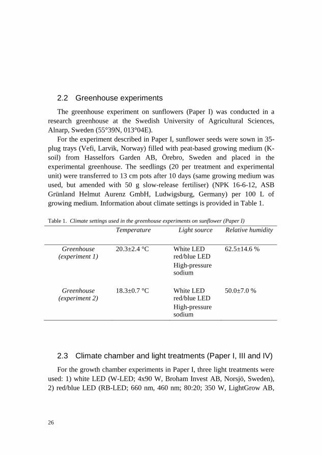
growing medium. Information about climate settings is provided in Table 1.
Table 1. Climate settings used in the greenhouse experiments on sunflower (Paper I)
Temperature Light source Relative humidity
Greenhouse (experiment 1)
20.3±2.4 °C
White LED red/blue LED
High-pressure sodium
62.5±14.6 %
Greenhouse (experiment 2)
18.3±0.7 °C
White LED red/blue LED
High-pressure sodium
50.0±7.0 %
2.3 Climate chamber and light treatments (Paper I, III and IV)
For the growth chamber experiments in Paper I, three light treatments were
used: 1) white LED (W-LED; 4x90 W, Broham Invest AB, Norsjö, Sweden),
2) red/blue LED (RB-LED; 660 nm, 460 nm; 80:20; 350 W, LightGrow AB,
27
Helsingborg, Sweden), and 3) high pressure sodium lamps (HPS 400 W,
Philips, Eindhoven, The Netherlands).
The photosynthetic photon flux density (PPFD) at canopy level was
adjusted to 70-120 μmol m-2 s-1 by adjusting the distance between the light
source and the top of the canopy. Artificial light was given for total of 16 h per
day, followed by exposure to natural daylight for 8 h per day.
To check the light transmission through different covering materials in
Paper III, the lids of 96-well PM microtitre plates were compared with six
other covering materials: Breath-easy sealing membrane, Titer-top sealing film
for microplates, sealing tape, household plastic film 1, household plastic film 2
and Greiner viewSeal for 96 plates) (Supplementary Table S1 in Paper I). For
this purpose, lid materials were exposed to three different LED sources: (i)
white LED, (ii) red LED (660 nm) and (iii) a combination (80/20) of red (660
nm) and blue (460 nm) light.
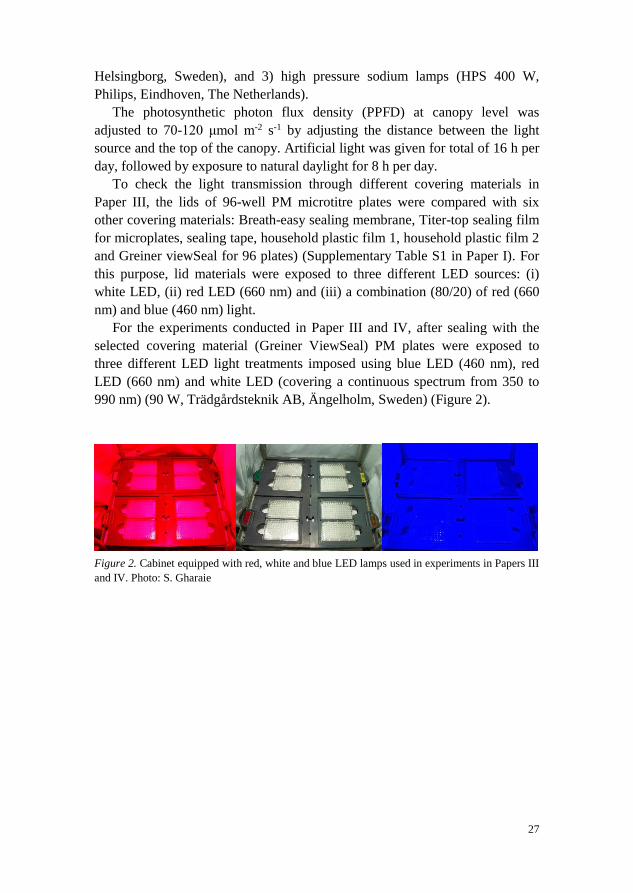
For the experiments conducted in Paper III and IV, after sealing with the
selected covering material (Greiner ViewSeal) PM plates were exposed to
three different LED light treatments imposed using blue LED (460 nm), red
LED (660 nm) and white LED (covering a continuous spectrum from 350 to
990 nm) (90 W, Trädgårdsteknik AB, Ängelholm, Sweden) (Figure 2).
Figure 2. Cabinet equipped with red, white and blue LED lamps used in experiments in Papers III
and IV. Photo: S. Gharaie
28
2.4 Microorganisms
Microbial strains and their culture conditions
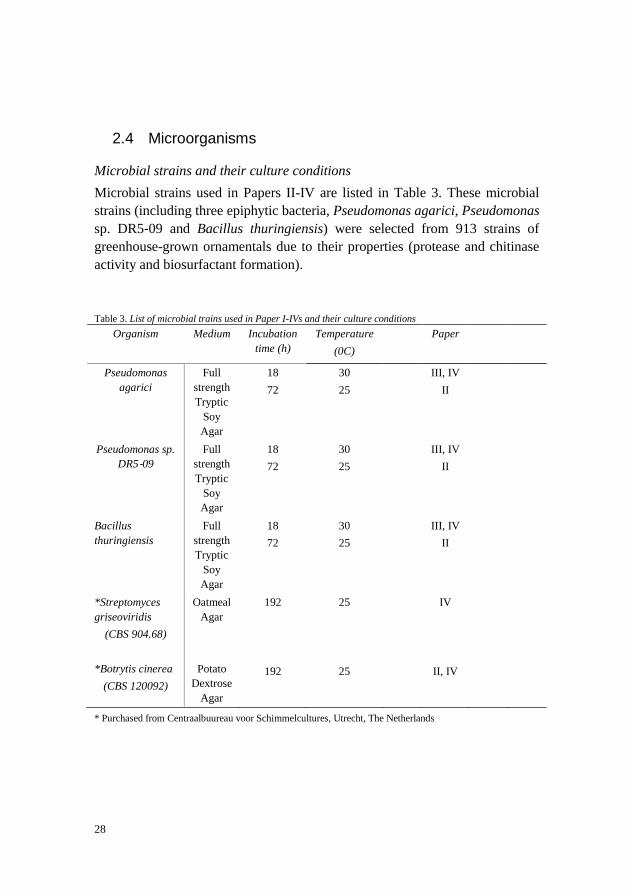
Microbial strains used in Papers II-IV are listed in Table 3. These microbial
strains (including three epiphytic bacteria, Pseudomonas agarici, Pseudomonas
sp. DR5‐09 and Bacillus thuringiensis) were selected from 913 strains of
greenhouse-grown ornamentals due to their properties (protease and chitinase
activity and biosurfactant formation).
Table 3. List of microbial trains used in Paper I-IVs and their culture conditions
Organism Medium Incubation
time (h)
Temperature
(0C)
Paper
Pseudomonas
agarici
Full
strength
Tryptic
Soy
Agar
18
72
30
25
III, IV
II
Pseudomonas sp.
DR5‐09
Full
strength
Tryptic
Soy
Agar
18
72
30
25
III, IV
II
Bacillus
thuringiensis
Full
strength
Tryptic
Soy
Agar
18
72
30
25
III, IV
II
*Streptomyces
griseoviridis
(CBS 904.68)
*Botrytis cinerea
(CBS 120092)
Oatmeal
Agar
Potato
Dextrose
Agar
192
192
25
25
IV
II, IV
* Purchased from Centraalbuureau voor Schimmelcultures, Utrecht, The Netherlands

29
2.5 Analyses
2.5.1 Plant analysis (Paper I)
Measurements of plant height, width, number of nodes and shoot length
(non-destructive measurements), and fresh and dry plant weight (destructive
measurements) in Paper I were performed at the end of the experiment on 10
plants per treatment. Dry weight was measured after two days of desiccation at
75 °C.
To have a consistent daily light integral for each treatment and experiment
in Paper I, light intensity was measured at the canopy level using a light meter
(Delta HD 2302.0, probe LP 471, Delta OHM, Padua, Italy) and light quality
was observed with a spectrophotometer (Delta HD 2302.0, probe LP 471,
Delta OHM, Padua, Italy). For measuring the rate of photosynthesis and
chlorophyll fluorescence, the third fully developed leaf from the apex was
used. The rate of photosynthesis was measured using a leaf chamber
photosynthesis meter (LC Pro+, ADC BioScientific Ltd, Hoddesdon, UK) and
chlorophyll fluorescence using a PAM-2500 fluorometer (Heinz Walz GmbH,
Effeltrich, Germany). Measurements of leaf temperature (top leaf) were
performed using an infrared camera (Flir IX, Flir systems Inc., Wilsonville OR,
USA).
2.5.2 Extraction of microbiota from the phyllosphere (Paper I and II)
For Papers I and II, apical and basal leaves were randomly removed from
selected plants. Further leaf samples were washed as described in Paper I and
II and used for plate counts, characterisation of enzyme activity and production
of biosurfactant, dual culture test, phenotypic microarray, sequencing and
meta-genomic analysis.
In order to store the microbial strains before testing (e.g. characterisation of
enzyme activity and production of biosurfactant, dual culture test, phenotypic
microarray and sequencing) in Paper II, strains were transferred to cryo
medium (described in detail in Paper II) and stored at -80 °C. In Paper I, for
metagenomics analysis the wash solution from leaf samples was directly
transferred to a sterile tube and after centrifugation and resuspension preserved
at -80 °C.

30
2.5.3 Culture-dependent microbial analyses
Viable count
Aliquots from the leaf wash-off were serially diluted and plated on various
growing media in order to assess a wide range of phyllosphere microorganisms
(Table 2). Total viable bacteria and fungi were counted as colony-forming
units (CFU) per mL and per g fresh weight (fw) (Papers I and II).
Table 2. . Overview of plate count analysis (semi-selective media and incubation conditions) for
enumeration of phyllosphere microorganisms (Papers 1 and II)
Diluted
Tryptic Soy
Agar (0.1x
TSA)
Standard I
nutrient
agar (SN1)
Standard II
nutrient
agar (SN2)
King B agar
(KB)*
Diluted
Malt
Extract
Agar
(0.5x MA)
Plate counts
Total
culturable
bacteria
Total
culturable
bacteria
Total
culturable
bacteria
Fluorescent
pseudomonas
Total fungi
Incubation time (h) 72 72 72 48 96
Incubation
temperature(0C)
25 25 25 25 25
Product number
Difco
218263
Merck
105450
Merck
107883
-
Difco
218630
* King et al., 1954.
Screening for enzyme activity (Paper II)
Isolated strains from the phyllosphere of greenhouse-grown ornamentals after
pure culturing and storing were screened for their enzyme activity (protease
and chitinase).
Figure 3. Production of clearing zone in skim milk agar plates by isolated strains. Photo: S.
Gharaie

31
For determination of protease (Figure 3) isolated strains were inoculated on
skim milk agar (SMA) (Smibert et al., 1994), while for chitinase activity they
were plotted on colloidal chitin minimal agar (CCMA) (Renwick et al., 1991),
with 24 strains spotted on each agar according to a prepared template. The 25th
position was used as a control. Plates were sealed, and halo formation assessed
after 24, 72 and 96 h at 30 °C.
Screening for biosurfactant formation (Papers II and IV)
For detection of biosurfactant production in Paper I, isolated strains after pure
culturing were assessed by drop-collapse test (flat drops indicated positive for
biosurfactant formation, whilst cultures with convex drops were recorded as
negative for biosurfactant formation) (Youssef et al., 2004).
In Paper IV, PM panels after inoculation with selected microorganisms
(Pseudomonas agarici, Pseudomonas sp. DR5‐09, Bacillus thuringiensis and
Streptomyces griseoviridis) and exposure to different types of LED light were
checked for detection of biosurfactant formation.
Dual culture test (Paper II)
Isolated strains that tested positive for chitinase activity and biosurfactant
formation were selected for dual culture test to evaluate their antagonistic
activity against Botrytis cinerea (CBS 1290092, Utrecht, Netherlands).
Candidate strains were inoculated on potato dextrose agar (PDA), King B agar
(KB) and 0.1x tryptic soy agar (TSA) (all Difco, Michigan, USA) on either
side of petri dishes (90 mm petri dish, 10 mm distance from the edge of the
petri dish) two days before inoculation of B. cinerea and thereafter the
pathogen (5 mm plug) was inoculated in the centre of the petri dishes. The
plates were incubated at 25 °C. The inhibition zone was measured after
mycelium reached the edge of the agar plates. Inhibition (%) was determined
according to an existing method (Skidmore & Dickinson, 1976).
Phenotypic microarray
In order to investigate the impact of light spectrum on utilisation of
different sole energy sources by phyllosphere microbiota, in Papers III and IV
microbial utilisation of energy sources was examined using Phenotype
MicroArray (PM) panels PM01, PM02, PM03 and PM04 (Biolog Inc., USA).
The PM panels are commercially available pre-filled 96-well microtitre plates

32
containing 190 different carbon sources (PM01 and PM02), 95 different
nitrogen sources (PM03), and 59 phosphorus and 35 sulphur sources (PM04).
The PM method was performed according to the manufacturer’s recommended
protocol. PM01 and PM02 were incubated as sole substrate, whereas PM03
and PM04 were supplemented with 2 mM sodium succinate and 2 μM ferric
citrate as additional carbon sources (enrichment). Microorganisms used for PM
assay and their culture conditions are described in detail in section 3.4. After
inoculation, the PM plates were sealed with selected covering material (Greiner
ViewSeal) and incubated in the Omnilog reader (Omnilog, Biolog Inc., USA)
in dark conditions or exposed to different light spectra (blue, red or white
LED). Detailed information regarding incubation time, temperature,
experimental set-up and data collection can be found in Papers III and IV.
2.5.4 Culture-independent analyses
Sequencing
The isolates displaying antagonistic activity were identified using 16S
rRNA gene sequencing in Paper II. The cryo-preserved cultures were grown on
full-strength TSA, incubated overnight at 30 °C and thereafter processed for
DNA extraction as described in Paper II. The PCR analysis of the 16S rRNA
genes was performed using the universal forward primer ENV1 and the reverse
primer ENV2. Amplicons with the correct size of amplified fragments (1500
bp) were sent to Eurofins MWG (Ebersberg, Germany). The primer used by
Eurofins MWG was ENV1.
Metagenomic analyses
Bacterial and fungal communities of sunflower leaves were investigated
using Illumina. For Illumina analysis, the wash solution was processed as
described in Paper I. The pellets obtained were used for the extraction of
genomic DNA (King Fisher Cell and Tissue DNA Kit, Product number:
97030196, Thermo Fisher Scientific Oy, Vantaa, Finland). The DNA
construction of amplicons of interest was determined by gel electrophoresis.
The amplicon pools were purified to remove primer, and additional purification
on MinElute columns (Qiagen) was also performed. Purified amplicon pool
DNA was used for constructing Illumina libraries. Illumina data were analysed
by the bioinformatics service of LGC Genomics, Berlin, Germany, using
QIIME1.8.0.

33
2.6 Calculations and statistics
General linear model (GLM) analysis, followed by Tukey’s test (p<0.05),
stepwise regression and cluster analysis (single linkage method), were
performed with Minitab (State College, PA, USA, Version 16.2.4) software
and biodiversity indices (Shannon H, Chao1) and Euclidian distance were
computed using the paleontological statistics software PAST (version 3) in
Paper I. Analysis of variance (ANOVA) in Papers II-IV was carried out using
Mintab (State College, PA, USA, Version 16.2.4) software. Phylogenetic
comparison in Paper II was conducted using Ribosomal Database Project,
release 11 (RDP, Michigan State University, East Lansing, USA). The PM data
in Papers III and IV were recorded using the OmniLog® PM kinetic analysis
software (Product Number UA24331-PMM, version 1.6), and thereafter
analysed using the R statistical software (Team, 2016) and functionality from
the dedicated R package opm (Vaas et al., 2013). Calculation of principal
component analysis (PCA) in Paper IV was performed using Minitab vers.17
(Minitab Inc., State College Pennsylvania). Detailed information on the
statistical methods used is provided in each individual Paper (I-IV).
34
3 Results and discussion
3.1 Microbial community structure in greenhouse-grown ornamentals (Papers I and II)
The phyllosphere is an ecologically and economically important ecosystem
that hosts a variety of microbial communities. Phyllosphere microbiota play a
critical role in protecting plants from diseases, as well as promoting their
growth by various mechanisms. There are gaps in our understanding of how
and why microbiota composition varies across spatial and temporal scales.
There is also a lack of knowledge regarding the ecology of leaf surface
colonisers, their interactions with their hosts and the genetic adaptations that
enable phyllosphere survival of microorganisms.
3.1.1 Effect of light spectrum on phyllosphere microbiota
In this thesis (Paper I), the effect of light spectrum on microbial
communities associated with the leaf microbiota of ornamental sunflower
(Helianthus annuus) grown in the greenhouse was examined.
The viable count results showed that light treatment had no effect on viable
counts of bacteria and fungi (Figure 5B in Paper I). However, there were
significant differences in viable counts between the different leaf positions on
all semi-selective media (Figure 5A in Paper I).
Leaves can be colonised by 103-106 culturable fungi and 106-109 bacteria
(Timms-Wilson et al., 2006). However, in Paper I the size of the bacterial
epiphytic populations was smaller, while the fungal counts were within the
reported range. The low viable counts of bacteria observed in Paper I might be
due to the extraction method used. Similar bacterial epiphytic population size
was found in Paper II using the same extraction method. Although viable
counts gave interesting information, it should be considered that culture-

35
dependent methods (viable counts) are inadequate to reflect the entire
phyllosphere microflora (Whipps et al., 2008; Yang et al., 2001). Therefore,
for investigation of microbial community composition exposed to different
light treatments in more detail, Illumina was used as a culture-independent
method, as a complement to the culture-dependent method.
Metasequencing of the fungal community indicated that different light
treatments affected species abundance and evenness, but not species richness
(Chao1) (Table 2 in Paper I). Irrespective of the light treatments, Ascomycota
was the dominant fungal phylum (Figure 6 in Paper I). On phylum level,
significant differences were observed between the two LED light treatments
(p=0.028; N=15) for Ascomycota. Its share within the fungal microbiome of
sunflower leaves was highest when exposed to white LEDs (98.1%) and lowest
when exposed to red-blue LED light (93.5%) (Figure 6B in Paper I). No
significant differences were observed for the relative abundance of Ascomycota
on sunflower leaves between HPS and LED treatments (red-blue and white
LED) (Figure 6A in Paper I). However, in the case of Basidiomycota,
significant differences were seen for the leaves exposed to white LED light for
relative abundance, and differences were seen between the two LED treatments
(p=0.036). There were, however, no differences in the case of Zygomycota or
miscellaneous phyla for either light treatment or leaf position. No interactions
between light treatment and leaf position were found for any of the phyla.
Distribution of fungal classes in the phyllosphere of sunflower was affected by
different light treatments (Figure 6A-C in Paper I) and leaf position (Figure
6D-E in Paper I).
The dominant class in the fungal microbiome of the sunflower phyllosphere
was Dothideomycetes when treated with HPS lamps. Its relative abundance
was decreased when exposed to LEDs. The share of both Leotimycetes and
Sordariomycetes was higher when exposed to LEDs (Figure 6A-C in Paper I).
In general few statistical differences were observed for the impact of light
treatment and the leaf position on the fungal microbiome of greenhouse-grown
sunflower.
On phylum level, there were no significant differences in bacterial
community in the phyllosphere except for the group of non-classified bacteria.
With respect to altered light treatment, Gammaproteobacteria (34-37%),
Alphaproteobacteria (18-23%), Betaproteobacteria (10-12%), Actinobacteria
(8.6-10.6%) and Sphingobacteria (5.2-5.7%) were the most dominant taxa. On
order level, no impact of light treatment or leaf position was observed, except
for Xanthomonadales. The impact of light treatment on some bacterial genera
associated with sunflower leaves was indirect, through the interaction between
36
leaf temperature, stomatal conductance and chlorophyll fluorescence (Figure 1
in Paper I).
The results from the first experiment performed in Paper I confirmed the
impact of light spectrum on the phyllosphere microbiota, which is consistent
with previous findings (Itagaki et al., 2016; Schuerger & Brown, 1994).
Interestingly, however, we found high colonisation by Golovinomyces and
Podosphaera (causative agents of powdery mildew; Mulpuri et al., 2016; Chen
et al., 2008; Braun, 1995) in the sunflower phyllosphere. Colonisation was
highest on canopies treated with white LEDs, and considerably lower when the
canopies were exposed to HPS and red-blue LEDs. A previous study
(Suthaparan et al., 2010) has reported a reduction in conidia germination of
Podosphaera pannosa on greenhouse roses when exposed to blue LEDs in
detached leaf assays, while a combination of red LED with 18 h of white LED
treatment followed by 6 h of red LEDs inhibited conidia formation in whole
plant tests. These results are in line with those in Paper I and support the initial
hypothesis regarding the effect of light spectrum quality on leaf microbiota.
The study reported in Paper I was the first to investigate the interaction
between light treatment, plant physiological properties and resident microbiota
of greenhouse-grown sunflower. It showed that the effect of light treatment on
phyllosphere microbiota (fungi species abundance and evenness) was mostly
due to different leaf temperatures under LEDs compared with HPS. Moreover,
no direct effects of light treatment were seen on photobiology parameters, but
there were correlations between these parameters and important bacterial and
fungal genera such as Bradyrhizobium, Sphingomonas, Brevibactericum,
Bacillus, Hypotrachyna and Aureobasidium. In addition, the effect of light
treatment on fungi was direct, whereas bacteria were affected indirectly
through plant environment fluctuations.
3.1.2 Occurrence of bacterial antagonistic to Botrytis cinerea (Paper II)
Botrytis cinerea is a necrotrophic fungal pathogen and causal agent of grey
mould, which is one of the most widespread fungal diseases, attacking over
200 plant species, including ornamentals. This pathogen causes substantial
commercial crop losses every year (Rupp et al., 2016; Hahn, 2014; Dean et al.,
2012; Williamson et al., 2007). It also has unlimited adaptability under broad
environmental conditions.
One of the aims of this thesis was to develop an optimal extraction
methodology to evaluate the phyllosphere microbiota of greenhouse-grown
ornamentals (Paper II). The method developed was then used for screening
37
bacteria with ability for enzyme activity, biosurfactant production and in vitro
antagonism towards Botrytis cinerea.
A suitable extraction procedure was selected through determination of the
impact of ultrasonic treatment, different buffers (PPB and TRIS) and
microbiological medium (TSA, SN1 and SN2) on the number of culturable
bacteria inhabiting the phyllosphere of model plant Begonia x elator (see
Materials & Methods section in Paper II). As can be seen from the results
(Figure 1 in Paper II), choice of buffer had a significant effect, with PPB
causing no difference in viable counts on the three nutrient media tested (0.1x
TSA, SN1 and SN2). Furthermore, following sonication the number of viable
counts significantly declined in comparison with the non-sonicated treatment.
Heterotrophic plate counts (HPC) displayed considerable differences
between the model crops within apical and basal leaves (Figure 2 in Paper II).
For instance, viable counts were significantly higher for Impatiens, in both
apical and basal leaves, but not for fluorescent pseudomonads. In the case of
HPC, higher counts were observed for basal leaves of conventionally grown
Poinsettia compared with its organically grown counterpart.
A total of 913 bacterial strains displaying morphological differences were
collected from apical and basal leaves of Poinsettia (conventionally and
organically grown), Begonia, Impatiens and Kalanchoë. All these bacterial
strains were screened for some antagonistic properties (protease and chitinase
activity and biosurfactant formation) against B. cinerea.
The phyllosphere is a dynamic environment, subjected to variations in
environmental factors, nutrient and water availability, plant species differences
and leaf age (Vorholt, 2012; Hunter et al., 2010; Redford & Fierer, 2009;
Whipps et al., 2008; Lindow & Brandl, 2003; Jager et al., 2001; Kinkel, 1997).
Resident microbial communities in the microenvironment provided by the
leaves differ considerably, depending on a variety of these factors affecting the
phyllosphere. Previous studies have suggested that bacterial colonisation and
bacterial distribution in the phyllosphere may also be governed by plant species
(Lambais et al., 2014; Vokou et al., 2012; Yadav et al., 2004; Yang et al.,
2001). Heterotrophic plate counts (Paper II) for Impatiens (apical and basal
leaves) and conventionally grown Poinsettia (basal leaves) had the highest
viable counts amongst the plants tested. It was therefore concluded that
significant differences in HPC between apical and basal leaves for different
model crops are probably due to species differences. These results,
representing the effect of plant species on bacterial counts on apical and basal
leaves, are in agreement with findings in previous studies (Knief et al., 2010;
Yang et al., 2001).

38
Most of bacteria (in terms of number) displaying enzyme (protease and
chitinase) and biosurfactant activity were retrieved from basal leaves of
Impatiens. Previous studies have suggested that bacteria displaying enzyme
and biosurfactant activity have potential as biocontrol agents (Kefi et al., 2015;
D'aes et al., 2010; Hultberg et al., 2010; Trotel-Aziz et al., 2008; Soberón-
Chávez et al., 2005). These bacterial strains can degrade fungal cell walls by
production of chitinase (Kefi et al., 2015; Kim et al., 2012; Yan et al., 2011) or
inhibit fungal growth through formation of biosurfactants (Varnier et al.,
2009), which are biologically produced amphiphilic compounds that exhibit
surface activity through the actions of their hydrophilic and hydrophobic
groups (Burch et al., 2011). As most of the epiphytic colonisers with
antagonistic properties identified in this thesis were retrieved from basal leaves
of Impatiens, an effect of plant species and leaf age was also observed. This
indicates that these factors might affect the abundance of bacterial strains with
biocontrol properties in the phyllosphere, but more research is necessary to
confirm this.
In Paper II, a dual culture test on the three semi-selective media (0.1x TSA,
KB and PDA) was employed to check the inhibitory effect of candidate strains
displaying chitinase activity and biosurfactant formation against B. cinerea. To
determine the nutritional effect of three semi-selective media on the inhibition
activity, comparisons were made between these media. The results showed that
mycelial growth of B. cinerea was inhibited by some selected strains among
the 67 strains examined. For instance, strain PCb52T extracted from the basal
leaves of conventionally grown Poinsettia showed the strongest inhibition
against B. cinerea on 0.1x TSA, whereas on KB, strain Ia176K, which was
isolated from apical leaves of Impatiens, displayed the strongest inhibition.
Mycelial growth of B. cinerea when grown on PDA was also strongly inhibited
by strain Ib44K extracted from basal leaves of Impatiens. On comparing the
different media, it was found that the inhibitory effect was significantly higher
on some media for some strains, such as Ib44K on PDA compared with KB
(Table 3 in Paper II). A previous study has also reported inhibitory effects on
fungi by bacterial biocontrol agents, in a dual culture test conducted on two
forms of nutrient media (PDA and 0.1x TSA) (Sylla et al., 2013). This
differential influence of various nutrient media on inhibitory effect might
indicate an effect of nutrient composition.
The strains with biosurfactant production, chitinase activity and B. cinerea-
inhibiting properties in vitro were identified with 16S rRNA sequencing.
Proteobacteria, Actinobacteria and Firmicutes were the three major phyla to
which the identified isolates belonged (Table 4 in Paper II). Furthermore, nine
isolates with in vitro properties to inhibit mycelial growth of B. cinerea were

39
identified (Pseudomonas vancouverensis, Pseudomonas asplenii,
Pseudomonas segetis, Pseudomonas mosselii, Pseudomonas reinekei, Bacillus
subtilis, Rhizobium rosettiformans, Paenibacillus taichungensis and
Enterobacter kobei). Among these nine strains, only B. subtilis has previously
been reported in this context (Kefi et al., 2015). Although that study also
concluded that strains with antagonistic properties might affect the suppression
of B. cinerea (Kefi et al., 2015), it should be noted that as there were no
interactions in planta in this thesis and as results were obtained from controlled
conditions, the underlying mechanisms need further research.
3.2 Impact of light spectrum on utilisation of energy sources by selected phyllosphere bacteria (Papers III and IV)
There is limited information about the impact of light spectrum on the
functionality and composition of non-phototrophic bacterial phyllosphere
biota. Therefore, Papers III and IV investigated the utilisation pattern of energy
sources in selected phyllosphere microbiota under different LED spectra. An in
vitro method was developed to study phenotypic profile responses of pure
bacterial cultures to different LED regimes by modification and optimisation of
a protocol for the Phenotype MicroArray™ technique (Paper III).
Pseudomonas sp. DR 5-09 was used as a representative bacterial strain
model in Paper III, as it tested positive for protease, chitinase and biosurfactant
properties in screening in Paper II. Paper III examined the utilisation of C, N, P
and S sources as a function of maximum curve height under different light
regimes. In addition to darkness (considered control conditions), blue (460
nm), red (660 nm) and white (350-990 nm) LEDs were used.
Substrate utilisation assays were conducted using four pre-fabricated panels
consisting of 379 substrates and conditions (190 C sources, 95 N, 59 P, 35 S).
Responses generated for the 190 C sources provided a basis for distinguishing
patterns for each light regime included in the study.
Carbon utilisation in darkness and in red light incubation regime had least
influence on the maximum curve height, and these clustered together compared
with white and blue light. In addition, blue light incubation for sole C sources
deviated from all other light treatments. In general, the blue light spectrum had
the most decisive impact on substrate utilisation of C sources.
Similarly, a strong impact of blue light spectrum on respiration was
recorded for N utilisation. Different N sources (e.g. L-theronine, D-asparagine,
L-isoleucine, cytosine, D,L-α-amino-N-butyric acid, D-mannosamine and
nitrate) were affected under all light conditions except blue light. Overall,

40
under blue light N utilisation was significantly lower in terms of maximum
curve height of dark conditions.
Similarly, P utilisation pattern showed significant differences for blue light
compared with red, white and dark incubation conditions. Of 59 P sources
included, 53 sources differed significantly under blue light incubation, while
the sources were metabolised to the same extent under all other light regimes.
Further analysis on the effect of selected wavelengths on energy source
utilisation by Pseudomonas sp. DR 5-09 revealed that reduced substrate
utilisation was due to the restrictive effect of light exposure on metabolic
pathways. Blue light interferes with several major critical metabolite pathways.
With respect to the 379 different substrate and conditions, no general effect of
light regime on substrate utilisation was observed. Through KEGG pathway
analyses, it might be possible to identify which pathways are affected. It is
important to understand the consequences of inhibited pathways on the general
utilisation of substrates.
Substrate utilisation by Pseudomonas sp. DR 5-09 was studied under
different light treatments. Some of the substrate sources were provided as sole
substrate and some were enriched with C sources (Figure 6 in Paper III). For
some of the sources, such as thymidine and D-aspartic acid, utilisation was not
affected irrespective of light regime and nutritional status. However, some
other sources, such as L-lysine, D-serine and D-glucose-6-phosphate, showed
considerable utilisation when they were provided as enriched compared with
sole substrate. Utilisation of substrate was the other way around, with higher
utilisation observed for sole substrate compared with enriched. Thus no general
pattern was observed for energy source utilisation of substrates when they were
provided as enriched sources. In a previous study on Pseudomonas aeruginosa,
succinate and some other sources, such as L-aspartate, glycerol, L-glutamate,
L-asparagine, fumarate, α-ketoglutarate and L-glutamine, were found to be the
main and most preferred C sources due to their positions in the citric acid cycle
(Li & Lu, 2007). In contrast, the results obtained in this thesis showed higher
levels of respiration for C sources such as L-aspartic acid, L-arginine,
putrescine, L-pyroglutamic acid, L-serine, L-glutamine, L-asparagine, L-
proline and L-glutamic acid, even in the absence of succinate. Hence there is a
need for further studies to identify the role of these C sources in different
conditions.
It is not possible to draw any conclusions based on nutrient utilisation, since
it also depends on other environmental factors. The results from this study
showed that utilisation was most likely higher in enriched conditions than sole
substrate conditions. This supports previous findings by Li and Lu (2007). Low
utilisation of some of the substrates when provided as the sole source in this

41
thesis might be explained by a lack of uptake mechanisms or absence of
essential nutrients. The ATP-binding cassette (ABC) uptake systems are the
largest transport systems in bacteria. Almost all of the less-preferred sugars in
bacteria are transported by the ABC uptake systems (Rees et al., 2009;
Higgins, 1992). A recent study (Maqbool et al., 2015) demonstrated that this
substrate-binding protein is substrate-specific and has high affinity for the
ABC uptake systems.
Carbon catabolite repression (CCR) is a regulatory process in many bacteria
such as the Pseudomonas genus (Rojo, 2010). This process assists in fast
adaptation to the competitive environment by utilising preferred C sources for
energy generation (Görke & Stülke, 2008). It works as a regulatory mechanism
that inhibits the synthesis of enzymes for less-preferred sources and
overexpression of virulence genes that help the bacteria to access new nutrient
sources (Moreno et al., 2009; Görke & Stülke, 2008). Hence, CCR has been
shown to be a driving force for evolution in many Gram-negative bacteria,
including both free-living and pathogenic strains. In this study succinate was
used as a preferred C source, as in other studies (Li & Lu, 2007; Collier et al.,
1996). The molecular mechanisms in the genus Pseudomonas are not
completely understood, but it is most likely that CCR or reverse CCR activates
a regulatory mechanism for nutrient uptake.
Nutritional status might also be an important factor for substrate utilisation.
Based on previous studies (Beier et al., 2015; Werner et al., 2014) and this
results, one could conclude that in addition to light spectrum, nutritional factors
play an important role in the microbial phenotypic response.
Paper IV examined light-dependent phenotypic plasticity in phyllosphere
bacterial strains in terms of substrate resource utilisation upon exposure to
different light regimes. The strains tested were Pseudomonas agarici,
Pseudomonas sp. DR 5-09 (results already discussed above), Bacillus
thuringeinsis serovar israeliensis and Streptomyces griseoviridis. The results
indicated a distinct impact of light regime on substrate utilisation by
Pseudomonas agarici, Pseudomonas sp. DR 5-09 and Streptomyces
griseoviridis (Figure 4 in Paper IV) as follows: (i) In the case of P. agarici (C
sources) and S. griseoviridis (C, N, P and S sources), blue LED and white
LEDs differed from red LED and dark incubation. (ii) In the case of P. agarici
(P and S sources), blue and red LED differed from white LED and dark
incubation. (iii) For N sources for P. agarici, dark incubation had a distinctly
different response from all LED regimes. (iv) In the case of B. thuringiensis
(all sources), no responses to light regime were observed.
Apart from these main findings, differences were seen for the different
substrate x light interactions for each specific strain. As previously mentioned,

42
for Pseudomonas sp. DR 5-09 light regime and enrichment conditions
significantly affected substrate utilisation. For example, in the case of P.
agarici, utilisation of substrate, when provided as both sole and enriched,
discriminated the impact of blue LED from that of all other light regimes.
No Impacts of light regime were observed for energy source utilisation of
sole and enriched substrates by B. thuringiensis. Better utilisation was
observed for most of the substrates when they were offered as sole C sources.
Regarding the fourth strain (S. griseoviridis), the impact of light regime on
substrate utilisation was less noticeable than for P. agarici.
In further analyses, the two Gram-negative strains (Pseudomonas) were
compared regarding their response as light-dependent phenotypic effects.
Differences were mostly found for C sources when they were incubated in dark
conditions. For S sources, exposure to red LED and incubation under blue LED
decreased the differences in substrate utilisation.
Comparisons between the four selected strains revealed that not all
differences between these strains were dependent on light regime. Overall, the
results indicated that blue light impairs the utilisation of sole and enriched
substrates by the two selected Pseudomonas strains and has similar, but less
pronounced, implications for S. griseoviridis. However, substrate utilisation by
B. thuringiensis appears to be indifferent to light regime during incubation
(Figures 4, 6-7).
Evolutionary responses to environmental changes are mainly influenced
by different environmental conditions (Salinas & Munch, 2012; Wong &
Ackerly, 2005). Therefore microorganisms, besides having a versatile
metabolism which is linked to tight but flexible regulation of the expression of
metabolic pathways that directly optimise efficiency and ecological fitness
(already discussed), try to use other modes of adaptation to variable
environments. Phenotype plasticity is one of these modes of adaptation used by
organisms and suggested in many other fields, such as zoology (Furness et al.,
2015; Miner et al., 2005). Adaptive phenotypic plasticity is a common
phenomenon by which a genotype can create different phenotypes from a
single genotype depending upon environment (Ghalambor et al., 2007; Sultan
& Stearns, 2005; Bradshaw, 1965). The hypothesis tested in this thesis was that
light, as one of the environmental cues, can affect the phenotype plasticity of
epiphytic phyllosphere colonisers. A previous study in another field reported
that environmental factors such as light and temperature can induce phenotype
plasticity (Furness et al., 2015). Others (e.g. Miner et al., 2005) highlight the
effect of phenotype plasticity on community structure due to phenotypic
alteration, in addition to direct and indirect effects of phenotype plasticity with
respect to adaptability and alterations in consumption pattern. In this thesis, it

43
was found that utilisation of sole and enriched substrates by the two Gram-
negative strains was impaired by blue light, but to a much lower extent for S.
griseoviridis and no effect of light regime on substrate utilisation by B.
thuringiensis was observed. These interactions and the effects of light on
substrate utilisation and phenotype plasticity of any of our selected bacterial
strains have not been studied previously, but the present findings supported our
hypothesis. Thus it might be tempting to draw the conclusion that light
spectrum affects the phenotype plasticity of phyllosphere residents, but at the
same time it should be keep in mind that of course this does not apply to all
epiphytic bacteria.
Light sensing as a signal for morphogenesis and the metabolites for this
process are well-known for many bacteria. Previous studies have also reported
that non-phototrophic bacteria can sense light, with blue light having an impact
on their lifestyle (Kraiselburd et al., 2012; Ondrusch & Kreft, 2011; Swartz et
al., 2007; van der Horst et al., 2007). As reported in previous reviews (Losi &
Gärtner, 2016; Yin et al., 2013), sensitivity of microbial cells to blue light has
been found. In this thesis, putative blue light receptor proteins were found
(only two Gram-negative strains were checked). However, knowledge of
molecular mechanisms for these selected model species is currently lacking.
Based on the results, one could draw the conclusion that blue light mediates
substrate utilisation under sole and enriched substrate conditions.
In the selected strains there were indications of light receptor activity, and
consequently of monitoring environmental fluctuations. Based on available
literature and the present thesis, it might be possible to draw the conclusion
that blue light receptors affect metabolic pathways in terms of affecting
bacterial lifestyle (biofilm formation, swarming activity etc.). This cascade of
events will eventually lead to the development of novel strategies to fight
bacterial pathogenesis. However, another aspect is that the two Pseudomonas
species in this thesis harboured regions for putative blue light receptor protein,
but some deviations were still observed between their utilisation of sole and
enriched substrates on exposure to different light regimes. This shows the
importance of nutritional status, beside light receptors, an important topic for
further analyses and future studies.
3.3 Impact of light spectrum on the formation of metabolites decisive for leaf colonisation (Paper IV)
Biosurfactants are a group of diverse molecules that have several biological
function (Banat et al., 2010). Biosurfactants share a basic structure, which

44
consists of a hydrophobic and a hydrophilic moiety (Mulligan, 2005). The
nature of biosurfactants is to produce amphiphilic compounds that exhibit
surface activity (Burch et al., 2011). These molecules act in three ways: (i)
adaptation of surface properties (Debode et al., 2007), (ii) alteration of
compound bio-availability (Das & Mukherjee, 2007) and (iii) interaction with
membranes (de Bruijn et al., 2007). Their action is a function of their specific
structure and production characteristics (D'aes et al., 2010)
Microorganisms that are able to form biosurfactants can be found in every
imaginable environment (Kefi et al., 2015; Hultberg et al., 2010; Haddad et al.,
2008). The frequency and versatility of surface-active compounds produced
displays the importance of biosurfactants for the functioning and survival of
the organisms (Banat et al., 2010). Epiphytic bacteria utilise biosurfactants to
increase the wettability of the leaf, to increase nutrient transmission across the
waxy cuticle and to move to favourable growth sites (D'aes et al., 2010;
Lindow & Brandl, 2003). The many possible contributions to epiphytic fitness
of biosurfactant-producing bacteria inspired the work in this thesis to check the
capacity of four selected bacterial strains to form biosurfactants as a response
to nutrient availability and light spectrum.
In Paper IV, the biosurfactant formation of four strains was evaluated using
the drop collapse test (see Materials & Methods section in Paper IV). The
results indicated that the effect of light regime on biosurfactant production was
lower and inconsistent for the two Gram-positive strains studied. However, the
two Gram-negative strains produced biosurfactants when exposed to different
light treatments. Biosurfactant production was decreased when the bacteria
were grown under a blue LED spectrum.
Another interesting finding in this thesis was that the four strains examined
behaved differently when utilising different substrates (Tween 20, Tween 40
and Tween 80), which themselves are surfactant agents. Moreover,
observations of drop collapse on these surfactant substrates even after exposure
to different light spectra and of different utilisation patterns on these substrates
as a function of light regime indicate an impact of light spectrum on
biosurfactant formation. It was also found that, besides effects of light
spectrum, substrate richness was decisive for biosurfactant formation. For
instance D‐alanine, L‐phenyl alanine, glycine, L‐threonine, α‐amino‐N valeric
acid and γ‐amino‐N‐valeric acid, when supplied as N source, were utilised and
affected the capacity of P. agarici to form biosurfactants, but not when
supplied as C source. However, for other substrates such as iso-leucine,
biosurfactant was formed in both sole and enriched substrate when exposed to
blue LED light.
45
Other strains of the four species examined in this thesis have already been
reported to display a capacity to form biosurfactants (Silva et al., 2016; Kefi et
al., 2015; Kalyani et al., 2014; Hue et al., 2001), and tested positive for this
property in Paper II when screened in vitro. As there is little published
information regarding biosurfactant formation by these selected strains and the
chemical nature of compound/s behind this activity, it is difficult to draw firm
conclusion on which compounds are decisive for biosurfactant formation.
In a plant ecology perspective, the role of biosurfactants is different
(Hultberg et al., 2010; Raaijmakers et al., 2006). For instance, biosurfactants
might promote bacterial colonisation of leaves to find new habitats in the
phyllosphere, or reduce the tension of the leaf surface caused by the waxy layer
of leaf cuticle. The findings in Paper IV suggest that the ability to investigate
new habitats in the plant phyllosphere is a function of both light spectrum and
presence of certain precursor compounds.
Another interesting finding in this thesis was that, under blue LED spectrum
biosurfactant formation was not supported in the presence of some enriched
substrates. This might indicate that the blue light spectrum and nutritional
factors affected bacterial ability to form biosurfactants, possibly through a
switch in metabolic pathway. Previous studies have reported that blue and
white light inhibit the swarming motility of Pseudomonas syringae pv tomato
DC3000 (Río‐Álvarez et al., 2014), and regulate the swarming motility of the
foliar pathogen Pseudomonas syringae (Wu et al., 2013). Blue light has also
antimicrobial activity against many bacterial and fungal pathogens (Bonomi et
al., 2016; El Din et al., 2016; Ricci et al., 2015; Maclean et al., 2014). All
these findings were supported by the results in this thesis. Together, these
findings raise the following questions: (i). How can new approaches with
respect to lighting strategy be exploited for better establishment of
Pseudomonas biocontrol strains? and (ii) How can light treatment be used to
suppress the impact of plant pathogenic bacteria?

46
4 Conclusions
The key findings of this thesis were that:
Choice of light for artificial assimilation lighting in the greenhouse
can affect sunflower plant shape and its phyllosphere microbial
community structure.
Several dominant bacterial leaf colonisers are dependent on leaf
temperature.
Light source affects phyllosphere-associated fungi (directly
through the physical properties of the chosen light source) and
bacteria (indirectly through alterations in the plant environment) on
sunflower leaves.
Complementary use of culture-dependent and culture-independent
methods is a suitable approach for investigating microbial
communities in the phyllosphere of greenhouse-grown
ornamentals.
There is a need for standard protocols to survey the phyllosphere of
greenhouse-grown ornamentals when mining for microbial
biocontrol agents with the potential to control diseases.
Phenotypic properties of microorganisms may change when
greenhouse-grown ornamentals are exposed to artificial lighting.
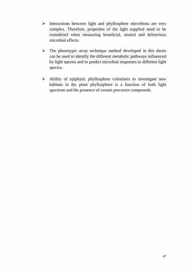
47
Interactions between light and phyllosphere microbiota are very
complex. Therefore, properties of the light supplied need to be
considered when measuring beneficial, neutral and deleterious
microbial effects.
The phenotypic array technique method developed in this thesis
can be used to identify the different metabolic pathways influenced
by light spectra and to predict microbial responses to different light
spectra.
Ability of epiphytic phyllosphere colonisers to investigate new
habitats in the plant phyllosphere is a function of both light
spectrum and the presence of certain precursor compounds.

48
References
Ávila-Pérez, M., Hellingwerf, K.J. & Kort, R. (2006). Blue light activates the σB-
dependent stress response of Bacillus subtilis via YtvA. Journal of
bacteriology, 188(17), pp. 6411-6414.
Bálint, M., Bartha, L., O'Hara, R.B., Olson, M.S., Otte, J., Pfenninger, M.,
Robertson, A.L., Tiffin, P. & Schmitt, I. (2015). Relocation, high‐latitude
warming and host genetic identity shape the foliar fungal microbiome of
poplars. Molecular ecology, 24(1), pp. 235-248.
Banat, I.M., Franzetti, A., Gandolfi, I., Bestetti, G., Martinotti, M.G., Fracchia, L.,
Smyth, T.J. & Marchant, R. (2010). Microbial biosurfactants production,
applications and future potential. Applied microbiology and
biotechnology, 87(2), pp. 427-444.
Beattie, G., Lindow, S., Hecht-Poinar, E. & Elliott, V. (2002). Leaf surface waxes
and the process of leaf colonization by microorganisms. Phyllosphere
microbiology, pp. 3-26.
Beier, S., Rivers, A.R., Moran, M.A. & Obernosterer, I. (2015). Phenotypic
plasticity in heterotrophic marine microbial communities in continuous
cultures. The ISME journal, 9(5), pp. 1141-1151.
Berg, G., Krechel, A., Ditz, M., Sikora, R.A., Ulrich, A. & Hallmann, J. (2005).
Endophytic and ectophytic potato-associated bacterial communities differ
in structure and antagonistic function against plant pathogenic fungi.
FEMS Microbiology Ecology, 51(2), pp. 215-229.
Bergstrand, K.-J. & Schüssler, H. Recent progresses on the application of LEDs in
the horticultural production. In: Proceedings of XXVIII International
Horticultural Congress on Science and Horticulture for People
(IHC2010): International Symposium on 9272010, pp. 529-534.
Bernard, F., Sache, I., Suffert, F. & Chelle, M. (2013). The development of a foliar
fungal pathogen does react to leaf temperature! New Phytologist, 198(1),
pp. 232-240.
Besnard, V., Federighi, M. & Cappelier, J. (2000). Development of a direct viable
count procedure for the investigation of VBNC state in Listeria
monocytogenes. Letters in applied Microbiology, 31(1), pp. 77-81.

49
Bian, Z.H., Yang, Q.C. & Liu, W.K. (2015). Effects of light quality on the
accumulation of phytochemicals in vegetables produced in controlled
environments: a review. Journal of the Science of Food and Agriculture,
95(5), pp. 869-877.
Bochner, B.R. (2003). New technologies to assess genotype–phenotype
relationships. Nature Reviews Genetics, 4(4), pp. 309-314.
Bochner, B.R. (2009). Global phenotypic characterization of bacteria. FEMS
microbiology reviews, 33(1), pp. 191-205.
Bochner, B.R., Gadzinski, P. & Panomitros, E. (2001). Phenotype microarrays for
high-throughput phenotypic testing and assay of gene function. Genome
research, 11(7), pp. 1246-1255.
Bodenhausen, N., Bortfeld-Miller, M., Ackermann, M. & Vorholt, J.A. (2014). A
synthetic community approach reveals plant genotypes affecting the
phyllosphere microbiota. PLoS Genet, 10(4), p. e1004283.
Bonomi, H.R., Toum, L., Sycz, G., Sieira, R., Toscani, A.M., Gudesblat, G.E.,
Leskow, F.C., Goldbaum, F.A., Vojnov, A.A. & Malamud, F. (2016).
Xanthomonas campestris attenuates virulence by sensing light through a
bacteriophytochrome photoreceptor. EMBO reports, p. e201541691.
Bourget, C.M. (2008). An introduction to light-emitting diodes. HortScience,
43(7), pp. 1944-1946.
Bradshaw, A.D. (1965). Evolutionary significance of phenotypic plasticity in
plants. Advances in genetics, 13, pp. 115-155.
Brandl, M.T. & Mandrell, R.E. (2002). Fitness of Salmonella enterica serovar
Thompson in the cilantro phyllosphere. Applied and environmental
microbiology, 68(7), pp. 3614-3621.
Braun, U. (1995). The powdery mildews (Erysiphales) of Europe: VEB Gustav
Fischer Verlag.
Brazaityte, A., Ulinskaite, R., Duchovskis, P., Samuoliene, G., Siksnianiene, J.,
Jankauskiene, J., Sabajeviene, G., Baranauskis, K., Staniene, G. &
Tamulaitis, G. (2006). Optimization of lighting spectrum for
photosynthetic system and productivity of lettuce by using light-emitting
diodes. Acta Horticulturae, 711, p. 183.
Bula, R., Morrow, R., Tibbitts, T., Barta, D., Ignatius, R. & Martin, T. (1991).
Light-emitting diodes as a radiation source for plants. HortScience, 26(2),
pp. 203-205.
Bunster, L., Fokkema, N.J. & Schippers, B. (1989). Effect of surface-active
Pseudomonas spp. on leaf wettability. Applied and environmental
microbiology, 55(6), pp. 1340-1345.
Burch, A.Y., Browne, P.J., Dunlap, C.A., Price, N.P. & Lindow, S.E. (2011).
Comparison of biosurfactant detection methods reveals hydrophobic
surfactants and contact‐regulated production. Environmental
Microbiology, 13(10), pp. 2681-2691.
Caporaso, J.G., Lauber, C.L., Walters, W.A., Berg-Lyons, D., Huntley, J., Fierer,
N., Owens, S.M., Betley, J., Fraser, L. & Bauer, M. (2012). Ultra-high-
throughput microbial community analysis on the Illumina HiSeq and
MiSeq platforms. The ISME journal, 6(8), pp. 1621-1624.

50
Carlsson-Kanyama, A. (1998). Food consumption patterns and their influence on
climate change: greenhouse gas emissions in the life-cycle of tomatoes
and carrots consumed in Sweden. Ambio, pp. 528-534.
Carvalho, S.D. & Folta, K.M. (2014). Sequential light programs shape kale
(Brassica napus) sprout appearance and alter metabolic and nutrient
content. Horticulture research, 1, p. 8.
Chen, M., Chory, J. & Fankhauser, C. (2004). Light signal transduction in higher
plants. Annu. Rev. Genet., 38, pp. 87-117.
Chen, R.-S., Chu, C., Cheng, C.-W., Chen, W.-Y. & Tsay, J.-G. (2008).
Differentiation of two powdery mildews of sunflower (Helianthus annuus)
by a PCR-mediated method based on ITS sequences. European journal of
plant pathology, 121(1), pp. 1-8.
Chen, X., Koumoutsi, A., Scholz, R., Schneider, K., Vater, J., Süssmuth, R., Piel,
J. & Borriss, R. (2009). Genome analysis of Bacillus amyloliquefaciens
FZB42 reveals its potential for biocontrol of plant pathogens. Journal of
biotechnology, 140(1), pp. 27-37.
Collier, D., Hager, P. & Phibbs, P. (1996). Catabolite repression control in the
Pseudomonads. Research in microbiology, 147(6), pp. 551-561.
D'aes, J., De Maeyer, K., Pauwelyn, E. & Höfte, M. (2010). Biosurfactants in
plant–Pseudomonas interactions and their importance to biocontrol.
Environmental microbiology reports, 2(3), pp. 359-372.
Das, K. & Mukherjee, A. (2007). Differential utilization of pyrene as the sole
source of carbon by Bacillus subtilis and Pseudomonas aeruginosa strains:
role of biosurfactants in enhancing bioavailability. Journal of applied
microbiology, 102(1), pp. 195-203.
Davis, C. & Brlansky, R. (1991). Use of immunogold labelling with scanning
electron microscopy to identify phytopathogenic bacteria on leaf surfaces.
Applied and environmental microbiology, 57(10), pp. 3052-3055.
de Bruijn, I., de Kock, M.J., Yang, M., de Waard, P., van Beek, T.A. &
Raaijmakers, J.M. (2007). Genome‐based discovery, structure prediction
and functional analysis of cyclic lipopeptide antibiotics in Pseudomonas
species. Molecular microbiology, 63(2), pp. 417-428.
de Carbonnel, M., Davis, P., Roelfsema, M.R.G., Inoue, S.-i., Schepens, I.,
Lariguet, P., Geisler, M., Shimazaki, K.-i., Hangarter, R. & Fankhauser,
C. (2010). The Arabidopsis PHYTOCHROME KINASE SUBSTRATE2
protein is a phototropin signaling element that regulates leaf flattening and
leaf positioning. Plant physiology, 152(3), pp. 1391-1405.
Dean, R., Van Kan, J.A., Pretorius, Z.A., Hammond‐Kosack, K.E., Di Pietro, A.,
Spanu, P.D., Rudd, J.J., Dickman, M., Kahmann, R. & Ellis, J. (2012).
The Top 10 fungal pathogens in molecular plant pathology. Molecular
plant pathology, 13(4), pp. 414-430.
Debode, J., Maeyer, K.D., Perneel, M., Pannecoucque, J., Backer, G.D. & Höfte,
M. (2007). Biosurfactants are involved in the biological control of
Verticillium microsclerotia by Pseudomonas spp. Journal of applied
microbiology, 103(4), pp. 1184-1196.
Delmotte, N., Knief, C., Chaffron, S., Innerebner, G., Roschitzki, B., Schlapbach,
R., von Mering, C. & Vorholt, J.A. (2009). Community proteogenomics

51
reveals insights into the physiology of phyllosphere bacteria. Proceedings
of the National Academy of Sciences, 106(38), pp. 16428-16433.
Devlin, P.F., Christie, J.M. & Terry, M.J. (2007). Many hands make light work.
Journal of experimental botany, 58(12), pp. 3071-3077.
Ding, T. & Melcher, U. (2016). Influences of plant species, season and location on
leaf endophytic bacterial communities of non-cultivated plants. PloS one,
11(3), p. e0150895.
Ding, T., Palmer, M.W. & Melcher, U. (2013). Community terminal restriction
fragment length polymorphisms reveal insights into the diversity and
dynamics of leaf endophytic bacteria. BMC microbiology, 13(1), p. 1.
Downs, R.J. & Thomas, J.F. (1982). Phytochrome regulation of flowering in the
long-day plant, Hyoscyamus niger. Plant physiology, 70(3), pp. 898-900.
El Din, S.N., El-Tayeb, T.A., Abou-Aisha, K. & El-Azizi, M. (2016). In vitro and
in vivo antimicrobial activity of combined therapy of silver nanoparticles
and visible blue light against Pseudomonas aeruginosa. International
journal of nanomedicine, 11, p. 1749.
Fankhauser, C. & Chory, J. (1997). Light control of plant development. Annual
review of cell and developmental biology, 13(1), pp. 203-229.
Folta, K.M. & Childers, K.S. (2008). Light as a growth regulator: controlling plant
biology with narrow-bandwidth solid-state lighting systems. HortScience,
43(7), pp. 1957-1964.
Foster, K.R. & Bell, T. (2012). Competition, not cooperation, dominates
interactions among culturable microbial species. Current biology, 22(19),
pp. 1845-1850.
Fukuda, N., Fujita, M., Ohta, Y., Sase, S., Nishimura, S. & Ezura, H. (2008).
Directional blue light irradiation triggers epidermal cell elongation of
abaxial side resulting in inhibition of leaf epinasty in geranium under red
light condition. Scientia Horticulturae, 115(2), pp. 176-182.
Furness, A.I., Lee, K. & Reznick, D.N. (2015). Adaptation in a variable
environment: Phenotypic plasticity and bet‐hedging during egg diapause
and hatching in an annual killifish. Evolution, 69(6), pp. 1461-1475.
Gadtke, L. (2010). Denmark-Sustainable horticulture crop production.
Ghalambor, C.K., McKay, J.K., Carroll, S.P. & Reznick, D.N. (2007). Adaptive
versus non‐adaptive phenotypic plasticity and the potential for
contemporary adaptation in new environments. Functional ecology, 21(3),
pp. 394-407.
Gomelsky, M. & Hoff, W.D. (2011). Light helps bacteria make important lifestyle
decisions. Trends in microbiology, 19(9), pp. 441-448.
Görke, B. & Stülke, J. (2008). Carbon catabolite repression in bacteria: many ways
to make the most out of nutrients. Nature Reviews Microbiology, 6(8), pp.
613-624.
Haddad, N.I., Wang, J. & Mu, B. (2008). Isolation and characterization of a
biosurfactant producing strain, Brevibacilis brevis HOB1. Journal of
industrial microbiology & biotechnology, 35(12), pp. 1597-1604.
Hahn, M. (2014). The rising threat of fungicide resistance in plant pathogenic
fungi: Botrytis as a case study. Journal of chemical biology, 7(4), pp. 133-
141.

52
Hakovirta, J.R., Prezioso, S., Hodge, D., Pillai, S.P. & Weigel, L.M. (2016).
Identification and analysis of informative single nucleotide
polymorphisms in 16S rRNA gene sequences of the Bacillus cereus
group. Journal of Clinical Microbiology, 54(11), pp. 2749-2756.
Heuvelink, E., Bakker, M., Hogendonk, L., Janse, J., Kaarsemaker, R. &
Maaswinkel, R. Horticultural lighting in the Netherlands: new
developments. In: Proceedings of V International Symposium on Artificial
Lighting in Horticulture 7112006, pp. 25-34.
Higgins, C.F. (1992). ABC transporters: from microorganisms to man. Annual
review of cell biology, 8(1), pp. 67-113.
Hodkinson, B.P. & Grice, E.A. (2015). Next-generation sequencing: a review of
technologies and tools for wound microbiome research. Advances in
wound care, 4(1), pp. 50-58.
Hogewoning, S.W., Trouwborst, G., Maljaars, H., Poorter, H., van Ieperen, W. &
Harbinson, J. (2010). Blue light dose–responses of leaf photosynthesis,
morphology, and chemical composition of Cucumis sativus grown under
different combinations of red and blue light. Journal of experimental
botany, p. erq132.
Huché-Thélier, L., Crespel, L., Le Gourrierec, J., Morel, P., Sakr, S. & Leduc, N.
(2015). Light signaling and plant responses to blue and UV radiations—
Perspectives for applications in horticulture. Environmental and
Experimental Botany, 121, pp. 22-38.
Huché-Thélier, L., Crespel, L., Le Gourrierec, J., Morel, P., Sakr, S. & Leduc, N.
(2016). Light signaling and plant responses to blue and UV radiations—
Perspectives for applications in horticulture. Environmental and
Experimental Botany, 121, pp. 22-38.
Hue, N., Serani, L. & Laprévote, O. (2001). Structural investigation of cyclic
peptidolipids from Bacillus subtilis by high‐energy tandem mass
spectrometry. Rapid communications in mass spectrometry, 15(3), pp.
203-209.
Hultberg, M., Alsberg, T., Khalil, S. & Alsanius, B. (2010). Suppression of disease
in tomato infected by Pythium ultimum with a biosurfactant produced by
Pseudomonas koreensis. BioControl, 55(3), pp. 435-444.
Hunter, P.J., Hand, P., Pink, D., Whipps, J.M. & Bending, G.D. (2010). Both leaf
properties and microbe-microbe interactions influence within-species
variation in bacterial population diversity and structure in the lettuce
(Lactuca species) phyllosphere. Applied and environmental microbiology,
76(24), pp. 8117-8125.
Hussa, E.A. & Goodrich-Blair, H. (2013). It takes a village: ecological and fitness
impacts of multipartite mutualism. Annual review of microbiology, 67, pp.
161-178.
Itagaki, K., Shibuya, T., Tojo, M., Endo, R. & Kitaya, Y. (2016). Early
Development of Powdery Mildew on Cucumber Leaves Acclimatized to
Illumination with Different Red-to-far-red Ratios. HortScience, 51(5), pp.
530-536.
Jager, E., Wehner, F. & Korsten, L. (2001). Microbial ecology of the mango
phylloplane. Microbial ecology, 42(2), pp. 201-207.
53
Jensen, B., Knudsen, I.M., Andersen, B., Nielsen, K.F., Thrane, U., Jensen, D.F. &
Larsen, J. (2013). Characterization of microbial communities and fungal
metabolites on field grown strawberries from organic and conventional
production. International journal of food microbiology, 160(3), pp. 313-
322.
Johkan, M., Shoji, K., Goto, F., Hahida, S. & Yoshihara, T. (2012). Effect of green
light wavelength and intensity on photomorphogenesis and photosynthesis
in Lactuca sativa. Environmental and Experimental Botany, 75, pp. 128-
133.
Johkan, M., Shoji, K., Goto, F., Hashida, S.-n. & Yoshihara, T. (2010). Blue light-
emitting diode light irradiation of seedlings improves seedling quality and
growth after transplanting in red leaf lettuce. HortScience, 45(12), pp.
1809-1814.
Joshi, J.R., Burdman, S., Lipsky, A. & Yedidia, I. (2015). Effects of plant
antimicrobial phenolic compounds on virulence of the genus
Pectobacterium. Research in microbiology, 166(6), pp. 535-545.
Kalyani, A., Naga Sireesha, G., Aditya, A., Girija Sankar, G. & Prabhakar, T.
(2014). Production Optimization of Rhamnolipid Biosurfactant by
Streptomyces coelicoflavus (NBRC 15399T) using Plackett-Burman
design. Eur J Biotechno l Biosci, 1, pp. 07-13.
Kefi, A., Slimene, I.B., Karkouch, I., Rihouey, C., Azaeiz, S., Bejaoui, M., Belaid,
R., Cosette, P., Jouenne, T. & Limam, F. (2015). Characterization of
endophytic Bacillus strains from tomato plants (Lycopersicon
esculentum) displaying antifungal activity against Botrytis cinerea Pers.
World Journal of Microbiology and Biotechnology, 31(12), pp. 1967-
1976.
Kemen, E. (2014). Microbe–microbe interactions determine oomycete and fungal
host colonization. Current opinion in plant biology, 20, pp. 75-81.
Kim, H.-S., Sang, M.K., Jung, H.W., Jeun, Y.-C., Myung, I.-S. & Kim, K.D.
(2012). Identification and characterization of Chryseobacterium
wanjuense strain KJ9C8 as a biocontrol agent of Phytophthora blight of
pepper. Crop Protection, 32, pp. 129-137.
Kim, J.J. & Sundin, G.W. (2000). Regulation of the rulAB mutagenic DNA repair
operon of Pseudomonas syringae by UV-B (290 to 320 nanometers)
radiation and analysis of rulAB-mediated mutability in vitro and in planta.
Journal of bacteriology, 182(21), pp. 6137-6144.
Kinkel, L., Wilson, M. & Lindow, S. (2000). Plant species and plant incubation
conditions influence variability in epiphytic bacterial population size.
Microbial ecology, 39(1), pp. 1-11.
Kinkel, L.L. (1997). Microbial population dynamics on leaves. Annual review of
phytopathology, 35(1), pp. 327-347.
Knief, C., Ramette, A., Frances, L., Alonso-Blanco, C. & Vorholt, J.A. (2010). Site
and plant species are important determinants of the Methylobacterium
community composition in the plant phyllosphere. The ISME journal,
4(6), pp. 719-728.
Kong, S.-G. & Okajima, K. (2016). Diverse photoreceptors and light responses in
plants. Journal of plant research, 129(2), pp. 111-114.
54
Kopsell, D.A. & Sams, C.E. (2013). Increases in shoot tissue pigments,
glucosinolates, and mineral elements in sprouting broccoli after exposure
to short-duration blue light from light emitting diodes. Journal of the
American society for horticultural science, 138(1), pp. 31-37.
Kraiselburd, I., Alet, A.I., Tondo, M.L., Petrocelli, S., Daurelio, L.D., Monzón, J.,
Ruiz, O.A., Losi, A. & Orellano, E.G. (2012). A LOV protein modulates
the physiological attributes of Xanthomonas axonopodis pv. citri relevant
for host plant colonization. PloS one, 7(6), p. e38226.
Laforest-Lapointe, I., Messier, C. & Kembel, S.W. (2016). Host species identity,
site and time drive temperate tree phyllosphere bacterial community
structure. Microbiome, 4(1), p. 27.
Lambais, M.R., Lucheta, A.R. & Crowley, D.E. (2014). Bacterial community
assemblages associated with the phyllosphere, dermosphere, and
rhizosphere of tree species of the Atlantic forest are host taxon dependent.
Microbial ecology, 68(3), pp. 567-574.
Lattanzio, V., Lattanzio, V.M. & Cardinali, A. (2006). Role of phenolics in the
resistance mechanisms of plants against fungal pathogens and insects.
Phytochemistry: Advances in research, 661, pp. 23-67.
Li, Q. & Kubota, C. (2009). Effects of supplemental light quality on growth and
phytochemicals of baby leaf lettuce. Environmental and Experimental
Botany, 67(1), pp. 59-64.
Li, S.-B., Fang, M., Zhou, R.-C. & Huang, J. (2012). Characterization and
evaluation of the endophyte Bacillus B014 as a potential biocontrol agent
for the control of Xanthomonas axonopodis pv. dieffenbachiae–Induced
blight of Anthurium. Biological Control, 63(1), pp. 9-16.
Li, W. & Lu, C.-D. (2007). Regulation of carbon and nitrogen utilization by
CbrAB and NtrBC two-component systems in Pseudomonas aeruginosa.
Journal of bacteriology, 189(15), pp. 5413-5420.
Lin, K.-H., Huang, M.-Y., Huang, W.-D., Hsu, M.-H., Yang, Z.-W. & Yang, C.-M.
(2013). The effects of red, blue, and white light-emitting diodes on the
growth, development, and edible quality of hydroponically grown lettuce
(Lactuca sativa L. var. capitata). Scientia Horticulturae, 150, pp. 86-91.
Lindahl, B.D., Nilsson, R.H., Tedersoo, L., Abarenkov, K., Carlsen, T., Kjøller, R.,
Kõljalg, U., Pennanen, T., Rosendahl, S. & Stenlid, J. (2013). Fungal
community analysis by high‐throughput sequencing of amplified
markers–a user's guide. New Phytologist, 199(1), pp. 288-299.
Lindow, S.E. & Brandl, M.T. (2003). Microbiology of the phyllosphere. Applied
and environmental microbiology, 69(4), pp. 1875-1883.
Line, J., Hiett, K., Guard, J. & Seal, B. (2011). Temperature affects sole carbon
utilization patterns of Campylobacter coli 49941. Current microbiology,
62(3), pp. 821-825.
Lopez-Velasco, G., Tydings, H.A., Boyer, R.R., Falkinham, J.O. & Ponder, M.A.
(2012). Characterization of interactions between Escherichia coli O157:
H7 with epiphytic bacteria in vitro and on spinach leaf surfaces.
International journal of food microbiology, 153(3), pp. 351-357.
Losi, A. (2004). The bacterial counterparts of plant phototropins. Photochemical &
Photobiological Sciences, 3(6), pp. 566-574.

55
Losi, A. & Gärtner, W. (2016). Solving Blue‐light Riddles: New Lessons from
Flavin‐binding LOV Photoreceptors. Photochemistry and Photobiology.
Luo, C., Tsementzi, D., Kyrpides, N., Read, T. & Konstantinidis, K.T. (2012).
Direct comparisons of Illumina vs. Roche 454 sequencing technologies on
the same microbial community DNA sample. PloS one, 7(2), p. e30087.
Macedo, A.F., Leal-Costa, M.V., Tavares, E.S., Lage, C.L.S. & Esquibel, M.A.
(2011). The effect of light quality on leaf production and development of
in vitro-cultured plants of Alternanthera brasiliana Kuntze. Environmental
and Experimental Botany, 70(1), pp. 43-50.
Maclean, M., McKenzie, K., Anderson, J.G., Gettinby, G. & MacGregor, S.J.
(2014). 405 nm light technology for the inactivation of pathogens and its
potential role for environmental disinfection and infection control.
Journal of Hospital Infection, 88(1), pp. 1-11.
Madigan, M., Martinko, J., Stahl, D. & Clark, D. (2012). Brock Biology of
Microorganisms.(13 Eth) Pearson Education. San Francisco, USA.
Maqbool, A., Horler, R.S., Muller, A., Wilkinson, A.J., Wilson, K.S. & Thomas,
G.H. (2015). The substrate-binding protein in bacterial ABC transporters:
dissecting roles in the evolution of substrate specificity. Biochemical
Society Transactions, 43(5), pp. 1011-1017.
Marcelis, L., Elings, A., Bakker, M., Brajeul, E., Dieleman, J., De Visser, P. &
Heuvelink, E. Modelling dry matter production and partitioning in sweet
pepper. In: Proceedings of III International Symposium on Models for
Plant Growth, Environmental Control and Farm Management in
Protected Cultivation 7182006, pp. 121-128.
Mardis, E.R. (2008). The impact of next-generation sequencing technology on
genetics. Trends in genetics, 24(3), pp. 133-141.
Mariano, R. & McCarter, S. (1993). Epiphytic survival of Pseudomonas viridiflava
on tomato and selected weed species. Microbial ecology, 26(1), pp. 47-58.
Mason, C.J., Pfammatter, J.A., Holeski, L.M. & Raffa, K.F. (2014). Foliar bacterial
communities of trembling aspen in a common garden. Canadian Journal
of Microbiology, 61(2), pp. 143-149.
Massa, G.D., Kim, H.-H., Wheeler, R.M. & Mitchell, C.A. (2008). Plant
productivity in response to LED lighting. HortScience, 43(7), pp. 1951-
1956.
Miner, B.G., Sultan, S.E., Morgan, S.G., Padilla, D.K. & Relyea, R.A. (2005).
Ecological consequences of phenotypic plasticity. Trends in Ecology &
Evolution, 20(12), pp. 685-692.
Mitchell, C.A., Both, A.-J., Bourget, C., Burr, J., Kubota, C., Lopez, R., Morrow,
R. & Runkle, E. (2012). Horticultural Science Focus-LEDs: The Future of
Greenhouse Lighting! Chronica Horticulturae-Subscription, 52(1), p. 6.
Mole, B. (2013). Microbiome research goes without a home. Nature, 500(7460),
pp. 16-16.
Moreno, R., Martínez‐Gomariz, M., Yuste, L., Gil, C. & Rojo, F. (2009). The
Pseudomonas putida Crc global regulator controls the hierarchical
assimilation of amino acids in a complete medium: evidence from
proteomic and genomic analyses. Proteomics, 9(11), pp. 2910-2928.

56
Morey, M., Fernández-Marmiesse, A., Castiñeiras, D., Fraga, J.M., Couce, M.L. &
Cocho, J.A. (2013). A glimpse into past, present, and future DNA
sequencing. Molecular genetics and metabolism, 110(1), pp. 3-24.
Morris, C., Kinkel, L., Lindow, S., Hecht-Poinar, E. & Elliott, V. (2002). Fifty
years of phyllosphere microbiology: significant contributions to research
in related fields. Phyllosphere microbiology, pp. 365-375.
Morris, C.E. & Monier, J.-M. (2003). The ecological significance of biofilm
formation by plant-associated bacteria. Annual review of phytopathology,
41(1), pp. 429-453.
Morrow, R.C. (2008). LED lighting in horticulture. HortScience, 43(7), pp. 1947-
1950.
Mulligan, C.N. (2005). Environmental applications for biosurfactants.
Environmental pollution, 133(2), pp. 183-198.
Mulpuri, S., Soni, P.K. & Gonela, S.K. (2016). Morphological and molecular
characterization of powdery mildew on sunflower (Helianthus annuus L.),
alternate hosts and weeds commonly found in and around sunflower fields
in India. Phytoparasitica, 44(3), pp. 353-367.
Nishio, J. (2000). Why are higher plants green? Evolution of the higher plant
photosynthetic pigment complement. Plant, Cell & Environment, 23(6),
pp. 539-548.
Nocker, A., Burr, M. & Camper, A.K. (2007). Genotypic microbial community
profiling: a critical technical review. Microbial ecology, 54(2), pp. 276-
289.
Ohashi-Kaneko, K., Takase, M., Kon, N., Fujiwara, K. & Kurata, K. (2007). Effect
of light quality on growth and vegetable quality in leaf lettuce, spinach
and komatsuna. Environmental Control in Biology, 45(3), pp. 189-198.
Ondrusch, N. & Kreft, J. (2011). Blue and red light modulates SigB-dependent
gene transcription, swimming motility and invasiveness in Listeria
monocytogenes. PloS one, 6(1), p. e16151.
Onstott, T., Phelps, T.J., Colwell, F., Ringelberg, D., White, D., Boone, D.,
Mckinley, J.P., Stevens, T.O., Long, P.E. & Balkwill, D. (1998).
Observations pertaining to the origin and ecology of microorganisms
recovered from the deep subsurface of Taylorsville Basin, Virginia.
Geomicrobiology Journal, 15(4), pp. 353-385.
Opdam, J., Schoonderbeek, G., Heller, E. & De Gelder, A. Closed greenhouse: a
starting point for sustainable entrepreneurship in horticulture. In:
Proceedings of International Conference on Sustainable Greenhouse
Systems-Greensys2004 6912004, pp. 517-524.
Opdam, J., Schoonderbeek, G., Heller, E. & De Gelder, A. Closed greenhouse: a
starting point for sustainable entrepreneurship in horticulture. In:
Proceedings of International Conference on Sustainable Greenhouse
Systems-Greensys2004 6912005, pp. 517-524.
Ouzounis, T., Rosenqvist, E. & Ottosen, C.-O. (2015). Spectral effects of artificial
light on plant physiology and secondary metabolism: a review.
HortScience, 50(8), pp. 1128-1135.
Paradiso, R., Meinen, E., Snel, J.F., De Visser, P., Van Ieperen, W., Hogewoning,
S.W. & Marcelis, L.F. (2011). Spectral dependence of photosynthesis and
57
light absorptance in single leaves and canopy in rose. Scientia
Horticulturae, 127(4), pp. 548-554.
Penuelas, J., Rico, L., Ogaya, R., Jump, A. & Terradas, J. (2012). Summer season
and long‐term drought increase the richness of bacteria and fungi in the
foliar phyllosphere of Quercus ilex in a mixed Mediterranean forest. Plant
Biology, 14(4), pp. 565-575.
Purcell, E.B., Siegal-Gaskins, D., Rawling, D.C., Fiebig, A. & Crosson, S. (2007).
A photosensory two-component system regulates bacterial cell
attachment. Proceedings of the National Academy of Sciences, 104(46),
pp. 18241-18246.
Quast, C., Pruesse, E., Yilmaz, P., Gerken, J., Schweer, T., Yarza, P., Peplies, J. &
Glöckner, F.O. (2013). The SILVA ribosomal RNA gene database
project: improved data processing and web-based tools. Nucleic acids
research, 41(D1), pp. D590-D596.
Raaijmakers, J.M., de Bruijn, I. & de Kock, M.J. (2006). Cyclic lipopeptide
production by plant-associated Pseudomonas spp.: diversity, activity,
biosynthesis, and regulation. Molecular Plant-Microbe Interactions,
19(7), pp. 699-710.
Rastogi, G., Coaker, G.L. & Leveau, J.H. (2013). New insights into the structure
and function of phyllosphere microbiota through high-throughput
molecular approaches. FEMS microbiology letters, 348(1), pp. 1-10.
Redford, A.J., Bowers, R.M., Knight, R., Linhart, Y. & Fierer, N. (2010). The
ecology of the phyllosphere: geographic and phylogenetic variability in
the distribution of bacteria on tree leaves. Environmental Microbiology,
12(11), pp. 2885-2893.
Redford, A.J. & Fierer, N. (2009). Bacterial succession on the leaf surface: a novel
system for studying successional dynamics. Microbial ecology, 58(1), pp.
189-198.
Rees, D.C., Johnson, E. & Lewinson, O. (2009). ABC transporters: the power to
change. Nature reviews Molecular cell biology, 10(3), pp. 218-227.
Reisberg, E.E., Hildebrandt, U., Riederer, M. & Hentschel, U. (2012).
Phyllosphere bacterial communities of trichome-bearing and trichomeless
Arabidopsis thaliana leaves. Antonie Van Leeuwenhoek, 101(3), pp. 551-
560.
Ricci, A., Dramis, L., Shah, R., Gärtner, W. & Losi, A. (2015). Visualizing the
relevance of bacterial blue‐and red‐light receptors during plant–pathogen
interaction. Environmental microbiology reports, 7(5), pp. 795-802.
Rigonato, J., Gonçalves, N., Andreote, A.P.D., Lambais, M.R. & Fiore, M.F.
(2016). Estimating genetic structure and diversity of cyanobacterial
communities in Atlantic forest phyllosphere. Canadian Journal of
Microbiology, 62(11), pp. 953-960.
Río‐Álvarez, I., Rodríguez‐Herva, J.J., Martínez, P.M., González‐Melendi, P.,
García‐Casado, G., Rodríguez‐Palenzuela, P. & López‐Solanilla, E.
(2014). Light regulates motility, attachment and virulence in the plant
pathogen Pseudomonas syringae pv tomato DC3000. Environmental
Microbiology, 16(7), pp. 2072-2085.

58
Rojo, F. (2010). Carbon catabolite repression in Pseudomonas: optimizing
metabolic versatility and interactions with the environment. FEMS
microbiology reviews, 34(5), pp. 658-684.
Romero, D., De Vicente, A., Olmos, J., Davila, J. & Pérez‐García, A. (2007).
Effect of lipopeptides of antagonistic strains of Bacillus subtilis on the
morphology and ultrastructure of the cucurbit fungal pathogen
Podosphaera fusca. Journal of applied microbiology, 103(4), pp. 969-976.
Runkle, E.S. & Heins, R.D. (2001). Specific functions of red, far red, and blue
light in flowering and stem extension of long-day plants. Journal of the
American society for horticultural science, 126(3), pp. 275-282.
Rupp, S., Weber, R.W., Detzel, P., Rieger, D. & Hahn, M. (2016). Spread of
Botrytis cinerea strains with multiple fungicide resistance in German
horticulture. Frontiers in Microbiology, 7, p. 2075.
Salinas, S. & Munch, S.B. (2012). Thermal legacies: transgenerational effects of
temperature on growth in a vertebrate. Ecology Letters, 15(2), pp. 159-
163.
Sandhu, A., Halverson, L.J. & Beattie, G.A. (2007). Bacterial degradation of
airborne phenol in the phyllosphere. Environmental Microbiology, 9(2),
pp. 383-392.
Schreiber, L., Krimm, U., Knoll, D., Sayed, M., Auling, G. & Kroppenstedt, R.M.
(2005). Plant–microbe interactions: identification of epiphytic bacteria
and their ability to alter leaf surface permeability. New Phytologist,
166(2), pp. 589-594.
Schubert, E.F. & Kim, J.K. (2005). Solid-state light sources getting smart. Science,
308(5726), pp. 1274-1278.
Schuerger, A. & Brown, C. (1994). Spectral quality may be used to alter plant
disease development in CELSS. Advances in Space Research, 14(11), pp.
395-398.
Schuerger, A.C., Brown, C.S. & Stryjewski, E.C. (1997). Anatomical features of
pepper plants (Capsicum annuum L.) grown under red light-emitting
diodes supplemented with blue or far-red light. Annals of Botany, 79(3),
pp. 273-282.
Shade, A., Hogan, C.S., Klimowicz, A.K., Linske, M., McManus, P.S. &
Handelsman, J. (2012). Culturing captures members of the soil rare
biosphere. Environmental Microbiology, 14(9), pp. 2247-2252.
Silva, M., Silva, A.F., Rufino, R.D., Luna, J.M., Santos, V.A. & Sarubbo, L.A.
(2016). Production of Biosurfactants by Pseudomonas Species for
Application in the Petroleum Industry. Water environment research: a
research publication of the Water Environment Federation.
Singh, D., Basu, C., Meinhardt-Wollweber, M. & Roth, B. (2015). LEDs for
energy efficient greenhouse lighting. Renewable and Sustainable Energy
Reviews, 49, pp. 139-147.
Sinha, R.P. & Häder, D.-P. (2002). UV-induced DNA damage and repair: a review.
Photochemical & Photobiological Sciences, 1(4), pp. 225-236.
Siqueira, J.F., Fouad, A.F. & Roˆcas, I.N. (2012). Pyrosequencing as a tool for
better understanding of human microbiomes. Journal of oral
microbiology, 4.
59
Skidmore, A. & Dickinson, C. (1976). Colony interactions and hyphal interference
between Septoria nodorum and phylloplane fungi. Transactions of the
British Mycological Society, 66(1), pp. 57-64.
Soberón-Chávez, G., Lépine, F. & Déziel, E. (2005). Production of rhamnolipids
by Pseudomonas aeruginosa. Applied microbiology and biotechnology,
68(6), pp. 718-725.
Stadler, B. & Müller, T. (2000). Effects of aphids and moth caterpillars on
epiphytic microorganisms in canopies of forest trees. Canadian journal of
forest research, 30(4), pp. 631-638.
Stefani, F.O., Bell, T.H., Marchand, C., Ivan, E., El Yassimi, A., St-Arnaud, M. &
Hijri, M. (2015). Culture-dependent and-independent methods capture
different microbial community fractions in hydrocarbon-contaminated
soils. PloS one, 10(6), p. e0128272.
Stutte, G.W., Edney, S. & Skerritt, T. (2009). Photoregulation of bioprotectant
content of red leaf lettuce with light-emitting diodes. HortScience, 44(1),
pp. 79-82.
Sultan, S.E. & Stearns, S.C. (2005). Environmentally contingent variation:
phenotypic plasticity and norms of reaction. Variation. A Central Concept
in Biology, pp. 303-332.
Sundin, G. & Jacobs, J. (1999). Ultraviolet radiation (UVR) sensitivity analysis
and UVR survival strategies of a bacterial community from the
phyllosphere of field-grown peanut (Arachis hypogeae L.). Microbial
ecology, 38(1), pp. 27-38.
Sundin, G., Lindow, S., Hecht-Poinar, E. & Elliott, V. (2002). Ultraviolet radiation
on leaves: its influence on microbial communities and their adaptations.
Phyllosphere microbiology, pp. 27-41.
Suthaparan, A., Torre, S., Stensvand, A., Herrero, M., Pettersen, R., Gadoury, D. &
Gislerød, H. (2010). Specific light-emitting diodes can suppress
sporulation of Podosphaera pannosa on greenhouse roses. Plant disease,
94(9), pp. 1105-1110.
Swartz, T.E., Tseng, T.-S., Frederickson, M.A., Paris, G., Comerci, D.J.,
Rajashekara, G., Kim, J.-G., Mudgett, M.B., Splitter, G.A. & Ugalde,
R.A. (2007). Blue-light-activated histidine kinases: two-component
sensors in bacteria. Science, 317(5841), pp. 1090-1093.
Sylla, J., Alsanius, B.W., Krüger, E., Becker, D. & Wohanka, W. (2013). In vitro
compatibility of microbial agents for simultaneous application to control
strawberry powdery mildew (Podosphaera aphanis). Crop Protection, 51,
pp. 40-47.
Team, R.C. (2016). R: A language and environment for statistical computing. R
Foundation for Statistical Computing, Vienna, Austria. 2015. URL h ttp.
www. R-project. org.
Thompson, I., Bailey, M., Fenlon, J., Fermor, T., Lilley, A., Lynch, J.,
McCormack, P., McQuilken, M., Purdy, K. & Rainey, P. (1993).
Quantitative and qualitative seasonal changes in the microbial community
from the phyllosphere of sugar beet (Beta vulgaris). Plant and Soil,
150(2), pp. 177-191.

60
Trotel-Aziz, P., Couderchet, M., Biagianti, S. & Aziz, A. (2008). Characterization
of new bacterial biocontrol agents Acinetobacter, Bacillus, Pantoea and
Pseudomonas spp. mediating grapevine resistance against Botrytis
cinerea. Environmental and Experimental Botany, 64(1), pp. 21-32.
Tukey Jr, H. (1970). The leaching of substances from plants. Annual review of
plant physiology, 21(1), pp. 305-324.
Vaas, L.A., Sikorski, J., Hofner, B., Fiebig, A., Buddruhs, N., Klenk, H.-P. &
Göker, M. (2013). opm: an R package for analysing OmniLog®
phenotype microarray data. Bioinformatics, 29(14), pp. 1823-1824.
van der Horst, M.A. & Hellingwerf, K.J. (2004). Photoreceptor proteins,“star
actors of modern times”: a review of the functional dynamics in the
structure of representative members of six different photoreceptor
families. Accounts of chemical research, 37(1), pp. 13-20.
van der Horst, M.A., Key, J. & Hellingwerf, K.J. (2007). Photosensing in
chemotrophic, non-phototrophic bacteria: let there be light sensing too.
Trends in microbiology, 15(12), pp. 554-562.
Van Ieperen, W. & Trouwborst, G. The application of LEDs as assimilation light
source in greenhouse horticulture: a simulation study. In: Proceedings of
International Symposium on High Technology for Greenhouse System
Management: Greensys2007 8012007, pp. 1407-1414.
Varnier, A.L., Sanchez, L., Vatsa, P., Boudesocque, L., GARCIA‐BRUGGER, A.,
Rabenoelina, F., Sorokin, A., RENAULT, J.H., Kauffmann, S. & Pugin,
A. (2009). Bacterial rhamnolipids are novel MAMPs conferring resistance
to Botrytis cinerea in grapevine. Plant, Cell & Environment, 32(2), pp.
178-193.
Vartoukian, S.R., Palmer, R.M. & Wade, W.G. (2010). Strategies for culture of
‘unculturable’bacteria. FEMS microbiology letters, 309(1), pp. 1-7.
Vokou, D., Vareli, K., Zarali, E., Karamanoli, K., Constantinidou, H.-I.A.,
Monokrousos, N., Halley, J.M. & Sainis, I. (2012). Exploring biodiversity
in the bacterial community of the Mediterranean phyllosphere and its
relationship with airborne bacteria. Microbial ecology, 64(3), pp. 714-
724.
Vorholt, J.A. (2012). Microbial life in the phyllosphere. Nature Reviews
Microbiology, 10(12), pp. 828-840.
Vox, G., Teitel, M., Pardossi, A., Minuto, A., Tinivella, F. & Schettini, E. (2010).
Sustainable greenhouse systems. Sustainable agriculture: technology,
planning and management. Nova Science Publishers, Inc., New York, NY,
USA, pp. 1-79.
Warren, R. (1972). The effect of pollen on the fungal leaf microflora of Beta
vulgaris L. and on infection of leaves by Phoma betae. Netherlands
Journal of Plant Pathology, 78(3), pp. 89-98.
Werner, G.D., Strassmann, J.E., Ivens, A.B., Engelmoer, D.J., Verbruggen, E.,
Queller, D.C., Noë, R., Johnson, N.C., Hammerstein, P. & Kiers, E.T.
(2014). Evolution of microbial markets. Proceedings of the National
Academy of Sciences, 111(4), pp. 1237-1244.

61
Whipps, J., Hand, P., Pink, D. & Bending, G.D. (2008). Phyllosphere microbiology
with special reference to diversity and plant genotype. Journal of applied
microbiology, 105(6), pp. 1744-1755.
Whitelam, G.C. & Halliday, K.J. (2008). Annual Plant Reviews, Light and Plant
Development30): John Wiley & Sons.
Williamson, B., Tudzynski, B., Tudzynski, P. & van Kan, J.A. (2007). Botrytis
cinerea: the cause of grey mould disease. Molecular plant pathology, 8(5),
pp. 561-580.
Wink, M. (2010). Functions and biotechnology of plant secondary metabolites.
Annual Plant Reviews 39. Blackwell Publishing Ltda, USA.
Wong, T.G. & Ackerly, D.D. (2005). Optimal reproductive allocation in annuals
and an informational constraint on plasticity. New Phytologist, 166(1), pp.
159-172.
Wu, L., McGrane, R.S. & Beattie, G.A. (2013). Light regulation of swarming
motility in Pseudomonas syringae integrates signaling pathways mediated
by a bacteriophytochrome and a LOV protein. MBio, 4(3), pp. e00334-13.
Yadav, R., Halley, J., Karamanoli, K., Constantinidou, H.-I. & Vokou, D. (2004).
Bacterial populations on the leaves of Mediterranean plants: quantitative
features and testing of distribution models. Environmental and
Experimental Botany, 52(1), pp. 63-77.
Yadav, R., Karamanoli, K. & Vokou, D. (2005). Bacterial colonization of the
phyllosphere of Mediterranean perennial species as influenced by leaf
structural and chemical features. Microbial ecology, 50(2), pp. 185-196.
Yan, L., Jing, T., Yujun, Y., Bin, L., Hui, L. & Chun, L. (2011). Biocontrol
efficiency of Bacillus subtilis SL-13 and characterization of an antifungal
chitinase. Chinese Journal of Chemical Engineering, 19(1), pp. 128-134.
Yang, C.-H., Crowley, D.E., Borneman, J. & Keen, N.T. (2001). Microbial
phyllosphere populations are more complex than previously realized.
Proceedings of the National Academy of Sciences, 98(7), pp. 3889-3894.
Yarza, P., Yilmaz, P., Pruesse, E., Glöckner, F.O., Ludwig, W., Schleifer, K.-H.,
Whitman, W.B., Euzéby, J., Amann, R. & Rosselló-Móra, R. (2014).
Uniting the classification of cultured and uncultured bacteria and archaea
using 16S rRNA gene sequences. Nature Reviews Microbiology, 12(9),
pp. 635-645.
Yashiro, E., Spear, R. & McManus, P. (2011). Culture‐dependent and culture‐independent assessment of bacteria in the apple phyllosphere. Journal of
applied microbiology, 110(5), pp. 1284-1296.
Yeh, N. & Chung, J.-P. (2009). High-brightness LEDs—Energy efficient lighting
sources and their potential in indoor plant cultivation. Renewable and
Sustainable Energy Reviews, 13(8), pp. 2175-2180.
Yin, R., Dai, T., Avci, P., Jorge, A.E.S., de Melo, W.C., Vecchio, D., Huang, Y.-
Y., Gupta, A. & Hamblin, M.R. (2013). Light based anti-infectives:
ultraviolet C irradiation, photodynamic therapy, blue light, and beyond.
Current opinion in pharmacology, 13(5), pp. 731-762.
Youssef, N.H., Duncan, K.E., Nagle, D.P., Savage, K.N., Knapp, R.M. &
McInerney, M.J. (2004). Comparison of methods to detect biosurfactant
62
production by diverse microorganisms. Journal of Microbiological
Methods, 56(3), pp. 339-347.
Zukauskas, A., Bliznikas, Z., Breivė, K., Novičkovas, A., Samuolienė, G.,
Urbonavičiūtė, A., Brazaitytė, A., Jankauskienė, J. & Duchovskis, P.
Effect of supplementary pre-harvest LED lighting on the antioxidant
properties of lettuce cultivars. In: Proceedings of VI International
Symposium on Light in Horticulture 9072009, pp. 87-90.

63
Acknowledgements
Although acknowledgement is the last part of the PhD thesis, still I believe it is
the most vital part of it. I am pleased to reflect on all my memories and feel
thankful to all people who has played important roles in this challenging
journey.
I would like to thank my main supervisor, Beatrix Alsanius for giving me the
opportunity to do my PhD under your supervision and inviting me to daily
challenges. I have learned a lot from you, not only about science but also about
life. You taught me how to improve my skills and I have become a better
professional in this field under your supervision. Thank you for all of the
enjoyable discussions during our walks. I have learned that life can be really
tough sometimes, but that I should not give up and that I have to fight for my
goals. Without your distinctive supervision, my project would not have been
materialized.
I wish to express my thanks to my co-supervisors, Sammar Khalil, Walter
Wohanka, Sofia Windstam and Håkan Asp for all your support and great input
to the projects and manuscripts.
Thanks to EU Interreg North Sea Program IVB; project “GreenGrowing:
Towards a smaller carbon footprint in the horticultural greenhouse industry”,
for funding this project.
I would like to sincerely express my warm regards and deep sense of
appreciation to Linda Tufvesson for all your endless support.
Many, many thanks to Lars Mogren for always being ready to listen to me,
give advice and encourage me even at difficult times. Thanks for your positive

64
energy and happiness, which you always brought not only to me but also to our
working environment. You are a bundle of greenish positive energy. Stay green
and happy!
Special thanks to Anna-Karin Rosberg. For your willingness to help whenever
asked for and giving me wise consult in different occasions. Simply, I enjoyed
sharing office with you.
My deep acknowledge to Christer Olsson, for helping me in the lab patiently
and keeping my heart warm by your smile. Stay blessed.
Many thanks to Karl-Johan Bergstrand for all scientific discussions and great
times during our business trips and conferences abroad.
Rahel, I was lucky enough to have your free hand during frustrating phase of
my night experiment, besides you were an amazing office mate. Thanks a lot!
I was very grateful to have to you both, Emina Mulaosmanovic and Maria
Sousa, beside me during the last year of my PhD. It has been a great time.
Thanks a lot for caring about me and being there for me even ʻafter workʼ and
keep asking if I needed any help. During these crucial stage of my PhD
education you were always there to cheer up when things weren’t working
well. I wish you all the best things in life!
A big thank to Lea Vaas! Meeting you in Florence, September 2015, opened
new doors in my PhD project. We wouldn’t have reached to these valuable
results without you.
I would also like to thank:
Maria Karlsson for support during literature exam, Julia Lindén and Maria
Grudén for making the work environment more enjoyable. Malin Hultberg for
help with the biosurfactant test. Andrea Kosiba Held, concerning scientific
discussions during her short stay. Helena Karlen, Peter Andersson and
Elisabeth Marling for all the fun around the coffee table!
Helena Person Hovmalm and Helene Larsson Jönsson for support concerning
PhD education.
Agnetha Karlberg and Gunilla Andersson for administrative support.

65
Christina Johansson, for help with the accommodation in Alnarp.
To the Microhort research school participants, for having great time during the
courses and nice chances to meet new PhD students.
I am thankful to Justine, Christine, Faraz, Shaheed, Lucie, Galen and
Theoharis for nice time and friendship.
My friends in Sweden, Mike (you always encouraged me and believed in me,
you never left me alone), Santosh (you were always there for me to listen) and
Suzan (you were always there to hear my woes and let me lighten my heart),
thanks for our great friendship, for making lots of good memories and for
being by my side in ups and downs. Stay friends!
Shahram, my wise uncle, you were next to me from the first day that I came to
Alnarp. After my parents, you are the one who always listened to me patiently,
gave me great advice, and kept me going.
Hengameh and Shahriyar, I will never forget that you kindly hosted me during
the first few months of my PhD study as a member of your family. I am really
thankful for all of your kindness!
My family in Sweden, My godmother Mahvash, I am so lucky and thankful to
have you close by. Thanks for treating me like (Pega and Pani). I will always
remember your song: Taghat biyar.
Many thanks for endless support and boundless fun!
Sara, I am so happy to have my old-gold friend not that far away. Thanks for
hosting me during Norouz and that you keep inviting me to Stockholm.
My friends back home, Shima, Khatoon, Sanaz. Thanks for giving me hope all
the time and make me smile.
Finally:
My wonderful parents, Hossein and Shohreh, my deepest gratitude is for you,
who encouraged me to continue my education, supported me financially during
my PhD study and pushed me forward in all hard situations. Without your
support, I could not reach to this point. I hope I could make you a little bit
more proud of me by my achievements. Sahar, my dear sister, even far away, I
always felt you standing by my side, talking with me on phone, trying to give
me strength. Thanks for taking care of our parents during my absence.