life cycle malarial species pathogenesis causes of · pdf filelife cycle malarial species...
TRANSCRIPT
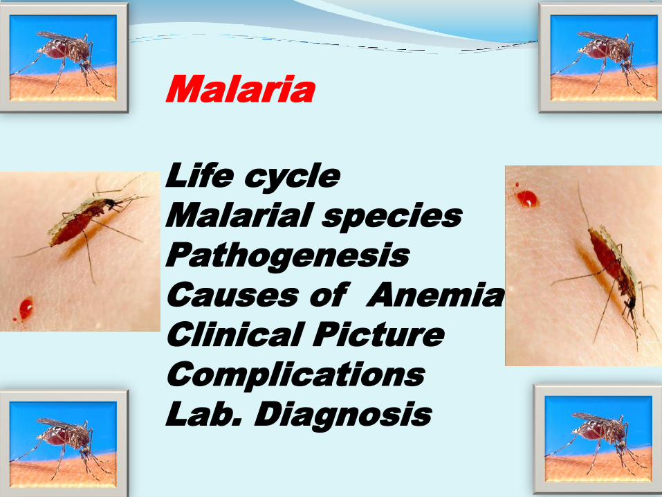
Malaria
Life cycle
Malarial species
Pathogenesis
Causes of Anemia
Clinical Picture
Complications
Lab. Diagnosis
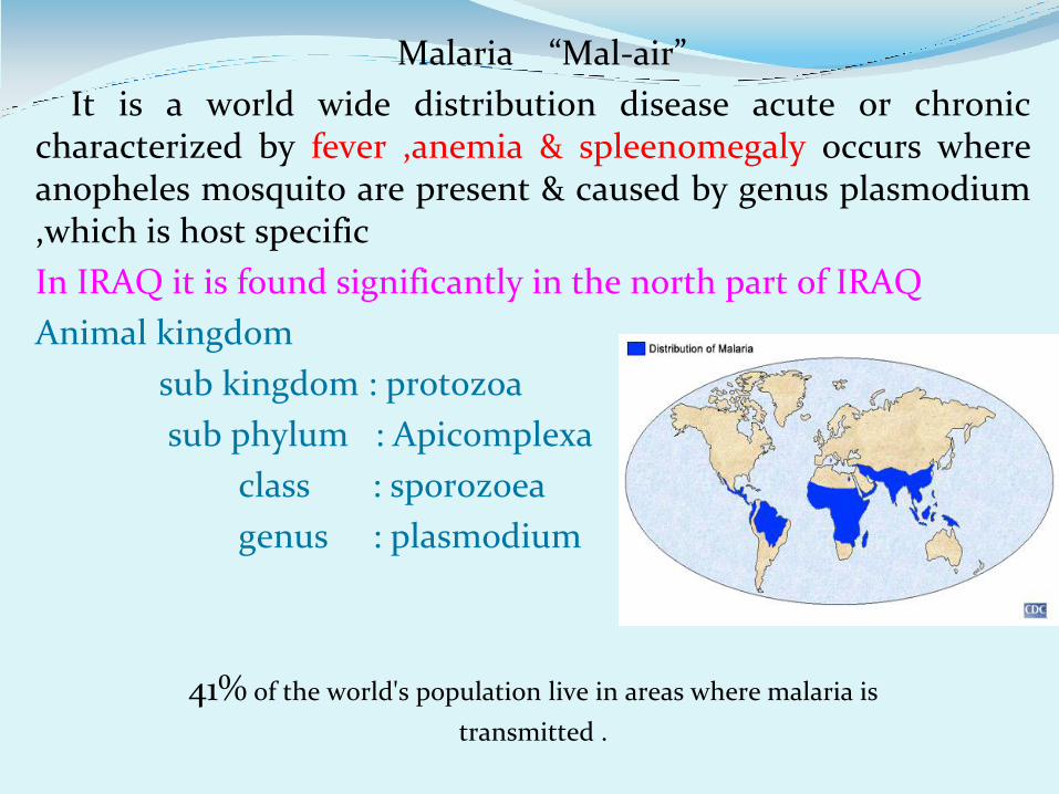
Malaria “Mal-air”
It is a world wide distribution disease acute or chronic characterized by fever ,anemia & spleenomegaly occurs where anopheles mosquito are present & caused by genus plasmodium ,which is host specific
In IRAQ it is found significantly in the north part of IRAQ
Animal kingdom
sub kingdom : protozoa
sub phylum : Apicomplexa
class : sporozoea
genus : plasmodium
41% of the world's population live in areas where malaria is
transmitted .
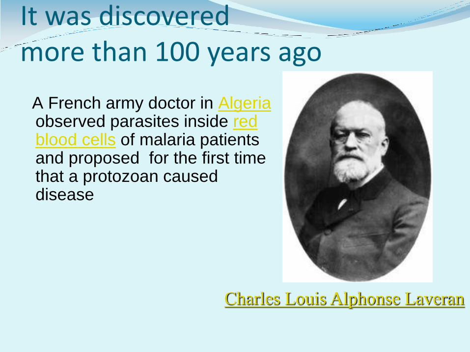
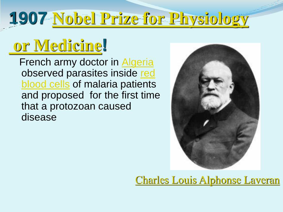
A French army doctor in Algeria observed parasites inside red blood cells of malaria patients and proposed for the first time that a protozoan caused disease
It was discovered more than 100 years ago
Charles Louis Alphonse Laveran
French army doctor in Algeria observed parasites inside red blood cells of malaria patients and proposed for the first time that a protozoan caused disease
Charles Louis Alphonse Laveran
1907 Nobel Prize for Physiology
or Medicine!
Around 300-500 million clinical cases of malaria are reported every year, of which more than a million die of severe and complicated cases of malaria.
Malaria ranks third among the major infectious diseases in causing deaths after HIV , pneumococcal acute respiratory infections and tuberculosis, then malaria.
Although malaria has been widely eradicated in many parts of the world, the global number of cases continues to rise. The most important reason for this alarming situation is the rapid spread of malaria parasites that are resistant to anti malarial drugs.
Malaria
Leading causes of death in Sub-Saharan Africa, South Asia, and Southeast Asia for persons age 0-44 (World Health Organization)
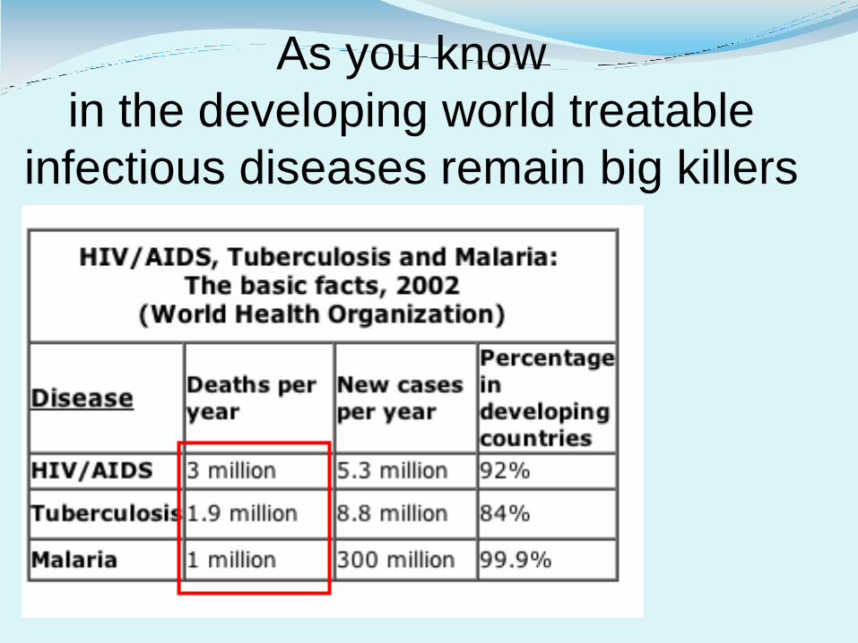
As you know
in the developing world treatable
infectious diseases remain big killers
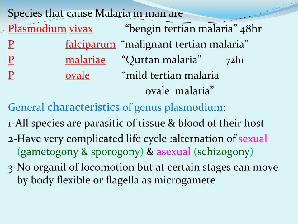
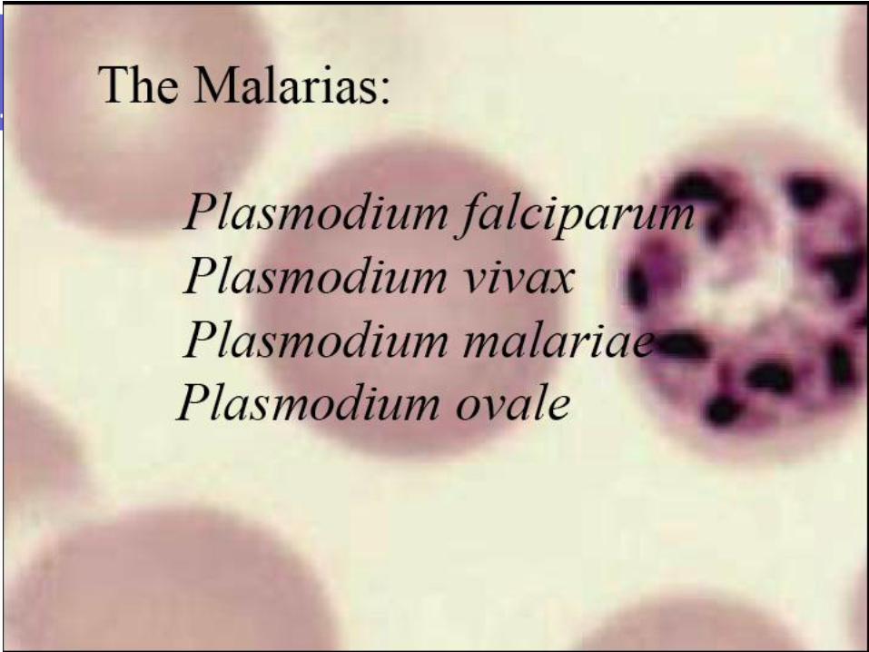
Species that cause Malaria in man are
Plasmodium vivax “bengin tertian malaria” 48hr
P falciparum “malignant tertian malaria”
P malariae “Qurtan malaria” 72hr
P ovale “mild tertian malaria
ovale malaria”
General characteristics of genus plasmodium:
1-All species are parasitic of tissue & blood of their host
2-Have very complicated life cycle :alternation of sexual (gametogony & sporogony) & asexual (schizogony)
3-No organil of locomotion but at certain stages can move by body flexible or flagella as microgamete
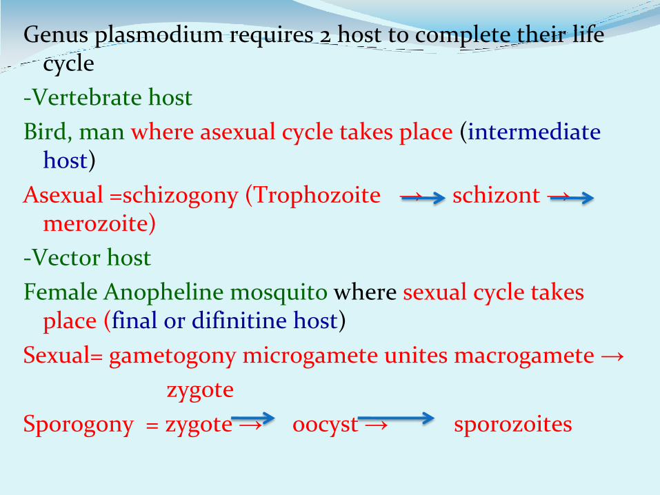
Genus plasmodium requires 2 host to complete their life cycle
-Vertebrate host
Bird, man where asexual cycle takes place (intermediate host)
Asexual =schizogony (Trophozoite → schizont → merozoite)
-Vector host
Female Anopheline mosquito where sexual cycle takes place (final or difinitine host)
Sexual= gametogony microgamete unites macrogamete →
zygote
Sporogony = zygote → oocyst → sporozoites
Each disease has a distinct course
“Tertian Malaria”
(P.falciparum, P.ovale and P.vivax)
fever occurs every third day.
“Quartan Malaria”
(P. malariae)
fever occurs every fourth day.
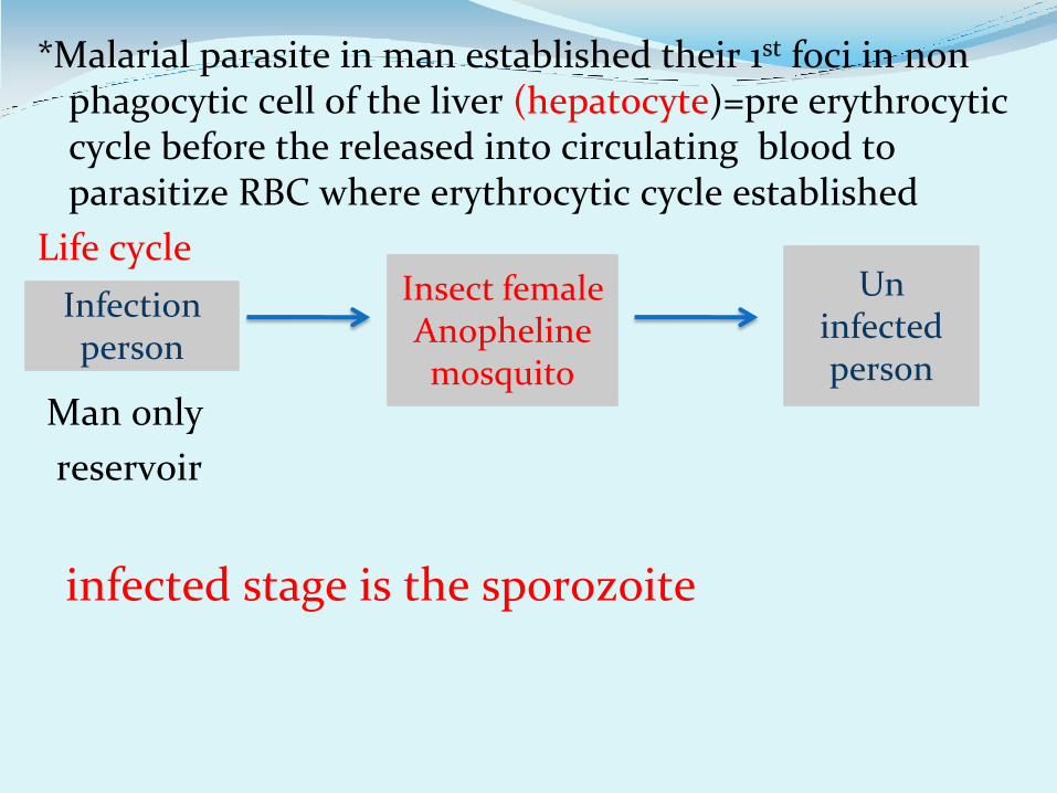
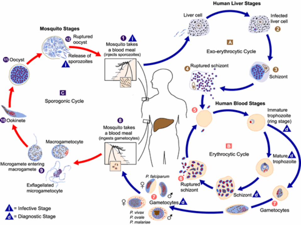
*Malarial parasite in man established their 1st foci in non phagocytic cell of the liver (hepatocyte)=pre erythrocytic cycle before the released into circulating blood to parasitize RBC where erythrocytic cycle established
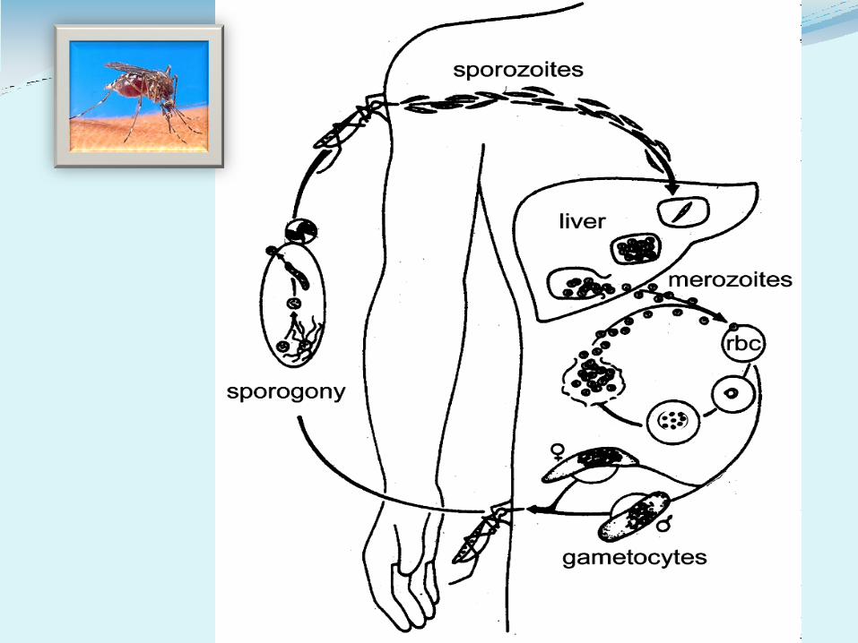
Life cycle
Man only
reservoir
infected stage is the sporozoite
Infection person
Insect female Anopheline mosquito
Un infected person
Malaria
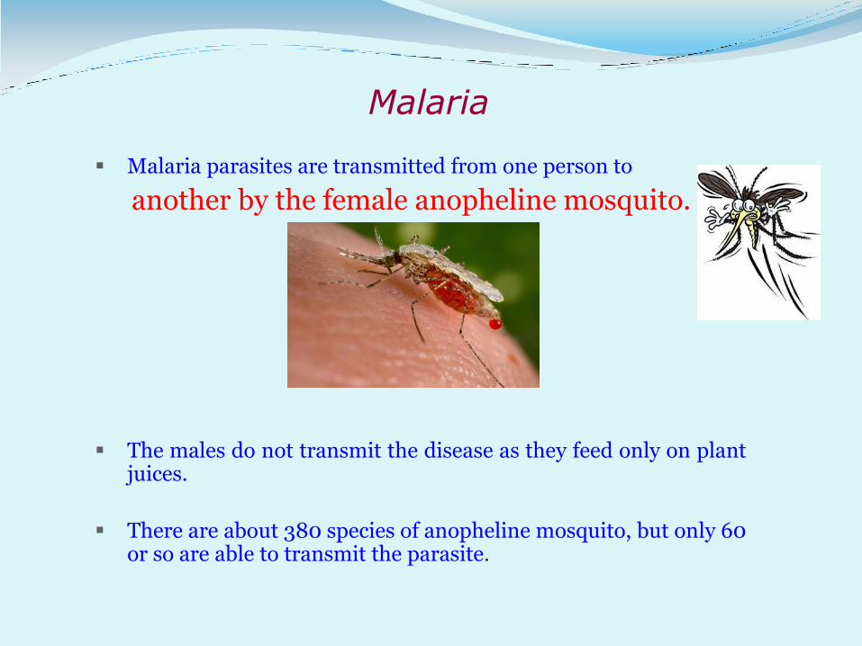
Malaria parasites are transmitted from one person to
another by the female anopheline mosquito.
The males do not transmit the disease as they feed only on plant juices.
There are about 380 species of anopheline mosquito, but only 60 or so are able to transmit the parasite.
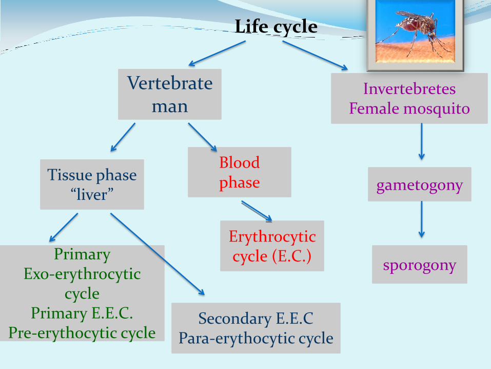
Life cycle
Vertebrate man
Invertebretes Female mosquito
Tissue phase “liver”
Blood phase
sporogony
gametogony
Erythrocytic cycle (E.C.)
Secondary E.E.C Para-erythocytic cycle
Primary Exo-erythrocytic
cycle Primary E.E.C.
Pre-erythocytic cycle
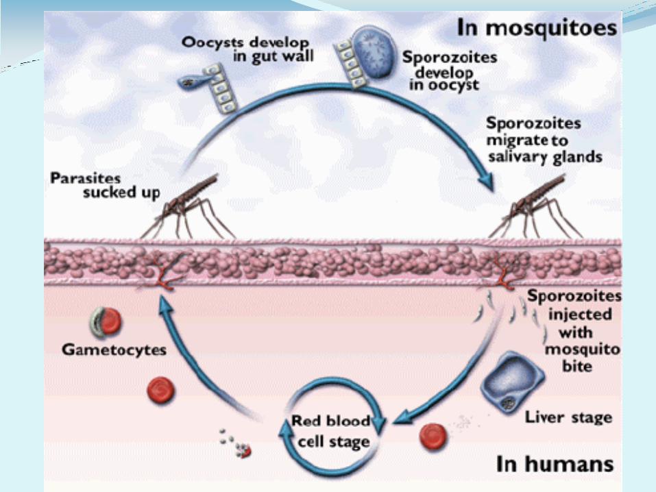
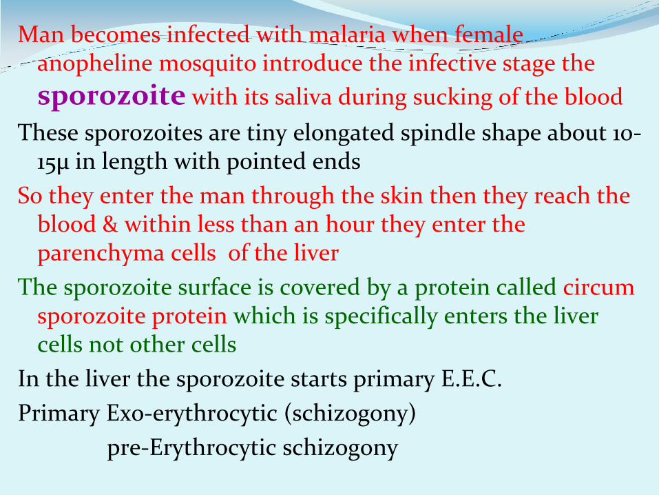
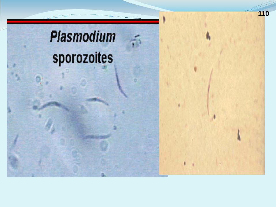
Man becomes infected with malaria when female anopheline mosquito introduce the infective stage the
sporozoite with its saliva during sucking of the blood
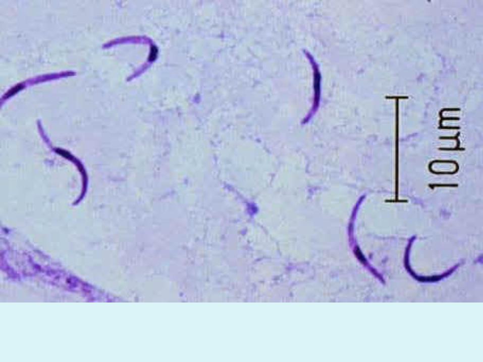
These sporozoites are tiny elongated spindle shape about 10-15µ in length with pointed ends
So they enter the man through the skin then they reach the blood & within less than an hour they enter the parenchyma cells of the liver
The sporozoite surface is covered by a protein called circum sporozoite protein which is specifically enters the liver cells not other cells
In the liver the sporozoite starts primary E.E.C.
Primary Exo-erythrocytic (schizogony)
pre-Erythrocytic schizogony
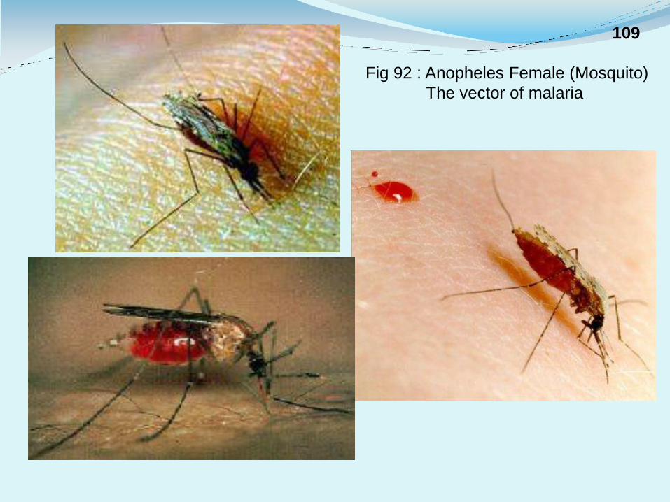
Fig 92 : Anopheles Female (Mosquito)
The vector of malaria
109
110
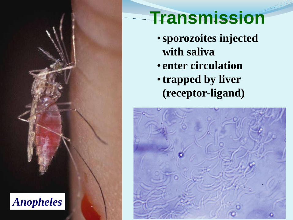
Anopheles
Transmission • sporozoites injected
with saliva
• enter circulation
• trapped by liver
(receptor-ligand)
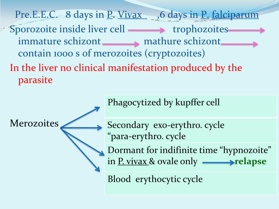
Pre.E.E.C. 8 days in P. Vivax ,6 days in P. falciparum
Sporozoite inside liver cell trophozoites immature schizont mathure schizont contain 1000 s of merozoites (cryptozoites)
In the liver no clinical manifestation produced by the parasite
Merozoites
Phagocytized by kupffer cell
Secondary exo-erythro. cycle “para-erythro. cycle
Dormant for indifinite time “hypnozoite” in P. vivax & ovale only relapse
Blood erythocytic cycle
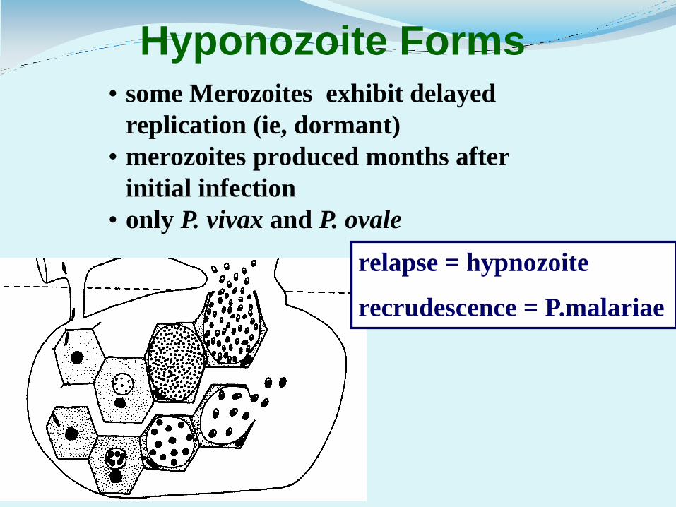
Hyponozoite Forms • some Merozoites exhibit delayed
replication (ie, dormant)
• merozoites produced months after
initial infection
• only P. vivax and P. ovale
relapse = hypnozoite
recrudescence = P.malariae
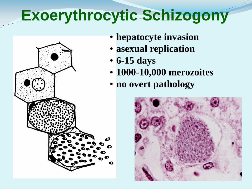
Exoerythrocytic Schizogony
• hepatocyte invasion
• asexual replication
• 6-15 days
• 1000-10,000 merozoites
• no overt pathology
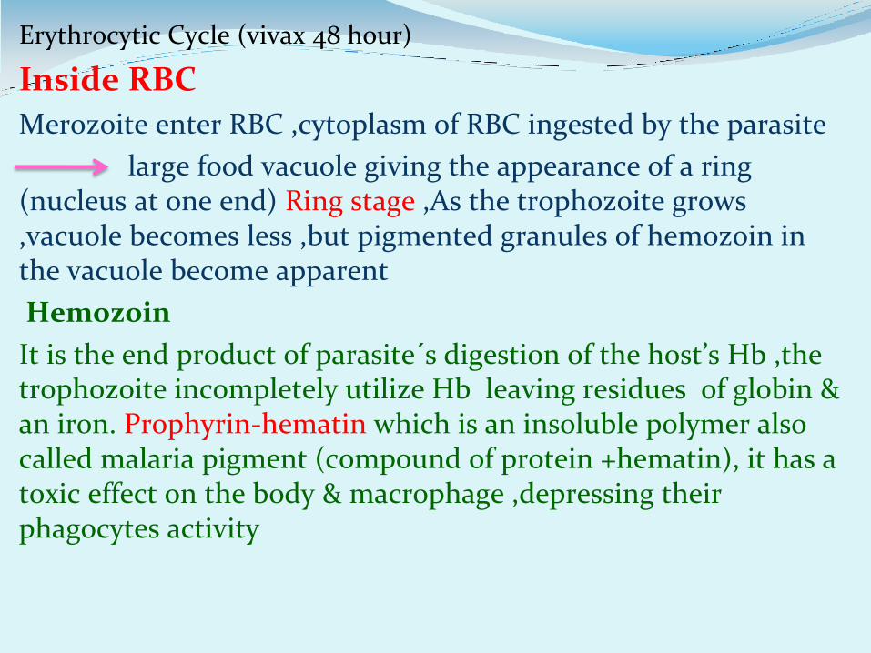
Erythrocytic Cycle (vivax 48 hour)
Inside RBC
Merozoite enter RBC ,cytoplasm of RBC ingested by the parasite
large food vacuole giving the appearance of a ring (nucleus at one end) Ring stage ,As the trophozoite grows ,vacuole becomes less ,but pigmented granules of hemozoin in the vacuole become apparent
Hemozoin
It is the end product of parasite´s digestion of the host’s Hb ,the trophozoite incompletely utilize Hb leaving residues of globin & an iron. Prophyrin-hematin which is an insoluble polymer also called malaria pigment (compound of protein +hematin), it has a toxic effect on the body & macrophage ,depressing their phagocytes activity
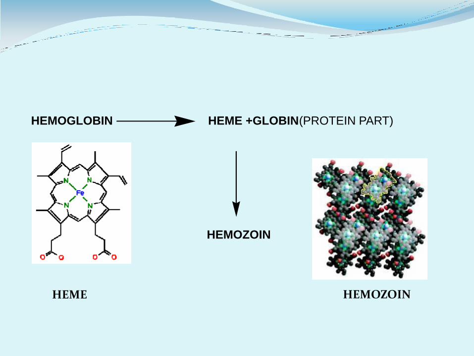
HEMOGLOBIN HEME +GLOBIN(PROTEIN PART)
HEMOZOIN
HEME HEMOZOIN
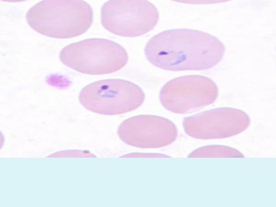
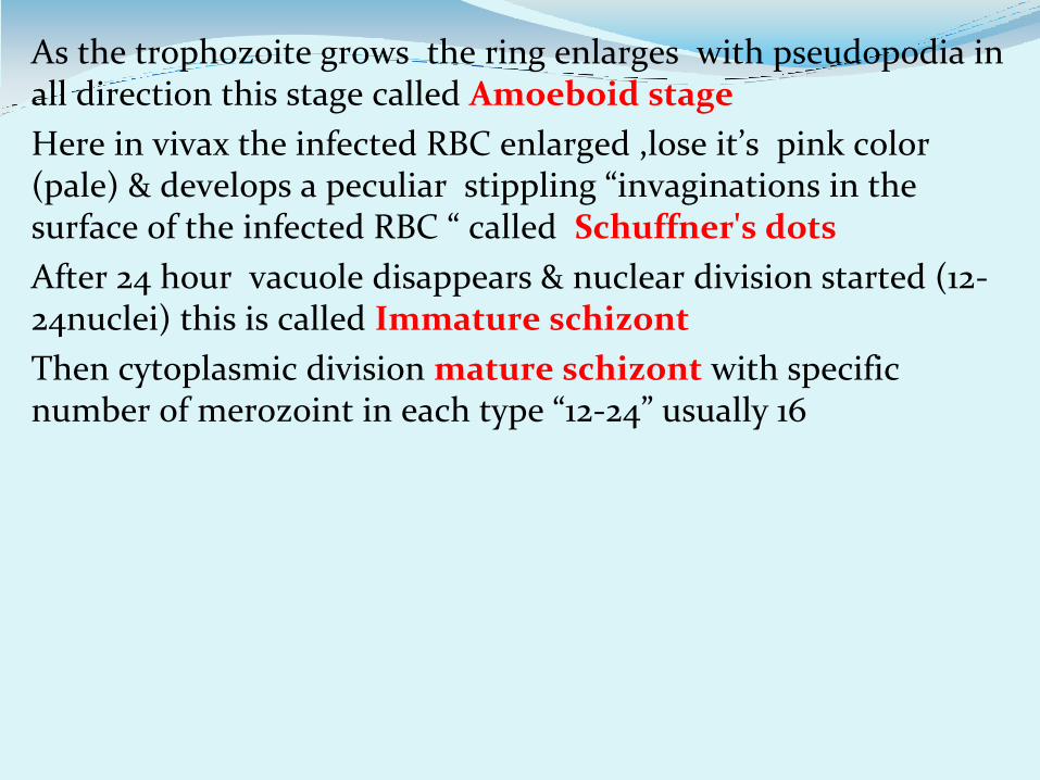
As the trophozoite grows the ring enlarges with pseudopodia in all direction this stage called Amoeboid stage
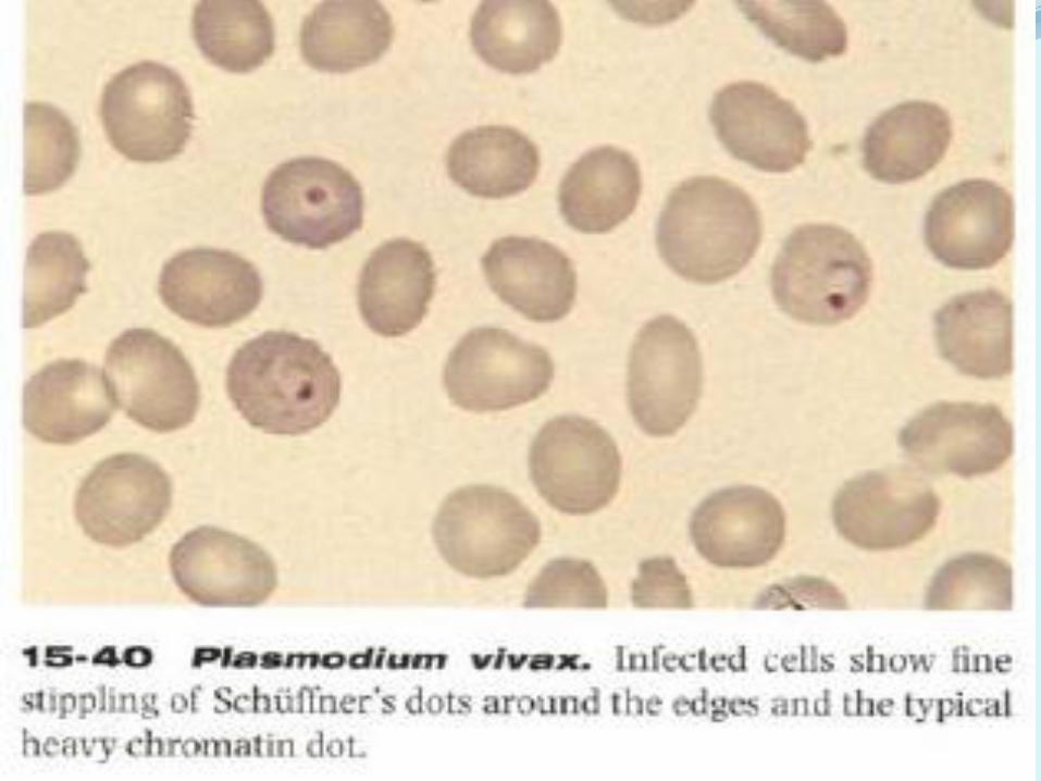
Here in vivax the infected RBC enlarged ,lose it’s pink color (pale) & develops a peculiar stippling “invaginations in the surface of the infected RBC “ called Schuffner's dots
After 24 hour vacuole disappears & nuclear division started (12-24nuclei) this is called Immature schizont
Then cytoplasmic division mature schizont with specific number of merozoint in each type “12-24” usually 16
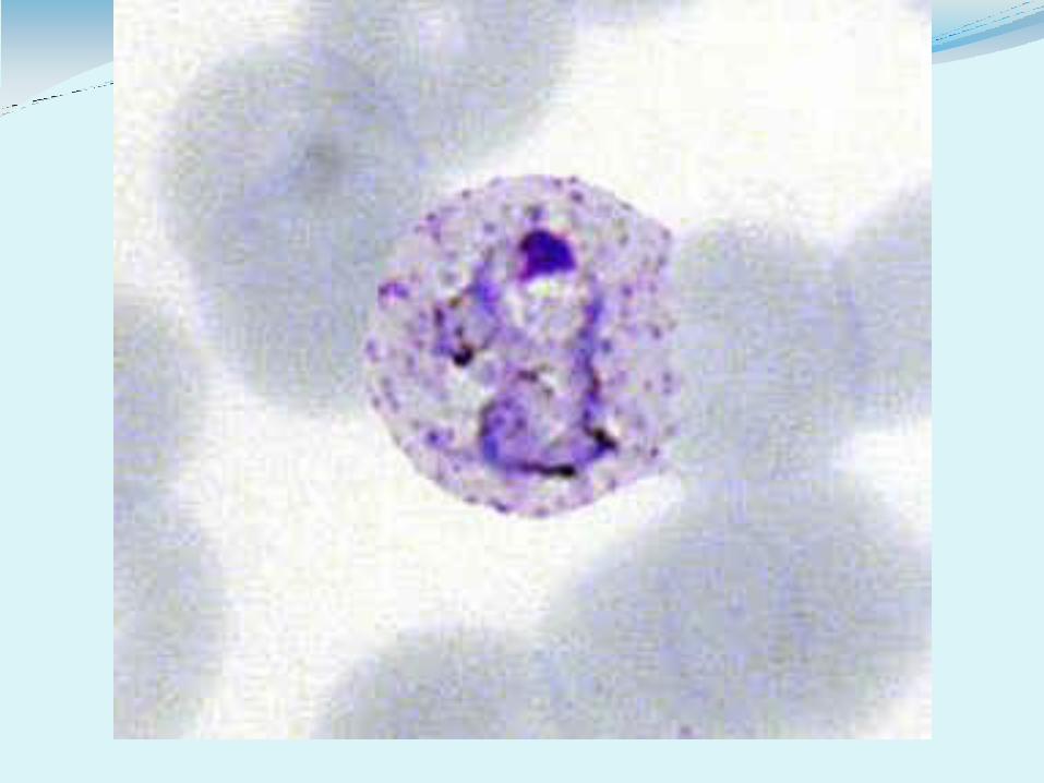
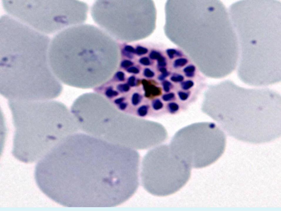
Then RBC rupture & releazing merozoites(parasites)+metabolic wastes inclucing hemozoin which responsible for symptoms of malaria
Merozoites enter new RBC & repeat eryth. cycle (every 48hr)
After an indeterminate number of asexual cycle (eryth. cycle ) some merozoites when enter RBC become microgametocyte( male) or macrogametocyte (female)
These gametocytes develope in RBC in the
capillary of internal organ (spleen + Bone
Marrow) & go to peripheral blood only when
become mature (96hr) twice the time of E.
schizogony
The mature gametocyte unless ingested by
female anopheline mosq. It will be die &
phagocytized
Individual who harbor gametocytes in his peripheral blood is called carrier
When female anopheline mosq. takes
erythrocytes containing gametocytes the sexual
cycle begin :
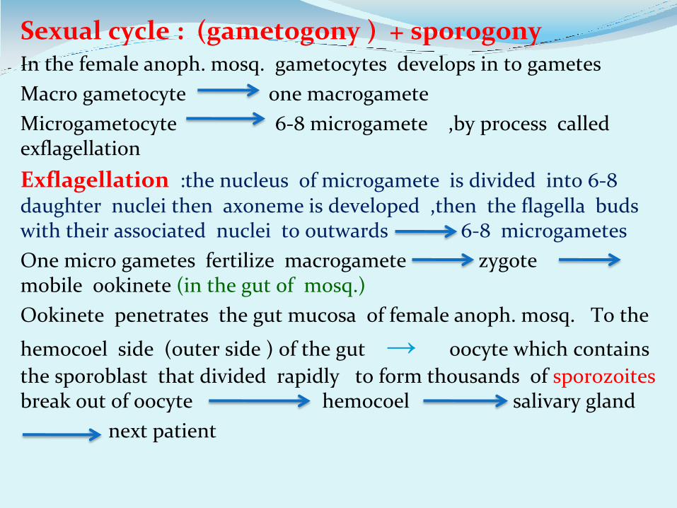
Sexual cycle : (gametogony ) + sporogony In the female anoph. mosq. gametocytes develops in to gametes
Macro gametocyte one macrogamete
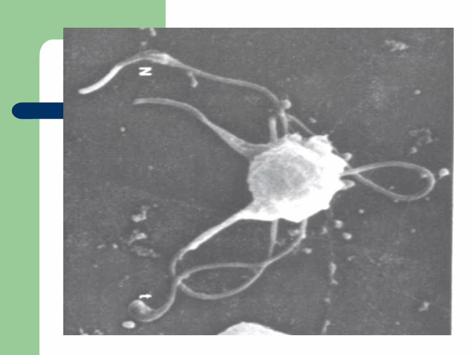
Microgametocyte 6-8 microgamete ,by process called exflagellation
Exflagellation :the nucleus of microgamete is divided into 6-8 daughter nuclei then axoneme is developed ,then the flagella buds with their associated nuclei to outwards 6-8 microgametes
One micro gametes fertilize macrogamete zygote mobile ookinete (in the gut of mosq.)
Ookinete penetrates the gut mucosa of female anoph. mosq. To the
hemocoel side (outer side ) of the gut → oocyte which contains
the sporoblast that divided rapidly to form thousands of sporozoites break out of oocyte hemocoel salivary gland
next patient
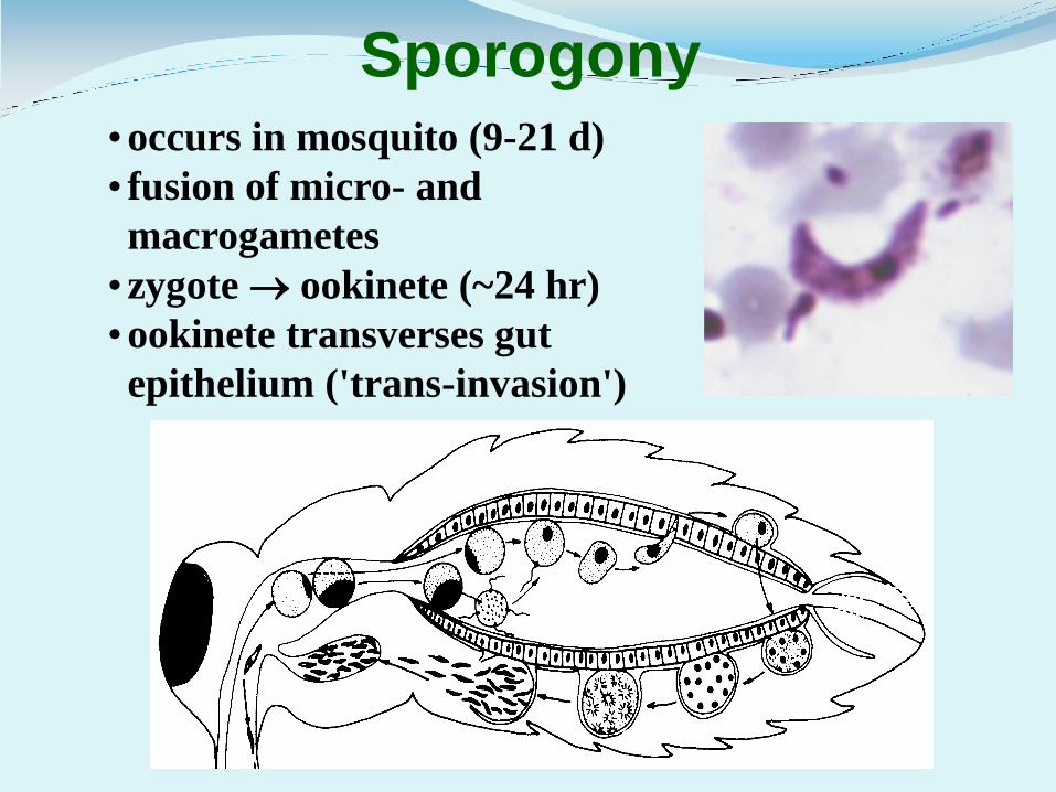
Sporogony •occurs in mosquito (9-21 d)
• fusion of micro- and
macrogametes
• zygote ookinete (~24 hr)
•ookinete transverses gut
epithelium ('trans-invasion')
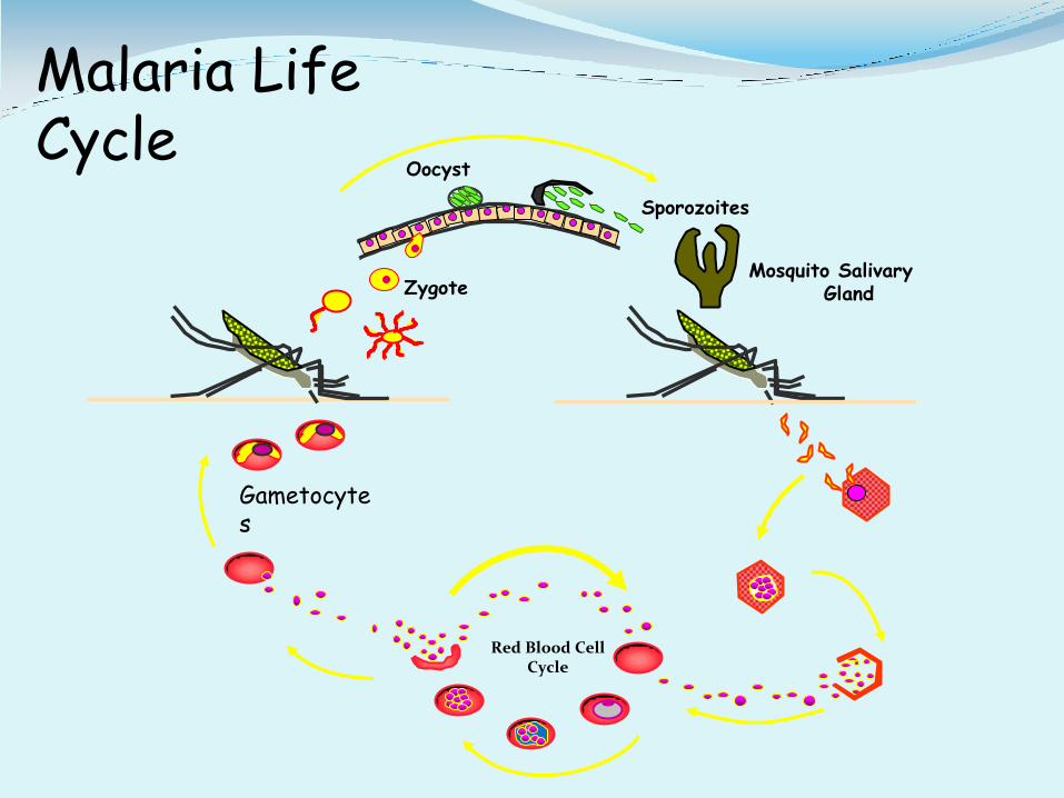
Liver stage
Sporozoites
Mosquito Salivary Gland
Malaria Life Cycle
Gametocytes
Oocyst
Red Blood Cell Cycle
Zygote
Method of transmission
*sporozoite induced
The infective stage is sporozoite by biting of female anoph. mosq. porter of entry =skin
*trophozoite induced
Blood transfusion especially P. malaria syringe ,lab. accident & rarely congenital infection
P. Vivax : 43% of malaria in the world some merozoites remain in the liver hypenozoite so relapse
Merozoite invades only young RBC (reticulocyte) unable to invade fully mature RBC ,black people have got natural resistance to p. vivax infection ,because merozoite enter RBC through receptor which is the duffy blood group protein Ag fy a ,fy b
Black people usually with no such Ag fy0
infected RBC in P. vivax enlarge ,pale ,with schuffner’s dots ,(fine dots)
Schizont in E. cycle with 12-24 hr usually 16 merozoits
E. cycle with 48 hr periodicity = tertian malaria
a
a
P. Falciparum = malignant ter. m.
50% of human malaria most virulent ,90% of mortality of malaria greater killer of humanity in tropical zone
No relapse (no hypnozoites)
Invade RBC at any age even reticulocyte so much higher parasitemia than other types (25% of RBC infected )
Soon after invasion of RBC the trophozoite produce protein that are deposite in the eryth. surface membrane in the
deformation called knobs, these protein bind to certain
glyco-protein on the post capillary venular endothelium this binding cause sequestration of the infected RBCs so stick to venular endothelium , also those RBCs stick to normal
RBC thrombosis
Gametocyte don’t produce these knobs so gametocyte infected RBCs don’t stick
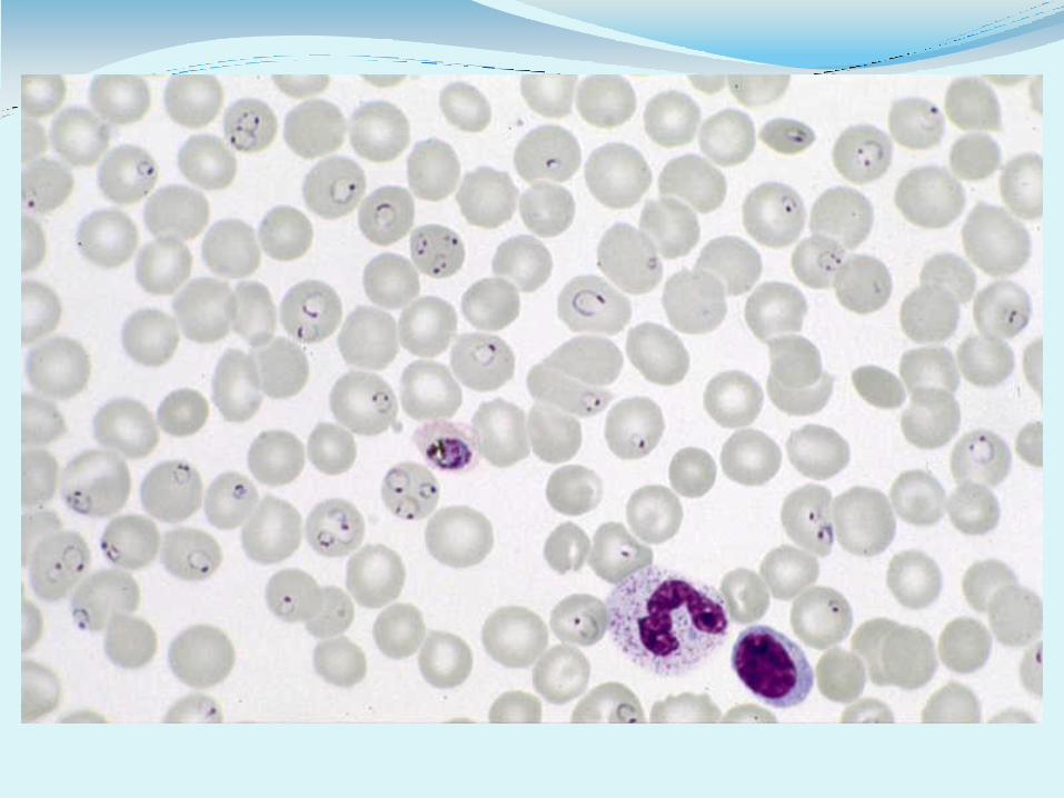
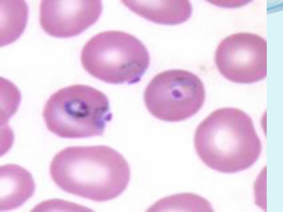
So in the peripheral blood only the early ring stages & gametocyte are seen in P. falciparum
Ring stage in P. falciparum are smallest than other species ,multiple infection of the same RBC is common ,rings with bi nucleated (2 chromatin dots) also present
may be division of the ring
RBC with Maurer's dots larger than the fine schiiffner’s dots of P. vivax
These dots for transport of nutrient
Amoeboid & schizont (8-32merozoites) ,not seen in the peripheral blood but in the capillaries of the internal organ (spleen , B.M.)
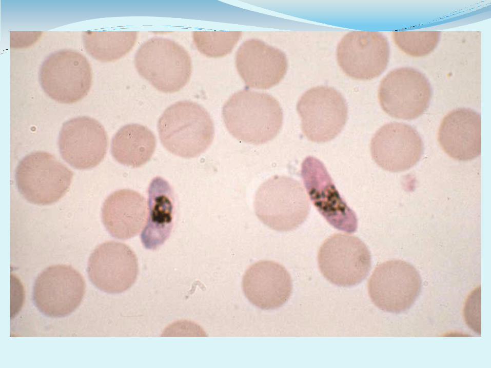
Gametocytes are crescent in shape
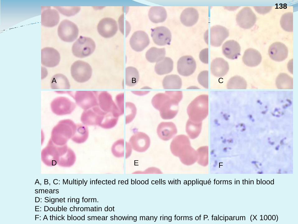
A, B, C: Multiply infected red blood cells with appliqué forms in thin blood
smears
D: Signet ring form.
E: Double chromatin dot
F: A thick blood smear showing many ring forms of P. falciparum (X 1000)
A B C
D E F
138
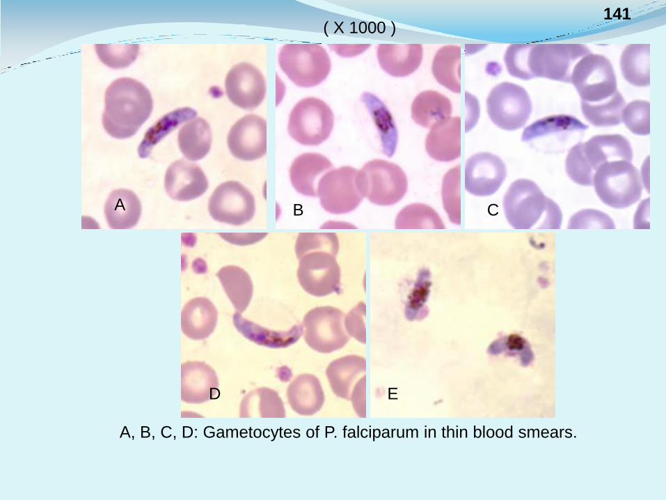
A, B, C, D: Gametocytes of P. falciparum in thin blood smears.
A B C
D E
( X 1000 ) 141
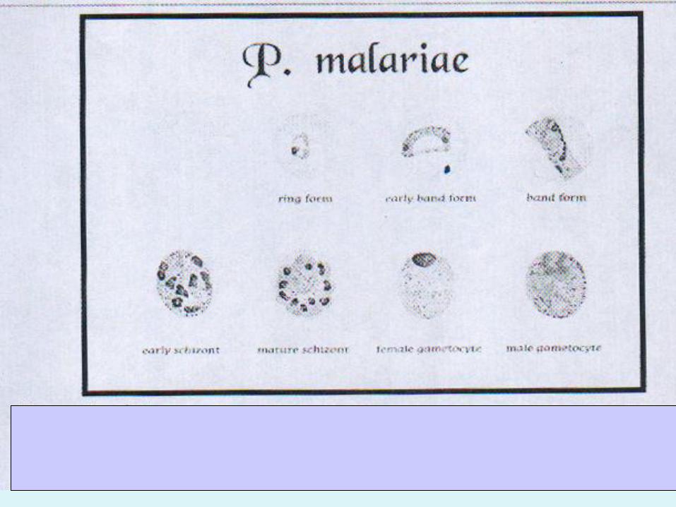
P. Malariae 7%
Quatrain malaria (paroxysm every 72hr), amoeboid band form schizont 6-12 (9) merozoites in a rosette shape ,infect only old (aging) RBC so low parasitemia
Can live in blood up to 50 years so important in blood transfusion ,transmission & cause recrudescence but no relapse
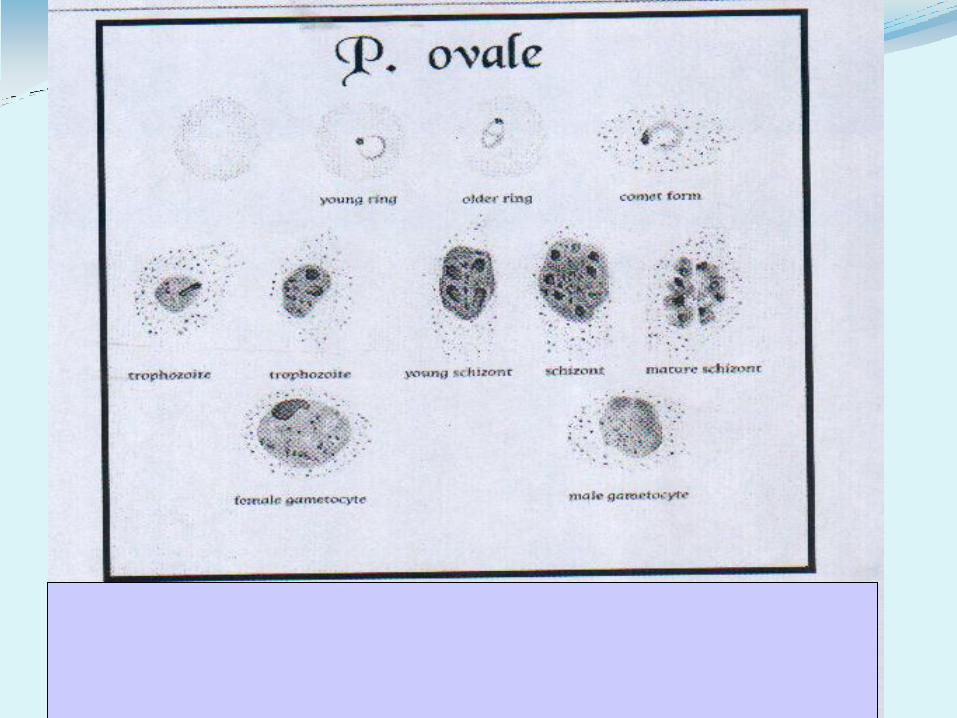
P. Ovale
Mild tertian malaria ,oval malaria
Rarest type ,RBC oval in shape schuffner’s dots appear earlyer ,larger more numerous than vivax , schizont
6-12 (9)
Pathogenesis of malaria
Major clinical manifestation is due to
1- Host inflammatory response which produce chills & fever
2-Anemia due to enormous destruction of RBC
*Severity depends on species of malaria ,most serious one is P. falciparum
*the fever in malaria is stimulated by the waste products of parasites which are released after lyses of RBSs ,which includs mainly malarial toxins “hemozoin or malarial pigments” into circulation trigger =TNF (tumor necrosis factor) fever
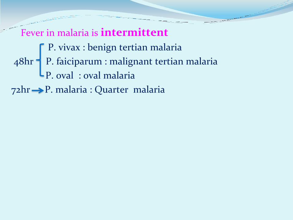
Fever in malaria is intermittent
P. vivax : benign tertian malaria
48hr P. faiciparum : malignant tertian malaria
P. oval : oval malaria
72hr P. malaria : Quarter malaria
Anemia :-
-Malaria causes destruction both infected & non infected RBCs
-In ability to recycle iron bounded in hemozoin
-Defective bone marrow response
-Spleen removes infected & non infected RBCs from blood ,due to TNF toxicity
Anemia may cause Juandice
Age No age limit
Pregnant women and children are most likely to get it.
People from non-malaria zones are at much higher risk than natives when they are in malaria zones.
Clinical feature of malaria :-
Incubation period in P. Vivax = 9-12 days (no symptoms)
Then the main clinical manifestation in a typical case of malaria started which include febrile paroxysms followed by anemia & splenomeglay
Febrile paroxysms :-
each paroxysms shows 3 stages
1-Cold stages (last 20-60 min. usually 1/2hr)
Chill , felling of intense cold ,although temp. 40˚C ,shivering
2-Hot stages (last 2-4hr)
Fever , 40 -41˚C ,headache ,mild delirium
3-sweating stages (last 2- 3 hr)
Perspiration
The total duration of febrile cycle ≈ 6-8 hr ,these paroxysms synchronies with the eryth. shizogony
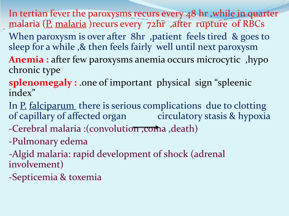
In tertian fever the paroxysms recurs every 48 hr ,while in quarter malaria (P. malaria )recurs every 72hr ,after rupture of RBCs
When paroxysm is over after 8hr ,patient feels tired & goes to sleep for a while ,& then feels fairly well until next paroxysm
Anemia : after few paroxysms anemia occurs microcytic ,hypo chronic type
splenomegaly : .one of important physical sign “spleenic index”
In P. falciparum there is serious complications due to clotting of capillary of affected organ circulatory stasis & hypoxia
-Cerebral malaria :(convolution ,coma ,death)
-Pulmonary edema
-Algid malaria: rapid development of shock (adrenal involvement)
-Septicemia & toxemia
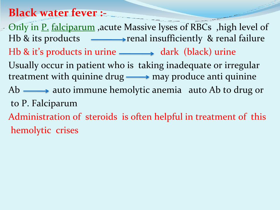
Black water fever :-
Only in P. falciparum ,acute Massive lyses of RBCs ,high level of Hb & its products renal insufficiently & renal failure
Hb & it’s products in urine dark (black) urine
Usually occur in patient who is taking inadequate or irregular treatment with quinine drug may produce anti quinine
Ab auto immune hemolytic anemia auto Ab to drug or
to P. Falciparum
Administration of steroids is often helpful in treatment of this
hemolytic crises
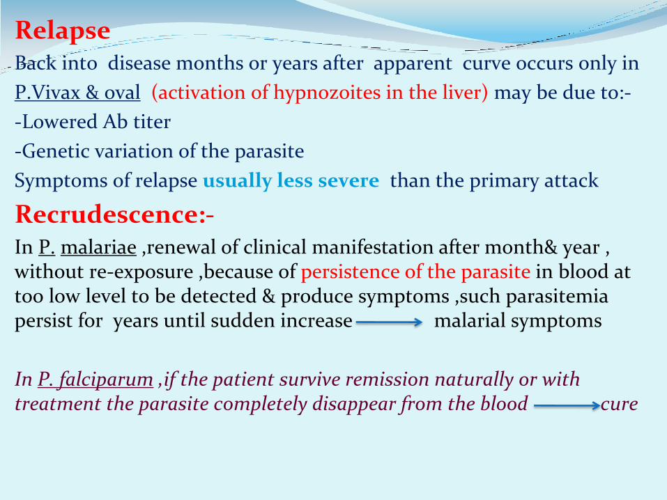
Relapse Back into disease months or years after apparent curve occurs only in
P.Vivax & oval (activation of hypnozoites in the liver) may be due to:-
-Lowered Ab titer
-Genetic variation of the parasite
Symptoms of relapse usually less severe than the primary attack
Recrudescence:- In P. malariae ,renewal of clinical manifestation after month& year , without re-exposure ,because of persistence of the parasite in blood at too low level to be detected & produce symptoms ,such parasitemia persist for years until sudden increase malarial symptoms
In P. falciparum ,if the patient survive remission naturally or with treatment the parasite completely disappear from the blood cure
Each disease has a distinct course
P.ovale and P.vivax
can cause chronic malaria,
reappearing after months or years
due to latent parasites in liver
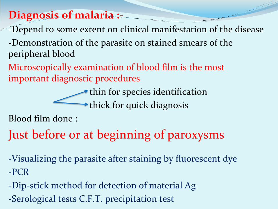
Diagnosis of malaria :-
-Depend to some extent on clinical manifestation of the disease
-Demonstration of the parasite on stained smears of the peripheral blood
Microscopically examination of blood film is the most important diagnostic procedures
thin for species identification
thick for quick diagnosis
Blood film done :
Just before or at beginning of paroxysms
-Visualizing the parasite after staining by fluorescent dye
-PCR
-Dip-stick method for detection of material Ag
-Serological tests C.F.T. precipitation test
Immunity to malaria is specific ,strain & variant specific
Genetic resistance to malaria
1-Black people (Duffy blood group) natural resistance to P. vivax
2-Sickle cell anemia
fauvism Abnormal Hb
Thalassemia
Treatment of malaria :-
Anti malarial drugs
Alkaloid :quinine ,chloroquine ,primequine
Chloroquine prevent digestion of Hb so no hemozoin ,it is not effective in exo-eryth. schizgony
primequine kill hypnozoint so prevent relapse
In resistant malaria :mefloquine , fansidar
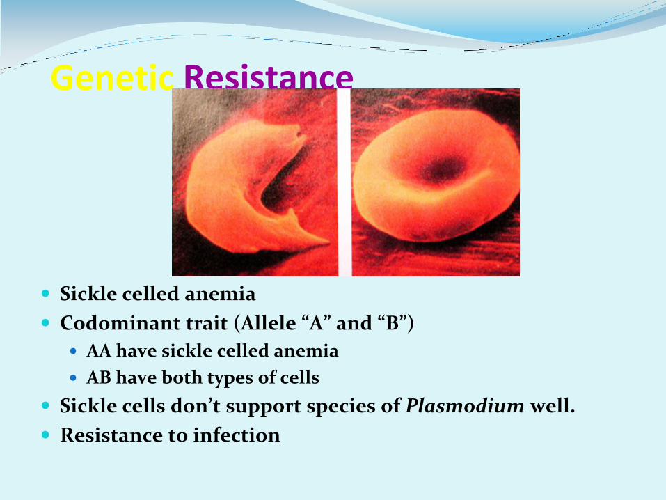
Genetic Resistance
Sickle celled anemia
Codominant trait (Allele “A” and “B”)
AA have sickle celled anemia
AB have both types of cells
Sickle cells don’t support species of Plasmodium well.
Resistance to infection
Prophylaxis :-
Vaccine :difficult (different stages ,changing their Ag )
Travelers to endemic take chloroquine 2 weeks before & contain to 6 weeks after leaving ,followed 2 weeks primequine
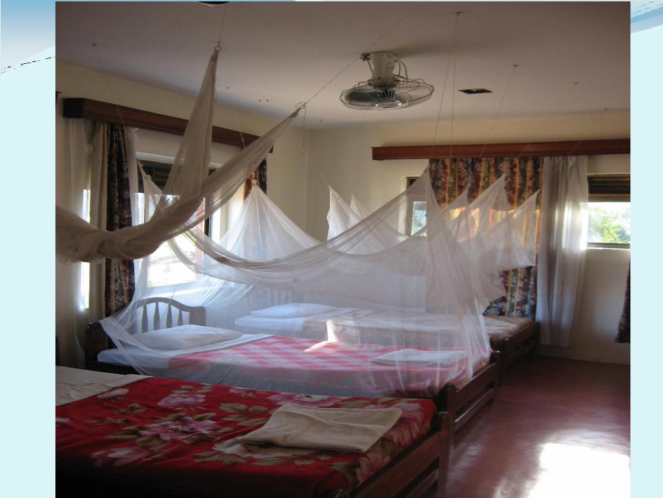
Control :-
Mosquito control (netting ,window screen ,destruction area of mosquito life cycle using predators (fishes) insecticides DDT
Drug resistance malaria & mosquito resistance so malaria will be with us as long in there is people