leukocyte composition of human breast cancer composition of human breast cancer brian ruffella,...
TRANSCRIPT

Leukocyte composition of human breast cancerBrian Ruffella, Alfred Aua,b, Hope S. Rugob,c, Laura J. Essermanb,d, E. Shelley Hwangb,d, and Lisa M. Coussensa,b,1
aDepartment of Pathology and bHelen Diller Family Comprehensive Cancer Center, University of California, San Francisco, CA 94143; and Departmentsof cMedicine and dSurgery, University of California, San Francisco, CA 94115
Edited by Kornelia Polyak, Dana–Farber Cancer Institute, Boston, MA, and accepted by the Editorial Board July 13, 2011 (received for review March 17, 2011)
Retrospective clinical studies have used immune-based biomarkers,alone or in combination, to predict survival outcomes for womenwith breast cancer (BC); however, the limitations inherent toimmunohistochemical analyses prevent comprehensive descrip-tions of leukocytic infiltrates, aswell as evaluation of the functionalstate of leukocytes in BC stroma. To more fully evaluate this com-plexity, and to gain insight into immune responses after chemo-therapy (CTX), we prospectively evaluated tumor and nonadjacentnormal breast tissue from women with BC, who either had or hadnot received neoadjuvant CTX before surgery. Tissues were evalu-atedbypolychromaticflowcytometry in combinationwith confocalimmunofluorescence and immunohistochemical analysis of tissuesections. These studies revealed that activated T lymphocytes pre-dominate in tumor tissue, whereas myeloid lineage cells are moreprominant in “normal” breast tissue. Notably, residual tumors froman unselected group of BC patients treated with neoadjuvant CTXcontained increased percentages of infiltrating myeloid cells, ac-companiedbyan increasedCD8/CD4T-cell ratioandhighernumbersof granzyme B-expressing cells, compared with tumors removedfrom patients treated primarily by surgery alone. These data pro-vide an initial evaluation of differences in the immune microenvi-ronmentof BC comparedwithnonadjacentnormal tissueand revealthe degree to which CTX may alter the complexity and presence ofselective subsets of immune cells in tumors previously treated in theneoadjuvant setting.
inflammation | macrophage
Several subtypes of CD45-expressing leukocytes infiltratebreast cancer (BC), including CD4+ and CD8+ T cells,
CD20+ B cells, and multiple myeloid-lineage cells includingtumor-associated macrophages (TAMs) that are often identifiedby immunohistochemical (IHC) detection of CD68 (1). Highlymphocyte infiltration is associated with increased survival inpatients <40 y of age (2) and with a favorable prognosis insubsets of patients whose tumors are also heavily infiltrated byTAMs (3). More specifically, large cohort studies of patients withBC have revealed that the presence of CD68+ cells in tumortissue correlates with poor prognostic features (4–6), higher tu-mor grade (7–9), increased angiogenesis (10–13), decreaseddisease-free survival (6, 11, 14, 15), and increased risk for sys-temic metastasis when assessed in conjunction with endothelialand carcinoma cell markers (16).The functional significance of specific leukocytes in BC de-
velopment has been implied based on experimental studies usingmurine models of mammary carcinogenesis where mice harboringhomozygous null mutations in genes specifying leukocyte de-velopment or recruitment have been evaluated. In transgenicmiceexpressing the polyoma virus middle T antigen regulated by themouse mammary tumor virus promoter (MMTV-PyMT mice),progression of mammary carcinomas and metastases to lungs arereduced inmice lacking the colony-stimulating factor-1 (csf1) gene,a cytokine critical for macrophage maturation and recruitment(17, 18). TAMs inmammary tumor tissue are often associatedwithvasculature (19), where their production of VEGFA fosters an-giogenic programming of tissue (20, 21), and their production ofEGF promotes invasive tumor growth and subsequent metastases(22, 23). Moreover, TAMs regulated by epithelial CSF1 expresshigher levelsof several hypoxia-inducedgenes (iNOSandarginase-1)that, in turn, mediate suppression of anti-tumor immunity byblocking cytotoxic T-cell proliferation and activation (6, 24). Thus,
TAM presence and bioactivity within mammary tumors corre-spond to their clinical activity, further indicating the importanceof TAMs, not only in promoting tumor development, but also insuppression of anti-tumor immunity.CD4+ T cells isolated from human BC produce high levels of
type II helper (TH2) cytokines including IL-4 and IL-13 (25, 26),which are significant in light of studies demonstrating that severalprotumor activities of TAMs are regulated by IL-4 derived fromCD4+T cells (1, 27). Based on these findings, we recently reportedthat infiltration by CD68+, CD4+, and CD8+ immune cells inhuman BC is predictive of overall survival, and that the ratio ofCD68 to CD8a mRNA in tumor tissue correlates with completepathologic response (pCR) in patients undergoing neoadjuvantchemotherapy (CTX) for early stage BC (6). Despite the clearcorrelation between these specific immune cell types and BCclinical outcome, leukocyte complexity within tumor tissueremains poorly described, with most studies relying on single-marker IHC detection. Furthermore, although some studies haveexamined the effects of CTX on the presence and function ofcirculating peripheral blood leukocytes (28), data regarding theeffect of CTX on tumor-infiltrating immune cells are limited (29).Herein, we evaluated leukocytic infiltrates in breast tissue
from predominantly hormone receptor positive patients whohad, or had not, received CTX before definitive surgery. In CTX-naïve patients, we found that activated T lymphocytes comprisedthe majority of immune cells within tumors, whereas myeloid-lineage cells predominate in nonadjacent normal breast tissue. Incontrast, tumors from patients with residual disease after neo-adjuvant CTX contained higher levels of infiltrating myeloidcells, with a simultaneous shift away from a TH2 dominatedlymphocyte response.
ResultsIncreased Presence of T Cells in Tumor Tissue. To evaluate thecomposition of tumor-infiltrating leukocytes in human BC,tumors from 20 patients were evaluated by polychromatic flowcytometry and IHC detection of leukocyte lineages in tissuesections as described in Materials and Methods. Nine invasiveductal carcinomas (IDC) and five invasive lobular carcinomas(ILC)—mostly histological grade two or three—were obtainedfrom patients with no prior exposure to CTX (CTX-naïve) at thetime of primary surgery for early stage BC, although one patienthad received neoadjuvant tamoxifen. Six tumor samples wereobtained from patients previously treated with neoadjuvant CTXbefore resection (CTX-treated), consisting entirely of grade twoor three IDC. Notably, three of six CTX-treated tumors wereHER2/neu-positive, compared with only 1 of 14 CTX-naïvetumors, whereas both groups contained roughly equivalent per-centages of tumors negative for estrogen, progesterone, andHER2 receptors (triple negative). Details of tumor pathologyare outlined in Table S1. Ipsilateral nonadjacent tissue was alsoobtained from seven CTX-naïve and four CTX-treated patients
Author contributions: B.R. and L.M.C. designed research; B.R. performed research; A.A.,H.S.R., L.J.E., and E.S.H. contributed new reagents/analytic tools; B.R. and L.M.C. analyzeddata; and B.R. and L.M.C. wrote the paper.
The authors declare no conflict of interest.
This article is a PNAS Direct Submission. K.P. is a guest editor invited by the EditorialBoard.1To whom correspondence should be addressed. E-mail: [email protected].
This article contains supporting information online at www.pnas.org/lookup/suppl/doi:10.1073/pnas.1104303108/-/DCSupplemental.
2796–2801 | PNAS | February 21, 2012 | vol. 109 | no. 8 www.pnas.org/cgi/doi/10.1073/pnas.1104303108

for use as “normal” tissue, in addition to tissues from two con-tralateral prophylactic mastectomies from patients with ipsilat-eral ductal carcinoma in situ (DCIS).Immune infiltrates detected with the pan-leukocyte marker CD45
were present in both normal and tumor tissue, but with substantiallyincreased density in BC (Fig. 1A). Leukocyte subsets were evaluatedby using a combination of lineage markers to identify specific sub-populations (Figs. S1 and S2), with the complexity of these pop-ulations shown in Fig. 1B as a percentage of the total number ofCD45+ cells in each sample. BC tissues from CTX-naïve patientscontained infiltrates dominated by T lymphocytes (CD3ε+), withminor populations of natural killer cells (CD3ε−CD56+NKG2D+)and B lymphocytes (CD19/20+HLA-DR+CD3−). In comparison,myeloid-lineage cells includingmacrophages (CD14hiCD11b+HLA-DR+), mast cells (FcεR1α+CD117+CD11b−CD49d+) and neu-trophils (CD15+CD11b+CD49d−) were more evident in the normaltissue from these patients. A similar immune profile was observed inbreast tissues obtained from the two prophylactic mastectomies(Fig. 1B).
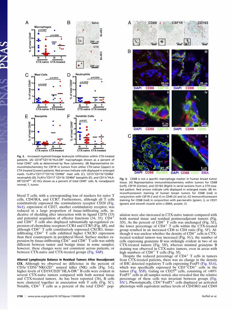
Increased Presence of Myeloid-Lineage Cells in Residual Tumors fromPatients Exposed to Neoadjuvant CTX. Comparative analysis ofresidual BC tissue removed from patients after neoadjuvant CTXrevealed an obvious difference in the percentages of myeloid-lineage cells compared with the CTX-naïve group. With someexceptions, this difference included an increased presence of mac-rophages as a percent of total leukocytes (Fig. 2A), as well as bydensity evaluation of CSF1 receptor (CSF1R)-positive cells in tissueby IHC (Fig. 2B). Increased percentages of mast cells (Fig. 2C) andneutrophils (Fig. 2D) were also evident in most CTX-treatedpatients, with an ≈14-fold increase in CTX-treated versus CTX-naïve groups. Basophils (FcεR1α+CD117−CD11b−CD49d+; Fig.2E) were highly increased in only one of six CTX-treated samples,whereas the percentage of myeloid dendritic cells (CD11c+HLA-DR+CD14lo/-; Fig. 2F) was unchanged. Evaluation of plasmacytoiddendritic cells expressing CD85g/ILT7 detected an insufficientnumber of events for analysis. Thus, with the exception of baso-phils, dendritic cells, and CD15+CD11b+CD49d+ eosinophils—which were present just at a detectable level in the tissues ex-amined—increased presence of myeloid-lineage cells typifiedresidual tumors of women treated with neoadjuvant CTX.
CD68 Is Not a Macrophage-Specific Marker in Human BC. Macro-phages are well established as regulators of murine mammarytumorigenesis (30), where they can represent up to 80% ofleukocytes present within late stage mammary carcinomas (1). Inhuman BC, immunoreactivity for CD68 has been used exten-sively for identification of macrophages, with CD68+ cell densityassociated with reduced overall survival (6, 11, 14, 15).The high number of CD68+ cells reported in the literature, and
shown in Fig. 3A, was in contrast to the limited number ofCD14hiCD11b+HLA-DR+ macrophages observed by flowcytometry in the BC suspensions examined (Figs. 1B and 2A). Tounderstand this discrepancy, wefirst evaluatedCD68 expression inBC tissue sections, compared with CD163 (a hemoglobin scav-enger receptor also commonly used as a marker for macrophages)and CSF1R (Fig. 3A). This comparative analysis revealed a lack ofcorrelation in cell density among the three markers. We nextevaluated frozen BC tissue sections by confocal microscopy afterimmunofluorescent detection of CD68 in combination withCSF1R or CD45 (Fig. 3B). Although all cells expressing high lev-els of the CSF1R also expressed CD68, there was a distinct pop-ulation of CD68+ cells that expressed neither CSF1R nor CD45.CD68 did not significantly colocalize with keratin+ epithelial cells,CD31+ endothelial cells, or smooth muscle actin α-expressingmural cells surrounding vasculature (Fig. 3C). This expressioncontrasted with murine mammary tumors isolated from MMTV-PyMT transgenic mice (17), where CD68+ cells coexpressedboth CSF1R and the murine macrophage marker F4/80 (Fig.S3). In agreement with historic literature (31, 32), these resultsthus indicate that CD68 is not a macrophage-specific marker inhuman BC.
Tumor-Infiltrating T Cells Display an Activated Phenotype. To revealthe phenotype of T cells infiltrating BCs, we examined surfacemarker and chemokine receptor expression of tissue-infiltratingCD4+ and CD8+ T cells (Fig. 4 A and B). Specifically, bothCD4+ and CD8+ T cells displayed increased expression of ac-tivation markers CD69 and HLA-DR compared with peripheral
Fig. 1. Leukocyte infiltration of human breast tumors. (A) Hematoxylin andeosin (H&E) staining of tissue sections (Left) with representative immunohis-tochemistry for CD45 (Right) shown for each. (B) Flow cytometric analysis ofleukocyte populations within human breast tumors. Results are shown asa percent of total CD45+ cells with markers used to define specific lineagesshown below.
Ruffell et al. PNAS | February 21, 2012 | vol. 109 | no. 8 | 2797
IMMUNOLO
GY
SPEC
IALFEATU
RE
blood T cells, with a corresponding loss of markers for naïve Tcells, CD45RA, and CCR7. Furthermore, although all T cellsconstitutively expressed the costimulatory receptor CD28 (Fig.S4A), expression of CD27, another costimulatory receptor, wasreduced in a large proportion of tissue-infiltrating cells, in-dicative of shedding after interaction with its ligand CD70 (33)and potential acquisition of effector functions (34, 35). CD4+and CD8+ T cells also displayed substantially up-regulated ex-pression of chemokine receptors CCR4 and CCR5 (Fig. 4B), andalthough CD8+ T cells constitutively expressed CXCR3, tissue-infiltrating CD4+ T cells exhibited higher CXCR3 expressionthan their counterparts in peripheral blood. Surface marker ex-pression by tissue-infiltrating CD4+ and CD8+ T cells was subtlydifferent between tumor and benign tissue in some samples;however, these changes were not consistent across patients, orbetween CTX-naïve and CTX-treated groups (Fig. S4B).
Altered Lymphocyte Balance in Residual Tumors After NeoadjuvantCTX. Although we observed no difference in the percent ofCD3e−CD56+NKG2D+ natural killer (NK) cells (Fig. 5A),higher levels of CD19/CD20+HLA-DR+ B cells were evident inseveral CTX-naïve tumors compared with both normal tissueand CTX-treated tumors. As has been reported (36), B cellswere clustered together in association with T cells (Fig. 5C).Notably, CD4+ T cells as a percent of the total CD45+ pop-
ulation were also increased in CTX-naïve tumors compared withboth normal tissue and residual postneoadjuvant tumors (Fig.5D). As the percent of CD8+ T cells was unchanged (Fig. 5E),the lower percentage of CD4+ T cells within the CTX-treatedgroup resulted in an increased CD8 to CD4 ratio (Fig. 5F). Al-though it was unclear whether the density of CD8+ cells in CTX-treated residual tumors was increased (Fig. 5G), the number ofcells expressing granzyme B was strikingly evident in two of sixCTX-treated tumors (Fig. 5H), whereas minimal granzyme Bstaining was observed in CTX-naïve tumors, even in areas withhigh numbers of CD8+ T cells (Fig. 5I).Despite the reduced percentage of CD4+ T cells in tumors
from CTX-treated patients, there was no change in the densityof IHC detected regulatory T cells expressing FoxP3 (Fig. S5A),which was specifically expressed by CD3+CD4+ cells in thetumor (Fig. S5B). Gating on CD25hi cells, consisting of >80%FoxP3+ cells in all samples tested, also revealed that the relativepercentage of these cells was invariant between groups (Fig.S5C). Phenotypically, CD4+FoxP3+ cells displayed an activatedphenotype with equivalent surface levels of CD45RO and CD69
Fig. 2. Increased myeloid-lineage leukocyte infiltration within CTX-treatedpatients. (A) CD14hiCD11b+HLA-DR+ macrophages shown as a percent oftotal CD45+ cells as determined by flow cytometry. (B) Representative im-munohistochemistry for CSF1R in tumors from either CTX-naïve (Upper) orCTX-treated (Lower) patients. Red arrows indicate cells displayed in enlargedinsets. FcεR1α+CD117+CD11b−CD49d+ mast cells (C), CD15+CD11b+CD49d−
neutrophils (D), FcεR1α+CD117−CD11b−CD49d+ basophils (E), and CD11c+HLA-DR+CD14lo/− (F) DCs shown as a percent of total CD45+ cells. N, nonadjacentnormal; T, tumor.
Fig. 3. CD68 is not a specific macrophage marker in human breast tumortissue. (A) Representative immunohistochemistry within tumors for CD68(Left), CSF1R (Center), and CD163 (Right) in serial sections from a CTX-trea-ted patient. Red arrows indicate cells displayed in enlarged insets. (B) Im-munofluorescent staining of human breast tumors for CD68 (red) inconjunction with CSF1R (i and ii) or CD45 (iii and iv). (C) Immunofluorescentstaining for CD68 (red) in conjunction with pan-keratin (green; i), or CD31(green) and smooth muscle actin-α (SMA; purple; ii).
2798 | www.pnas.org/cgi/doi/10.1073/pnas.1104303108 Ruffell et al.

to CD4+FoxP3− cells and, as has been reported for cells in pe-ripheral blood (37), expressed lower levels of CD127 (Fig. S5D).Interestingly, although not all FoxP3+ cells expressed HLA-DR,they did comprise the majority of HLA-DR–expressing CD4+ Tcells, in addition to coexpressing high levels of CD25.These data collectively reveal a shift within tumors toward
a TH2-type response in BC characterized by increased presence ofB cells and CD4+ T cells, in comparison with nonadjacent normalbreast tissue. This shift is reversed in tumors obtained from CTX-treated patients, with additional evidence of a cytotoxic T-cellresponse through a more favorable CD8/CD4 T-cell ratio andincreased presence of granyzme B-expressing lymphocytes; thus,even residual tumors from patients with a poor response to CTXmay contain immune microenvironments that are more favorablyskewed towards an anti-tumor, TH1-type immune response.
DiscussionHerein, we present a detailed description of leukocyte com-plexity in BC as evaluated in a cohort of CTX-naïve patients withstage 2/3 tumors, compared with patients with significant residualdisease after neoadjuvant CTX. T lymphocytes were the majorpopulation within both CTX-naïve and CTX-treated tumors,found almost exclusively in an activated state as determined byincreased expression of CD69 and chemokine receptors, withsimultaneous loss of naïve markers CCR7 and CD45RA. Thepresence of activation markers, however, does not definitivelydemonstrate that intratumoral T cells are functionally active. Infact, granzyme B expression was minimal within tumors fromCTX-naïve patients, suggesting negligible cytotoxic activity byinfiltrating CD8+ T cells. In comparison, granzyme B was highlyexpressed in one-third of the CTX-treated tumors, suggestive ofa more cytotoxic T-cell response within some tumors after ex-posure to CTX.
Importantly, residual tumors from CTX-treated patients alsocontained reduced percentages of B cells and CD4+ T cells.Tumor-infiltrating CD4+ T cells in BC are known to express theTH2 cytokines IL-4 and IL-13 concomitantly with the productionof IFN-γ (25, 26), consistent with coexpression of CXCR3 andCCR4 (38, 39) as we observed herein. It remains to be de-termined whether cytokine production by CD4+ T cells is alteredby neoadjuvant CTX; however, the combined reduction in bothCD4+ T cells and B cells is indicative of a favorable shift awayfrom a TH2 microenvironment. This shift could be relevant forTAM function, as has been described in the MMTV-PyMTmodel where TAMs are programmed by IL-4 toward a TH2phenotype (1), and more recently in pancreatic ductal adeno-carcinoma during treatment where an agonist CD40 monoclonalantibody fostered cytolytic macrophage activities (40).Although the extent of lymphocyte infiltration has been as-
sociated with improved prognosis in subsets of patients (2, 3),and with pCR after CTX (41, 42), information regarding therelationship between individual lymphocyte subsets to survival islimited. High FoxP3 counts correlate with reduced overall andrelapse-free survival in estrogen receptor (ER)-positive tumors(43), and pCR to neoadjuvant CTX is associated with reducedFoxP3 grading (44, 45). Although two studies examining T-cellinfiltration by flow cytometry found conflicting results regardingthe CD8:CD4 ratio and lymph node metastasis (46, 47), thenumber of CD8+ T cells within tissue has been associated withimproved patient survival (48). We have also reported a CD68/CD4/CD8 immune signature predicting overall and relapse-freesurvival, with inverse correlations evident for CD4 when used inconjunction with other markers (6). There is thus an urgent needfor additional prospective investigations where multiple param-eters of lymphocytic infiltration and functionality are evaluatedto determine the most significant biomarker comparisons thatpredict outcome and guide specific therapy.
Fig. 4. Tissue-infiltrating T cells display an activated pheno-type. (A and B) Representative histograms of CD3+CD4+ (Upper)or CD3+CD8+ (Lower) T cells isolated from a single CTX-treatedpatient with both normal (blue) and tumor (red) tissue. Ex-pression of activation markers CD69 (Left), HLA-DR (CenterLeft), CD45RA (Center Right), and CD27 (Right) are shown in A,and expression of chemokine receptors CCR4 (Left), CCR5(Center Left), CCR7 (Center Right) and CXCR3 (Right) are shownin B.
Ruffell et al. PNAS | February 21, 2012 | vol. 109 | no. 8 | 2799
IMMUNOLO
GY
SPEC
IALFEATU
RE

Although used successfully in multiple studies to relate TAMinfiltration with clinically relevant outcomes, our results indicatethat CD68 alone cannot accurately evaluatemacrophage presencein human breast tissue given that multiple stromal cells express itand that a subset of these are CSF1R- and CD45-negative. Weobserved that the nonleukocytic CD68+ cells were predominantlylocated within tumor stroma and, thus, based on this localizationand morphology, we speculate that CSF1R−CD68+ cells likelyreflect tumor-associated fibroblasts or monocyte-derived fibro-cytes in agreement with other reports (31, 32, 49–52). Our findingsdo not invalidate CD68 as a clinically relevant marker and, im-portantly, CSF1-response gene signatures have been identified inbreast adenocarcinomas that are predictive of recurrence risk andmetastasis (53, 54). However, given the important role thatfibroblasts (and perhaps fibrocytes) play in fostering aspects oftumorigenesis (55–57), differentiating among macrophages,fibroblasts, and other stromal populations within tumors has thepotential to improve diagnostic information currently generatedby immunodetection of CD68.As we have reported for expression of csf1 mRNA (6), mul-
tiple genes encoding myeloid cell chemoattractants are differ-entially expressed by human BC cell lines, with variableinduction of these genes in response to CTX (Fig. S6). Althoughdifferential expression between cell lines corresponding to par-ticular subtypes of BC is evident, it is doubtful these cell linesaccurately represent the response of BC tumor tissue; thus, weare investigating whether differences in myeloid cell infiltrates
reflect distinct molecular subtypes of BC and to what extentthese differ in residual tumors from CTX-treated patients.It is important to acknowledge that leukocyte composition
within tumors responding to CTX likely differs substantially fromresidual or recurrent tumors from patients that have receivedCTX, given what is known regarding immune responses to CTX-induced cell death (28). However, we recently reported that inmammary carcinomas of MMTV-PyMT mice, blockade of theCSF1-CSF1R pathway critical for TAM recruitment improvedresponse to CTX through a CD8+ T-cell–dependent effect (6).Thus, even though the findings presented herein are based ona small dataset of heterogeneous tumor subtypes, and our resultsmay be biased because of sample selection favoring large and/orless CTX-responsive tumors among the CTX-treated group, theclear distinctions in the myeloid profiles between CTX-naive andCTX-treated tumors is provocative and indicates that a CSF1-targeted strategy may be a promising approach to enhancetherapeutic efficacy of cytotoxic CTX, particularly for treatmentof refractory BC. Moreover, given the increase in granulocyticpopulations within tumors resistant to CTX, and the involvementof these cells in regulating immune responses in chronic in-flammatory diseases (58–62), these populations may also befunctionally relevant, and targeting common pathways of im-mune suppression within the tumor microenvironment mayprovide additional therapeutic opportunities to increase efficacyof neoadjuvant CTX.
Fig. 5. Improved cytotoxic T-cell response in CTX-treated tumors. CD3ε −CD56+NKG2D+ natural killer cells (A) and CD3ε−CD19/20+HLA-DR+ B cells (B) shown asa percent of total CD45+ cells as determined by flow cytometry. (C) Immunofluorescent staining of tumors for CD20 (green) and CD3 (red). CD3ε+CD4+ T cells(D) and CD3ε+CD8+ T cells (E) are shown as a percent of total CD45+ cells. (F) Ratio of CD8+ to CD4+ T cells within CTX-naive versus CTX-treated tumors.Number of CD8-positive (G) and granzyme B-positive (H) cells per area as determined by automated counting. (I) Representative sections stained with CD8 orgranzyme B from CTX-naive (Left) or CTX-treated (Right) tumors. Red arrows indicate cells displayed in enlarged insets. N, nonadjacent normal; T, tumor.
2800 | www.pnas.org/cgi/doi/10.1073/pnas.1104303108 Ruffell et al.

Materials and MethodsTissues were collected at the time of surgery from consenting patients at theUniversity of California, San Francisco under approval from the institutionalreviewboard.Tumorand ipsilateralnonadjacentnormal tissueswere collectedby a certified pathologist (A.A.) and were prepared for analysis on the day ofresection. The percent ofmacrophages and CD8+ T cells has been reported fora subset of the patients described here (6). Flow cytometry, immunohisto-chemistry, and immunofluorescence were performed as described (6), withdetailed methods contained in SI Materials and Methods, and a list of anti-bodies available in Tables S2 and S3. Statistical differences between two in-
dependent groups were determined by using Student’s t test via Prism 4.0software (GraphPad Software).
ACKNOWLEDGMENTS. Wethank Erin Bowlby for compiling patient data. Thiswork was supported by a Department of Defense Breast Cancer ResearchProgram Fellowship (to B.R.); a grant from the Breast Cancer ResearchFoundation (to H.S.R.); National Institutes of Health/National Cancer InstituteGrants R01CA130980, R01CA132566, R01CA140943, and P50CA58207; a Dr.Susan Love Research Foundation Instructional grant; and Department ofDefense Grants W81XWH-06-1-0416 and PR080717 (to L.M.C.).
1. DeNardo DG, et al. (2009) CD4(+) T cells regulate pulmonary metastasis of mammarycarcinomas by enhancing protumor properties ofmacrophages. Cancer Cell 16:91–102.
2. Ménard S, et al. (1997) Lymphoid infiltration as a prognostic variable for early-onsetbreast carcinomas. Clin Cancer Res 3:817–819.
3. Pupa SM, et al. (1996) Macrophage infiltrate and prognosis in c-erbB-2-overexpressingbreast carcinomas. J Clin Oncol 14:85–94.
4. Bingle L, Brown NJ, Lewis CE (2002) The role of tumour-associated macrophages intumour progression: Implications for new anticancer therapies. J Pathol 196:254–265.
5. Mukhtar RA, Nseyo O, Campbell MJ, Esserman LJ (2011) Tumor-associated macro-phages in breast cancer as potential biomarkers for new treatments and diagnostics.Expert Rev Mol Diagn 11:91–100.
6. DeNardoDG, et al. (2011) Leukocyte complexity inbreast cancerpredicts overall survivaland functionally regulates response to chemotherapy. Cancer Discovery 1:54–67.
7. Esserman LJ, et al. (2006) Magnetic resonance imaging captures the biology of ductalcarcinoma in situ. J Clin Oncol 24:4603–4610.
8. Volodko N, Reiner A, Rudas M, Jakesz R (1998) Tumour-associated macrophages inbreast cancer and their prognostic correlations. Breast 7:99–105.
9. Lee AH, Happerfield LC, Bobrow LG, Millis RR (1997) Angiogenesis and inflammationin invasive carcinoma of the breast. J Clin Pathol 50:669–673.
10. Uzzan B, Nicolas P, Cucherat M, Perret GY (2004) Microvessel density as a prognosticfactor in women with breast cancer: A systematic review of the literature and meta-analysis. Cancer Res 64:2941–2955.
11. Tsutsui S, et al. (2005) Macrophage infiltration and its prognostic implications inbreast cancer: The relationship with VEGF expression and microvessel density. OncolRep 14:425–431.
12. Bolat F, et al. (2006) Microvessel density, VEGF expression, and tumor-associatedmacrophages in breast tumors: Correlations with prognostic parameters. J Exp ClinCancer Res 25:365–372.
13. Chen JJ, et al. (2005) Tumor-associated macrophages: The double-edged sword incancer progression. J Clin Oncol 23:953–964.
14. Leek RD, et al. (1996) Association of macrophage infiltration with angiogenesis andprognosis in invasive breast carcinoma. Cancer Res 56:4625–4629.
15. Campbell MJ, et al. (2010) Proliferating macrophages associated with high grade,hormone receptor negative breast cancer and poor clinical outcome. Breast CancerRes Treat 128:703–711.
16. Robinson BD, et al. (2009) Tumor microenvironment of metastasis in human breastcarcinoma: A potential prognostic marker linked to hematogenous dissemination.Clin Cancer Res 15:2433–2441.
17. Guy CT, Cardiff RD, Muller WJ (1992) Induction of mammary tumors by expression ofpolyomavirus middle T oncogene: A transgenic mouse model for metastatic disease.Mol Cell Biol 12:954–961.
18. Lin EY, Nguyen AV, Russell RG, Pollard JW (2001) Colony-stimulating factor 1 pro-motes progression of mammary tumors to malignancy. J Exp Med 193:727–740.
19. Lin EY, et al. (2006) Macrophages regulate the angiogenic switch in a mouse model ofbreast cancer. Cancer Res 66:11238–11246.
20. Stockmann C, et al. (2008) Deletion of vascular endothelial growth factor in myeloidcells accelerates tumorigenesis. Nature 456:814–818.
21. Lin EY, et al. (2007) Vascular endothelial growth factor restores delayed tumor pro-gression in tumors depleted of macrophages. Mol Oncol 1:288–302.
22. Wyckoff J, et al. (2004) A paracrine loop between tumor cells and macrophages isrequired for tumor cell migration in mammary tumors. Cancer Res 64:7022–7029.
23. Wyckoff JB, et al. (2007) Direct visualization of macrophage-assisted tumor cell in-travasation in mammary tumors. Cancer Res 67:2649–2656.
24. Doedens AL, et al. (2010) Macrophage expression of hypoxia-inducible factor-1 alphasuppresses T-cell function and promotes tumor progression. Cancer Res 70:7465–7475.
25. Aspord C, et al. (2007) Breast cancer instructs dendritic cells to prime interleukin 13-secreting CD4+ T cells that facilitate tumor development. J Exp Med 204:1037–1047.
26. Pedroza-Gonzalez A, et al. (2011) Thymic stromal lymphopoietin fosters humanbreast tumor growth by promoting type 2 inflammation. J Exp Med 208:479–490.
27. Gocheva V, et al. (2010) IL-4 induces cathepsin protease activity in tumor-associatedmacrophages to promote cancer growth and invasion. Genes Dev 24:241–255.
28. Zitvogel L, Apetoh L, Ghiringhelli F, Kroemer G (2008) Immunological aspects ofcancer chemotherapy. Nat Rev Immunol 8:59–73.
29. Allan CP, Turtle CJ, Mainwaring PN, Pyke C, Hart DN (2004) The immune response tobreast cancer, and the case for DC immunotherapy. Cytotherapy 6:154–163.
30. Qian BZ, Pollard JW (2010) Macrophage diversity enhances tumor progression andmetastasis. Cell 141:39–51.
31. Pulford KA, Sipos A, Cordell JL, Stross WP, Mason DY (1990) Distribution of the CD68macrophage/myeloid associated antigen. Int Immunol 2:973–980.
32. Kunz-Schughart LA, et al. (2003) [The “classical” macrophage marker CD68 is stronglyexpressed in primary human fibroblasts]. Verh Dtsch Ges Pathol 87:215–223.
33. Hintzen RQ, et al. (1994) Characterization of the human CD27 ligand, a novel memberof the TNF gene family. J Immunol 152:1762–1773.
34. Hamann D, et al. (1997) Phenotypic and functional separation of memory and ef-fector human CD8+ T cells. J Exp Med 186:1407–1418.
35. Okada R, Kondo T, Matsuki F, Takata H, Takiguchi M (2008) Phenotypic classificationof human CD4+ T cell subsets and their differentiation. Int Immunol 20:1189–1199.
36. Nelson BH (2010) CD20+ B cells: The other tumor-infiltrating lymphocytes. J Immunol185:4977–4982.
37. Liu W, et al. (2006) CD127 expression inversely correlates with FoxP3 and suppressivefunction of human CD4+ T reg cells. J Exp Med 203:1701–1711.
38. Kunkel EJ, et al. (2002) Expression of the chemokine receptors CCR4, CCR5, and CXCR3by human tissue-infiltrating lymphocytes. Am J Pathol 160:347–355.
39. Kim CH, et al. (2001) Rules of chemokine receptor association with T cell polarizationin vivo. J Clin Invest 108:1331–1339.
40. Beatty GL, et al. (2011) CD40 agonists alter tumor stroma and show efficacy againstpancreatic carcinoma in mice and humans. Science 331:1612–1616.
41. Demaria S, et al. (2001) Development of tumor-infiltrating lymphocytes in breastcancer after neoadjuvant paclitaxel chemotherapy. Clin Cancer Res 7:3025–3030.
42. Denkert C, et al. (2010) Tumor-associated lymphocytes as an independent predictor ofresponse to neoadjuvant chemotherapy in breast cancer. J Clin Oncol 28:105–113.
43. Bates GJ, et al. (2006) Quantification of regulatory T cells enables the identification ofhigh-risk breast cancer patients and those at risk of late relapse. J Clin Oncol 24:5373–5380.
44. Ladoire S, et al. (2008) Pathologic complete response to neoadjuvant chemotherapyof breast carcinoma is associated with the disappearance of tumor-infiltrating foxp3+regulatory T cells. Clin Cancer Res 14:2413–2420.
45. de Kruijf EM, et al. (2010) The predictive value of HLA class I tumor cell expression andpresence of intratumoral Tregs for chemotherapy in patients with early breast cancer.Clin Cancer Res 16:1272–1280.
46. Macchetti AH, et al. (2006) Tumor-infiltrating CD4+ T lymphocytes in early breastcancer reflect lymph node involvement. Clinics (Sao Paulo) 61:203–208.
47. Sheu BC, et al. (2008) Clinical significance of tumor-infiltrating lymphocytes in neo-plastic progression and lymph node metastasis of human breast cancer. Breast 17:604–610.
48. Mahmoud SM, et al. (2011) Tumor-infiltrating CD8+ lymphocytes predict clinicaloutcome in breast cancer. J Clin Oncol 29:1949–1955.
49. Gottfried E, et al. (2008) Expression of CD68 in non-myeloid cell types. Scand J Im-munol 67:453–463.
50. Pilling D, Fan T, Huang D, Kaul B, Gomer RH (2009) Identification of markers thatdistinguish monocyte-derived fibrocytes from monocytes, macrophages, and fibro-blasts. PLoS ONE 4:e7475.
51. Shao DD, Suresh R, Vakil V, Gomer RH, Pilling D (2008) Pivotal Advance: Th-1 cyto-kines inhibit, and Th-2 cytokines promote fibrocyte differentiation. J Leukoc Biol 83:1323–1333.
52. Azambuja D, et al. (May 2, 2011) Lack of association of tumor-associated macro-phages with clinical outcome in patients with classical Hodgkin’s lymphoma. AnnOncol, 10.1093/annonc/mdr157.
53. Sharma M, et al. (2010) Analysis of stromal signatures in the tumor microenvironmentof ductal carcinoma in situ. Breast Cancer Res Treat 123:397–404.
54. Beck AH, et al. (2009) The macrophage colony-stimulating factor 1 response signaturein breast carcinoma. Clin Cancer Res 15:778–787.
55. Trimboli AJ, et al. (2009) Pten in stromal fibroblasts suppresses mammary epithelialtumours. Nature 461:1084–1091.
56. Erez N, Truitt M, Olson P, Arron ST, Hanahan D (2010) Cancer-associated fibroblastsare activated in incipient neoplasia to orchestrate tumor-promoting inflammation inan NF-kappaB-dependent manner. Cancer Cell 17:135–147.
57. Kalluri R, Zeisberg M (2006) Fibroblasts in cancer. Nat Rev Cancer 6:392–401.58. Schmielau J, Finn OJ (2001) Activated granulocytes and granulocyte-derived hydro-
gen peroxide are the underlying mechanism of suppression of T-cell function in ad-vanced cancer patients. Cancer Res 61:4756–4760.
59. Fridlender ZG, et al. (2009) Polarization of tumor-associated neutrophil phenotype byTGF-beta: “N1” versus “N2” TAN. Cancer Cell 16:183–194.
60. Eller K, et al. (2011) IL-9 production by regulatory T cells recruits mast cells that areessential for regulatory T cell-induced immune suppression. J Immunol 186:83–91.
61. Galli SJ, Nakae S, Tsai M (2005) Mast cells in the development of adaptive immuneresponses. Nat Immunol 6:135–142.
62. Perrigoue JG, et al. (2009) MHC class II-dependent basophil-CD4+ T cell interactionspromote T(H)2 cytokine-dependent immunity. Nat Immunol 10:697–705.
Ruffell et al. PNAS | February 21, 2012 | vol. 109 | no. 8 | 2801
IMMUNOLO
GY
SPEC
IALFEATU
RE