letters to the editor - journal of medical genetics - bmj
TRANSCRIPT

Letters to the Editor
J Med Genet 2000;37:449–451
Fine molecular mapping of the 4p16.3aneuploidy syndromes in fourtranslocation families
EDITOR—Deletions of 4p16.3 have attracted considerableattention, particularly since the introduction of FISH andmolecular techniques, and are associated with a variety ofclinical pictures. Although all aVected subjects are mentallyretarded, this can vary from profound to mild and thephysical manifestations may be those of the severe, oftenfatal, Wolf-Hirschhorn syndrome (WHS) or of therelatively milder, usually non-fatal Pitt-Rogers-Danks syn-drome (PRDS). Genotype-phenotype correlations are notconsistent except for the broad generalisation that the mostsevere physical abnormalities are more likely to be seenwith the largest deletions. There is less information about4p16.3 duplications. Before FISH, patients with 4ptrisomy or duplications were reported to have profoundmental retardation with microcephaly, short stature, andother marked physical abnormalities.1 By contrast, in twotranslocation families where we have described index caseswith PRDS, those sibs with the 4p16.3 duplication had
relatively mild mental retardation and late onset physicalovergrowth.2 Here we describe the fourth family we haveencountered with a translocation in which the index casehas PRDS. This boy’s father and older brother carried thetranslocation in a balanced form and his younger brotherhad an unbalanced karyotype with 4p16.3 duplication.
Patient 1 is the proband, born in 1986, who wasdiagnosed clinically at the age of 10 years. He was born at35 weeks’ gestation with a birth weight of 1800 g (10thcentile). There were early feeding problems and he was inhospital for three months. Pyloric and ureteric stenosiswere found and operated on. He was left with only onefunctioning kidney. In the second year of life, he developedgrand mal seizures, up to six per day, but these stopped atthe age of 5 years. At 12 years 9 months he was an aVablechild with some limited conversation. He had developedsome expertise in bowling. His height (136 cm) was belowthe 3rd centile and his head circumference (HC) was 48cm, some 3.5 SD below the mean. He had abundant curlyhair on the head, apparent hypotelorism, slightly promi-nent eyes with some fullness of the lower lids and the scleravisible below the iris, a pointed nose, some prominence ofthe glabella, a short philtrum, wide mouth, and small chin(fig 1). He was very slender with little subcutaneous fat.
Figure 1 The PRDS proband (left) and his OGS sib, aged 12 years and 51⁄2 years, respectively.
Letters, Correction, Notice 449
on 15 January 2019 by guest. Protected by copyright.
http://jmg.bm
j.com/
J Med G
enet: first published as 10.1136/jmg.37.6.454 on 1 June 2000. D
ownloaded from
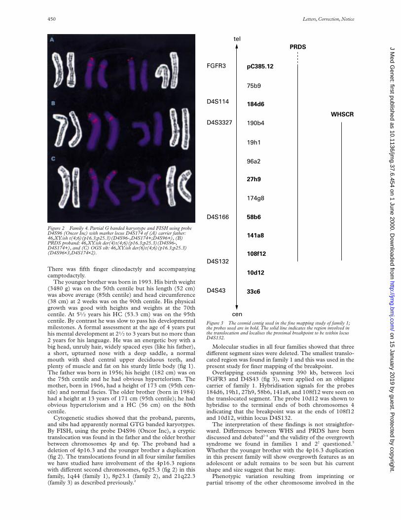
There was fifth finger clinodactyly and accompanyingcamptodactyly.
The younger brother was born in 1993. His birth weight(3480 g) was on the 50th centile but his length (52 cm)was above average (85th centile) and head circumference(38 cm) at 2 weeks was on the 90th centile. His physicalgrowth was good with heights and weights at the 70thcentile. At 51⁄2 years his HC (53.3 cm) was on the 95thcentile. By contrast he was slow to pass his developmentalmilestones. A formal assessment at the age of 4 years puthis mental development at 21⁄2 to 3 years but no more than2 years for his language. He was an energetic boy with abig head, unruly hair, widely spaced eyes (like his father),a short, upturned nose with a deep saddle, a normalmouth with shed central upper deciduous teeth, andplenty of muscle and fat on his sturdy little body (fig 1).The father was born in 1956; his height (182 cm) was onthe 75th centile and he had obvious hypertelorism. Themother, born in 1966, had a height of 173 cm (95th cen-tile) and normal facies. The older brother (born in 1984)had a height at 13 years of 171 cm (95th centile); he hadobvious hypertelorism and a HC (56 cm) on the 80thcentile.
Cytogenetic studies showed that the proband, parents,and sibs had apparently normal GTG banded karyotypes.By FISH, using the probe D4S96 (Oncor Inc), a cryptictranslocation was found in the father and the older brotherbetween chromosomes 4p and 6p. The proband had adeletion of 4p16.3 and the younger brother a duplication(fig 2). The translocations found in all four similar familieswe have studied have involvement of the 4p16.3 regionswith diVerent second chromosomes, 6p25.3 (fig 2) in thisfamily, 1q44 (family 1), 8p23.1 (family 2), and 21q22.3(family 3) as described previously.2
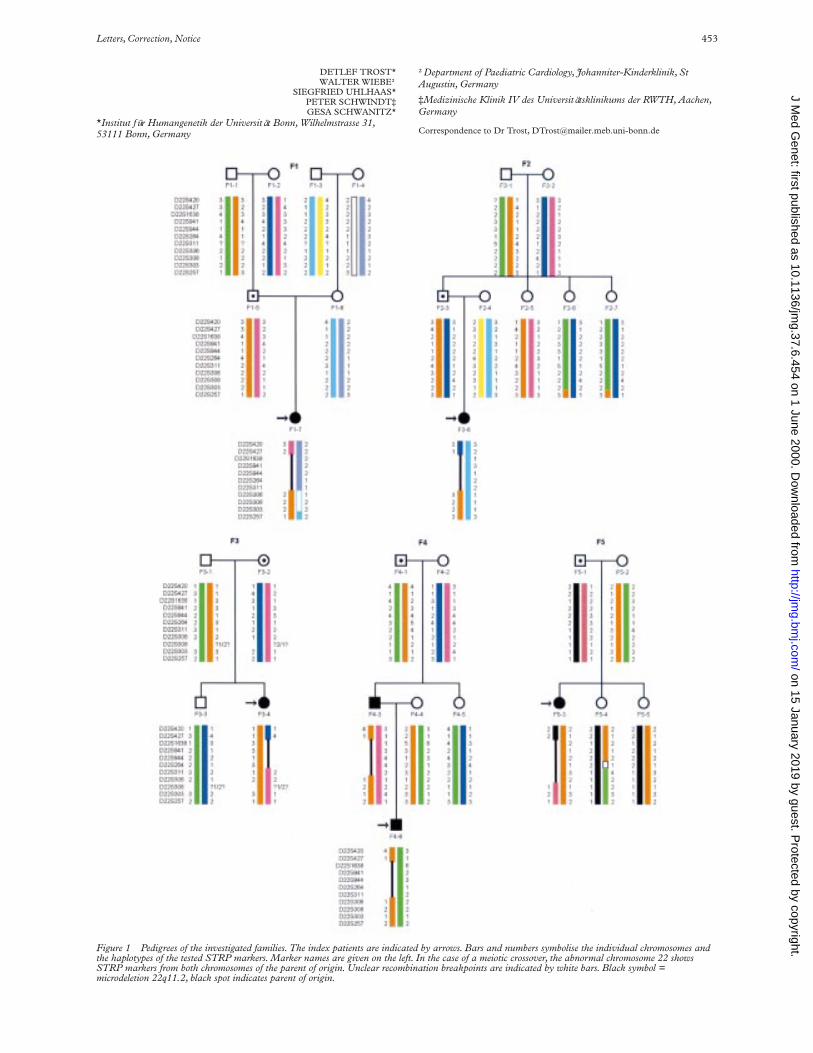
Molecular studies in all four families showed that threediVerent segment sizes were deleted. The smallest translo-cated region was found in family 1 and this was used in thepresent study for finer mapping of the breakpoint.
Overlapping cosmids spanning 390 kb, between lociFGFR3 and D4S43 (fig 3), were applied on an obligatecarrier of family 1. Hybridisation signals for the probes184d6, 19h1, 27h9, 58b6, 141a8, and 108f12 were seen onthe translocated segment. The probe 10d12 was shown tohybridise to the terminal ends of both chromosomes 4indicating that the breakpoint was at the ends of 108f12and 10d12, within locus D4S132.
The interpretation of these findings is not straightfor-ward. DiVerences between WHS and PRDS have beendiscussed and debated2–4 and the validity of the overgrowthsyndrome we found in families 1 and 22 questioned.5
Whether the younger brother with the 4p16.3 duplicationin this present family will show overgrowth features as anadolescent or adult remains to be seen but his currentshape and size suggest that he may.
Phenotypic variation resulting from imprinting orpartial trisomy of the other chromosome involved in the
Figure 2 Family 4. Partial G banded karyotype and FISH using probeD4S96 (Oncor Inc) with marker locus D4S174 of (A) carrier father:46,XY.ish t(4;6)(p16.3;p25.3)(D4S96-,D4S174+;D4S96+), (B)PRDS proband: 46,XY.ish der(4)t(4;6)(p16.3;p25.3)(D4S96-,D4S174+), and (C) OGS sib: 46,XY.ish der(6)t(4;6)(p16.3;p25.3)(D4S96×3,D4S174×2).
Figure 3 The cosmid contig used in the fine mapping study of family 1;the probes used are in bold. The solid line indicates the region involved inthe translocation and localises the proximal breakpoint to be within locusD4S132.
cen
telPRDS
WHSCR
pC385.12
75b9
184d6
190b4
19h1
96a2
27h9
174g8
58b6
141a8
108f12
10d12
33c6D4S43
D4S132
D4S166
D4S3327
D4S114
FGFR3
450 Letters, Correction, Notice
on 15 January 2019 by guest. Protected by copyright.
http://jmg.bm
j.com/
J Med G
enet: first published as 10.1136/jmg.37.6.454 on 1 June 2000. D
ownloaded from

translocation was not evident in our families wherethe probands had PRDS. At the molecular levelboth WHS and PRDS seem to have very similar deletionsas the proximal breakpoint we have found is the same asthat recently defined in WHS patients, that is, withinD4S132.7
Two recent publications6 7 describe molecular studies ofone WHS and six PRDS patients with a deletion of 4p16.3in all cases. Only one of these resulted from an obvioustranslocation; no details of molecular family studies weregiven in the other cases. The breakpoints found wereproximal to locus D4S180, that is, similar to thetranslocation breakpoints in our families 2 and 3 (fig 4).One cell line of a PRDS patient (MA117)7 showed a
breakpoint between loci D4S166 and D4S43, similar toour family 1.
The overlap in some of the clinical features of WHS andPRDS is probably the result of the overlap in the two criti-cal regions (fig 3). The PRDS critical region may beentirely within the WHS critical region (WHSCR) butsmaller, involving fewer gene(s); this could account for therelatively milder phenotype of PRDS compared to WHS.Another explanation might be that the critical regions ofWHS and PRDS overlap in the middle, leaving out thedistal end of WHS and the proximal end of the PRDScritical regions; the PRDS critical region could theninclude FGFR3 which may be relevant for overgrowth. Itcould be that point mutations in diVerent genes or in a dif-ferent region of the same gene within the overlapped areaof the PRDS and WHS critical regions might account forthe diVerence in severity of the two syndromes. So far,WHS families have not been described with 4p transloca-tions that have sibs with the overgrowth resulting from aduplication of 4p16.3.
There are reports of patients with possible WHS/PRDSwho were not deleted for locus D4S96.4 8 9 As the criticalregion of WHS has been recently reduced to 260 kb byWHS patient data,7 this now excludes the locus D4S96 andprovides one explanation for these patients. However, asthe distal breakpoint for PRDS has not been determined, itmay be that D4S96 is not included in the critical regioneither. Just as the WHSCR has been reduced in size to 260kb, the same could be done with PRDS patients when suchare found with interstitial deletions.
Cosmid clones were obtained from Dr T Wright, Los Alamos National Labora-tories (from the laboratory of Dr M Altherr), and VS was supported by an Aus-tralian PHRDC Program grant No 954614.
K FAGAN*V SOUBJAKI*
*Cytogenetics Unit, Hunter Area Pathology Service, Locked Bag No 1,Newcastle Mail Exchange, New South Wales 2310, Australia
P DONALD†
†Mount Pleasant Medical Centre, Maitland, NSW, Australia
G TURNER‡M PARTINGTON‡
‡Hunter Genetics, PO Box 84, Waratah, NSW 2301, Australia
Correspondence to: Ms Fagan, [email protected]
1 Gonzalez CH, Sommer A, Meisner LF, Elejalde BR, Opitz JM. The trisomy4p syndrome: case report and review. Am J Med Genet 1977;1:137-56.
2 Partington MW, Fagan K, Soubjaki V, Turner G. Translocations involving4p16.3 in three families: deletion causing the Pitt-Rogers-Danks syndromeand duplication resulting in a new overgrowth syndrome. J Med Genet1997;34:719-28.
3 Wright TJ, Altherr MR, Callen D, Hirschhorn K. Reply to the Letter to theEditor by Partington and Turner on Wolf-Hirschhorn and Pitt-Rogers-Danks syndromes Am J Med Genet 1999;82:89-90.
4 Petit P, Schmidt J, Van den Berghe H, Fryns JP. On two patients with andwithout the classical Wolf-Hirschhorn syndrome (WHS) sharing the samechromosome 4p16.3 specific probe deletion: evidence of a contiguous genedeletion syndrome. Clin Genet 1996;50:19-22.
5 Cohen MM, Neri G. New overgrowth syndrome and FGFR3 dosage eVect.J Med Genet 1998;35:348-9.
6 Kant SG, van Haeringen A, Bakker E, et al. Pitt-Rogers-Danks syndromeand Wolf-Hirschhorn syndrome are caused by a deletion in the same regionon chromosome 4p16.3. J Med Genet 1997;34:569-72.
7 Wright TJ, Ricke DO, Denison K, et al. A transcript map of the newlydefined 165 kb Wolf-Hirschhorn syndrome critical region. Hum Mol Genet1997;6:317-24.
8 Donnai D. Brief clinical report. A further patient with the Pitt-Rogers-Danks syndrome of mental retardation, unusual face, and intrauterinegrowth retardation. Am J Med Genet 1986;24:29-32.
9 Zollino M, Bova R, Neri G. From Pitt-Rogers-Danks to Wolf-Hirschhornand back? Am J Med Genet 1996; 66:113-15.
Figure 4 Physical map of 4p16.3. The loci used are indicated in bold.Solid patterns indicate the regions translocated and the broken lines showundefined areas. The size of the region involved in each translocation isshown for our families 1, 2, 3, and 4. All deletions overlap with the newlyrefined WHSCR.7
cen
tel
D4S10
D4S81
D4S126
D4S125
D4S180
D4S224
D4S127
D4S95
D4S182
D4S181D4S43
D4S132
D4S166D4S3327D4S114
FGFR3
D4S113
D4S168
D4S115
D4S97
D4S96
D4S111
D4S90
D4F26
D4S142
16.316.2
6.0 Mb
5.8 Mb
3.0 Mb
2.6 Mb
2.4 Mb
1.1 Mb
300 Kb
150 Kb
80 Kb
WHSCR
2431
Letters, Correction, Notice 451
on 15 January 2019 by guest. Protected by copyright.
http://jmg.bm
j.com/
J Med G
enet: first published as 10.1136/jmg.37.6.454 on 1 June 2000. D
ownloaded from

J Med Genet 2000;37:452–454
Investigation of meioticrearrangements in DGS/VCFS patientswith a microdeletion 22q11.2
EDITOR—Microdeletions in 22q11.2 are associated in80-90% of cases with DiGeorge syndrome (DGS, MIM188400) or velocardiofacial syndrome (VCFS, MIM192430) and occur with an estimated frequency of 1/4000live births.1 Most deletions are the result of a de novo event,although probably 6-28% of them are familial.2 Thephenotype of the patients is mainly characterised byconotruncal heart defect, cleft palate, immune deficiency,neonatal hypocalcaemia, and facial dysmorphism. Thenumber of clinical symptoms varies substantially and theirreduced expression can lead to a mild phenotype.3 Theredoes not seem to be a correlation between the presence orthe size of a microdeletion and the clinical manifestation ofthe syndrome. Molecular analyses have shown that mostpatients have a deletion of about 1.5 or 3 Mb.4 5 The lengthof the delineated minimal critical region for a DGS/VCFSphenotype, however, is only 480 kb.6 Reports of patientswith a DGS/VCFS-like phenotype having a deletion in10p7 led to the definition of a second critical region,DGSII. However, the incidence of 10p deletions is low incomparison to the rate of microdeletions in 22q.8 9 Thehigh rate of sporadic microdeletions in 22q11.2 providesevidence for frequent meiotic rearrangements as a molecu-lar basis for the development of this structural aberration.
In order to ascertain such rearrangements in patientswith a 22q11.2 deletion, we performed haplotype analyseson five patients and their unaVected relatives using 11polymorphic STRP markers from the DGS/VCFS criticalregion in 22q11.2 (fig 1). Furthermore, the haplotypeanalyses enabled us to determine the extent of the deletionsin deletion carriers and the parental origin of the abnormalchromosome.
Microdeletion analysis was performed by fluorescence insitu hybridisation (FISH) on metaphase chromosomesprepared from fresh peripheral blood samples using DNAprobes D22S75 (Oncor, Illkirch) or TUPLE1 (Vysis,Downers Grove, IL) from the DGS/VCFS critical regionon 22q11.2.
In order to haplotype patients and their family membersthe parents and, if available, the grandparents of origin wereanalysed with 11 STRP markers using standard methods.Primer information was obtained from the Genome DataBase (GDB). If the grandparents of origin were notavailable, haplotyping was performed with the resultsobtained from healthy sibs of the patient and the parent oforigin. A total of 30 family members were included in thestudy. The family pedigrees are shown in fig 1.
The STRP analyses allowed us to determine the deletionsizes in 22q11.2 of the investigated probands (fig 1).Patients F1-7, F2-8, F4-3, F4-8, and F5-3 had deletions ofthe 3 Mb type and patient F3-4 has a 1.5 Mb deletion. Theparental origin of the aberrant chromosome was deter-mined to be four times paternal and once maternal.
Haplotype analyses were performed to ascertain thedevelopmental mechanism of the microdeletion. In fourfamilies (F1, F2, F3, F5), a parental unequal crossover wasproven by the exchange of parental marker alleles flankingthe deleted region (fig 1 and 2). In family F4, the underly-ing mechanism could be either an unequal crossover or anintrachromosomal rearrangement, as the microsatellite
markers proximal to the monosomic area did not allow usto distinguish between these mechanisms.
Additional crossover events are present in family F5 whereproband F5-4 shows a rearrangement between D22S264and D22S311 (fig 1) and in family F2 in probands F2-6 andF2-7 between D22S303 and D22S257 (fig 1).
In this study we were able to analyse in detail the22q11.2 deletions present in five patients and one fatherfrom five unrelated families. Although there are diVerencesin deletion size, it was not possible to delineate any signifi-cant correlation with the phenotypic manifestations. This isespecially conspicuous in family F4 in which the father andson share an identical deletion. In this case the father dis-plays only slight dysmorphic facial features and a cleft pal-ate, while his son is more severely aVected with ventricularseptal defect (VSD), cerebral malformations, T cell defect,and marked developmental delay. This variation may bethe result of the diVerent maternal haplotype in each ofthem (fig 1, patients F4-3 and F4-6).
All proximal deletion breakpoints are flanked by theSTRP marker D22S427. The distal breakpoint, however,is variable, being flanked in four cases by D22S306 and inone case each by D22S311 or D22S308. These findingsare in agreement with previously defined deletionbreakpoints where most patients showed a deleted intervalof approximately 3 Mb flanked by D22S247 andD22S306.6 The genetic distance over this region isapproximately 6 cM according to the Généthon and GDBlinkage maps.
In our study the deletions were of maternal (n=1) as wellas of paternal (n=4) origin which does not confirm the biastowards maternally derived deletions found in otherstudies.10 The haplotype analyses of the investigated fami-lies show that four of five deletions are the result of anunequal meiotic crossover event. In these cases the markersflanking the deletion breakpoints are derived from diVerentparental chromosomes (fig 1 and 2). In family F4, it is notclear from the present data if the underlying event involvedhomologous pairing of chromosomes or exchanges be-tween sister chromatids (fig 1). In our sample of five fami-lies, no statistical significance can be calculated, but thedata confirm the findings from a previous investigation thatthe DiGeorge critical region (DGCR), though located nearthe centromere of chromosome 22, is subject to numerousmeiotic recombinations, many of which lead to the forma-tion of a microdeletion.11 Patient F5-4 displays a crossovernear D22S264 and D22S311 (fig 1). This is an interestingfinding because these markers are located at common dis-tal deletion breakpoints6 and underlines the presence ofcrossover mediating elements at the breakpoints of22q11.2 deletions.5 12–14
The mechanisms of microdeletion formation have beeninvestigated in other syndromes as well. The critical regionfor Prader-Willi/Angelman syndrome (PWS/AS) (15q11-q13) is subject to above average rates of recombination andsex specific hotspots have been described.15 The deletionswere caused by both intra- and interchromosomal recom-bination in the PWS/AS families investigated.16 17 Theresults obtained for deletions in 7q11.23 associated withWilliams-Beuren syndrome (WBS) suggest that the major-ity of microdeletions in this region are caused by unequalcrossover events.11 18 19 In comparison, in most informativeDGS/VCFS families, the microdeletion 22q11.2 was asso-ciated with a crossover but an intrachromosomal rear-rangement cannot be excluded in the remaining cases.5 11
We thank all families participating in this investigation. The study was supportedby “Richard-Winter-Stiftung”, Stuttgart, Germany.
452 Letters, Correction, Notice
on 15 January 2019 by guest. Protected by copyright.
http://jmg.bm
j.com/
J Med G
enet: first published as 10.1136/jmg.37.6.454 on 1 June 2000. D
ownloaded from
DETLEF TROST*WALTER WIEBE†
SIEGFRIED UHLHAAS*PETER SCHWINDT‡GESA SCHWANITZ*
*Institut für Humangenetik der Universität Bonn, Wilhelmstrasse 31,53111 Bonn, Germany
†Department of Paediatric Cardiology, Johanniter-Kinderklinik, StAugustin, Germany
‡Medizinische Klinik IV des Universitätsklinikums der RWTH, Aachen,Germany
Correspondence to Dr Trost, [email protected]
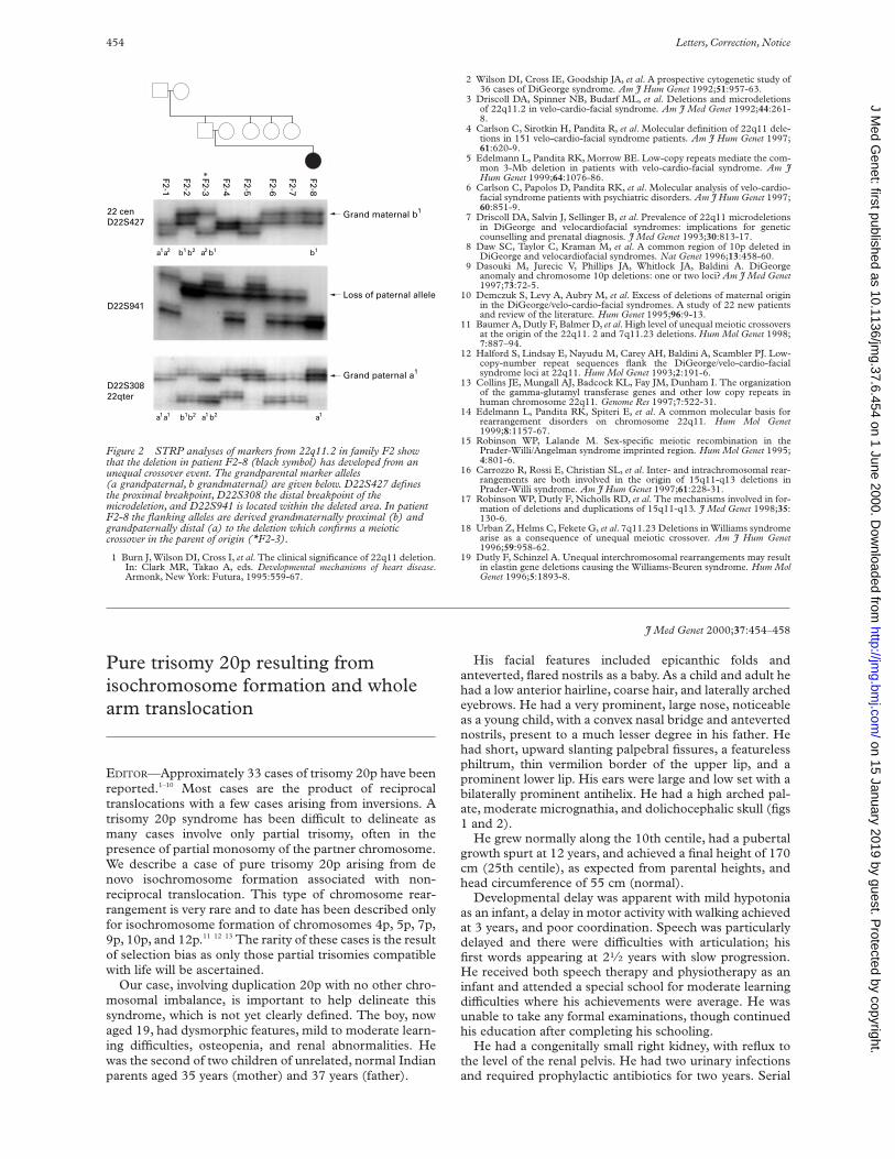
Figure 1 Pedigrees of the investigated families. The index patients are indicated by arrows. Bars and numbers symbolise the individual chromosomes andthe haplotypes of the tested STRP markers. Marker names are given on the left. In the case of a meiotic crossover, the abnormal chromosome 22 showsSTRP markers from both chromosomes of the parent of origin. Unclear recombination breakpoints are indicated by white bars. Black symbol =microdeletion 22q11.2, black spot indicates parent of origin.
Letters, Correction, Notice 453
on 15 January 2019 by guest. Protected by copyright.
http://jmg.bm
j.com/
J Med G
enet: first published as 10.1136/jmg.37.6.454 on 1 June 2000. D
ownloaded from
1 Burn J, Wilson DI, Cross I, et al. The clinical significance of 22q11 deletion.In: Clark MR, Takao A, eds. Developmental mechanisms of heart disease.Armonk, New York: Futura, 1995:559-67.
2 Wilson DI, Cross IE, Goodship JA, et al. A prospective cytogenetic study of36 cases of DiGeorge syndrome. Am J Hum Genet 1992;51:957-63.
3 Driscoll DA, Spinner NB, Budarf ML, et al. Deletions and microdeletionsof 22q11.2 in velo-cardio-facial syndrome. Am J Med Genet 1992;44:261-8.
4 Carlson C, Sirotkin H, Pandita R, et al. Molecular definition of 22q11 dele-tions in 151 velo-cardio-facial syndrome patients. Am J Hum Genet 1997;61:620-9.
5 Edelmann L, Pandita RK, Morrow BE. Low-copy repeats mediate the com-mon 3-Mb deletion in patients with velo-cardio-facial syndrome. Am JHum Genet 1999;64:1076-86.
6 Carlson C, Papolos D, Pandita RK, et al. Molecular analysis of velo-cardio-facial syndrome patients with psychiatric disorders. Am J Hum Genet 1997;60:851-9.
7 Driscoll DA, Salvin J, Sellinger B, et al. Prevalence of 22q11 microdeletionsin DiGeorge and velocardiofacial syndromes: implications for geneticcounselling and prenatal diagnosis. J Med Genet 1993;30:813-17.
8 Daw SC, Taylor C, Kraman M, et al. A common region of 10p deleted inDiGeorge and velocardiofacial syndromes. Nat Genet 1996;13:458-60.
9 Dasouki M, Jurecic V, Phillips JA, Whitlock JA, Baldini A. DiGeorgeanomaly and chromosome 10p deletions: one or two loci? Am J Med Genet1997;73:72-5.
10 Demczuk S, Levy A, Aubry M, et al. Excess of deletions of maternal originin the DiGeorge/velo-cardio-facial syndromes. A study of 22 new patientsand review of the literature. Hum Genet 1995;96:9-13.
11 Baumer A, Dutly F, Balmer D, et al. High level of unequal meiotic crossoversat the origin of the 22q11. 2 and 7q11.23 deletions. Hum Mol Genet 1998;7:887–94.
12 Halford S, Lindsay E, Nayudu M, Carey AH, Baldini A, Scambler PJ. Low-copy-number repeat sequences flank the DiGeorge/velo-cardio-facialsyndrome loci at 22q11. Hum Mol Genet 1993;2:191-6.
13 Collins JE, Mungall AJ, Badcock KL, Fay JM, Dunham I. The organizationof the gamma-glutamyl transferase genes and other low copy repeats inhuman chromosome 22q11. Genome Res 1997;7:522-31.
14 Edelmann L, Pandita RK, Spiteri E, et al. A common molecular basis forrearrangement disorders on chromosome 22q11. Hum Mol Genet1999;8:1157-67.
15 Robinson WP, Lalande M. Sex-specific meiotic recombination in thePrader-Willi/Angelman syndrome imprinted region. Hum Mol Genet 1995;4:801-6.
16 Carrozzo R, Rossi E, Christian SL, et al. Inter- and intrachromosomal rear-rangements are both involved in the origin of 15q11-q13 deletions inPrader-Willi syndrome. Am J Hum Genet 1997;61:228-31.
17 Robinson WP, Dutly F, Nicholls RD, et al. The mechanisms involved in for-mation of deletions and duplications of 15q11-q13. J Med Genet 1998;35:130-6.
18 Urban Z, Helms C, Fekete G, et al. 7q11.23 Deletions in Williams syndromearise as a consequence of unequal meiotic crossover. Am J Hum Genet1996;59:958-62.
19 Dutly F, Schinzel A. Unequal interchromosomal rearrangements may resultin elastin gene deletions causing the Williams-Beuren syndrome. Hum MolGenet 1996;5:1893-8.
J Med Genet 2000;37:454–458
Pure trisomy 20p resulting fromisochromosome formation and wholearm translocation
EDITOR—Approximately 33 cases of trisomy 20p have beenreported.1–10 Most cases are the product of reciprocaltranslocations with a few cases arising from inversions. Atrisomy 20p syndrome has been diYcult to delineate asmany cases involve only partial trisomy, often in thepresence of partial monosomy of the partner chromosome.We describe a case of pure trisomy 20p arising from denovo isochromosome formation associated with non-reciprocal translocation. This type of chromosome rear-rangement is very rare and to date has been described onlyfor isochromosome formation of chromosomes 4p, 5p, 7p,9p, 10p, and 12p.11 12 13 The rarity of these cases is the resultof selection bias as only those partial trisomies compatiblewith life will be ascertained.
Our case, involving duplication 20p with no other chro-mosomal imbalance, is important to help delineate thissyndrome, which is not yet clearly defined. The boy, nowaged 19, had dysmorphic features, mild to moderate learn-ing difficulties, osteopenia, and renal abnormalities. Hewas the second of two children of unrelated, normal Indianparents aged 35 years (mother) and 37 years (father).
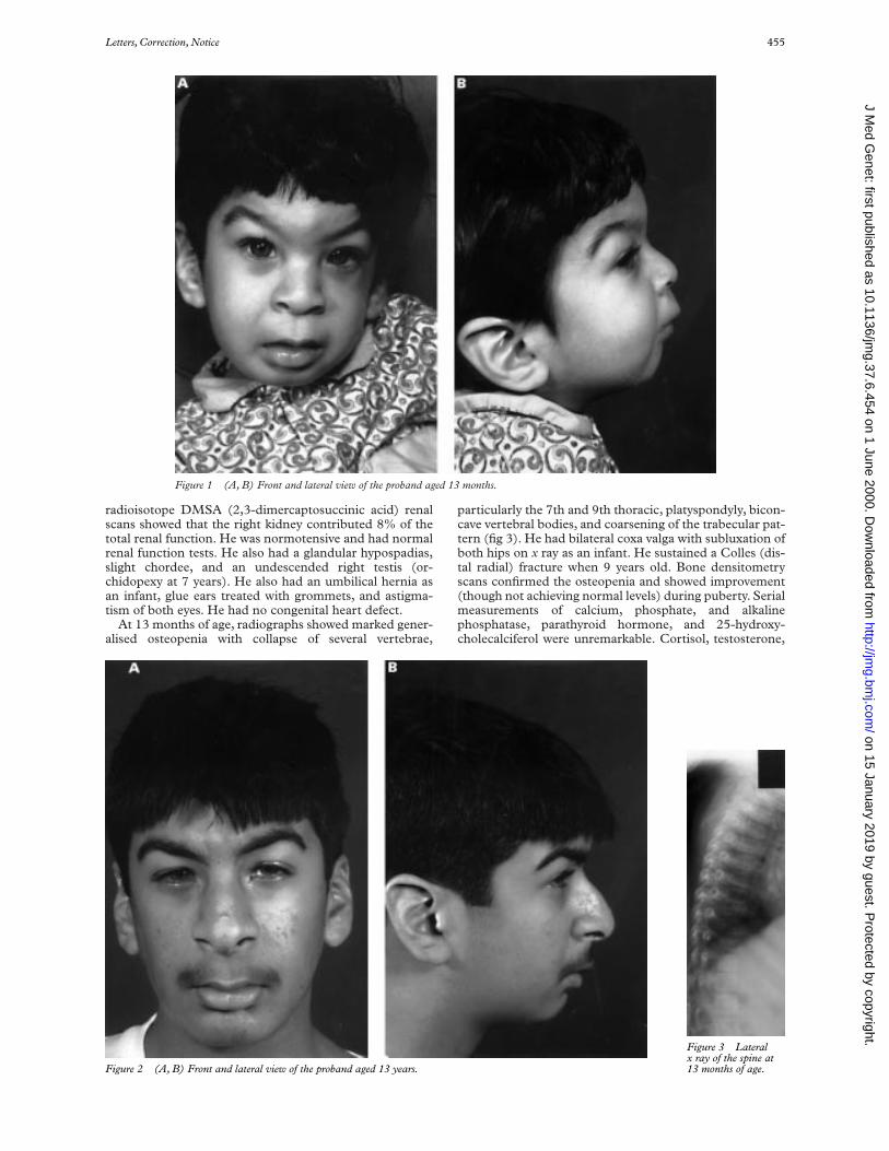
His facial features included epicanthic folds andanteverted, flared nostrils as a baby. As a child and adult hehad a low anterior hairline, coarse hair, and laterally archedeyebrows. He had a very prominent, large nose, noticeableas a young child, with a convex nasal bridge and antevertednostrils, present to a much lesser degree in his father. Hehad short, upward slanting palpebral fissures, a featurelessphiltrum, thin vermilion border of the upper lip, and aprominent lower lip. His ears were large and low set with abilaterally prominent antihelix. He had a high arched pal-ate, moderate micrognathia, and dolichocephalic skull (figs1 and 2).
He grew normally along the 10th centile, had a pubertalgrowth spurt at 12 years, and achieved a final height of 170cm (25th centile), as expected from parental heights, andhead circumference of 55 cm (normal).
Developmental delay was apparent with mild hypotoniaas an infant, a delay in motor activity with walking achievedat 3 years, and poor coordination. Speech was particularlydelayed and there were diYculties with articulation; hisfirst words appearing at 21⁄2 years with slow progression.He received both speech therapy and physiotherapy as aninfant and attended a special school for moderate learningdiYculties where his achievements were average. He wasunable to take any formal examinations, though continuedhis education after completing his schooling.
He had a congenitally small right kidney, with reflux tothe level of the renal pelvis. He had two urinary infectionsand required prophylactic antibiotics for two years. Serial
Figure 2 STRP analyses of markers from 22q11.2 in family F2 showthat the deletion in patient F2-8 (black symbol) has developed from anunequal crossover event. The grandparental marker alleles(a grandpaternal, b grandmaternal) are given below. D22S427 definesthe proximal breakpoint, D22S308 the distal breakpoint of themicrodeletion, and D22S941 is located within the deleted area. In patientF2-8 the flanking alleles are derived grandmaternally proximal (b) andgrandpaternally distal (a) to the deletion which confirms a meioticcrossover in the parent of origin (*F2-3).
22 cenD22S427
Grand maternal b1
D22S30822qter
Grand paternal a1
a1a1
a1a2 a2 b1b1 b1b2
a1a1 b1b2 b2
D22S941
F2-1
F2-2
F2-3*
F2-4
F2-5
F2-6
F2-7
F2-8
Loss of paternal allele
454 Letters, Correction, Notice
on 15 January 2019 by guest. Protected by copyright.
http://jmg.bm
j.com/
J Med G
enet: first published as 10.1136/jmg.37.6.454 on 1 June 2000. D
ownloaded from
radioisotope DMSA (2,3-dimercaptosuccinic acid) renalscans showed that the right kidney contributed 8% of thetotal renal function. He was normotensive and had normalrenal function tests. He also had a glandular hypospadias,slight chordee, and an undescended right testis (or-chidopexy at 7 years). He also had an umbilical hernia asan infant, glue ears treated with grommets, and astigma-tism of both eyes. He had no congenital heart defect.
At 13 months of age, radiographs showed marked gener-alised osteopenia with collapse of several vertebrae,
particularly the 7th and 9th thoracic, platyspondyly, bicon-cave vertebral bodies, and coarsening of the trabecular pat-tern (fig 3). He had bilateral coxa valga with subluxation ofboth hips on x ray as an infant. He sustained a Colles (dis-tal radial) fracture when 9 years old. Bone densitometryscans confirmed the osteopenia and showed improvement(though not achieving normal levels) during puberty. Serialmeasurements of calcium, phosphate, and alkalinephosphatase, parathyroid hormone, and 25-hydroxy-cholecalciferol were unremarkable. Cortisol, testosterone,
Figure 1 (A, B) Front and lateral view of the proband aged 13 months.
Figure 2 (A, B) Front and lateral view of the proband aged 13 years.
Figure 3 Lateralx ray of the spine at13 months of age.
Letters, Correction, Notice 455
on 15 January 2019 by guest. Protected by copyright.
http://jmg.bm
j.com/
J Med G
enet: first published as 10.1136/jmg.37.6.454 on 1 June 2000. D
ownloaded from

24 hour urinary calcium, and urinary amino acid measure-ments were all normal.
G banded metaphase chromosomes were karyotypedafter routine PHA stimulated peripheral blood culture.Chromosome analysis showed a male karyotype with anisochromosome for the short arm of chromosome 20 andtranslocation of the chromosome 20 long arm to the shortarm of one chromosome 4 (fig 4A).
Examination of 100 cells showed no evidence oftelocentric 20p, 20q, or other mosaicism. Both parents ofthe patient had a normal (46,XX and 46,XY) karyotype.
Further characterisation of the chromosome rearrange-ment was obtained from fluorescence in situ hybridisation(FISH) studies, in all cases performed following the manu-facturer’s instructions. Application of whole chromosomepaints (WCP) (Cambio) for chromosomes 4 and 20showed hybridisation of WCP 20 to the isochromosomeand to the distal p arm of the der(4)t(4;20) confirming thatthe translocated material was derived from chromosome20 (fig 4B).
FISH with probes (Cytocell) mapping to the subte-lomere regions of 20p and 20q (fig 4C) showed 20q subte-lomeric sequences on the der(4)t(4;20), confirming thatthe translocated material was derived from the chromo-some 20 long arm, and 20p signals were seen at both endsof the isochromosome, confirming it to be an isochromo-some for the 20 short arm.
FISH with probes (Cytocell) mapping to the subte-lomere regions of 4p and 4q (fig 4D) showed the signalnear the breakpoint junction suggesting the possibility of atelomeric breakpoint in 4p. FISH with a probe specific tothe Wolf-Hirschhorn region, mapping to D4S96 (Oncor),showed signal on both the normal and abnormal copies of
chromosome 4, indicating that there was no deletion of theWolf-Hirschhorn critical region.
The all human telomeres probe (Oncor) showed aninterstitial signal in addition to the expected terminalsignals on the der(4)t(4;20), confirming the presence ofinterstitial telomeric TTAGGG repeat sequence at thet(4;20) breakpoint junction (fig 4E). The presence ofinterstitial telomeric sequence confirmed that the rear-rangement involved either breakage of the 4p telomere orfusion between the 4p telomere and sequence fromchromosome 20.
C banding suggested a monocentric isochromosomealthough C band positive material was present in botharms of the isochromosome and was therefore larger thanin the normal 20. No C band positive material was seen onthe der(4) at the t(4;20) breakpoint junction.
Application of a chromosome 20 alpha satellite probe(D20Z1, Oncor), showed similar signals on the normalchromosome 20 and the isochromosome 20p, againsuggesting a monocentric isochromosome. Theder(4)t(4;20), identified by hybridisation with a chromo-some 4 alpha satellite probe (D4Z1 Oncor), showed nosignal with D20Z1 at the breakpoint junction (fig 4F).
The all human centromeres probe labelled with FITC(Oncor) gave a very small but unambiguous signal at theder(4)t(4;20) breakpoint junction in a proportion of cells(fig 4G). Signal was seen on both chromatids in nine of the27 cells, on one chromatid in seven cells, and six cells werenegative. This variation in signal is almost certainlybecause of its small size and is not considered to besuggestive of mosaicism. Der(4)t(4;20) was identified bysequential hybridisation with the rhodamine labelled chro-mosome 4 alpha satellite probe resulting in a yellow signal.
Figure 4 Der(4)t(4;20)(pter;q11.1),i(20)(q11.1) shown by (A) G banding. (B) Whole chromosome paints for chromosomes 4 (Cy3) and 20 (FITC).(C) Subtelomeric probes for 20p (FITC) and 20q (Cy3) showing 20q signal on der(4)t(4;20). (D) Subtelomeric probes for 4p (FITC) and 4q (Cy3),showing interstitial 4p signal near the breakpoint junction on der(4)t(4;20). (E) All human telomeres probe (FITC) showing interstitial signal onder(4)t(4;20). (F) Probe for D20Z1 (rhodamine) showing similar signals on the normal and isochromosome 20, but lack of signal at the breakpoint junctionon der(4)t(4;20). Chromosome 4 was identified by the centromeric signal of D4Z1 (rhodamine). (G) All human centromeres probe (FITC) showing signal atthe breakpoint junction of der(4)t(4;20). Chromosome 4 was identified by previous hybridisation with D4Z1 (rhodamine), resulting in a yellow signal.
456 Letters, Correction, Notice
on 15 January 2019 by guest. Protected by copyright.
http://jmg.bm
j.com/
J Med G
enet: first published as 10.1136/jmg.37.6.454 on 1 June 2000. D
ownloaded from

The karyotype was interpreted as 46,XY,der(4)t(4;20)(pter;q11.1),i(20)(q11.1). The translocation isnon-reciprocal and the patient therefore appears to be tri-somic for 20p without any concomitant loss of materialfrom 4p.
We have described a patient with pure trisomy 20p as aresult of a rare type of de novo non-reciprocal chromosomerearrangement involving formation of an isochromosomeby a whole p arm and translocation of the residual q arm toanother chromosome. Rearrangements of this type arethought to arise from centromere fission, or pericentricbreakage followed by isochromosome formation by the parm and translocation (or fusion) of the acentric ortelocentric q arm to the telomere of anotherchromosome.14 They are thought to be mediated by thepresence of intrachromosomal telomere-like repeats whichhave been detected at many sites including the centromeresof chromosomes 1-6, 8-11, 16, 17, and 20.15 The presenceof both alphoid sequences and interstitial telomeresequence has been shown in three cases of telomere-centromere fusion. In two cases the translocation involvedan acrocentric chromosome and loss of the acrocentricshort arm.16 The third case involved translocation of 12q to8p with i(12p) formation.13
We were able to show both centromeric repeat sequenceand interstitial telomeric sequence at the t(4;20) break-point junction. Our results support the interpretation of abreak in the pericentric long arm of chromosome 20 closeto the end of the alphoid sequences, leading to formation ofan isochromosome with fused centromeres and therefore amonocentric appearance. The absence of mosaicisminvolving normal cells or telocentric 20p or 20q supports asingle step aetiology.
As our patient is an example of pure trisomy 20p, thefeatures are of particular importance in helping to deline-ate the syndrome. Although there are many similaritieswith previously described patients with trisomy 20p, themost striking diVerences in our patient are the very promi-nent nose and the osteopenia (table 1). Van Langen et al17
produced a table of the clinical picture seen in trisomy 20p;however, 15 cases were included twice in their table
(Centerwall and Francke,1 13 cases; Delicado et al,18 onecase; Schinzel,19 one case), thus producing inaccuratefigures.
Moderate mental retardation and poor coordination arefairly consistent findings, as is a marked speech delay (withmutism reported in two cases by Taylor et al20), all of whichwere present in our case. The dysmorphic features of ourcase have similarities with some previously reported cases.The distinctive nose is an unusual feature, as many othercases had a short, upturned nose (10 reported in youngerchildren and seven in older children or adults). However,the nose in our patient has a similar appearance to the casesdescribed by Grammatico et al4 (case 1) and Balesstrazzi etal.21
The thick, high arched eyebrows seen in our case werealso noted by Grammatico et al.4 Thick eyebrows werenoted by Rudd et al22 and thin, high arched eyebrows byFunderburk et al.23 Epicanthic folds and upward slantingpalpebral fissures are frequently seen. Short palpebralfissures have been occasionally described.9 19 24
Renal anomalies have been seen before, but acongenitally hypoplastic kidney, as in our case, has notbeen seen. Previously reported renal anomalies includeduplicated urinary tract and hydronephrosis,20 bilateralpolycystic kidneys,8 25 ectopic kidney,24 and duplication ofthe left renal collecting system.2 Hypospadias, as seenhere, has been reported in two previous cases.20 26
Cryptorchidism was previously reported by Schinzel etal.19 A case involving macro-orchidism has beendescribed,21 and one involving a ventrally positioned clito-ris and anus.22
Our patient had striking osteopenia first noted at a veryyoung age (13 months). Two previous reports exist of ageneralised osteoporosis.2 19 Coxa valga deformity of thehips as seen in our case has been previously reported.19 20 25
Skeletal anomalies in trisomy 20p appear to be a variablephenomenon, with vertebral abnormalities the most com-monly reported, including fusion of vertebrae, reductionof intervertebral spaces, spina bifida, scoliosis, andkyphosis.
Table 1 Clinical features of 32 cases of trisomy 20p*
Fraction Percentage Our case
Sex 16F/16M MGestation 21/25 (21=term) TermBirth weight (g) 1800–4300 2500Growth normal 15/21 71 +Mental retardation 29/31 (moderate/severe) 94 + (moderate)Poor coordination 18/19 95 +Language delay 21/22 95 +Occipital flattening 11/22 50 −Low posterior hairline 3/4 75 −Coarse hair 14/18 78 +Thick, arched eyebrows 2/2 100 +Epicanthus 8/26 31 +Strabismus 9/23 39 −Increased inner canthal distance 11/21 52 −Upward slanting palpebral fissures 19/29 66 +Round face, prominent cheeks 24/29 83 −Short, upturned nose 17/30 57 −Large flared nostrils 16/30 53 +Large/abnormal ears 11/11 100 +Moderate microganthia 16/22 73 +High arched/cleft palate 8/11 73 +Short neck 5/5 100 −Cardiac anomalies 10/28 36 −Renal anomalies 6/8 75 +Vertebral anomalies 17/21 81 +Osteopenia 2/2 100 +Hip/pelvis anomalies 6/6 100 +Digital anomalies 12/13 92 −Feet anomalies 14/15 93 −Dental anomalies 13/19 68 −Umbilical/inguinal hernia 6/7 86 +
+ : present, − : absent.*Taken from 32 cases described.1–6 8–10
Letters, Correction, Notice 457
on 15 January 2019 by guest. Protected by copyright.
http://jmg.bm
j.com/
J Med G
enet: first published as 10.1136/jmg.37.6.454 on 1 June 2000. D
ownloaded from
Our case is unusual in being the first reported case of acentromere-telomere fusion resulting in trisomy 20p.
R U SIDWELL*M-P PINSON†
B GIBBONS†S-A BYATT†
E C SVENNEVIK‡R J HASTINGS‡
D M FLYNN**Department of Paediatrics, Royal Free Hospital, London, UK†Department of Clinical Cytogenetics, Royal Free Hospital, London, UK‡Department of Clinical Cytogenetics, University College London, London, UK
Correspondence to: Dr Sidwell, c/o Dr D M Flynn, Department of Paediatrics,Royal Free Hospital, Pond Street, London NW3 2QG, UK.
1 Centerwall W, Franke U. Familial trisomy 20p. Five cases and two carriersin three generations. A review. Ann Genet 1977;20:77-83.
2 Chen H, HoVman WH, Tyrrus M, Al Saadi A, Bawle E. Partial trisomy 20psyndrome and maternal mosaicism. Ann Genet 1983;26:21-5.
3 Lurie IW, Rumyantseva NV, Zaletajev DV, Gurevich DB, Korotkova IA.Trisomy 20p: case report and genetic review. J Genet Hum 1985;33:67-75.
4 Grammatico P, Culpilari F, Di Rosa C, Falcolini M, Del Porto G. 20pduplication as a result of parental translocation: familial case report and acontribution to the clinical delineation of the syndrome. Clin Genet1992;41:285-9.
5 Lucas J, Le Mee F, Le Marec B, Pluquailec K, Journel H, Picard F.Trisomie 20p derivee d’une inversion pericentrique maternelle etbrachymesophalangie de l’index. Ann Genet 1985;28:167-71.
6 Bown N, Cross I, Davison EV, Burn J. Partial trisomy 20p resulting from afamilial recombination of a pericentric inversion. Hum Genet 1986;74:417-19.
7 Zumel RM, Darnaude A, Delicado A, Diaz de Bustamante A, de TorresML, Lopez Pajares I. Trisomy 20p from maternal translocation and anen-cephaly. Case report and genetic review. Ann Genet 1989;32:247-9.
8 Clark P, Jones KL, Freidenberg GR. Duplication (20p) in association withthyroid carcinoma. Am J Med Genet 1993:45:14-16.
9 Le Chien KA, McPherson E, Estop AM. Duplication 20p identified viafluorescence in situ hybridization. Am J Med Genet 1994;50:187-9.
10 Wuu SW, Hwang B, Wuu KD. A partial duplication of the short arm ofchromosome 20. Taiwan I Hsuch Hui Tsu Chin 1983;82:1088-92.
11 Petit P, Devriendt K, Vermeesch JR, Meireleire J, Fryns JP. Localisation byFISH of centric fission breakpoints in a de novo trisomy 9p patient withi(9p) and t(9q;11p). Genet Couns 1998; 9:215-21.
12 Lurie IW, Schwartz MF, Schwartz S, Cohen MM. Trisomy 7p resultingfrom isochromosome formation and whole-arm translocation. Am J MedGenet 1995;55:62-6.
13 Rivera H, Vasquez AI, Perea FJ. Centromere-telomere (12;8p) fusion, telo-meric 12q translocation, and I(12p) trisomy. Clin Genet 1999;55:122-6.
14 Fujita M, Flori E, Lemaire F, Casanova R, Astruc D. A new case of completetrisomy 5p with isochromosome 5p associated with a de novo translocationt(5;8)(q11;p23). Clin Genet 1994;45:305-7.
15 Azzalin CM, Mucciolo E, Bertoni L, Giulotto E. Fluorescence in situhybridization with a synthetic (T2AG3)n polynucleotide detects severalintrachromosomal telomere-like repeats on human chromosomes. Cy-togenet Cell Genet 1997;78:112-15.
16 Rossi E, Floridia G, Casali M, Danesimo C, Chiumello G, Bernardi F,Magnani I, Papi L, Mura M, ZuVardi O. Types, stability and phenotypicconsequences of chromosome rearrangements leading to interstitial telom-eric sequences. J Med Genet 1993;30:926-31.
17 Van Langen IM, Otter MA, Aronson DC, Overweg-Plandsoen WCG, Hen-nekam RCM, Leschot NJ. Supernumerary ring chromosome 20 character-ised by fluorescence in situ hybridization. Clin Genet 1996;49:49-53.
18 Delicado A, Lopez Pajares I, Vicente P, Gracia R. Partial trisomy 20. AnnGenet 1981;24:54-6.
19 Schinzel A. Trisomy 20pter-q11 in a malformed boy from a t(13;20)(p11;q11) translocation carrier mother. Hum Genet 1980;53:169-72.
20 Taylor KM, Wolfinger HL, Brown MG, Chadwick DL, Franke U. Partialtrisomy 20p derived from a t(18;20) translocation. Hum Genet 1976;34:155-62.
21 Balestrazzi P, Virdis R, Frassi C, Negri V, Rigoli E, Bernasconi S. ‘De novo’trisomy 20p with macroorchidism in prepubertal boy. Ann Genet 1984;27:58-9.
22 Rudd NL, Bain HW, Giblett E, Chen SH, Worten RG. Partial trisomy 20confirmed by gene dosage studies. Am J Med Genet 1979;4:357-64.
23 Funderburk SJ, Sparkes RS, Sparkes MC. Trisomy 20p due to a paternalreciprocal translocation. Ann Genet 1983;26:94-7.
24 Archidiachono N, Telcilazich D, Tonini G, Rocchi M, Philippi G. Trisomy20p from maternal t(3;20) translocation. J Med Genet 1979;16:229-32.
25 Francke U. Abnormalities of chromosomes 11 and 20. In: Yunis JJ, ed. Newchromosomal syndromes. New York: Academic Press, 1977:262-72.
26 Marcus ES, Furler B, Riccardi RM. Triplication of chromosome arm 20pdue to inherited translocation and secondary non-disjunction. Am J MedGenet 1979;4:47-50.
J Med Genet 2000;37:458–460
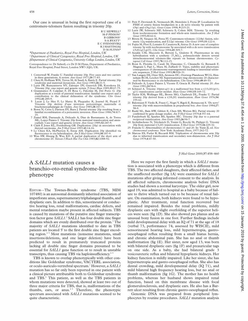
A SALL1 mutation causes abranchio-oto-renal syndrome-likephenotype
EDITOR—The Townes-Brocks syndrome (TBS, MIM107480) is an autosomal dominantly inherited association ofimperforate anus, supernumerary/triphalangeal thumbs, anddysplastic ears. In addition to this, sensorineural or conduc-tive hearing loss, renal malformations, cardiac defects, andmental retardation maybe present in aVected subjects. TBSis caused by mutations of the putative zinc finger transcrip-tion factor gene SALL1.2 SALL1 has four double zinc fingerdomains which are evenly distributed over the protein.3 Themajority of SALL1 mutations identified to date in TBSpatients are located 5' to the first double zinc finger encod-ing region.4 5 Most mutations (nonsense mutations, smallinsertions/deletions, and one larger deletion) have beenpredicted to result in prematurely truncated proteinslacking all double zinc finger domains presumed to beessential for SALL1 gene function or to result in unstabletranscripts, thus causing TBS via haploinsuYciency.4 5
TBS is known to overlap phenotypically with other con-ditions like Goldenhar syndrome, VACTERL association,or oculo-auriculo-vertrebral spectrum.1 However, a SALL1mutation has so far only been reported in one patient witha clinical picture attributable both to Goldenhar syndromeand TBS.6 This patient, as well as the TBS patients inwhom mutations were detected, showed at least two out ofthree major criteria for TBS, that is, malformations of thethumbs, ears, or anus.4 5 Therefore, the phenotypicspectrum associated with SALL1 mutations seemed to bequite characteristic.
Here we report the first family in which a SALL1 muta-tion is associated with a phenotype which is diVerent fromTBS. The two aVected daughters, their aVected father, andthe unaVected mother (fig 1A) were examined for SALL1mutations after giving informed consent to the analysis. Inall aVected subjects, chromosome analysis before DNAstudies had shown a normal karyotype. The older girl, nowaged 19, was admitted to hospital as a baby because of fail-ure to thrive which turned out to be because of renal fail-ure. On examination, both kidneys were found to be hypo-plastic. After treatment, renal function recovered butremained impaired. Besides the renal problems, mildlydysplastic ears with slight overfolding of the superior heli-ces were seen (fig 1D). She also showed pes planus and anunusual bony fusion in one foot. Further findings includemild developmental delay with an IQ of 71 at the age of 18(verbal 73, performance 74, assessed by WISCII), mildsensorineural hearing loss, mild hypermetropia, gastro-oesophageal reflux resulting from a small hiatus hernia,and chronic abdominal pain. She has no anal or thumbmalformation (fig 1E). Her sister, now aged 13, was bornwith bilateral dysplastic ears (fig 1F) and preauricular tagson one side. As a baby, she had bilateral grade 3vesicoureteric reflux and bilateral hypoplastic kidneys. Herkidney function is mildly impaired. Like her sister, she hashypermetropia and gastro-oesophageal reflux. She also hasdental crowding, mild developmental delay (IQ 71), andmild bilateral high frequency hearing loss, but no anal orthumb malformation (fig 1G). The mother has no healthproblems, whereas her husband shows impaired renalfunction, based on thin membrane disease with focalglomerulosclerosis, and dysplastic ears. He also has a Bar-rett ulcer resulting from chronic gastro-oesophageal reflux.
Genomic DNA was prepared from peripheral lym-phocytes by routine procedures. SALL1 mutation analysis
458 Letters, Correction, Notice
on 15 January 2019 by guest. Protected by copyright.
http://jmg.bm
j.com/
J Med G
enet: first published as 10.1136/jmg.37.6.454 on 1 June 2000. D
ownloaded from

was performed by PCR amplification and direct sequenc-ing of PCR products as described previously.5 BothaVected children as well as the aVected father had aheterozygous 1819delG SALL1 mutation, which is locatedin exon 2 between the coding regions for the first and thesecond double zinc finger unit (fig 1C).
Most SALL1 mutations previously reported in TBS residein exon 2 5' of the coding region for the first double zinc fin-ger domain and are predicted to result in SALL1haploinsuYciency.4 5 1819delG is yet another short deletionbut is located 3' of the region where most previously knownmutations cluster. While this mutation could result in a pre-maturely terminated SALL1 protein lacking double zinc fin-ger domains 2-4, it remains unclear if the mutated transcriptand the corresponding protein are indeed expressed. Muta-tions at similar positions have been reported by Marlin et al.4
Here, the patients with the mutations 1565delC or1966del10 showed at least two out of three major criteria forTBS. Both mutations should have a similar eVect (transla-tion stop at nucleotides 1624-1626 and 2074-2076) as com-pared to the mutation reported here (translation stop atnucleotides 1924-1926). Therefore, if proteins were trans-lated from these mutated alleles, all would be missing thedouble zinc finger domains 2-4. The most likely eVect, how-ever, is that such mutations would lead to an unstable tran-script and thereby to SALL1 haploinsuYciency.
The clinical presentation in the family reported here isquite unusual, since all aVected family members do nothave thumb or anal malformations. Therefore, TBS couldnot be diagnosed. While the features in the patients
carrying the 1565delC or 1966del10 mutation4 can beexplained by haploinsuYciency for SALL1, the clinicalpicture in the family shown here could suggest that the1819delG mutation has a diVerent eVect. As seen withmutations in GLI3, truncating mutations at similarpositions may lead to either Pallister-Hall syndrome(PHS), diVerent polydactylies, or Greig cephalopolysyn-dactyly syndrome (GCPS), suggesting that some mightresult in GLI3 haploinsuYciency while others lead totruncated proteins with aberrant functions.7 8
Based on these observations one could speculate that,unlike 1565delC or 1966del10, the 1819delG mutationdoes not result in an unstable mRNA but in a truncatedSALL1 protein. On the other hand, TBS is known to varyeven between families with the same mutation, thus point-ing to a strong influence of modifying genes or environ-mental factors. Yet, while families with SALL1 mutationsmay diVer from each other with respect to the occurrenceof renal or cardiac malformations, no family has beenreported to date in which all aVected members have aSALL1 mutation but not TBS.
Patients like those reported here may instead carry thediagnosis of branchio-oto-renal (BOR) syndrome (MIM113650), as was initially suspected in our patients based onthe combination of dysplastic ears, hearing loss, and hypo-plastic kidneys. However, BOR syndrome patients com-monly present with cup shaped ears, preauricular pits, andbranchial fistulae,9 none of which was seen in our patients.In addition, gastro-oesophageal reflux and borderlinemental retardation are not typical of BOR syndrome.9
Figure 1 (A) Pedigree of the family reported here. Both daughters (II.1 and II.2) and their aVected father (I.1) show the SALL1 mutation (B, upperpart), whereas the mother (I.2) shows the wild type sequence (B, lower part). The position of the mutation with respect to the SALL1 protein is shown in(C) possibly resulting in a truncated protein lacking double zinc finger domains 2-4 (zinc fingers are indicated by oval symbols). (D-G) Ears and hands ofthe aVected girls: (D, E) II.1 aged 19, (F, G) II.2 aged 13. Note typical overfolded superior helices (D, F), but absence of thumb malformations (E, G) inboth patients, which was also confirmed by x ray (not shown).
WT
AC CTAG30 40
GT G GGC T C CCAGA GAG A
1819delG
1819del G
COOHNH2
AC GCTA30 40GT G GNT
I
A C
D
F
E
G
B
II
1
1
2
2
TC CAA GAA A A
Letters, Correction, Notice 459
on 15 January 2019 by guest. Protected by copyright.
http://jmg.bm
j.com/
J Med G
enet: first published as 10.1136/jmg.37.6.454 on 1 June 2000. D
ownloaded from
Therefore, our report shows that a phenotypic overlap notonly exists between TBS and VACTERL association,oculo-auriculo-vertebral spectrum, or Goldenhar syn-drome, but also between TBS and BOR syndrome. SALL1mutation analysis should therefore be considered forpatients who present with dysplastic ears, hearing loss, andrenal malformations but do not have a causative mutationin the EYA1 gene.10
The cooperation of the family described in this paper is greatly appreciated. Wewould also like to thank Wolfgang Engel for his support. This work was fundedby the Wilhelm Sander-Stiftung (grant No 98.075.1 to JK). The first twoauthors contributed equally to this work.
SASKIA ENGELS*JÜRGEN KOHLHASE*
JULIE MCGAUGHRAN†*Institute for Human Genetics, University of Göttingen,Heinrich-Düker-Weg 12, D-37073 Göttingen, Germany†Department of Medical Genetics, University of Auckland, Auckland,New Zealand
Correspondence to: Dr Kohlhase, [email protected]
1 Powell CM, Michaelis RC. Townes-Brocks syndrome. J Med Genet1999;36:89-93.
2 Kohlhase J, Wischermann A, Reichenbach H, Froster U, Engel W.Mutations in the SALL1 putative transcription factor gene cause Townes-Brocks syndrome. Nat Genet 1998;18:81-3.
3 Kohlhase J, Schuh R, Dowe G, Kühnlein RP, Jäckle H, Schroeder B,Schulz-SchaeVer W, Kretzschmar HA, Köhler A, Müller U, Raab-Vetter M,Burkhardt E, Engel W, Stick R. Isolation, characterization, and organ-specific expression of two novel human zinc finger genes related to the Dro-sophila gene spalt. Genomics 1996;38:291-8.
4 Marlin S, Blanchard S, Slim R, Lacombe D, Denoyelle F, Alessandri JL,Calzolari E, Drouin-Garraud V, Ferraz FG, Fourmaintraux A, Philip N,Toublanc JE, Petit C. Townes-Brocks syndrome: detection of a SALL1mutation hotspot and evidence for a position eVect in one patient. HumMut 1999;14:377-86.
5 Kohlhase J, Taschner PEM, Burfeind P, Pasche B, Newman B, Blanck C,Breuning MH, ten Kate LP, Maaswinkel-Mooy P, Mitulla B, Seidel J,Kirkpatrick SJ, Pauli RM, Wargowski DS, Devriendt K, Proesmans W,Gabrielli O, Coppa GV, Wesby-vanSwaay E, Trembath RC, Schinzel AA,Reardon W, Seemanova E, Engel W. Molecular analysis of SALL1mutations in Townes-Brocks syndrome. Am J Hum Genet 1999;64:435-45.
6 Gabrielli O, Bonifazi V, OYdani AM, Cellini A, Coppa GV, Giorgi PL.Description of a patient with diYcult nosological classification: Goldenharsyndrome or Townes-Brocks syndrome. Minerva Pediatr 1993;45:459-62.
7 Radhakrishna U, Bornholdt D, Scott HS, Patel UC, Rossier C, Engel H,Bottani A, Chandal D, Blouin JL, Solanki JY, Grzeschik KH, AntonarakisSE. The phenotypic spectrum of GLI3 morphopathies includes autosomaldominant preaxial polydactyly type-IV and postaxial polydactyly type-A/B:no phenotype prediction from the position of GLI3 mutations. Am J HumGenet 1999;65:645-55.
8 KalV-Suske M, Wild A, Topp J, Wessling M, Jacobsen EM, Bornholdt D,Engel H, Heuer H, Aalfs CM, Ausems MG, Barone R, Herzog A, HeutinkP, Homfray T, Gillessen-Kaesbach G, König R, Kunze J, Meinecke P,Müller D, Rizzo R, Strenge S, Superti-Furga A, Grzeschik KH. Pointmutations throughout the GLI3 gene cause Greig cephalopolysyndactylysyndrome. Hum Mol Genet 1999;8:1769-77.
9 Chen A, Francis M, Ni L, Cremers CW, Kimberling WJ, Sato Y, PhelpsPD, Bellman SC, Wagner MJ, Pembrey M, Smith RJH. Phenotypicmanifestations of branchiootorenal syndrome. Am J Med Genet 1995;58:365-70.
10 Abdelhak S, Kalatzis V, Heilig R, Compain S, Samson D, Vincent C, Weil D,Cruaud C, Sahly I, Leibovici M, Bitner-Glindzicz M, Francis M, LacombeD, Vigneron J, Charachon R, Boven K, Bedbeder P, Van Regemorter N,Weissenbach J, Petit C. A human homologue of the Drosophila eyes absentgene underlies branchio-oto-renal (BOR) syndrome and identifies a novelgene family. Nat Genet 1997;15:157-64.
J Med Genet 2000;37:460–462
New MR/MCA syndrome with distinctfacial appearance and general habitus,broad and webbed neck, hypoplasticinverted nipples, epilepsy, andpachygyria of the frontal lobes
EDITOR—We present the clinical histories and physicalfindings in two unrelated, severely mentally retarded males,now 14 and 11 years old.
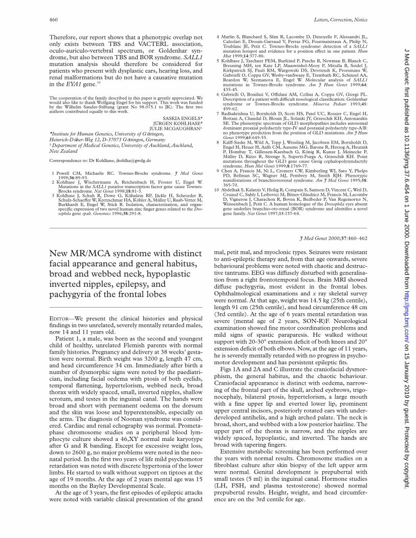
Patient 1, a male, was born as the second and youngestchild of healthy, unrelated Flemish parents with normalfamily histories. Pregnancy and delivery at 38 weeks’ gesta-tion were normal. Birth weight was 3200 g, length 47 cm,and head circumference 34 cm. Immediately after birth anumber of dysmorphic signs were noted by the paediatri-cian, including facial oedema with ptosis of both eyelids,temporal flattening, hypertelorism, webbed neck, broadthorax with widely spaced, small, inverted nipples, shallowscrotum, and testes in the inguinal canal. The hands werebroad and short with permanent oedema on the dorsumand the skin was loose and hyperextensible, especially onthe arms. The diagnosis of Noonan syndrome was consid-ered. Cardiac and renal echography was normal. Prometa-phase chromosome studies on a peripheral blood lym-phocyte culture showed a 46,XY normal male karyotypeafter G and R banding. Except for excessive weight loss,down to 2600 g, no major problems were noted in the neo-natal period. In the first two years of life mild psychomotorretardation was noted with discrete hypertonia of the lowerlimbs. He started to walk without support on tiptoes at theage of 19 months. At the age of 2 years mental age was 15months on the Bayley Developmental Scale.
At the age of 3 years, the first episodes of epileptic attackswere noted with variable clinical presentation of the grand
mal, petit mal, and myoclonic types. Seizures were resistantto anti-epileptic therapy and, from that age onwards, severebehavioural problems were noted with chaotic and destruc-tive tantrums. EEG was diVusely disturbed with generalisa-tion from a right frontotemporal focus. Brain MRI showeddiVuse pachygyria, most evident in the frontal lobes.Ophthalmological examinations and x ray skeletal surveywere normal. At that age, weight was 14.5 kg (25th centile),length 91 cm (25th centile), and head circumference 48 cm(3rd centile). At the age of 6 years mental retardation wassevere (mental age of 2 years, SON-R)F. Neurologicalexamination showed fine motor coordination problems andmild signs of spastic paraparesis. He walked withoutsupport with 20-30° extension deficit of both knees and 20°extension deficit of both elbows. Now, at the age of 11 years,he is severely mentally retarded with no progress in psycho-motor development and has persistent epileptic fits.
Figs 1A and 2A and C illustrate the craniofacial dysmor-phism, the general habitus, and the chaotic behaviour.Craniofacial appearance is distinct with oedema, narrow-ing of the frontal part of the skull, arched eyebrows, trigo-nocephaly, bilateral ptosis, hypertelorism, a large mouthwith a fine upper lip and everted lower lip, prominentupper central incisors, posteriorly rotated ears with under-developed antihelix, and a high arched palate. The neck isbroad, short, and webbed with a low posterior hairline. Theupper part of the thorax is narrow, and the nipples arewidely spaced, hypoplastic, and inverted. The hands arebroad with tapering fingers.
Extensive metabolic screening has been performed overthe years with normal results. Chromosome studies on afibroblast culture after skin biopsy of the left upper armwere normal. Genital development is prepubertal withsmall testes (5 ml) in the inguinal canal. Hormone studies(LH, FSH, and plasma testosterone) showed normalprepubertal results. Height, weight, and head circumfer-ence are on the 3rd centile for age.
460 Letters, Correction, Notice
on 15 January 2019 by guest. Protected by copyright.
http://jmg.bm
j.com/
J Med G
enet: first published as 10.1136/jmg.37.6.454 on 1 June 2000. D
ownloaded from

Patient 2, a male, is the third child of a non-consanguineous European couple. He has two normalbrothers, 16 and 9 years old respectively, and a 7 year oldnormal sister. Pregnancy was unremarkable. Birth weightwas 3200 g. He had facial oedema and it was noted that hehad significant redundant skin over the nape of the neck.Furthermore, he had significant weight loss in the neonatalperiod, losing more than 500 g in weight. He did not haveany feeding diYculties. He crawled at 9 months and walkedon tiptoes by 18 months. He had mild speech delay and hisdevelopment was reasonable until the age of 5 years when hebegan to have persistent seizures which so far are not totallycontrolled with anti-epileptic medication. A brain CT scanshowed pachygyria of the frontal lobes. His mental develop-ment deteriorated from the onset of the seizures andcurrently his mental function is at the 5 year level. In the pasthe has had surgery for squint, as well as for ptosis. Bilateralvesicoureteral reflux with hydronephrosis was also diagnosedin infancy and required bilateral ureteral reimplantation.
At present (figs 1B and 2B, D), height is 136 cm (3rdcentile is 143 cm) and head circumference 53.5 cm (25thcentile). Craniofacial appearance is distinct with thick hair,low frontal hairline, significant narrowing of the frontalpart of the skull, facial oedema, bushy, arched eyebrows, abroad root and bridge of the nose, and persistent bilateralptosis. He has a long, flat philtrum, a thin upper lip, micro-gnathia, and everted lower lip. The palate is high archedand the upper central incisors are prominent. The ears areprotuberant, posteriorly rotated, and with underdevelopedantihelices. The neck is broad, short, and webbed with alow posterior hairline. The upper part of the thorax is rela-tively small with broadly spaced, hypoplastic, and invertednipples. The hands are broad with proximally placedthumbs. He is able to walk without support, with 20-30°extension deficit of the knees and some diYculties in fullyextending the elbows.
Chromosome studies on a peripheral blood lymphocyteculture showed a 46,XY normal male karyotype on Gbanding.
The two unrelated males present, as described above, aremarkably similar MCA/MR syndrome and their clinical
history is also identical. Patient 1 has been followed inLeuven since the neonatal period and patient 2 has beenfollowed in Auckland. The striking resemblance betweenthe two patients was recognised almost by coincidence onthe occasion of the exchange of data on patients with dis-tinct but hitherto unidentified MCA/MR syndromes. Asdescribed, their craniofacial appearance is particularlysimilar. Both males presented at birth with facial oedema,and it is still evident at their present respective ages of 14and 11 years. The significant weight loss in both patientsin the neonatal period indicates that they had more gener-alised fetal oedema and, especially in patient 1, in thepostnatal period the skin was loose and hyperextensible,most evident on the arms. Based on the combination offacial oedema, ptosis of the eyelids, webbed neck with lowposterior hairline, and broad thorax with widely spacednipples, the diagnosis of Noonan syndrome was consid-ered in patient 1.1 However, the clinical follow up andevolution with age were not compatible with this diagno-sis. Another remarkable finding in both males was theirlack of psychomotor evolution with age. At the respectiveages of 3 and 5 years, epileptic attacks began, which so farcannot be controlled despite a great variety of anti-epileptic medication. In both patients brain CT and MRIscan showed pachygyria, most pronounced in the frontallobes. Up to the start of the complex epileptic fits,psychomotor development was only mildly to moderatelyretarded, but since the onset of seizures mental develop-ment has deteriorated. At the present time, both males areseverely to profoundly mentally retarded and, especially inpatient 1, major behavioural problems are now present.Also the general habitus of both males is identical.Whereas no specific neurological abnormalities arepresent, except walking on tiptoes with mild signs of spas-tic diplegia at a young age, both males walk independentlybut with 20-30° extension deficit of both knees, and bothhave diYculties in fully extending theirelbows.
The MCA/MR syndrome present in these two malesthus combines the following major symptoms: (1) distinctfacies with oedema and notable postnatal weight loss;
Figure 1 Similar craniofacial dysmorphism in both males, (A) patient 1 and (B) patient 2. Note the facial oedema.
Letters, Correction, Notice 461
on 15 January 2019 by guest. Protected by copyright.
http://jmg.bm
j.com/
J Med G
enet: first published as 10.1136/jmg.37.6.454 on 1 June 2000. D
ownloaded from

(2) broad and webbed neck; (3) hypoplastic, inverted nip-ples; (4) limited extension of elbows and knees resulting ina characteristic general habitus; and (5) complex epilepsyin early childhood with deterioration of mental develop-ment and pachygyria on brain imaging.
The authors thank Dr N Goeman (Paediatric Department, University HospitalLeuven, Belgium) and Dr E Carmichael (Paediatrician, Hamilton, NewZealand) for referring the patients.
J-P FRYNS*S AFTIMOS†
*Centre for Human Genetics, University Hospital Gasthuisberg,Herestraat 49, B-3000 Leuven, Belgium†Northern Regional Genetic Services, Auckland, New Zealand
Correspondence to: Professor Fryns, [email protected]
1 Noonan JA, Ehmke DA. Associated noncardiac malformations in childrenwith congenital heart disease. J Pediatr 1963;63:469-74.
Figure 2 The general habitus with similar dysmorphic signs and behaviour, (A, C)patient 1 and (B, D) patient 2.
462 Letters, Correction, Notice
on 15 January 2019 by guest. Protected by copyright.
http://jmg.bm
j.com/
J Med G
enet: first published as 10.1136/jmg.37.6.454 on 1 June 2000. D
ownloaded from

J Med Genet 2000;37:463–466
The mitochondrial genome in Wolframsyndrome
EDITOR—Wolfram syndrome is the association of juvenileonset diabetes mellitus and optic atrophy,1 also known asDIDMOAD (diabetes insipidus, diabetes mellitus, opticatrophy, and deafness). This is a progressive, neurodegen-erative disorder, with diabetes mellitus and optic atrophypresenting in the first decade,2 cranial diabetes insipidus,and sensorineural deafness in the second, and neuropathicbladder in the third, followed by neurological complica-tions (cerebellar ataxia, myoclonus) and psychiatric illnessin the fourth decade. The clinical phenotype is consistentwith an ATP supply defect, suggesting a mitochondrialmediated disease.3 Mitochondrial genome deletions andpathogenic point mutations4 5 have been described inWolfram patients. A nuclear gene WFS1, wolframin,6 7 wasrecently identified, encoding a polypeptide of 890 aminoacids. Wolfram syndrome thus appears to be geneticallyheterogeneous. Recently, a distinct “mitochondrial haplo-type” was described.8 Because recombination does notappear to be characteristic of mtDNA, the accumulation ofpolymorphisms can be used as a “genetic clock” toestimate diversity within and between populations.9 Acluster of nucleotide exchanges at nucleotide positions4216 and 11 251 roughly distinguished a series of 6/8Wolfram patients from controls and patients with Leberhereditary optic neuropathy (LHON). The authorssuggested that these mtDNA variants may predispose toWolfram syndrome.8 We investigated our cohort of 50Wolfram syndrome patients10 for evidence of a distinctmitochondrial haplotype and mitochondrial DNA rear-rangements.
Patients for this study were included from a cohortrecruited nationally in the UK.10 Minimal ascertainmentcriteria were juvenile onset (less than 30 years of age)diabetes mellitus and optic atrophy. These were chosen asthe only features consistently present and earliest todevelop in 166/168 case reports.11 Diabetes mellitus wasdefined as a fasting plasma glucose of more than 6.0mmol/l (>3 SD above the mean of the normal population).All aVected patients had been examined, with pupilsdilated, by an experienced ophthalmologist. All patientswere visited at home; blood samples were obtained from allavailable family members after informed consent wasgiven. DNA was extracted from whole blood usingPuregene DNA extraction kits (Gentra Systems), accord-ing to the manufacturers’ instructions, and diluted to stocksolutions of 500 ng/µl.
The mitochondrial haplotype of the Wolfram patientswas investigated using PCR and direct sequencing of theregion between 16 050 and 16 400 in the first hypervari-able region of the large non-coding region as previouslydescribed.12 Mitochondrial haplotype analysis was carriedout on 32 Wolfram families of European origin. The pres-ence of mtDNA variants at bp 4216, 11 251, and 15 257were assessed using PCR and restriction digestion.9 13
Mean pairwise diVerences14 were estimated and comparedwith a control data set comprising 10015 and 60 UK whitesubjects.16 An unrooted tree was drawn based on previousstudies of Europeans. A PCR ApaI restriction site assaywas used to screen for the mitochondrial tRNA Leu(UUR) A to G (3243) mutation17; this has been associatedwith maternally transmitted diabetes and deafness.18 Thefinal cycle of PCR was labelled with 33P-dCTP permitting
detection of the mutation at levels of heteroplasmy below1%. DNA from whole blood was also screened for the11778A:T and 3460A:T mutations associated with Leber’shereditary optic neuropathy (LHON) using allele specificPCR.19 These mutations were chosen as they account for75% of all cases of LHON. The 14484 mutation was notinvestigated as it is phenotypically milder and has a betteroutcome.20 Mitochondrial DNA was also analysed for thepresence of major rearrangements by long range PCR21;PCRs were performed in a final volume of 50 µl with 0.25µl each of primers L1 (nt2695-2720) and H3 (nt16459-16436), 2.5 mmol/l MgCl2, 0.25 mmol/l of each dNTP, 1× Bio-Optiform 111 buVer (Bioline), and 1.5 U Bio-X-ACT Taq polymerase(Bioline). PCRs were hot started at80°C by the addition of 20-50 ng DNA and denatured at95°C for 10 seconds and 68°C for 10 minutes plus 30 sec-onds for each subsequent cycle (25 cycles). PCR productswere run on 0.7% Seakem agarose gels. The resolution forlong range PCR is about 1 kb (sometimes 0.5 kb depend-ing on the gel).
Needle muscle biopsies under local anaesthetic wereundertaken on nine adult Wolfram patients. Informed,written consent was obtained, after a written and verbalexplanation. Respiratory chain activity was analysed infresh muscle biopsy samples from six patients by two sepa-rate methodologies. Flux experiments measuring rotenonesensitive cytochrome c reductase with glutamate,2-oxoglutarate, and pyruvate + malate as substrates toassay complexes I and III, and antimycin A sensitive succi-nate cytochrome reductase for complexes II and III, werecarried out.22 23 Flux experiments on cytochrome oxidasefor complex IV were also performed. Specific assays usingn-decylubiquinone for complex I and the reduced form forcomplex III were also used.24 The reference values werecalculated from a control population of 100, whichincluded normal muscle obtained from orthopaedic proce-dures, and muscle biopsy tissue from patients with neuro-muscular disease including non-mitochondrial metabolicabnormalities, such as McArdle disease. Histochemistryresults were available for three additional patients for cyto-chrome oxidase, succinate dehydrogenase, and NADHreductase. The study was approved by the ethicscommittee of South Birmingham district health authority.
The clinical features of our cohort of 45 patients havebeen described.10 There were 29 index patients (14 male,15 female) and 16 secondary patients (all sibs, seven maleand nine female). Twenty seven of the 29 families werewhite UK patients. In addition, we included three aVectedsibs from Ireland and two aVected sibs from New Zealand.The mitochondrial haplotypes of 32 white UK Wolframpatients are shown in the “unrooted tree” (fig 1). Therewas no evidence of clustering of the Wolfram haplotypes byeye or by calculating the mean number of diVerencesbetween subjects in the Wolfram and control populations.14
Only 4/32 (12%) fell into haplogroups 2A and 2B (equiv-alent to J and T of Torroni et al25 and collectively to4216+11251 of Hofmann et al8), which was less than theexpected 6 based on prevalence of these lineages in ourcontrol population. This proportion was significantlydiVerent from the 7/8 (88% p=0.0001, Fisher’s exact test)Wolfram patients found in this haplogroup by Hofmann etal.8 Hence there was no evidence of a founder mitochon-drial haplotype in our patients. The patients were thendivided into those from families showing genetic linkage tothe Wolfram locus on chromosome 4p and sporadic cases.Neither group showed evidence of haplotype clusteringeither on the unrooted tree or by comparison of the mean
Letters, Correction, Notice 463
on 15 January 2019 by guest. Protected by copyright.
http://jmg.bm
j.com/
J Med G
enet: first published as 10.1136/jmg.37.6.454 on 1 June 2000. D
ownloaded from

pairwise diVerences. Mutations in WFS1 have been identi-fied in 15/16 patients investigated; there is no evidence ofhaplotype clustering of patients with WFS1 mutations.Lymphocyte derived DNA was available from 40 patientsin the cohort from 28 unrelated white families. There wasno evidence for the mitochondrial tRNA Leu 3243G:Cmutation or the 11778 A:T and 3460 A:T mutations asso-ciated with LHON. In addition, there was no evidence formajor mitochondrial rearrangements. Nine Wolfram pa-tients had muscle biopsies; of these, six showed normalrespiratory chain complex activity and three normal histo-
chemistry (table 1). The respiratory chain complex activityof four of these patients has been reported previously.26
Sixteen of the 32 families were included in a mutationanalysis of WFS1.27 Loss of function mutations in WFS1were found in 15 of these families, including nonsense,missense, in frame deletions, in frame insertions, andframeshift mutations.
We found no evidence supporting a role for mtDNA inWolfram syndrome. Firstly, our data show no evidence fordistinct mitochondrial haplotypes in Wolfram syndrome aspreviously described.8 A cluster of nucleotide exchanges
25
10
8
1
15
13
3129
3
17
7
5
23
6
24
12
19
14
11
18
16
21
22
9
4
Cambridge referencesequence
4
5
2A
2B
1B
1A
3
26
32
20
28192
399
126
69
189
294
311
189
296
298
256
192
256
356
134223
298
263
278
192
270
222
223
185311
224
23493
224
311
93
172
316 293
140
29231 189
278
286
296
309
320
362
189
192
270
399
2
27
30
Figure 1 Unrooted mitochondrial phylogenetic tree of UK Wolfram patients. Each numbered open circle represents a UK Wolfram patient. Shaded circlesrepresent patients in whom a (nuclear) WFS1 mutation has been identified. Each solid line represents a change in one base relative to the Cambridgereference sequence. Numbers in italics indicate the base change, for example, 298 represents a base change at nucleotide 16 298. Dashed lines representdivisions between Clades (for example, 1-5) and within Clades (for example, 1A, 1B).
464 Letters, Correction, Notice
on 15 January 2019 by guest. Protected by copyright.
http://jmg.bm
j.com/
J Med G
enet: first published as 10.1136/jmg.37.6.454 on 1 June 2000. D
ownloaded from

were reported at nucleotide positions 4216 and 11 251(designated haplogroup B by Hofmann et al,8 haplogroup2B by Richards et al,28 and haplogroup T by Torroni etal25), which was found in 6/8 (75%) of Wolfram patientsbut in only 13/67 (19%) controls. Only 2/28 (7%) of ourpatients fell within this haplogroup and there was no evi-dence of polymorphism clustering in the remainder of ourcohort of 40 patients. We conclude that because of thesmall size of the previous study, the results most probablyoccurred by chance.8 On the other hand, it is clear that inLHON the 11 778 A:T mutation is associated withsequence changes at base pairs 4216 and 13 708 (whichdefine haplogroup A of Hofmann et al,8 2A of Richards etal,28 and J of Torroni et al25). This suggests that mtDNAsequence changes found in this haplogroup (not necessar-ily the 4216 and 13 708 variants) may play an aetiologicalrole in LHON. Similarly, recent studies suggest that acommon variant in the non-coding region of mtDNA atbp 16189 is associated with insulin resistance inadult life.29 It remains possible that an unrecognisedmitochondrial DNA variant may predispose to Wolframsyndrome.
Secondly, we found no abnormal mitochondrial func-tion or mtDNA mutations in our series. Our search formtDNA rearrangements only excluded them from onetissue (lymphocytes); one might expect patients toharbour mutations in other non-dividing tissues such asbrain or pancreas. Wolfram syndrome is a neurodegenera-tive disease; it is known that mtDNA mutations normallyaccumulate with age in postmitotic tissues, and this maybe increased in neurodegenerative disease.30 We believethat the phenotypes of patients in whom mtDNAmutations have been reported are not typical of themajority of our cases and will not prove to have mutationsin WFS1. The recent identification of a Wolframsyndrome gene (WFS1, wolframin), allowed us to screenour patients for mutations.27 We found loss of functionmutations in WFS1 in 16 of the 17 families investigated.At present, the intracellular location of the WFS1 proteinis not known. It is expressed in most organs, but exactlocalisation awaits the development of specific antibodies.The amino acid sequence does not have significanthomology with other proteins in the databases and there isno evidence that the WFS1 protein has a mitochondrialtargeting sequence.
Our study has implications for clinical practice;definition of inherited diabetes syndromes at a molecularlevel will help us to distinguish overlapping clinical pheno-types such as Wolfram syndrome and mitochondrialdiabetes and deafness. In addition, exclusion of a role formitochondrial DNA should simplify genetic counsellingfor families with Wolfram syndrome.
The authors wish to thank Ann Clark, Gary Brown, and Sabine Hofman fortheir helpful comments during the preparation of this paper. In addition, theauthors wish to acknowledge their debt to Sarah Bundey, who initiated thestudy. TGB is supported by Children Nationwide, the John William Clark awardof The British Medical Association, and The William A Cadbury Trust.
TIMOTHY G BARRETT*MARTIN SCOTT-BROWN†
ANNEKE SELLER‡AMY BEDNARZ‡
KELVIN POULTON§JOANNA POULTON†
*Department of Endocrinology, Birmingham Children’s Hospital,Steelhouse Lane, Birmingham B4 6NH, UK†Department of Paediatrics, John RadcliVe Hospital, Headington, OxfordOX3 9DU, UK‡Molecular Genetics Laboratory, The Churchill Hospital, OxfordOX3 7LJ, UK§Department of Neuropathology, The Medical School, University ofBirmingham, Birmingham, UK
Correspondence to: Dr Barrett, [email protected]
1 Wolfram D, Wagener, H. Diabetes mellitus and simple optic atrophy amongsiblings: report of four cases. Mayo Clin Proc 1938;13:715-18.
2 Barrett TG, Bundey S. Wolfram (DIDMOAD) syndrome. J Med Genet1997;34:838-41.
3 Bu X, Rotter, J. Hypothesis. Wolfram syndrome: a mitochondrial mediateddisorder Lancet 1993;342:598-600.
4 Barrientos A, Volpini V, Casademont J, Genis D, Manzanares J, Ferrer I,Corral J, Cardellach F, Urbano-Marquez A, Estivill X, Nunes V. A nucleardefect in the 4p16 region predisposes to multiple mitochondrial DNAdeletions in families with Wolfram syndrome. J Clin Invest 1996;97:1570-6.
5 Pilz D, Quarrell O, Jones. E. Mitochondrial mutation commonly associatedwith Leber’s hereditary optic neuropathy observed in a patient with Wolf-ram syndrome (DIDMOAD). J Med Genet 1994;31:328-30.
6 Inoue H, Tanizawa Y, Wasson J, Behn P, Kalidas K, Bernal-Mizrachi E,Mueckler M, Marshall H, Donis-Keller H, Crock P, Rogers D, Mikuni M,Kumashiro H, Higashi K, Sobue G, Oka Y, Permutt A. A gene encoding atransmembrane protein is mutated in patients with diabetes mellitus andoptic atrophy (Wolfram syndrome). Nat Genet 1998;20:143-8.
7 Strom TM, Hortnagel K, Hofmann S, Gekeler F, Scharfe C, Rabl W, Ger-bitz KD, Meitinger T. Diabetes insipidus, diabetes mellitus, optic atrophyand deafness (DIDMOAD) caused by mutations in a novel gene(wolframin), coding for a predicted transmembrane protein. Hum Mol Genet1998;7:2021-8.
8 Hofmann S, Bezold R, Jaksch M, Obermeier-Kusser B, Mertens S,Kaufhold P, Rable W, Hecker W, Gerbitz, K. Wolfram (DIDMOAD) syn-drome and Leber hereditary optic neuropathy (LHON) are associated withdistinct mitochondrial DNA haplotypes. Genomics 1997;39:8-18.
9 Hofmann S, Jaksch M, Bezold R, Mertens S, Aholt S, Paprotta A, Gerbitz K.Population genetics and disease susceptibility: characterisation of centralEuropean haplogroups by mtDNA gene mutations, correlation with D loopvariants and association with disease. Hum Mol Genet 1997;6:1835-46.
10 Barrett T, Bundey S, Macleod A. Neurodegeneration and diabetes: UKnationwide study of Wolfram syndrome. Lancet 1995;346:1458-63.
11 Blasi C, Pierelli E, Rispoli E, Saponara M, Vingolo E, Andreani D.Wolfram’s syndrome: a clinical, diagnostic and interpretive contribution.Diabetes Care 1986;9:521-8.
12 Morten K, Poulton J, Sykes B. Multiple independent occurrences of the3243 mutation in mitochondrial tRNAleuUUR in patients with the MELASphenotype. Hum Mol Genet 1995;4:1689-91.
13 Hofmann S, Bezold R, Jaksch M, Kaufhold P, Obermaier-Kusser B, GerbitzK. Disease relevance of the so-called secondary Leber hereditary opticneuropathy mutations. Am J Hum Genet 1997;60:1539-42.
14 Macaulay V. MULTIPAIR. Department of Cellular Science, University ofOxford, 1996.
15 Piercy R, Sullivan K, Benson N, Gill P. The application of mitochondrialDNA typing to the study of white Caucasian genetic identification. Int JLegal Med 1993;106:85-90.
16 Poulton J, Marchington D, Brown M, Phillips D, Hagelberg E. Does a com-mon mitochondrial DNA polymorphism underlie susceptibility to diabetesand the thrifty genotype? Trends Genet 1998;14:385-7.
Table 1 Mitochondrial respiratory chain activities
Assays
Patients
Reference value4 14 30 32 23 34
Flux experimentsComplexes I and IIIPyruvate + malate cytochrome oxidase 0.245 0.327 0.426 0.164 0.418 0.139 0.1–1.0Glutamate cytochrome reductase 0.009 0.019 0.098 0.147 0.262 0.025 0.1–1.0Oxoglutarate cytochrome reductase 0.296 0.164 0.426 0.115 0.303 0.115 0.1–1.0
Complexes II and IIISuccinate cytochrome reductase 0.475 0.139 0.672 0.409 0.409 0.254 0.1–6.0
Complex IVCytochrome oxidase 0.395 0.299 0.901 0.655 0.984 0.59 0.1–5.0
Specific assaysComplex I 0.836 0.409 0.861 0.091 0.1–1.0Complex II 4.918 9.835 10.359 1.967 3.0–15.0
Respiratory chain activities expressed as µmol/g/min (rate constant k/g/min for COX). Complexes I and III are specific assays. Patients 4, 14, 30, and 32 have beenpreviously reported.26
Letters, Correction, Notice 465
on 15 January 2019 by guest. Protected by copyright.
http://jmg.bm
j.com/
J Med G
enet: first published as 10.1136/jmg.37.6.454 on 1 June 2000. D
ownloaded from
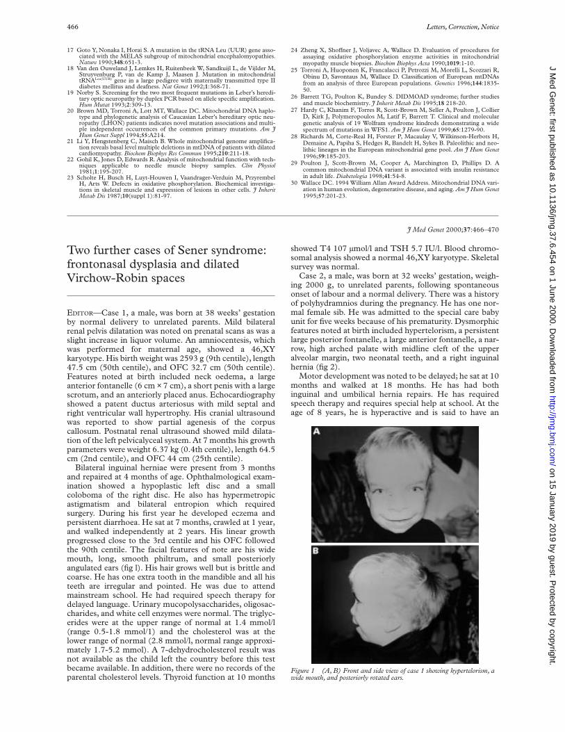
17 Goto Y, Nonaka I, Horai S. A mutation in the tRNA Leu (UUR) gene asso-ciated with the MELAS subgroup of mitochondrial encephalomyopathies.Nature 1990;348:651-3.
18 Van den Ouweland J, Lemkes H, Ruitenbeek W, Sandkuijl L, de Vijlder M,Struyvenburg P, van de Kamp J, Maasen J. Mutation in mitochondrialtRNALeu(UUR) gene in a large pedigree with maternally transmitted type IIdiabetes mellitus and deafness. Nat Genet 1992;1:368-71.
19 Norby S. Screening for the two most frequent mutations in Leber’s heredi-tary optic neuropathy by duplex PCR based on allele specific amplification.Hum Mutat 1993;2:309-13.
20 Brown MD, Torroni A, Lott MT, Wallace DC. Mitochondrial DNA haplo-type and phylogenetic analysis of Caucasian Leber’s hereditary optic neu-ropathy (LHON) patients indicates novel mutation associations and multi-ple independent occurrences of the common primary mutations. Am JHum Genet Suppl 1994;55:A214.
21 Li Y, Hengstenberg C, Maisch B. Whole mitochondrial genome amplifica-tion reveals basal level multiple deletions in mtDNA of patients with dilatedcardiomyopathy. Biochem Biophys Res Commun 1995;210:211-18.
22 Gohil K, Jones D, Edwards R. Analysis of mitochondrial function with tech-niques applicable to needle muscle biopsy samples. Clin Physiol1981;1:195-207.
23 Scholte H, Busch H, Luyt-Houwen I, Vaandrager-Verduin M, PrzyrembelH, Arts W. Defects in oxidative phosphorylation. Biochemical investiga-tions in skeletal muscle and expression of lesions in other cells. J InheritMetab Dis 1987;10(suppl 1):81-97.
24 Zheng X, ShoVner J, Voljavec A, Wallace D. Evaluation of procedures forassaying oxidative phosphorylation enzyme activities in mitochondrialmyopathy muscle biopsies. Biochim Biophys Acta 1990;1019:1-10.
25 Torroni A, Huoponen K, Francalacci P, Petrozzi M, Morelli L, Scozzari R,Obinu D, Savontaus M, Wallace D. Classification of European mtDNAsfrom an analysis of three European populations. Genetics 1996;144:1835-50.
26 Barrett TG, Poulton K, Bundey S. DIDMOAD syndrome; further studiesand muscle biochemistry. J Inherit Metab Dis 1995;18 218-20.
27 Hardy C, Khanim F, Torres R, Scott-Brown M, Seller A, Poulton J, CollierD, Kirk J, Polymeropoulos M, Latif F, Barrett T. Clinical and moleculargenetic analysis of 19 Wolfram syndrome kindreds demonstrating a widespectrum of mutations in WFS1. Am J Hum Genet 1999;65:1279-90.
28 Richards M, Corte-Real H, Forster P, Macaulay V, Wilkinson-Herbots H,Demaine A, Papiha S, Hedges R, Bandelt H, Sykes B. Paleolithic and neo-lithic lineages in the European mitochondrial gene pool. Am J Hum Genet1996;59:185-203.
29 Poulton J, Scott-Brown M, Cooper A, Marchington D, Phillips D. Acommon mitochondrial DNA variant is associated with insulin resistancein adult life. Diabetologia 1998;41:54-8.
30 Wallace DC. 1994 William Allan Award Address. Mitochondrial DNA vari-ation in human evolution, degenerative disease, and aging. Am J Hum Genet1995;57:201-23.
J Med Genet 2000;37:466–470
Two further cases of Sener syndrome:frontonasal dysplasia and dilatedVirchow-Robin spaces
EDITOR—Case 1, a male, was born at 38 weeks’ gestationby normal delivery to unrelated parents. Mild bilateralrenal pelvis dilatation was noted on prenatal scans as was aslight increase in liquor volume. An amniocentesis, whichwas performed for maternal age, showed a 46,XYkaryotype. His birth weight was 2593 g (9th centile), length47.5 cm (50th centile), and OFC 32.7 cm (50th centile).Features noted at birth included neck oedema, a largeanterior fontanelle (6 cm × 7 cm), a short penis with a largescrotum, and an anteriorly placed anus. Echocardiographyshowed a patent ductus arteriosus with mild septal andright ventricular wall hypertrophy. His cranial ultrasoundwas reported to show partial agenesis of the corpuscallosum. Postnatal renal ultrasound showed mild dilata-tion of the left pelvicalyceal system. At 7 months his growthparameters were weight 6.37 kg (0.4th centile), length 64.5cm (2nd centile), and OFC 44 cm (25th centile).
Bilateral inguinal herniae were present from 3 monthsand repaired at 4 months of age. Ophthalmological exam-ination showed a hypoplastic left disc and a smallcoloboma of the right disc. He also has hypermetropicastigmatism and bilateral entropion which requiredsurgery. During his first year he developed eczema andpersistent diarrhoea. He sat at 7 months, crawled at 1 year,and walked independently at 2 years. His linear growthprogressed close to the 3rd centile and his OFC followedthe 90th centile. The facial features of note are his widemouth, long, smooth philtrum, and small posteriorlyangulated ears (fig l). His hair grows well but is brittle andcoarse. He has one extra tooth in the mandible and all histeeth are irregular and pointed. He was due to attendmainstream school. He had required speech therapy fordelayed language. Urinary mucopolysaccharides, oligosac-charides, and white cell enzymes were normal. The triglyc-erides were at the upper range of normal at 1.4 mmol/l(range 0.5-1.8 mmol/1) and the cholesterol was at thelower range of normal (2.8 mmol/l, normal range approxi-mately 1.7-5.2 mmol). A 7-dehydrocholesterol result wasnot available as the child left the country before this testbecame available. In addition, there were no records of theparental cholesterol levels. Thyroid function at 10 months
showed T4 107 µmol/l and TSH 5.7 IU/l. Blood chromo-somal analysis showed a normal 46,XY karyotype. Skeletalsurvey was normal.
Case 2, a male, was born at 32 weeks’ gestation, weigh-ing 2000 g, to unrelated parents, following spontaneousonset of labour and a normal delivery. There was a historyof polyhydramnios during the pregnancy. He has one nor-mal female sib. He was admitted to the special care babyunit for five weeks because of his prematurity. Dysmorphicfeatures noted at birth included hypertelorism, a persistentlarge posterior fontanelle, a large anterior fontanelle, a nar-row, high arched palate with midline cleft of the upperalveolar margin, two neonatal teeth, and a right inguinalhernia (fig 2).
Motor development was noted to be delayed; he sat at 10months and walked at 18 months. He has had bothinguinal and umbilical hernia repairs. He has requiredspeech therapy and requires special help at school. At theage of 8 years, he is hyperactive and is said to have an
Figure 1 (A, B) Front and side view of case 1 showing hypertelorism, awide mouth, and posteriorly rotated ears.
466 Letters, Correction, Notice
on 15 January 2019 by guest. Protected by copyright.
http://jmg.bm
j.com/
J Med G
enet: first published as 10.1136/jmg.37.6.454 on 1 June 2000. D
ownloaded from

attention deficit disorder. Ophthalmological assessmentshowed hypermetropic astigmatism. He was also noted tohave tortuosity of the fundal vessels. On examination aged8 years, his weight (24.3 kg) and head circumference (51cm) were on the 25th centile and his height was <25thcentile (118.5 cm). He had coarse hair with two crowns. Inaddition, he was noted to have abnormally shaped, slenderteeth. An orthopantomogram showed abnormalities ofseveral of the teeth in the position of the incisors andcanines. Many were slender and canine-like. In addition,some deciduous teeth were absent. At the age of 8, none ofhis adult teeth have erupted. The possibility of an ectoder-mal dysplasia was raised, although following consultationwith a dermatologist this was excluded.
Urinary amino and organic acids (including gly-cosaminoglycans), performed on two separate occasions,excluded a mucopolysaccharide disorder. Cholesterol andtriglyceride levels, measured on two occasions, werenormal. A 7-dehydrocholesterol level was also normal. Askeletal survey was normal. Chromosomal analysis showeda normal 46,XY karyotype.
In case 1, a CT scan was reported to show diVuse lowdensity areas in the cerebral white matter bilaterally. Thecorpus callosum was hypoplastic. MRI scan aged 18months showed multiple cystic areas within the white mat-ter radiating out at right angles from the ventricles into alllobes, but especially the parietal, occipital, and temporallobes (fig 3) Their signal intensity paralleled that of CSF.
Figure 2 (A, B, C) Front and side view of case 2 aged7 months and 5 years showing hypertelorism, a widemouth, and posteriorly rotated ears.
Letters, Correction, Notice 467
on 15 January 2019 by guest. Protected by copyright.
http://jmg.bm
j.com/
J Med G
enet: first published as 10.1136/jmg.37.6.454 on 1 June 2000. D
ownloaded from

The basal ganglia and brain stem were spared with very lit-tle involvement of the corpus callosum. A repeat MRI scanaged 4 years showed an increase in the number and size ofthe spaces suggesting a progressive process. The ventricleswere mildly dilated suggesting some atrophic change.
In case 2, CT brain scan showed multiple cystic spaces(fig 4). MRI scan at the age of 6 years confirmed the pres-ence of these cystic spaces. These were noted to be moreconfluent in the occipital region (fig 5). The ventricles wereof normal size. The findings were similar to case 1.
Figure 3 Case 1, aged 4 years. MRI of the head/sagittalT1W + coronal/axial T2W, showing multiple, well definedareas of CSF signal intensity throughout both cerebralhemispheres, especially in the periventricular white matter.They are of varying sizes, being seen as linear streaks inthe cortex but oval or rounded areas in the white matter.All tend to be orientated at right angles to the lateralventricles.
Figure 4 (A, B) Case 2 aged 6 years 9 months. CT of the head showing multiple areas of low density within the cerebralwhite matter bilaterally, most prominent posteriorly.
468 Letters, Correction, Notice
on 15 January 2019 by guest. Protected by copyright.
http://jmg.bm
j.com/
J Med G
enet: first published as 10.1136/jmg.37.6.454 on 1 June 2000. D
ownloaded from

Frontonasal dysplasia is a developmental field defect ofmidfacial development. Clinical features include a broadnose, hypertelorism, low anterior hairline, and sometimesbony defects of the forehead. It is usually a sporadicmalformation and is known to occur with a high frequencyin twins. There are reports of this malformation occurringwith CNS anomalies, in particular frontal encephalocelesand agenesis of the corpus callosum. Dobyns et al1 havereported the association of frontonasal dysplasia withbilateral periventricular nodular heterotopia. Additionalfeatures including mental retardation and epilepsy werepresent in their cases. However, the CNS findings in ourcases are distinct with no evidence of nodular heterotopia.
Virchow-Robin spaces are invaginations of the sub-arachnoid space containing cerebrospinal fluid thataccompanies small arteries and arterioles as they perforatethe surface of the brain. The distribution of theabnormalities seen on the MRI scans of these two casesmirrored the perivascular distribution of the Virchow-Robin spaces. Dilatation of Virchow-Robin spaces havebeen described in a number of conditions including oldage dementia, HIV encephalopathy, and multiple sclerosis(MS).2 In children, similar MRI findings are seen withmucopolysaccharidosis.3 Neither of our cases had anyother clinical features of a mucopolysaccharide disorderand screening of urinary amino acids was negative. Inaddition, the basal ganglia and corpus callosum arefrequently involved in the mucopolysaccharide disorders.In our cases, only case 1 had some involvement of the cor-pus callosum and the basal ganglia were spared in bothcases. The pathogenesis of large Virchow-Robin spacesremains unknown. Increased CSF pulsations, vascularectasia, or an abnormality of arterial wall permeabilityhave all been postulated as possible mechanisms.2 Thesespaces may also have an important immunological role inreactions to foreign antigens that gain access to them.2 Itis felt that this immunological role may have implicationsfor the understanding of the pathogenesis of a number of
neuropathological conditions including encephalitis, MS,and HIV encephalopathy. Both HIV encephalopathy andMS are progressive in nature. The fact that case 1 hadfeatures suggesting a progressive process may indicate thatthe abnormality seen in our cases has a metabolic orautoimmune basis.
Rollins et al4 described the prevalence of dilatedVirchow-Robin spaces in 1250 children having consecutiveMRI scans. Thirty seven children (3%) were shown to havesuch abnormalities on MRI scanning. Five of thesechildren were said to have coarse or dysmorphic featuresand the remainder were referred with headaches, seizures,or developmental delay. All of the children with dysmor-phic features had a negative urinary amino and organicacid screen. Four of the 37 were said to be hyperactive,although none of these had dysmorphic features. In theirstudy, the size and number of the dilated Virchow-Robinspaces appeared to be static in contrast to case 1. Unfortu-nately, the authors did not describe the dysmorphicfeatures of the five cases in detail, nor were there any clini-cal photographs so it is not possible to determine whethertheir cases had similar clinical features to our own. Anumber of other case reports have described similar MRIfindings but without a dysmorphic phenotype.5–8
It is diYcult to establish the significance of the low nor-mal cholesterol level in association with high normaltriglyceride level in case 1. Cholesterol has a role in myeli-nation of the brain in early fetal life. The child has left thecountry and is therefore no longer available for furthertesting. The fact that case 2 had consistently normal levelssuggests that the association may be coincidental.
Blepharocheilodontic syndrome shares some of theectodermal characteristics in common with our cases, butthe degree of hypertelorism is milder and cleft lip/palate isa common finding in BCD.9 In contrast, Sener10 describeda female child with similar dysmorphic features and identi-cal MRI findings to our own cases. This child was said tohave normal development up to the age of 5 years. Subse-quently she developed mild developmental delay and hadto leave normal school aged 13 years. She was noted tohave dental anomalies with hypodontia, dental occlusions,and several buccal frenulae. She was also noted to have thinhair and nail abnormalities. The author suggested that herfeatures were consistent with ectodermal dysplasia and thather condition was slowly progressive in nature. No karyo-type was reported for this patient. Slaney et al11 reported aboy with dysmorphic features similar to the case reportedby Sener.10 However, this child, a boy, had diVerent abnor-malities on MRI scan with evidence of periventricular greymatter heterotopia. All three cases (that of Sener10 and ourtwo) have occurred sporadically with no parental consan-guinity. Both males and females have been aVected. Thegenetic basis remains unknown although the similarities onscan to the mucopolysaccharide storage disorders and thesuggestion of a progressive nature raises the possibility ofautosomal recessive inheritance.
S A LYNCH*K HALL†
S PRECIOUS‡A O M WILKIE§
J A HURST§
*Department of Human Genetics, 19 Claremont Place, Newcastle on TyneNE2 4AA, UK
†Department of Neuroradiology, Newcastle General Hospital, WestgateRoad, Newcastle on Tyne, UK
‡Department of Paediatrics, West Cumberland General Hospital,Whitehaven, Cumbria, UK
§Department of Clinical Genetics, Oxford RadcliVe Hospital, TheChurchill, Oxford OX3 7LJ, UK
Correspondence to: Dr Lynch, [email protected]
Figure 5 (A, B) MRI of the head (sagittal/coronal T1W and axialT2W) showing a similar appearance to that shown in fig 3. The CSFdensity areas in the cerebral white matter are most prominent in theparieto-occipital regions where there is associated localised ventriculardilatation. Some involvement of the corpus callosum is also noted. Thebrain is otherwise normal.
Letters, Correction, Notice 469
on 15 January 2019 by guest. Protected by copyright.
http://jmg.bm
j.com/
J Med G
enet: first published as 10.1136/jmg.37.6.454 on 1 June 2000. D
ownloaded from

1 Dobyns WB, Guerrini R, Czapansky-Beilman DK, et al. Bilateralperiventricular nodular heterotopia with mental retardation and syndactylyin boys: a new X-linked mental retardation syndrome. Neurology1997;49:1042-7.
2 Esiri MM, Gay D. Immunological and neuropathological significance of theVirchow-Robin space. J Neurol Sci 1990;100:3-9.
3 Wraith JE. The mucopolysaccharidoses: a clinical review and guide to man-agement. Arch Dis Child 1995;72:263-7.
4 Rollins N, Deline C, Morris MC. Prevalence and clinical significance ofdilated Virchow-Robin spaces in childhood. Radiology 1993;189:53-7.
5 Afifi AK. Enlarged Virchow-Robin spaces along the medullary perforatorsin a child with seizures. J Neuroimag 1996;6:197-8.
6 Ogawa T, Okudera T, Fukasawa H, et al. Unusual widening ofVirchow-Robin spaces: MR appearance. AJNR 1995;16:1238-40.
7 Bacheschi LA, Magalhaes ACA, Mathias SC. Multiple cystic lesions inwhite matter without clinical manifestations (unidentified black holes).Proceedings of the XV symposium Neuroradiologicum. Kumamoto Neuro-radiol 1995;37:(suppl).
8 Artigas, Poo P, Rovira A, Cardo E. Macrocephaly and dilated Virchow-Robin spaces in childhood. Pediatr Radiol 1999;29:188-90.
9 Gorlin RJ, Zellweger H, Waziri Curtis W, et al. Blepharo-cheilo-dontic syn-drome. Am J Med Genet 1996;65:109-11.
10 Sener RN. Polycystic brain (cerebrum polycystica vera) associated withectodermal dysplasia: a new neurocutaneous syndrome. Pediatr Radiol1994;24:116-18.
11 Slaney SF, WK Chong, Winter RM. A new syndrome of short stature, dis-tinctive facial features and periventricular grey matter heterotopia. ClinDysmorphol 1999;8:5-9.
J Med Genet 2000;37:470–472
The psychological impact of a cancerfamily history questionnaire completedin general practice
EDITOR—On the basis of family history, it is possible toidentify subjects at significantly increased genetic risk ofbreast or colorectal cancer.1 2 Evaluation of the benefits ofscreening these patients to facilitate early diagnosis andtreatment forms the subject of continuing studies. Forcolorectal cancer, the benefits of colonoscopic surveillancehave been reported,3 but for breast cancer more data areneeded to confirm the value of mammographic screening.4
At present, patients with a significant family history whoseek advice from their general practitioner are likely to bereferred to a cancer genetics clinic and oVered screening. Iffurther research confirms the benefits of screening forpatients at increased genetic risk, eVective strategies fortheir ascertainment in primary care will be needed. Onepossible method is a postal family history questionnairesent to the patient by their general practitioner. We reportelsewhere on the eVectiveness of this approach.5 An impor-tant issue is whether this method of ascertainment raisesanxieties, particularly among the majority of patients whodo not have a significant family history. The collection ofcancer family history information constitutes a form ofscreening. There is a large body of evidence that healthrelated screening can have unintended adverse eVects, themost studied of which is raised anxiety, particularly amongthose found to be at an increased risk.6 As knowledge of thegenetic component of common diseases increases,7 morepatients may be asked to provide information about theirfamily history. It is therefore timely to consider whethersuch a task may inadvertently raise general levels of anxietyor worries about the disease in question. To our knowledge,there have been no previous studies of the psychologicalconsequences of screening using a postal questionnaire toobtain information about relatives aVected by cancer. Thepurpose of the present study was to determine the psycho-logical impact of completing a cancer family history ques-tionnaire and receiving an assessment of personal geneticrisk of breast or colorectal cancer.
General anxiety was assessed using the six item, shortform of the state scale of the Spielberger State-Trait
Anxiety Inventory (STAI).8 This yields a single scoreranging from 20 to 80. The mean for the adult populationis 36. Worry about cancer was measured using theshortened version of the Cancer Worries Scale,9 whichassesses (1) people’s perceptions of their own chances ofdeveloping cancer, (2) their frequency of cancer relatedthoughts, (3) the frequency with which they perceive theirmood to be aVected by such thoughts, and (4) thefrequency with which such thoughts aVect the perform-ance of their daily tasks.
The participants in this study were patients completing acancer family history questionnaire as part of a separatestudy to evaluate its use in general practice.5 For that study,patients aged between 35 and 65 years registered with asingle general practice in Cambridge were invited toparticipate. They were sent an information sheet explain-ing that the purpose of the study was to identify the smallminority of patients whose family history would put themat suYciently increased risk of breast or colorectal cancerto warrant the oVer of screening to facilitate early diagno-sis and treatment. A consent form and short questionnaireto measure baseline levels of general anxiety and worryabout cancer were also enclosed. Those wishing to partici-pate were asked to complete and return the consent formand the baseline measure. They were then sent the familyhistory questionnaire (for details see http://www.jmedgenet.com). On the basis of their responses, themajority of patients were judged not to be at significantlymore than the population risk of breast or colorectal cancer(lower risk group). These patients were sent a letter tellingthem that, on the basis of their stated family history, theirpersonal risk of developing breast or colorectal cancer wasbelow the level at which extra screening tests would be re-commended. A small number of patients were assessed tobe at potentially increased risk where one of the followingapplied: (1) their family history as reported met localscreening criteria for breast or colorectal cancer (table 1) or(2) their family history approached screening criteria soclosely that it was considered advisable to check crucialdetails such as age at onset in relatives, or (3) theinformation provided on the questionnaire was ambiguousor incomplete and there remained a possibility that thescreening criteria might be met, or (4) their family historydid not meet screening criteria but suggested an increasedrisk to the GP assessor. Almost all of these patients were
Table 1 Criteria used to define increased genetic risk suYcient to warrant referral and the oVer of screening
For breast cancer, females with one of the following:(1) Three first or second degree relatives with breast or ovarian cancer(2) Two first or second degree relatives with breast cancer diagnosed under 60 years of age or ovarian cancer at any age(3) One first degree relative with (i) breast cancer diagnosed under 40 years of age, or (ii) bilateral breast cancer, or (iii) male breast cancer
For colorectal cancer, one of the following:(1) A first degree relative plus two other relatives with colorectal cancer and (i) one case diagnosed under 50 years of age, and (ii) one case a first degree
relative of the other two, and (iii) at least two generations aVected(2) A first degree relative with colorectal cancer diagnosed under 45 years of age
470 Letters, Correction, Notice
on 15 January 2019 by guest. Protected by copyright.
http://jmg.bm
j.com/
J Med G
enet: first published as 10.1136/jmg.37.6.454 on 1 June 2000. D
ownloaded from

interviewed but in a few cases minor uncertainties wereresolved over the telephone. Most were confirmed to be atsignificantly increased genetic risk (higher risk group) buta minority were deemed not to be at increased risk (falsepositive group). An explanation of their final riskassessment was given to all these patients, usually atpersonal interview (all patients assigned to higher risk as aresult of the study were informed of this at interview), butin a few cases by telephone or letter. Patients in the higherrisk group who had not previously received genetic advicewere oVered referral to the cancer genetics clinic. All par-ticipants were asked to complete a follow up anxiety andcancer worries questionnaire four to six weeks afterfeedback of their personal risk. In the group of patientsreferred for genetic advice, this measure was completedafter the consultation at which referral was oVered butbefore the appointment at the cancer genetics clinic.Statistical comparisons were made using the Wilcoxonsigned rank test for paired data and the Mann-Whitney Utest for independent samples. The Cambridge LocalResearch Ethics Committee gave ethical approval for thestudy.
The eVective practice population for the study of thecancer family history questionnaire was 2265 patients. Atotal of 666 patients (29%) completed that questionnaireand are the participants in the present study. They diVeredfrom the practice population in terms of both gender andage.5 A total of 62.2% were women, compared to 50.2% ofthe practice population (÷2=37.8, p<0.001) and a lowerproportion were aged 35-44 (30.2% compared with40.7%, ÷2=30.3, p<0.001).
A total of 604 patients (91%) returned baseline and fol-low up measures of anxiety and cancer worry. The genderand age distribution of respondents did not diVersignificantly from the study population. Paired responseswere obtained from 568 patients assessed not to be at sig-nificantly increased risk of breast or colorectal cancer(lower risk group) and 36 patients judged to be atpotentially increased genetic risk on the basis of their fam-ily history questionnaires. The latter group comprised 25patients who were subsequently confirmed to be at signifi-cantly increased risk (higher risk group) and 11 deemednot to be at significantly increased risk after further inves-tigation of their family history (false positive group). Somepatients failed to answer all six items of the SpielbergerState-Trait Anxiety Inventory or all four items of the Can-
cer Worries Scale. Table 2 gives the numbers of valid pairedresponses in each patient group.
The scores for general anxiety and cancer worries atbaseline (before completion of the family history question-naire) and follow up (four to six weeks after receipt of theirrisk assessment) for all three groups are shown in table 2.In the lower risk group, the only diVerence in pairedresponses between the two time points assessed was inpatients’ perceptions of their personal risk of developingcancer, which showed a small reduction (p<0.001). For theother two groups there were no diVerences in pairedresponses for general anxiety or cancer worries. For boththe higher risk group and the false positive group, baselineresponses showed that their pre-existing perception of theirrisk of developing cancer was higher than that of the lowerrisk group (p<0.001 and p=0.003, respectively). For thefalse positive group, the frequency with which cancerrelated thoughts aVected their mood was also higher(p=0.02).
The results of this study suggest that completion of acancer family history questionnaire and receipt of anassessment of personal genetic risk for breast and colorec-tal cancer does not make patients more anxious or worriedabout cancer. This conclusion is based on a substantialnumber of subjects, but should be tempered by the factthat only a minority of practice patients returned the fam-ily history questionnaire and constituted a self-selectedgroup.
Responses to the Cancer Worries Scale showed thatmost patients rarely worried about their risks of developingcancer either before or after the study. Indeed, receipt ofinformation that their personal risk was below the level atwhich extra screening tests would be oVered was associatedwith a small but significant reduction in perceived risks ofdeveloping cancer. This raises the question of whether thisknowledge could influence health related behaviour. Forexample, it might reduce the incentive to participate inhealth related activities, such as attendance for routinemammographic screening or eating a fibre rich diet. ThiseVect has been reported for other forms of screening10 11
and merits further investigation.For patients assessed to be at potentially increased risk
on the basis of their family history questionnaire, baselineresponses showed that their pre-existing perceptions oftheir own risk of developing cancer were significantlyhigher than those of other patients. This suggests that many
Table 2 General anxiety and cancer worries at baseline and follow up for paired responses in (1) patients assessed not to be at significantly increased risk(lower risk group), (2) patients with potentially significant family histories subsequently shown not to be at significantly increased risk (false positivegroup), (3) patients confirmed to be at significantly increased risk (higher risk group)
Mean scores (95% CI)
Lower risk group False positive group Higher risk group
Baseline Follow up Baseline Follow up Baseline Follow up
Speilberger State-Trait Anxiety InventoryGeneral anxiety 35.8 35.1 34.8 34.3 36.3 38.9
(34.6–36.9) (33.9–36.2) (26.6–42.9) (24.1–44.5) (31.7–40.9) (32.8–45.0)n=427 n=7 n=18
Cancer Worries ScalePerception of own chances of developing cancer 2.95 2.83 3.55 3.27 3.56 3.40
(2.88–3.01) (2.76–2.91) (3.19–3.90) (2.53–4.01) (3.18–3.94) (2.99–3.81)n=534 n=11 n=25
Frequency of cancer related thoughts 1.63 1.61 1.73 1.64 1.96 1.76(1.57–1.69) (1.55–1.67) (1.29–2.16) (1.09–2.18) (1.59–2.33) (1.36–2.16)n=564 n=11 n=25
EVect on mood 1.21 1.22 1.45 1.27 1.29 1.25(1.17–1.26) (1.17–1.26) (1.10–1.81) (0.84–1.71) (1.10–1.49) (1.03–1.47)n=560 n=11 n=24
EVect on performance of daily tasks 1.12 1.11 1.00 1.09 1.17 1.08(1.08–1.15) (1.08–1.15) * (0.89–1.29) (1.01–1.33) (0.96–1.20)n=562 n=11 n=24
CI = confidence interval.*All respondents scored 1 for this item.
Letters, Correction, Notice 471
on 15 January 2019 by guest. Protected by copyright.
http://jmg.bm
j.com/
J Med G
enet: first published as 10.1136/jmg.37.6.454 on 1 June 2000. D
ownloaded from
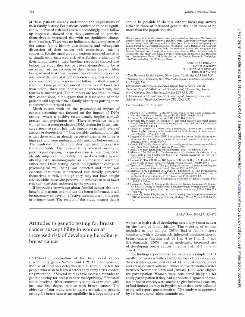
of these patients already understood the implications oftheir family history. For patients confirmed to be at signifi-cantly increased risk and advised accordingly, their followup responses showed that they continued to perceivethemselves at increased risk with no significant changefrom baseline. There was no indication that completion ofthe cancer family history questionnaire and subsequentdiscussion of their cancer risk exacerbated existingconcerns. For the small group of patients assessed not to beat significantly increased risk after further evaluation oftheir family history, their baseline responses showed thatbefore the study they too perceived themselves to be atincreased risk on account of their family history. Afterbeing advised that their personal risk of developing cancerwas below the level at which extra screening tests would berecommended, their responses at follow up show a mixedreaction. Four patients regarded themselves at lower riskthan before, three saw themselves at increased risk, andfour were unchanged. The numbers are too small to drawfirm conclusions, but suggest that at least some of thesepatients still regarded their family history as putting themat somewhat increased risk.
Much recent work on the psychological impact ofgenetic screening has focused on the impact of DNAtesting12 where a positive result usually implies a muchgreater than population risk. There is evidence that, inwomen undergoing predictive DNA testing for breast can-cer, a positive result has little impact on general levels ofanxiety or depression.13 14 One possible explanation for thisis that these women already perceived themselves to be athigh risk and were understandably anxious before testing.The result did not, therefore, alter their psychological sta-tus appreciably. The present study assessed anxiety inpatients participating in a questionnaire survey designed toidentify subjects at moderately increased risk with a view tooVering extra mammographic or colonoscopic screeningrather than DNA testing. Again, no significant change inpsychological well being was detected and there wasevidence that those at increased risk already perceivedthemselves at risk, although they may not have soughtadvice, while those who perceived themselves to be at lowerrisk had their view endorsed by the process.
If improving knowledge about familial cancer risk is tobenefit all patients and not just the better informed, it willbe necessary to develop eVective ascertainment strategiesin primary care. The results of this study suggest that it
should be possible to do this without increasing anxietyeither in those at increased genetic risk or in those at nomore than the population risk.
We are grateful to all the patients who participated in this study. We thank thedoctors and staV at East Barnwell Health Centre, Cambridge for their supportand encouragement. We are grateful to Steve Jones for help with computing andElaine Farrell for secretarial assistance. We thank Hilarie Bateman for help withplanning the study and Chris Todd for statistical advice. We are grateful toMartin Bobrow, Ann Louise Kinmonth, and Fawzia Khan for their helpfulcomments. This study was funded by the Oxford and Anglia Regional Researchand Development Fund. JM is funded by the Cancer Research Campaign.TMM is funded by The Wellcome Trust.
VIRGINIA LEGGATT*JAMES MACKAY†
THERESA M MARTEAU‡JOHN R W YATES§
*East Barnwell Health Centre, Ditton Lane, Cambridge CB5 8SP, UK†Department of Oncology, Box 193, Addenbrooke’s Hospital, CambridgeCB2 2QQ, UK‡Psychology and Genetics Research Group, Guy’s, King’s, and StThomas’ Hospitals’ Medical and Dental School, Thomas Guy House,Guy’s Campus, Guy’s Hospital, London SE1 9RT, UK§Department of Medical Genetics, University of Cambridge, Box 134,Addenbrooke’s Hospital, Cambridge CB2 2QQ, UK
Correspondence to: Dr Leggatt
1 Pharoah PDP, Stratton JF, Mackay J. Screening for breast and ovarian can-cer: the relevance of family history. Br Med Bull 1998;54:823-8.
2 Dunlop MG. Colorectal cancer. BMJ 1997;314:1882-5.3 Jarvinen HJ, Mecklin JP, Sistonen P. Screening reduces colorectal cancer
rate in families with hereditary non-polyposis colorectal cancer. Gastroenter-ology 1995;108:1405-11.
4 Lalloo F, Boggis CR, Evans DG, Shenton A, Threlfall AG, Howell A.Screening by mammography, women with a family history of breast cancer.Eur J Cancer 1998;34:937-40.
5 Leggatt V, Mackay J, Yates JRW. Evaluation of a cancer family history ques-tionnaire for identifying patients at increased genetic risk in generalpractice. BMJ 1999;319:757-8.
6 Croyle RT, ed. Psychosocial eVects of screening for disease prevention and detec-tion. New York: Oxford University Press, 1995.
7 Bell J. The new genetics in clinical practice. BMJ 1998;316:618-20.8 Marteau TM, Bekker H. The development of a six-item short-form of the
state scale of the Spielberger State-Trait Anxiety Inventory (STAI). Br JClin Psychol 1992;31:301-6.
9 Lerman C, Trock B, Rimer BK, Jepson C, Brody D, Boyce A. Psychologicalside eVects of breast cancer screening. Health Psychol 1991;10:259-67.
10 Tymstra T, Bielman B. The psychological impact of mass screening forcardiovascular risk factors. Family Practice 1987;4:287-90.
11 Marteau TM, Kinmonth AL, Pyke S, Thompson S. The psychologicalimpact of a cardiovascular risk reduction programme in primary care: aproblem of false reassurance? Br J Gen Pract 1996;46:577-82.
12 Marteau TM, Croyle RT. Psychological responses to genetic testing. BMJ1998;316:693-6.
13 Lerman C, Narod S, Shulman K, Hughes C, Gomez-Caminero A, BonneyG. BRCA1 testing in families with hereditary breast-ovarian cancer. A pro-spective study of patient decision making and outcomes. JAMA 1996;275:1885-92.
14 Croyle RT, Smith KR, Botkin JR, Baty B, Nash J. Psychological responses toBRCA1 mutation testing: preliminary findings. Health Psychol 1997;16:63-72.
J Med Genet 2000;37:472–476
Attitudes to genetic testing for breastcancer susceptibility in women atincreased risk of developing hereditarybreast cancer
EDITOR—The localisation of the two breast cancersusceptibility genes BRCA1 and BRCA2 made possiblethe use of mutation detection as a susceptibility test forpeople who wish to learn whether they carry a risk confer-ring mutation.1–4 Several studies have assessed attitudes togenetic testing for breast cancer susceptibility,5–11 most ofwhich involved either community samples or women withjust one first degree relative with breast cancer. Theobjective of our study was to assess attitudes to genetictesting for breast cancer susceptibility in a large sample of
women at high risk of developing hereditary breast canceron the basis of family history. The majority of womenincluded in our sample (80%) had a family historyconsistent with a dominantly inherited predisposition tobreast cancer (lifetime risk of 1 in 4 to 1 in 2),12 andthe remainder (20%) was at moderately increased riskof developing breast cancer (lifetime risk of 1 in 8 to1 in 4).12
The findings reported here are based on a sample of 461unaVected women with a family history of breast cancer.Women who approached one of 14 familial cancer clinicsand six associated outreach clinics in five Australian statesbetween November 1996 and January 1999 were eligiblefor participation. Women were considered ineligible forstudy participation if they had a previous diagnosis of ovar-ian or breast cancer, were unable to give informed consent,or had limited literacy in English, since data were collectedusing self-report questionnaires. The study was approvedby 16 institutional ethics committees.
472 Letters, Correction, Notice
on 15 January 2019 by guest. Protected by copyright.
http://jmg.bm
j.com/
J Med G
enet: first published as 10.1136/jmg.37.6.454 on 1 June 2000. D
ownloaded from

Familial cancer clinic staV invited women to participatein the study during the preclinic telephone call, where pos-sible. Questionnaires, consent forms, and reply paid enve-lopes were then mailed out by the coordinating researchcentre. Women were subsequently telephoned by the cen-tral research staV and given further information about thestudy and issues of informed consent. Women were askedto return the completed questionnaire and consent formbefore attending the familial cancer clinic, if possible.Reminder calls were made as required.
Sex, age, educational level, marital status, number andsex of biological children, and referral source wereassessed. To provide an estimate of objective risk, clinicstaV were asked to make a judgment on whether a partici-pant’s family history was either consistent or not consistentwith a dominantly inherited predisposition to breastcancer, and participants were thus classified as being at“high risk” or “moderately increased risk”, respectively.Following the risk assessment interview at the familial can-cer clinic and once pedigree information13 14 and relatives’diagnoses confirmed by medical records were available,clinic staV categorised participants into objective riskgroups. For women from high risk families, clinic staVmade a judgment on whether the participant was at either50% or 25% mutation carrier risk. Risk of being a mutationcarrier, rather than the estimated lifetime risk of developingbreast cancer, was used as a measure of objective risk. Theexpert opinion of clinical geneticists was used as a goldstandard, since there are currently no universally acceptedstandards to estimate breast cancer risk in high riskwomen. The number of first and second degree relativeswho developed breast or ovarian cancer were collectedfrom study participants.
One item asked participants to select their approximateperceived lifetime breast cancer risk from the followingresponse options: 1%, 4%, 8%, 12%, 16%, 25%, 33%,50%, 85%, and 100%. Risk was expressed both as apercentage and as odds (for example, 1 in 8).
One item measured perceived likelihood of being amutation carrier, with five response options ranging from“Certain that I will not have gene” to “Certain I will havegene”. The phrase “have the gene” was used, because ofthe common misconceptions regarding heterozygosityamong the lay population and to facilitate ease ofunderstanding.
The Impact of Event Scale is a 15 item, validated scalethat measures anxiety responses about a specific stressfulevent.15 16 Subjects at increased risk of developing heredi-tary breast cancer may construe their being at risk as acontinuous, rather than specific, trauma. Although thescale has not previously been specifically validated amongsubjects at high risk of developing breast cancer, it has beenused in several studies as a measure of breast canceranxiety7 17 and has previously been shown to be predictiveof interest in genetic testing.18 In the current study the par-ticular stressor was concern about being at risk of develop-ing breast cancer. Participants were asked to ratesymptoms of anxiety (for example, “I had strong waves offeelings about being at risk of breast cancer”) on a scaleranging from “Not at all” to “Often”.
The Monitoring-Blunting Style Scale is an eight item,validated scale measuring individual diVerences in copingstyles in threatening situations.19 The scale measures a per-son’s tendency either actively to seek out threatening infor-mation (“monitoring”) or to ignore it and distract oneself(“blunting”). The Monitoring-Blunting Style scale was
Figure 1 Percentages of women endorsing perceived benefits of genetic testing.
...to help me understand how to reduce myrisk of developing cancer
...to learn about my children's risk
...to be certain about my risk
...to help research
...to plan for the future
...to make child bearing decisions
0 20 40
A very important factor in deciding about whether I would have a gene test or not wouldbe...
60 80 100
Figure 2 Percentages of women endorsing perceived limitations of genetic testing.
...I am concerned about the effectsof gene testing on the family
...I don't believe that I can preventgetting cancer
...I am worried about losing insurance
...I couldn't handle knowledge emotionally
...the test result may not be accurate
...I don't trust modern medicine
0 5 10 15 20
A very important factor in deciding about whether I would have a gene test or not wouldbe that...
Letters, Correction, Notice 473
on 15 January 2019 by guest. Protected by copyright.
http://jmg.bm
j.com/
J Med G
enet: first published as 10.1136/jmg.37.6.454 on 1 June 2000. D
ownloaded from

included to assess individual diVerences in responding togenetic risk information.
Six items each assessed perceived benefits and limita-tions of genetic testing for breast cancer susceptibility. Figs1 and 2 show items relating to benefits and limitationsrespectively. These items have been used in relatedstudies.20 21 Women endorsed perceived importance of eachbenefit or limitation on three response options rangingfrom “Not at all important” to “Very important”.
Interest in genetic testing was the outcome variable andasked women whether they would be interested in a genetictest. Four response options ranged from “Yes, definitely” to“No, definitely not”.
The outcome variable “interest in genetic testing” wasrecoded as a binary variable. Only eight participants (2%)reported being “probably not” interested in genetic testingand none reported being “definitely not” interested. It isgenerally recommended to collapse categories whichcontain less than 5% of cases to achieve a more balanceddistribution.22 Therefore, rather than collapsing the two“yes” and two “no” categories, the new variable wasdefined as “yes, definitely” versus “yes, probably”,“probably not” and “definitely not”.
To assess associations of predictor variables with theoutcome variables, contingency table analysis with ÷2 testswere performed for categorical predictor variables, Mann-Whitney U, and Kruskal-Wallis tests for ordinal andnon-normal interval variables and two sample t tests fornormally distributed interval data. For the outcomevariable “interest in genetic testing” hierarchical logisticregression was used to identify variables associatedindependently with interest in genetic testing. All inde-pendent variables with a bivariate association of p<0.1 with
the outcome variable were entered as predictors and ageand objective risk as covariates.
Of the 520 women who met the eligibility criteria, 59women declined participation or never returned the ques-tionnaire (response rate of 89%). Table 1 summarisessociodemographic and family history variables of the studysample.
The mean age of breast cancer onset in the youngestperson in the family was 41 years (SD=9.6). The numberof self-reported first and second degree relatives with adiagnosis of breast or ovarian cancer ranged from 1 to 18,with a median of three. Ninety five women (21%) had afamily history which included ovarian cancer in addition tobreast cancer. All women were assessed before receiving agenetic testing result. Seventy percent of women reportedbeing “definitely”, 22% “probably”, and 2% “probablynot” interested in genetic testing, with the remainder (6%)being unsure.
Bivariate analyses between predictor variables and inter-est in genetic testing showed that subjective carrier risk wassignificantly associated with interest: 86% of women whoreported being “quite certain” or “certain” that they werecarriers were “definitely interested” in testing, compared to71% of those who were uncertain (÷2=8.20, p=0.017).Women who were “definitely interested” in genetic testinghad significantly higher breast cancer risk perceptions(Z=−2.08, p=0.038). Neither educational level (÷2=0.82,p=0.37), referral status (÷2=0.32, p=0.57), having daugh-ters or not (÷2=0.22, p=0.63), breast cancer anxiety(Z=−1.48, p=0.14), nor monitoring score (Z=−0.062,p=0.95) were significantly associated with interest ingenetic testing. Table 2 shows the final regression model.Only perceived mutation carrier risk was significantly asso-ciated with interest in genetic testing (p=0.0017), in thatwomen who were uncertain if they were mutation carrierswere less likely to be definitely interested in genetic testing(OR 0.37, 95% CI 0.18-0.73, p=0.0044), compared towomen who were quite certain or certain that they werecarriers.
Fig 1 shows the percentages of participants whoendorsed each perceived benefit as a “very important” fac-tor in deciding about whether or not to have a genetic test.The item endorsed by the highest percentage of women asvery important (87%) related to perceiving genetic testingas helpful in understanding what steps to take to reduceone’s cancer risk. Learning about one’s children’s risk wasthe second most frequently endorsed item, with 77% ofwomen who had children reporting it to be a veryimportant factor.
Fig 2 shows the percentages of participants whoendorsed each perceived limitation as a “very important”factor in deciding about whether to have a genetic test. Theitem endorsed by the highest percentage of women as veryimportant (16%) related to concerns about the eVect ofgenetic testing on the family. Interestingly, only 4% and11% of women reported that worry about losing insurancewas a very important or somewhat important factor,respectively.
Table 3 provides an overview of the results of bivariateanalyses to identify associations between demographic andpsychological variables and individual perceived benefitsand shortcomings of testing. It shows that women who haddaughters, compared to women who had sons only, weresignificantly more likely to report that learning about one’schildren’s risk was an important factor (84% versus 61%,÷2=29.31, p<0.0001). Women who reported that not beingable to handle the knowledge emotionally was a very orsomewhat important factor had significantly higher breastcancer anxiety than women who reported that this was notat all important (Z=−6.91, p<0.0001). None of the other
Table 1 Sociodemographic and family history variables of study sample(n=461)
Variable Level No %
SociodemographicsAge <30 97 21
30–39 157 3440–49 124 2750+ 81 18
Marital status Married 295 64Not married 164 36
Biological children Yes 329 72No 130 28
Educational level Post-school qualifications 301 68No post-school qualifications 143 32
Family history variablesObjective risk Moderate risk 85 20
High risk - 25% MCR* 48 10High risk - 50% MCR* 303 70
No of FDR & SDR with BrCaor OvCa†
1–2 135 343–4 176 445–18 87 22
*MCR = mutation carrier risk.†Number of first and second degree relatives with breast or ovarian cancer.
Table 2 Hierarchical regressions on attitude to genetic testing for breastcancer predisposition (n=384)
Model variableReferencegroup
−2 loglikelihood(p value) OR* 95% CI OR p
Final model 428.31(p<0.040)
Objective risk 0.5725% MCR† 0.71 0.30, 1.72 0.4550% MCR† 0.73 0.40, 1.32 0.30Moderate risk
Perceivedcarrier risk
0.017
Not have gene 0.38 0.11, 1.29 0.12Unsure 0.37 0.18, 0.73 0.0044Have gene
*OR = odds ratio, refers to comparisons with the category listed last.†MCR = Mutation carrier risk.
474 Letters, Correction, Notice
on 15 January 2019 by guest. Protected by copyright.
http://jmg.bm
j.com/
J Med G
enet: first published as 10.1136/jmg.37.6.454 on 1 June 2000. D
ownloaded from
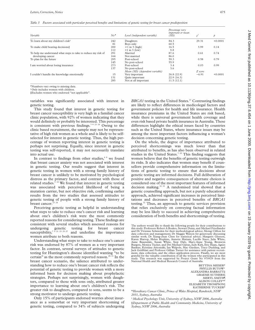
variables was significantly associated with interest ingenetic testing.
This study found that interest in genetic testing forbreast cancer susceptibility is very high in a familial cancerclinic population, with 92% of women indicating that theywould definitely or probably be interested. This percentageis consistent with previous findings.7–9 23 24 Owing to theclinic based recruitment, the sample may not be represen-tative of high risk women as a whole and is likely to be self-selected for interest in genetic testing. Thus, the high per-centage of women reporting interest in genetic testing isperhaps not surprising. Equally, since interest in genetictesting was self-reported, it may not necessarily translateinto actual use.
In contrast to findings from other studies,6 8 we foundthat breast cancer anxiety was not associated with interestin genetic testing. Our results suggest that interest ingenetic testing in women with a strong family history ofbreast cancer is unlikely to be motivated by psychologicaldistress as the primary factor, and contrast with those ofrelated studies.9 18 We found that interest in genetic testingwas associated with perceived likelihood of being amutation carrier, but not objective risk, confirming earlierresults from the two studies that assessed attitudes togenetic testing of people with a strong family history ofbreast cancer.23 24
Perceiving genetic testing as helpful in understandingwhat steps to take to reduce one’s cancer risk and learningabout one’s children’s risk were the most commonlyreported reasons for considering testing. These findings areconsistent with several studies which assessed reasons forundergoing genetic testing for breast cancersusceptibility,7 9 21 23 25–27 and underline the importancewomen attribute to both reasons.
Understanding what steps to take to reduce one’s cancerrisk was endorsed by 87% of women as a very importantfactor. In contrast, several surveys of attitudes to genetictesting for Huntington’s disease identified wanting “to becertain” as the most commonly reported reason.28–31 In thebreast cancer scenario, the salience attributed to under-standing how to reduce one’s breast cancer risk reflects thepotential of genetic testing to provide women with a moreinformed basis for decision making about prophylacticstrategies. Perhaps not surprisingly, women with daugh-ters, compared to those with sons only, attributed greaterimportance to learning about one’s children’s risk. Thegreater risk to daughters, compared to sons, seems to be astrong motivator to undergo genetic testing.
Only 15% of participants endorsed worries about insur-ance as a somewhat or very important shortcoming ofgenetic testing, compared to 34% of subjects undergoing
BRCA1 testing in the United States.21 Contrasting findingsare likely to reflect diVerences in medicolegal factors andgovernment policies for health and life insurance. Healthinsurance premiums in the United States are risk based,while there is universal government health coverage andeven risk based private health insurance in Australia. ThesediVerences highlight the ethical issues faced by countriessuch as the United States, where insurance issues may beamong the most important factors influencing a woman’sdecision concerning genetic testing.
On the whole, the degree of importance attributed toperceived shortcomings was much lower than thatattributed to benefits, as has also been observed in relatedstudies in the United States.7 25 This finding suggests thatwomen believe that the benefits of genetic testing outweighits risks. It also indicates that women may benefit if coun-sellors provide comprehensive information on the limita-tions of genetic testing to ensure that decisions aboutgenetic testing are informed decisions. Full deliberation ofpositive and negative consequences of alternate choices isconsidered one of the most important features of informeddecision making.32 33 A randomised trial showed that agenetic counselling approach, but not a purely educationalapproach, achieved significant increases in perceived limi-tations and decreases in perceived benefits of BRCA1testing.32 Thus, an approach to genetic services provisionthat relies exclusively on conveying factual informationmay be less likely to succeed in achieving comprehensiveconsideration of both benefits and shortcomings of testing.
The authors would like to thank the following people for their contributions tothis study: Professors Robert A Boakes, Stewart Dunn, and Michael Friedlanderand Dr Vivienne Schnieden for their methodological advice; Morag Clifton fordata collection and management; Dr Maggie Watson for generously discussingsimilar work; Dr Xiang-chun Chen for statistical advice; Margaret Gleeson,Karen Harrop, Helen Hopkins, Annette Hattam, Lucille Stace, Julie White,Anne Baxendale, Susan White, Step Daly, Mary-Anne Young, BronwynBurgess, Monica Tucker, and Drs Michael Gattas, Judy Kirk, Eric Haan, AgnesBankier, Kristiina Aittomäki, Ian Walpole, Mac Gardner, Tracy Dudding, andJack Goldblatt and Professor Gillian Turner for assistance with patient recruit-ment, data collection, and the ethics application process. Finally, we are mostgrateful for the valuable contribution of all the women who participated in thisstudy. This research was supported by Project Grant No 970929 from theNational Health and Medical Research Council of Australia.
BETTINA MEISER*PHYLLIS BUTOW†
ALEXANDRA BARRATT‡GRAEME SUTHERS§
MERYL SMITH¶ALISON COLLEY**
ELIZABETH THOMPSON§KATHERINE TUCKER*
*Hereditary Cancer Clinic, Prince of Wales Hospital, Randwick, NSW2031, Sydney, Australia†Medical Psychology Unit, University of Sydney, NSW 2006, Australia‡Department of Public Health and Community Medicine, University ofSydney, NSW 2006, Australia
Table 3 Factors associated with particular perceived benefits and limitations of genetic testing for breast cancer predisposition
Variable No* Level (independent variable)
Percentage veryimportant or mean(SD) ÷2 p
To learn about my children’s risk† 242 Daughters 84.3 29.31 <0.000181 Boys only 60.5
To make child bearing decisions‡ 200 >1 in 3 (high) 16.5 3.99 0.14112 <1 in 3 (low) 9.8
To help me understand what steps to take to reduce my risk ofdeveloping cancer
291 Married 87.6 0.61 0.74164 Not married 86.0
To plan for the future 295 Post-school 59.3 0.58 0.79145 No post-school 55.9
I am worried about losing insurance 273 Post-school 3.4 0.03 0.99137 No post-school 3.6
Mean (SD) (dependent variable) Z scoreI couldn’t handle the knowledge emotionally 15 Very important 26.8 (22.9) −6.91 <0.0001
131 Quite important 22.9 (16.3)285 Not at all important 11.5 (12.1)
*Numbers vary owing to missing data.†Only includes women with children.‡Excludes women who endorsed “not applicable”.
Letters, Correction, Notice 475
on 15 January 2019 by guest. Protected by copyright.
http://jmg.bm
j.com/
J Med G
enet: first published as 10.1136/jmg.37.6.454 on 1 June 2000. D
ownloaded from

§South Australian Clinical Genetics Service, Women’s and Children’sHospital, North Adelaide, SA 5006, Australia¶Familial Cancer Clinic, Westmead Hospital, Westmead, NSW 2145,Australia**Department of Clinical Genetics, Liverpool Hospital, Elizabeth Street,Liverpool, NSW 2170, Australia
Correspondence to: Dr Meiser, [email protected]
1 Hall JM, Lee MK, Newman B, Morrow J, Anderson L, Huey B, King M.Linkage analysis of early-onset familial breast cancer to chromosome17q12. Science 1990;250:1684-9.
2 Miki Y, Swensen J, Shattuck-Eidens D, Futreal A, Harshman K, Tavtigian S,et al. A strong candidate for the breast and ovarian cancer susceptibilitygene BRCA1. Science 1994;266:66-71.
3 Wooster R, Neuhausen SL, Mangion J, Quirk Y, Ford D, Collins N, et al.Localization of a breast cancer susceptibility gene, BRCA2, to chromosome13q12-13. Science 1994;265:2088-90.
4 Wooster R, Bignell G, Lancaster J, Swift S, Seal S, Mangion J, et al. Identifi-cation of the breast cancer susceptibility gene BRCA2. Nature 1995;378:789-92.
5 Chaliki H, Loader S, Levenkron JC. Women’s receptivity to testing for agenetic susceptibility to breast cancer. Am J Public Health 1995;85:1133-5.
6 Andrykowski MA, Munn RK, Studts JL. Interest in learning of personalgenetic risk for cancer: a general population survey. Prev Med 1996;25:527-36.
7 Lerman C, Seay J, Balshem A, Audrain J. Interest in genetic testing amongfirst-degree relatives of breast cancer patients. Am J Med Genet1995;57:385-92.
8 Jacobsen PB, Valdimarsdottir HB, Brown KL, OYt K. Decision-makingabout genetic testing among women at familial risk for breast cancer. Psy-chosom Med 1997;59:459-66.
9 Lerman C, Daly M, Masny A, Balshem A. Attitudes about genetic testingfor breast-ovarian cancer susceptibility. J Clin Oncol 1994;12:843-50.
10 Lerman C, Hughes C, Lemon SJ, Main D, Snyder C, Durham C, et al. Whatyou don’t know can hurt you: adverse psychological eVects in members ofBRCA1-linked and BRCA2-linked families who decline genetic testing. JClin Oncol 1998;16:1650-4.
11 Audrain J, Rimer B, Cella D, Garber J, Peshkin BN, Ellis J, et al. Geneticcounseling and testing for breast-ovarian cancer susceptibility: what dowomen want? J Clin Oncol 1998;16:133-8.
12 National Breast Cancer Centre. Current best advice about familial aspects ofbreast cancer. Sydney: NHMRC National Breast Cancer Centre, 1997.
13 Ford D, Easton DF, Bishop DT. Risks of cancer in BRCA1-mutation carri-ers. Lancet 1994;343:692-5.
14 Easton DF, Bishop DT, Ford D, Crockford GP, and the Breast CancerLinkage Consortium. Genetic linkage analysis in familial breast and ovar-ian cancer: results from 214 families. Am J Hum Genet 1993;52:678-701.
15 Horowitz MJ, Wilner N, Alvarez W. Impact of Event Scale: a measure ofsubjective stress. Psychosom Med 1979;41:209-18.
16 Zilberg NJ, Weiss DS, Horowitz MJ. Impact of Event Scale: across-validation study and some empirical evidence supporting aconceptual model of stress response syndromes. J Consult Clin Psychol1982;50:407-14.
17 Lerman C, Daly M, Sands C, Balshem A, Lustbader E, Heggan T,Goldstein L, James J, Engstrom P. Mammography adherence and psycho-logical distress among women at risk for breast cancer. J Natl Cancer Inst1993;85:1074-80.
18 Croyle R, Lerman C. Interest in genetic testing for colon cancersusceptibility: cognitive and emotional correlates. Prev Med 1993;22:284-92.
19 Miller SM. Monitoring and blunting: validation of a questionnaire to assessstyles of information seeking under threat. J Pers Soc Psychol 1987;52:345-53.
20 Watson M, Lloyd SM, Eeles R, Ponder B, Easton D, Seal S, Averill D, DalyP, Ormiston W, Murday V. Psychosocial impact of testing (by linkage) forthe BRCA1 breast cancer gene: an investigation of two families in theresearch setting. Psycho-Oncol 1996;5:233-9.
21 Lerman C, Narod S, Schulman K, Hughes C, Gomez-Caminero A, BonneyG, Gold K, Trock B, Main D, Lynch J, Fulmore C, Snyder C, Lemon SJ,Conway T, Tonin P, Lenoir G, Lynch H. BRCA1 testing in families withhereditary breast-ovarian cancer. JAMA 1996;275:1885-92.
22 Tabachnik BG, Fidell LS. Using mulivariate statistics. 2nd ed. New York:Harper Collins, 1989.
23 Struewing JP, Lerman C, Kase RG, Giambarresi TR, Tucker MA.Anticipated uptake and impact of genetic testing in hereditary breast andovarian cancer. Cancer Epidemiol Biomarkers Prev 1995;4:169-73.
24 Julian Reynier C, Eisinger F, Vennin P. Attitudes towards cancer predictivetesting and transmission of information to the family. J Med Genet 1996;33:731-6.
25 Hughes C, Gomez-Caminero A, Benkendorf J, Kerner J, Isaacs C, Barter J,Lerman C. Ethnic diVerences in knowledge and attitudes about BRCA1testing in women at increased risk. Pat Educ Counsel 1997;32:51-62.
26 Craufurd D, Binchy A, Evans G. Uptake and impact of predictive testing - acomparison of breast-ovarian cancer and Huntington’s disease. 5thEuropean Meeting on Psychosocial Aspects of Genetics, Rome, Italy, 1996.
27 Lynch HT, Lemon SJ, Durham C, Tinley ST, Connolly C, Lynch JF, Sur-dam J, Orinion E, Slominski-Caster S, Watson P, Lerman C, Tonin P,Lenoir G, Serova O, Narod S. A descriptive study of BRCA1 testing andreactions to disclosure of testing results. Cancer 1997;79:2219-28.
28 Evers Kiebooms G, Cassiman JJ, Van den Berghe H. Attitudes towards pre-dictive testing in Huntington’s disease: a recent survey in Belgium. J MedGenet 1987;24:275-9.
29 Jacobpini GA, D’Amico R, Frontali M, Vivona G. Attitudes of persons atrisk and their partners toward predictive testing. In: Evers-Kiebooms G,Fryns JP, Cassiman JJ, Van den Berghe H, eds. Psychosocial aspects of geneticcounseling. New York: Wiley-Liss, 1992:113-7.
30 Mastromauro C, Myers RH, Berkman B. Attitudes toward presymptomatictesting in Huntington’s disease. Am J Med Genet 1987;26:271-82.
31 Decruyenaere M, Evers-Kiebooms G, Boogaerts A. Predictive testing forHuntington’s disease: risk perception, reasons for testing and psychologicalprofile of test applicants. Genet Couns 1995;6:1-13.
32 Lerman C, Biesecker B, Benkendorf JL. Controlled trial of pretest educationapproaches to enhance informed decision-making for BRCA1 gene testing.J Natl Cancer Inst 1997;89:148-57.
33 Janis IL, Mann L. Decision making: a psychological analysis of conflict, choiceand commitment. New York: The Free Press, 1977.
J Med Genet 2000;37:476–478
Attitudes of von Hippel-Lindau diseasepatients towards presymptomaticgenetic diagnosis in children andprenatal diagnosis
EDITOR—Von Hippel-Lindau (VHL) disease is an auto-somal dominant disorder characterised by a predispositionto develop a wide variety of benign tumours and malignantneoplasms, most frequently haemangioblastomas of thecerebellum and spinal cord, retinal haemangioblastomas,phaeochromocytomas, renal cysts, and clear cell carcino-mas, pancreatic cysts and tumours, epididymal cystadeno-mas, and endolymphatic sac tumours.1–3 The combinationof aVected organs and the sequence of organ involvementvary considerably among families and even amongindividual patients in a family. Two types of families may,however, be distinguished according to the presence orabsence of phaeochromocytoma.4 Central nervous systemhaemangioblastoma remains the main cause of deathalthough renal cell carcinoma could represent a majorproblem in the future.5 Recent progress in research meth-ods will render possible the identification of VHL genemutations in virtually all families.6
Between 1995 and 1997, we have evaluated the views of24 women aged 20 to 41 and 17 men aged 20 to 50 withVHL about presymptomatic genetic diagnosis in their chil-dren as well as prenatal diagnosis and termination of preg-nancy. All were informed of the hereditary nature of theirdisease. Patients, having previously agreed over thetelephone, were interviewed in the French VHL CareCentre (Necker Hospital in Paris and Kremlin-BicêtreHospital) where they were currently being followed. It wasdecided to have a full discussion with each subject aftercompletion of the questionnaire, but not to interrupt itscompletion or correct erroneous answers immediately. Theoral discussion allowed us not to ask a specific question, oreven to interrupt the interview, if the patient showed astrong emotional reaction. All were included in the nationalFrench VHL register which includes 650 patients to date.Family pedigrees and medical records were reviewed.
The participants belonged to 34 families. In 28 families,one subject was interviewed. In the remaining six families,two or three sibs (two brothers in two families, a brotherand sister in three families, and two sisters and a brother inone family) were interviewed. At the time of interview, 31patients (76%) had developed central nervous system hae-mangioblastoma, 25 (61%) pancreatic cysts, 19 (46%)retinal haemangioblastoma (one woman was blind, an-other was amblyopic, and three had unilateral blindness),14 (34%) renal cell carcinoma (one brother-sister pair
476 Letters, Correction, Notice
on 15 January 2019 by guest. Protected by copyright.
http://jmg.bm
j.com/
J Med G
enet: first published as 10.1136/jmg.37.6.454 on 1 June 2000. D
ownloaded from

being treated by dialysis because of bilateral nephrectomyand another man showing chronic renal failure), and seven(17%) phaeochromocytoma (one man and four womenrequiring substitutive treatment because of bilateraladrenalectomy). DiVerences between men and womenwere not significant. Percentages of retinal haemangioblas-toma were, however, higher in women. According to a per-sonal medical score (evaluating the severity of the diseaseconsidering the harm possibly resulting from symptoms,periodic examination, treatment, and residual disabilities),VHL disease was considered mild in five patients, moder-ate in 15, intense in 17, and severe in four. During theinterview, 75% of women and 47% of men declared thattheir lives were modified by manifestations directly linkedto the illness and treatments and/or to psychologicaldistress, that is, anxiety or depression, sometimes promi-nent and requiring specific treatment. When comparingthe patient’s personal perception of burden with personalmedical score, it appears that life modifications wereacknowledged by many, but not all patients who had severeor intense disease and also by patients (especially women)who had moderate or mild disease and mainly suVeredfrom psychiatric symptoms.
In studying family structures, it appears that VHLdisease had an impact on the patients’ reproductive inten-tions. Only three men and 11 women (34%) have had chil-dren. There were more childless men than women (82% v54%). Most patients had only one child, but one womanhad two children, two others have had three children, anda fourth woman with two children was pregnant at the timeof interview. Two thirds of the pregnancies had occurredeither when the parent was not ill or when he (or she) didnot know that the disease was hereditary and that childrenwould be at risk. Furthermore, four patients (three womenand one man) who already have children and six childlesspatients (three women and three men) have decided not tohave children in the future because of the potential risk forthem. Although all patients claimed that they had heard ofinheritance of the disease, half of them did not correctlyremember the genetic risks (table 1). This misunderstand-ing was not influenced by educational background. Onepart of the discussion we had with each subject after com-
pletion of the questionnaire focused on helping them tounderstand the present state of medical and geneticknowledge so as to be able to make better informeddecisions in the future.
Before the recent progress in DNA based testing, thediagnosis of VHL led to recommending screening of at riskmembers throughout their lifetime to determine whetherthey had manifestations of the disease. This recommen-dation imposed a considerable burden on asymptomaticfamily members.1 On the one hand, it is not known at whatage screening can be discontinued for at risk members,assuming that they do not have the disease. On the otherhand, it is not known at what age screening should begin.It may be guided by the possible age at onset of symptomssince each lesion has its own onset age.1 2 7 In this series,disease onset occurred between 10 and 15 years in sixpatients. Arterial hypertension related to a phaeochromo-cytoma was discovered in a 10 year old boy and neurologi-cal symptoms related to a cerebellar haemangioblastomaoccurred in a 9 year old boy. A 12 year old girl and two 13and 15 year old boys presented with ocular symptoms. Inaddition, visual field loss was discovered at school in a 12year old girl. Similarly, there are several other publishedreports of childhood onset. For example, retinal haeman-gioblastoma, phaeochromocytoma, and cerebellar haem-angioblastoma have been detected in children as young as4,8 5,9 and 10 years,10 respectively. Furthermore, renal cellcarcinoma was discovered in a 12 year old child included inthe French VHL register (Richard, personal communica-tion). Consequently it could be recommended to startregular screening of asymptomatic patients for retinallesions and phaeochromocytoma at the age of 5, for renalcell carcinoma at the age of 10, and for central nervoussystem haemangioblastomas at the age of 15.
The rapid development in genetic technology ischanging the practice of medicine. Once the VHL genemutation has been identified for a given family, at riskmembers can be tested. If no mutation is identified, it is notnecessary to subject a person to periodic medical testing. Ifa mutation is identified, that person should be periodicallyexamined for manifestations of the disease, taking the spe-cific mutation into account, as well as the age specific inci-dence of VHL tumours. As indicated by the AmericanSociety for Clinical Oncology, genetic testing constitutes ahelp for families presenting with syndromes predisposingto cancer (for example, familial adenomatous polyposis,multiple endocrine neoplasia type 2, retinoblastoma, andVHL disease) for which either a positive or negative resultwill change medical care, and for which it may be consid-ered part of the standard management of aVectedfamilies.11 Various situations have to be distinguished,however. Whereas presymptomatic surgical treatment isproposed for patients with familial adenomatous polyposisand multiple endocrine neoplasia type 2, preventive treat-ment cannot be oVered for VHL. Carriers with a mutantgene causing VHL syndrome are treated only after expres-sion of the disease. However, identification of the mutationmay have implications for guiding specific screening forphaeochromocytoma and renal cell carcinoma.12–14
In our group of 14 VHL patients with children, most, butnot all, were willing to have their children tested as soon aspossible or had already asked for testing (table 2). In six ofthese families, nine children aged between 3 and 18 yearshad already been clinically tested (no abnormality hadbeen detected). However, there was a discrepancy betweenattitudes regarding themselves and their children sincethree of the 10 patients with onset after 20 years declaredthat they would not have appreciated knowing earlier thatthey were aVected and three others did not know whetherthey would have appreciated it. Asked if they would tell the
Table 1 Knowledge about transmission and risk for children to be aVectedat the time of interview
Men(n=17)
Women(n=24)
Current knowledge about transmissionThat both genders can be aVected 15 21That both parents can transmit the disease 15 19
Current knowledge about the risk in childrenThat risk for the first child is 50% 12 15That risk for a second child is identical 10 13
Overall adequate knowledge* of transmission and risk 9(53%) 11(46%)
*Four correct answers.
Table 2 Attitudes of the 14 patients with children towards genetic testingfor their children
Men Women
Willing to have their children studied n=3 n=11Yes 2 9Undecided 1 2No 0 0
Would tell the truth to their children if the test is positive n=2* n=9*Yes 2 8No 0 1
Would have liked to have known earlier through genetictesting whether they were aVected n=2† n=8†Yes 0 4Did not know 2 1No 0 3
*Patients willing to have their children studied.†Patients in whom initial symptoms had occurred after 20 years of age.
Letters, Correction, Notice 477
on 15 January 2019 by guest. Protected by copyright.
http://jmg.bm
j.com/
J Med G
enet: first published as 10.1136/jmg.37.6.454 on 1 June 2000. D
ownloaded from
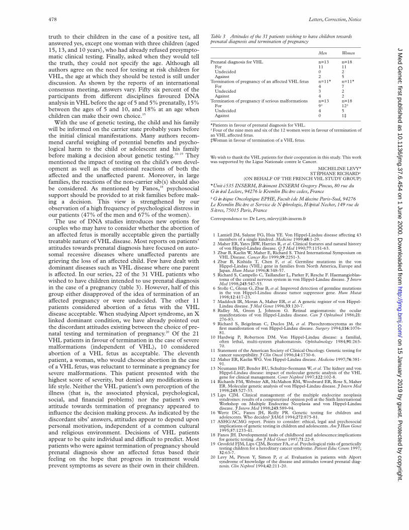
truth to their children in the case of a positive test, allanswered yes, except one woman with three children (aged15, 13, and 10 years), who had already refused presympto-matic clinical testing. Finally, asked when they would tellthe truth, they could not specify the age. Although allauthors agree on the need for testing at risk children forVHL, the age at which they should be tested is still underdiscussion. As shown by the reports of an internationalconsensus meeting, answers vary. Fifty six percent of theparticipants from diVerent disciplines favoured DNAanalysis in VHL before the age of 5 and 5% prenatally, 15%between the ages of 5 and 10, and 18% at an age whenchildren can make their own choice.15
With the use of genetic testing, the child and his familywill be informed on the carrier state probably years beforethe initial clinical manifestations. Many authors recom-mend careful weighing of potential benefits and psycho-logical harm to the child or adolescent and his familybefore making a decision about genetic testing.16–19 Theymentioned the impact of testing on the child’s own devel-opment as well as the emotional reactions of both theaVected and the unaVected parent. Moreover, in largefamilies, the reactions of the non-carrier sib(s) should alsobe considered. As mentioned by Fanos,18 psychosocialsupport should be provided to at risk families before mak-ing a decision. This view is strengthened by ourobservation of a high frequency of psychological distress inour patients (47% of the men and 67% of the women).
The use of DNA studies introduces new options forcouples who may have to consider whether the abortion ofan aVected fetus is morally acceptable given the partiallytreatable nature of VHL disease. Most reports on patients’attitudes towards prenatal diagnosis have focused on auto-somal recessive diseases where unaVected parents aregrieving the loss of an aVected child. Few have dealt withdominant diseases such as VHL disease where one parentis aVected. In our series, 22 of the 31 VHL patients whowished to have children intended to use prenatal diagnosisin the case of a pregnancy (table 3). However, half of thisgroup either disapproved of the idea of termination of anaVected pregnancy or were undecided. The other 11patients considered abortion of a fetus with the VHLdisease acceptable. When studying Alport syndrome, an Xlinked dominant condition, we have already pointed outthe discordant attitudes existing between the choice of pre-natal testing and termination of pregnancy.20 Of the 21VHL patients in favour of termination in the case of severemalformations (independent of VHL), 10 consideredabortion of a VHL fetus as acceptable. The eleventhpatient, a woman, who would choose abortion in the caseof a VHL fetus, was reluctant to terminate a pregnancy forsevere malformations. This patient presented with thehighest score of severity, but denied any modifications inlife style. Neither the VHL patient’s own perception of theillness (that is, the associated physical, psychological,social, and financial problems) nor the patient’s ownattitude towards termination of pregnancy appeared toinfluence the decision making process. As indicated by thediscordant sibs’ answers, attitudes appear to depend uponpersonal motivation, independent of a common culturaland religious environment. Decisions of VHL patientsappear to be quite individual and diYcult to predict. Mostpatients who were against termination of pregnancy shouldprenatal diagnosis show an aVected fetus based theirfeeling on the hope that progress in treatment wouldprevent symptoms as severe as their own in their children.
We wish to thank the VHL patients for their cooperation in this study. This workwas supported by the Ligue Nationale contre le Cancer.
MICHELINE LEVY*STÉPHANE RICHARD†
(ON BEHALF OF THE FRENCH VHL STUDY GROUP)
*Unité 535 INSERM, Bâtiment INSERM Gregory Pincus, 80 rue duGénéral Leclerc, 94276 le Kremlin Bicêtre cedex, France
†Génétique Oncologique EPHE, Faculté de Médecine Paris-Sud, 94276Le Kremlin Bicêtre et Service de Néphrologie, Hôpital Necker, 149 rue deSèvres, 75015 Paris, France
Correspondence to: Dr Levy, [email protected]
1 Lamiell JM, Salazar FG, Hsia YE. Von Hippel-Lindau disease aVecting 43members of a single kindred. Medicine 1989;68:1-29.
2 Maher ER, Yates JRW, Harries R, et al. Clinical features and natural historyof von Hippel-Lindau disease. Q J Med 1990;77:1151-63.
3 Zbar B, Kaelin W, Maher E, Richard S. Third International Symposium onVHL Disease. Cancer Res 1999;59:2251-3.
4 Zbar B, Kishida T, Chen F, et al. Germline mutations in the vonHippel-Lindau (VHL) gene in families from North America, Europe andJapan. Hum Mutat 1996;8:348-57.
5 Richard S, Campello C, Taillandier L, Parker F, Resche F. Haemangioblas-toma of the central nervous system in von Hippel-Lindau disease. J InternMed 1998;243:547-53.
6 Stolle C, Glenn G, Zbar B, et al. Improved detection of germline mutationsin the von Hippel-Lindau disease tumor suppressor gene. Hum Mutat1998;12:417-23.
7 Maddock IR, Moran A, Maher ER, et al. A genetic register of von Hippel-Lindau disease. J Med Genet 1996;33:120-7.
8 Ridley M, Green J, Johnson G. Retinal angiomatosis: the ocularmanifestations of von Hippel-Lindau disease. Can J Ophtalmol 1986;21:276-83.
9 Richard S, Beigelman C, Duclos JM, et al. Pheochromocytoma as thefirst manifestation of von Hippel-Lindau disease. Surgery 1994;116:1076-81.
10 Hardwig P, Robertson DM. Von Hippel-Lindau disease: a familial,often lethal, multi-system phakomatosis. Ophthalmology 1984;91:263-70.
11 Statement of the American Society of Clinical Oncology. Genetic testing forcancer susceptibility. J Clin Oncol 1996;14:1730-6.
12 Maher ER, Kaelin WG. Von Hippel-Lindau disease. Medicine 1997;76:381-91.
13 Neumann HP, Bender BU, Schultze-Seemann W, et al. The kidney and vonHippel-Lindau disease: impact of molecular genetic analysis of the VHLgene for clinical management. Contr Nephrol 1997;122:102-8.
14 Richards FM, Webster AR, McMahon RM, Woodward ER, Rose S, MaherER. Molecular genetic analysis of von Hippel-Lindau disease. J Intern Med1998;243:527-33.
15 Lips CJM. Clinical management of the multiple endocrine neoplasiasyndromes: results of a computerized opinion poll at the Sixth InternationalWorkshop on Multiple Endocrine Neoplasia and von Hippel-Lindaudisease. J Intern Med 1998;243:589-94.
16 Wertz DC, Fanos JH, Reilly PR. Genetic testing for children andadolescents. Who decides? JAMA 1994;272:875-81.
17 ASHG/ACMG report. Points to consider: ethical, legal and psychosocialimplications of genetic testing in children and adolescents. Am J Hum Genet1995;57:1233-41.
18 Fanos JH. Developmental tasks of childhood and adolescence:implicationsfor genetic testing. Am J Med Genet 1997;71:22-8.
19 Grosfeld FJM, Lips CJM, Beemer FA, et al. Psychological risks of geneticallytesting children for a hereditary cancer syndrome. Patient Educ Couns 1997;32:63-7.
20 Levy M, Pirson Y, Simon P, et al. Evaluation in patients with Alportsyndrome of knowledge of the disease and attitudes toward prenatal diag-nosis. Clin Nephrol 1994;42:211-20.
Table 3 Attitudes of the 31 patients wishing to have children towardsprenatal diagnosis and termination of pregnancy
Men Women
Prenatal diagnosis for VHL n=13 n=18For 11 11Undecided 0 2Against 2 5
Termination of pregnancy of an aVected VHL fetus n=11* n=11*For 4 7Undecided 3 2Against 4 2
Termination of pregnancy if serious malformations n=13 n=18For 9† 12†Undecided 4 5Against 0 1‡
*Patients in favour of prenatal diagnosis for VHL.†Four of the nine men and six of the 12 women were in favour of termination ofan VHL aVected fetus.‡Woman in favour of termination of a VHL fetus.
478 Letters, Correction, Notice
on 15 January 2019 by guest. Protected by copyright.
http://jmg.bm
j.com/
J Med G
enet: first published as 10.1136/jmg.37.6.454 on 1 June 2000. D
ownloaded from

CORRECTION
The following is a correction to the paper by Loukola et al (J MedGenet 1999;36:819-22). An algorithm was introduced by Wijnen et al1
to predict the probability of finding a disease causing MLH1 or MSH2mutation in patients with HNPCC or possible HNPCC. The variablesof this formula are the mean age of colorectal cancer (CRC) diagnosisin the family, fulfilment of the so called Amsterdam criteria, and thepresence of endometrial cancer in the family. In addition to this basicformula, an alternative formula to be used in small families was intro-duced. The variables of this alternative formula are the mean age ofCRC diagnosis in the family, the number of patients with CRC in thefamily, and the number of patients with endometrial cancer in thefamily. Applying these algorithms, a probability (p1 for the basicformula and p2 for the alternative formula) of 20% or higher for agermline mutation was proposed to justify mutation analyses. Wetested these formulae in a series of 509 consecutive colorectal adeno-
carcinoma samples.2 MSI analysis and genomic sequencing of MLH1and MSH2 for the MSI positive samples had been previouslyperformed.3
Unfortunately, we used an inaccurate factor in the alternativeformula. We used L=1.4 + (−0.09)V1 + 0.27 V2 + 0.75 V3, when thefirst factor should have been 1.8. Thus, the results obtained using thealternative formula were not accurate. We proposed that the alterna-tive formula was able to identify only three out of 10 mutation positivepatients when first degree pedigrees (on average eight familymembers) were used and six out of 10 when extensive pedigrees (onaverage 39 family members) were used. The correct results are: thealternative formula was able to detect six out of 10 (with first degreepedigrees) and eight out of 10 (with extensive pedigrees) mutationcarriers.
To test the algorithm of Wijnen et al1 further, we have now analysed535 additional consecutive colorectal adenocarcinoma samples. Thesubjects ranged in age from 29 to 91 years with a mean age of 67 years.MSI analysis and genomic sequencing of MLH1 and MSH2 for MSIpositive samples were performed previously (Salovaara et al,submitted); 66 out of 535 (12%) samples were MSI positive and 18 ofthese patients had an MLH1 or MSH2 germline mutation. We then
Table 1 Calculations using the algorithms of Wijnen et al1 to determine the probabilities of finding a germline MLH1 or MSH2 mutation in 28 mutationcarrier probands
Patient
Mean age ofdiagnosis of CRC inthe family
Fulfilment ofAmsterdam criteria
Endometrial cancer inthe family
No of patientswith CRC
No of patients withendometrial cancer
No of family memberswith verified cancerstatus p1 p2
(A) Limited pedigrees (proband and first degree relatives)c35 49 No No 2 0 6 3 11c54 36 No No 1 0 4 10 24c64 53 No No 2 0 5 2 8c125 52 No No 2 0 9 2 9c145 42 Yes Yes 3 1 12 78 40c219 50 No Yes 1 1 7 13 16c340 61 No Yes 3 2 7 5 20c430 38 No No 2 0 4 8 25c558 57 No Yes 4 2 8 7 32c564 45 No Yes 2 1 7 19 28c138 39 No No 1 0 5 8 19c275 55 No No 2 0 9 2 7c587 36 No Yes 2 1 4 37 46c614 50 No Yes 3 2 6 13 40c615 38 No No 2 0 5 8 25c661 54 No Yes 1 1 6 9 12c676 46 No Yes 2 1 7 18 26c698 43 No Yes 1 1 6 22 26c700 61 No No 1 0 5 1 3c811 54 No No 2 0 14 2 7c870 56 No No 2 0 5 1 6c899 50 No No 2 0 6 3 10c952 52 No Yes 2 1 10 11 17c953 45 No Yes 6 3 9 19 83c955 53 No No 2 0 7 2 8c1027 53 No No 1 0 8 2 6c1077 45 Yes No 3 0 8 34 19c1095 53 Yes Yes 3 1 8 55 20
(B) Extended pedigreesc35 47 Yes No 4 0 24 30 21c54 35 No No 1 0 13 11 25c64 53 Yes No 4 0 24 10 16c125 48 Yes No 4 0 14 28 19c145 56 Yes Yes 13 3 85 48 93c219 52 No Yes 7 2 49 11 62c340 56 Yes Yes 5 2 33 48 40c430 44 Yes Yes 9 1 33 75 73c558 57 Yes Yes 5 3 39 45 57c564 45 Yes Yes 12 4 77 73 98c138 38 No Yes 1 1 8 32 35c275 46 Yes Yes 10 4 87 71 97c587 42 No Yes 3 1 14 24 40c614 49 No Yes 3 2 42 14 42c615 40 Yes No 3 0 16 46 27c661 54 No Yes 1 1 24 9 12c676 44 Yes Yes 12 4 74 75 98c698 46 Yes Yes 10 4 87 71 97c700 49 No Yes 13 5 97 14 99c811 48 Yes No 5 0 29 28 24c870 56 No No 2 0 20 1 6c899 50 No No 2 0 8 3 10c952 64 No Yes 3 1 11 3 8c953 44 Yes Yes 12 4 74 75 98c955 47 No No 3 0 25 4 17c1027 43 No Yes 2 1 20 22 31c1077 44 Yes No 7 0 46 37 43c1095 53 Yes Yes 3 1 12 55 20
p1 = probability of finding a germline mutation in MLH1 or MSH2.p2 = probability of finding a germline mutation in MLH1 or MSH2; the alternative formula.
Letters, Correction, Notice 479
on 15 January 2019 by guest. Protected by copyright.
http://jmg.bm
j.com/
J Med G
enet: first published as 10.1136/jmg.37.6.454 on 1 June 2000. D
ownloaded from

combined the results with the previous set of samples and describehere the results from 1044 consecutive colorectal cancer patients. Theresults of all 28 mutation positive samples are presented in table 1.The basic formula identified five out of 28 (18%) mutation carrierswhen first degree pedigrees (on average eight family members) wereused, and 18 out of 28 (64%) when extensive pedigrees (on average 39family members) were used. The corresponding results for the alter-native formula are 13 out of 28 (46%) and 21 out of 28 (75%). Inaddition, these formulae identified 11 patients in whom no MLH1 orMSH2 mutations could be found. They have all been tested and foundnegative for the two most common Finnish founder mutations. One ofthem was found to be a FAP patient and another one had juvenilepolyposis. Three were sequenced for MLH1 and MSH2 mutationswith negative results. The remaining six patients were all MSInegative, diagnosed before the age of 40, and had no family history ofcancer.
Extensive pedigree data are diYcult to obtain in clinical practice.When relying on first degree pedigrees, which are generally easilyobtained during patient interview, the mathematical algorithmsproposed by Wijnen et al1 were able to detect 18% (p1) and 46% (p2)of mutation carriers. Even with extensive pedigree information, theformulae were able to detect only 61% (p1) and 75% (p2) of carriers,meaning that every third or every fourth were missed. Based on theanalysis of 1044 colorectal cancer probands, we conclude that thesemathematical formulae alone are of limited value and use of additionaltools such as MSI screening is warranted.
ANU LOUKOLA*ALBERT DE LA CHAPELLE†
LAURI A AALTONEN**Department of Medical Genetics, PO Box 21 (Haartmaninkatu 3),FIN-00014 University of Helsinki, Helsinki, Finland
†Human Cancer Genetics Program, Comprehensive Cancer Center, TheOhio State University, Medical Research Facility, Room 646, 420 W 12thAvenue, Columbus, Ohio 43210, USA
1 Wijnen JT, Vasen HFA, Khan PM, Zwinderman AH, van der Klift H,Mulder S, Tops C, Moller P, Fodde R. Clinical findings with implicationsfor genetic testing in families with clustering of colorectal cancer. N Engl JMed 1998;339:511-18.
2 Loukola A, de la Chapelle A, Aaltonen L. Strategies to screen for hereditarynonpolyposis colorectal cancer. J Med Genet 1999;36:819-22.
3 Aaltonen LA, Salovaara R, Kristo P, Canzian F, Hemminki A, Peltomäki P,Chadwick RB, Kääriäinen H, Eskelinen M, Järvinen H, Mecklin JP, de laChapelle A. Incidence of hereditary nonpolyposis cancer and the feasibilityof molecular screening for the disease. N Engl J Med 1998;338:1481-7.
NOTICE
The Tenth International Clinical Genetics Seminar
“Genetics in Primary Care” is the main theme of the TenthInternational Clinical Genetics Seminar to be held in Amman, Jordanon 20-24 October 2000. Further information may be obtained fromProfessor Mohammed El-Khateeb, Department of Pathology andMicrobiology, University of Jordan/NCDEG, PO Box 13002, Amman11943, Jordan. Fax: 00 962 6 535 5655. E-mail: [email protected]
480 Letters, Correction, Notice
on 15 January 2019 by guest. Protected by copyright.
http://jmg.bm
j.com/
J Med G
enet: first published as 10.1136/jmg.37.6.454 on 1 June 2000. D
ownloaded from