letter to the editor: 1h, 13c and 15n resonance assignment and secondary structure of mycobacterium...
TRANSCRIPT
Journal of Biomolecular NMR, 19: 89–90, 2001.KLUWER/ESCOM© 2001Kluwer Academic Publishers. Printed in the Netherlands.
89
Letter to the Editor: 1H, 13C and 15N resonance assignment and secondarystructure of Mycobacterium tuberculosisadenylate kinase
Simona Mirona, Helene Munier-Lehmannb & Constantin T. Craescua,∗aInstitut National de la Sant´e et de la Recherche M´edicale U350 & Institut Curie-Recherche, Centre Universitaire,Bâtiments 110–112, F-91405 Orsay, France;bLaboratoire de Chimie Structurale des Macromol´ecules, InstitutPasteur, Paris, France
Received 18 October 2000; Accepted 31 October 2000
Key words:adenylate kinase, resonance assignment, triple resonance NMR
Biological context
Adenylate kinases (AKs) are ubiquitous small en-zymes which catalyze the reversible transfer of theterminal phosphate group from ATP:Mg2+ to AMPand play a key role in the energetic metabolism andnucleic acid synthesis (Noda, 1973). Comparison ofavailable AK sequences has suggested a first classifi-cation in two main forms: a short form, having about190 residues, and a long one composed of 214–238residues. Generally, the eukaryotic cytosolic enzymesare short variants, whereas the bacterial, yeast andmitochondrial AKs contain an additional segment, ex-posed to the solvent and highly flexible. However,several bacterial AKs were shown to have a short se-quence while displaying a weak similarity with thecytosolic AK counterpart. This group, including AKfrom Mycobacterium tuberculosis, was proposed asa new subfamily of short bacterial variants (Munier-Lehmann et al., 1999). Several 3D structures of short(cytosolic) and long variants of adenylate kinases havebeen solved at high resolution by X-ray crystallogra-phy. In contrast, no detailed structural information isavailable for short bacterial variants.
We therefore focused our attention onM. tuber-culosisadenylate kinase (AKmt) with the aim of ob-taining its 3D structure in solution and of analysingthe structure/function relationship. Tuberculosis con-tinues to represent a major public health problem, dueto the emergence of multiple-drug-resistant strains andto coinfection with HIV. As AKmt plays a critical rolein M. tuberculosissurvival (Kohiyama et al., 1966),and displays catalytic properties significantly differ-
∗To whom correspondence should be addressed.
ent from the eukaryotic AKs (Munier-Lehmann et al.,1999), it may be expected that the bacterial enzymemight be a new target for antibacterial drugs.
In this Letter we report almost complete backboneand partial (over 80%) side-chain resonance assign-ment, as well as the secondary structure predicted bythe NOE interactions and chemical shift analysis.
Methods and experiments
Uniformly labeled recombinantM. tuberculosisAK was overproduced inEscherichia coli strainBli5/pHL20 (Munier-Lehmann et al., 1999) usingM9 minimal medium containing 1.5 g/l 99% (15N-ammonium sulfate and 3.0 g/l 99% (13C)-glucose asthe sole nitrogen and carbon source, respectively. Theprotein was purified as described earlier (Bârzu andMichelson, 1983).
NMR samples at a concentration of 1.2 mM(pH 7.1) were obtained by dissolving the lyophilizedprotein in potassium phosphate buffer (50 mM) in 95%1H2O/5% 2H2O or in 100%2H2O. 2D homonuclear,and double- and triple-resonance (HSQC, NOESY-HSQC, TOCSY-HSQC, HNCA and HN(CO)CA)NMR experiments (Wüthrich, 1986; Cavanaghet al., 1996) were performed on a Varian Unity-500 NMR spectrometer. The HCCH-TOCSY andtriple-resonance 3D CBCA(CO)NH, HNCACB wereacquired on a Varian Inova 750 MHz spectrometer(European SON NMR Large-Scale Facility, Utrecht).Proton chemical shifts (in ppm) were referenced rela-tive to internal DSS and15N and13C references wereset indirectly relative to DSS using frequency ratios(Wishart et al., 1995).

90
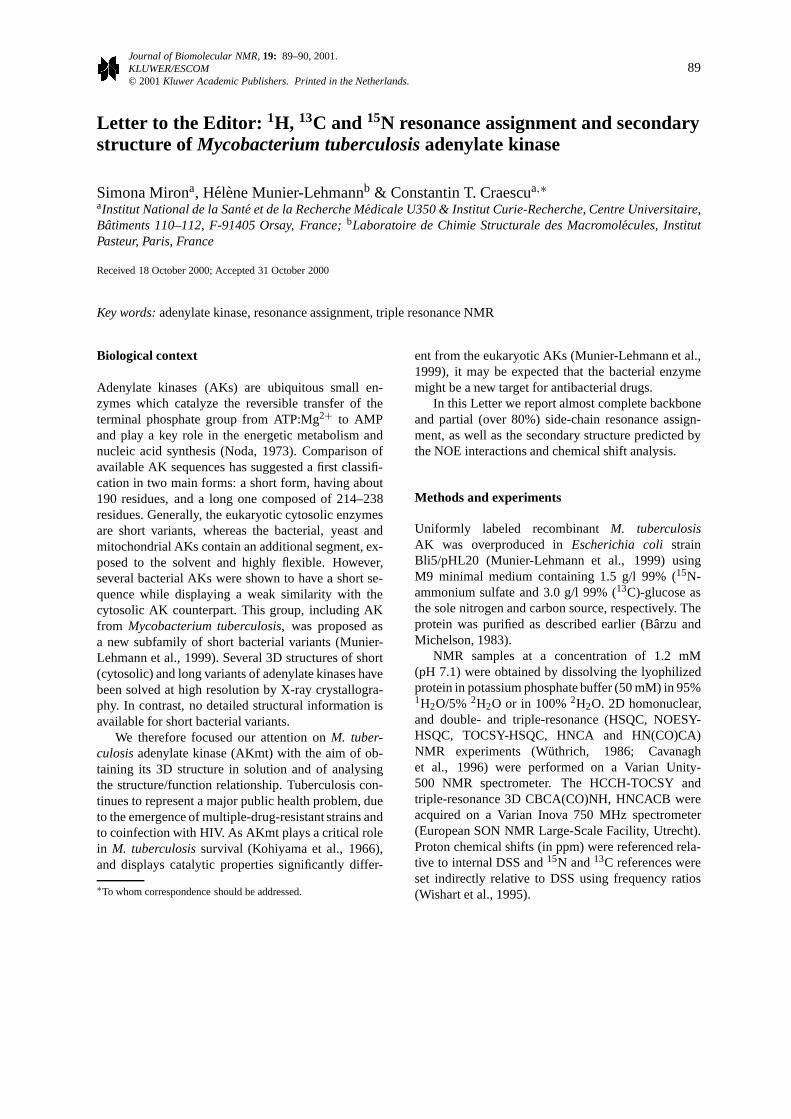
Figure 1. (A) 2D 1H-15N HSQC spectrum ofM. tuberculosisadenylate kinase at 308 K. Assignments of resonances are indicatedusing one-letter codes for amino acids. Pairs of peaks connected byhorizontal lines represent Asn and Gln side chain NH2 groups. (B)The consensus chemical shift index (CSI) plot for AKmt obtainedusing1Hα,13Cα and13Cβ chemical shift values.
The NMR data were processed and analyzed usingFelix98 software (MSI, San Diego, CA), running ona Silicon Graphics Indigo. The chemical shift indices(CSI) were obtained using the CSI software (Wishartand Sykes, 1994).
Extent of assignments and data deposition
The15N-HSQC spectrum (Figure 1A) of AKmt illus-trates the good dispersion of the proton and nitrogenresonances in the amide groups.
1H, 15N, 13Cα backbone resonances and the side-chain 13Cβ resonances of 170 from the 174 non-proline residues as well as the13C resonances of
4 proline residues were assigned. Resonances cor-responding to the segment G12–T15 were not ob-served, neither in the HSQC nor in the triple resonancespectra, and remain unassigned. The correspondingresidues belong to the ATP binding P-loop (G10-T15),a flexible site which is considered to be in interme-diate exchange (on the NMR chemical shift scale)between two or more conformations in the absence ofany bound ligands (Burlacu-Miron et al., 1999).
The secondary structure prediction based on CSI(Figure 1B) and short-range NOEs analysis shows theexistence of eightα-helices and fiveβ-strands. Thelong-range NOEs indicate that these strands consti-tute a single parallelβ-sheet. The topology of theβ-sheet is similar to the other adenylate kinases, andonly differs in theβ5 strand, which is one residueshorter. The chemical shift values of the proton, ni-trogen and carbon resonances have been deposited inthe BioMagResBank (accession number: 4840).
Acknowledgements
This work was supported by the Centre National dela Recherche Scientifique URA 2185, the InstitutNational de la Santé et de la Recherche Médicale,the Institut Curie, the Institut Pasteur and the Eu-ropean Community (TMR program). S.M. gratefullyacknowledges a postdoctoral fellowship from Insti-tut Curie. We thank Rainer Wechselberger for spectraacquisition on the 750 MHz spectrometer.
References
Bârzu, O. and Michelson, S. (1983)FEBS Lett., 153, 280–284.Burlacu-Miron, S., Perrier, V., Gilles, A.-M., Mispelter, J., Bârzu,
O. and Craescu, C.T. (1999)J. Biomol. NMR, 13, 93–94.Cavanagh, J., Fairbrother, W.J., Palmer III, A.G. and Skelton,
N.J. (1996)Protein NMR Spectroscopy. Principles and Practice,Academic Press, San Diego, CA.
Kohiyama, M., Cousin, D., Ryter, A. and Jacob, F. (1966)Ann. Inst.Pasteur, 110, 465–486.
Munier-Lehmann, H., Burlacu-Miron, S., Craescu, C.T., Mantsch,H.H. and Schultz, C.P. (1999)Proteins, 36, 238–248.
Noda, L.H. (1973)Adenylate Kinase, Academic Press, New York,NY.
Wishart, D.S., Bigam, C.G., Yao, J., Abildgaard, F.H., Dyson, J.,Oldfield, E., Markley, J.L. and Sykes, B.D. (1995)J. Biomol.NMR, 6, 135–140.
Wishart, D.S. and Sykes, B.D. (1994)J. Biomol. NMR, 4, 171–180.Wüthrich, K. (1986)NMR of Proteins and Nucleic Acids, Wiley,
New York, NY.