lessons in human biology from a monogenic pancreatic β ... · in patients with breast cancer....
TRANSCRIPT

commentaries
TheJournalofClinicalInvestigation http://www.jci.org Volume 121 Number 10 October 2011 3821
work may well be reduced in favor of mean-ingful, objective data. Just as importantly, these gene expression patterns may help refocus research efforts to understand the pathogenesis of IBD and the mechanisms of treatment resistance.
ImplicationsThe fact that the gene expression signa-tures identified for AAV and SLE (10), which were very similar to those used by Lee and colleagues (9), could be used to divide normal controls into the same two groups that it did the patients with these various diseases indicates that these gene expression signatures stratify something beyond just a propensity to a particular dis-ease. This suggests that these data indicate some preexisting set point for the inflam-matory response that may be independent of any specific disease. We speculate that this non-Gaussian (bimodal) distribution of the population indicates that, in the past, natural selection favored the coor-dinated increase in gene expression for enhanced immunity and that this pattern of gene expression is now associated with more severe disease in IBD, SLE, and AAV vascular-type inflammation and perhaps
other autoimmune diseases. Coordinat-ing future studies with genome-wide sur-veys of genetic polymorphisms in larger populations may help identify the master regulator(s) of this particular gene expres-sion signature; again, the bimodal distri-bution may hint that the number of these master regulators is smaller than expected.
In conclusion, if follow-up studies show that prestratifying IBD patients for differ-ent treatment strategies based on CD8+ T cell gene expression signatures can lead to improved outcomes, management of IBD will take a giant leap forward. Furthermore, the general approach of Lee et al. (9) may become a useful paradigm for managing potentially hazardous immunotherapies for this and other autoimmune diseases.
Address correspondence to: Laurence A. Turka, Transplant Institute, Beth Israel Deaconess Medical Center, 330 Brookline Avenue - E/CLS 607, Boston, Massachusetts 02215, USA. E-mail: [email protected]. Or to: Simon C. Robson, Transplant Institute, Beth Israel Deaconess Medical Center, 330 Brookline Avenue - E/CLS 612, Boston, Massachusetts 02215, USA. E-mail: [email protected].
1. Chang JC, et al. Gene expression profiling for the prediction of therapeutic response to docetaxel in patients with breast cancer. Lancet. 2003; 362(9381):362–369.
2. Tanaka Y, et al. Genome-wide association of IL28B with response to pegylated interferon-alpha and ribavirin therapy for chronic hepatitis C. Nat Genet. 2009;41(10):1105–1109.
3. Podolsky DK. Inflammatory bowel disease. N Engl J Med. 2002;347(6):417–429.
4. Khor B, Gardet A, Xavier RJ. Genetics and pathogen-esis of inflammatory bowel disease. Nature. 2011; 474(7351):307–317.
5. Strober W, Fuss I, Mannon P. The fundamental basis of inflammatory bowel disease. J Clin Invest. 2007; 117(3):514–521.
6. Lees CW, Barrett JC, Parkes M, Satsangi J. New IBD genetics: common pathways with other diseases [published online ahead of print February 7, 2011]. Gut. doi:gut.2009.199679.
7. Lettre G, Rioux JD. Autoimmune diseases: insights from genome-wide association studies. Hum Mol Genet. 2008;17(R2):R116–R121.
8. Baranzini SE. The genetics of autoimmune dis-eases: a networked perspective. Curr Opin Immunol. 2009;21(6):596–605.
9. Lee JC, et al. Gene expression profiling of CD8+ T cells predicts prognosis in patients with Crohn disease and ulcerative colitis. J Clin Invest. 2011; 121(10):4170–4179.
10. McKinney EF, et al. A CD8+ T cell transcription sig-nature predicts prognosis in autoimmune disease. Nat Med. 2010;16(1):586-591.
11. Bonen DK, Cho JH. The genetics of inflammatory bowel disease. Gastroenterology. 2003;124(2):521–536.
12. Stockinger B, Veldhoen M, Martin B. Th17 T cells: linking innate and adaptive immunity. Semin Immunol. 2007;19(6):353–361.
Lessons in human biology from a monogenic pancreatic β cell disease
Benjamin Glaser
Endocrinology and Metabolism Service, Internal Medicine Department, Hadassah-Hebrew University Medical Center, Jerusalem, Israel.
Decipheringthecomplexitiesofhumanβcellphysiologyiscriticaltoourunderstandingofthepathophysiologybehindbothtype1andtype2diabe-tes.Onewaytodothisistostudyindividualswithcongenitalhyperinsulin-ism(CHI),araregeneticdiseasecharacterizedbydysregulationofinsulinsecretionresultinginhypoglycemia.InthisissueoftheJCI,Henquinetal.reportinvitrostudiesofpancreatictissueobtainedfromCHIpatientsdur-ingtherapeuticpancreatectomythathaveyieldedexcitingnewinsightsintohumanβcellphysiology.Thedatavalidateandextendobservationsmadeinmodelorganisms.
β Cell dysfunction lies at the center of both major forms of diabetes, the incidence of which is increasing at an alarming rate in both the developed and the develop-ing world (1). In type 1 diabetes mellitus
(T1DM), autoimmune β cell destruction results in complete insulin deficiency, whereas in type 2 diabetes mellitus (T2DM), subtle defects in β cell functional mass result in progressive disease. Studies using animal models and cell lines from various sources have yielded invaluable information that has allowed us to better understand the processes responsible for β cell func-tion, dysfunction, replication, and survival.
However, since the model systems may not always accurately reflect in vivo human physiology, validation studies in humans are crucial. The crunch comes, however, in that performing such studies is at best diffi-cult and often impossible. Genetic manipu-lation in vivo, a hugely powerful tool in ani-mal-based research, is obviously impossible in humans, and in vitro genetic manipula-tions of primary human tissues are difficult and in themselves fraught with pitfalls.
One way to overcome some of these prob-lems is to identify and study individuals with naturally occurring genetic mutations. These cases are rare and often difficult to study, and the results may be difficult to interpret. With animal models, experiments can be performed in genetically identical animals under controlled conditions. In human studies, however, each patient typi-
Conflictofinterest: The author has declared that no conflict of interest exists.
Citationforthisarticle: J Clin Invest. 2011; 121(10):3821–3825. doi:10.1172/JCI60002.

commentaries
3822 TheJournalofClinicalInvestigation http://www.jci.org Volume 121 Number 10 October 2011
cally has a different mutation and studies are done at different ages, under different experimental circumstances, and after dif-ferent environmental exposures. Patients, or their guardians, frequently do not agree to perform detailed in vivo studies unless they are clinically required, and relevant tis-sues are often not available for study. These issues make such studies infinitely more difficult, yet because of the limitations of alternative approaches, they are absolutely critical to advancing our understanding of human (as opposed to rodent) physiology.
In this issue of the JCI, Henquin and col-leagues describe their attempts to decipher the complexities of human β cell physiology through detailed in vitro physiologic anal-ysis of tissues obtained from 24 patients with congenital hyperinsulinism (CHI) (2), a rare genetic disorder that is essentially the
opposite of diabetes, since it is character-ized by hypoglycemia, not hyperglycemia (3). CHI is no less relevant to β cell biol-ogy than the monogenic forms of diabe-tes, which are extremely difficult to study, since pancreatic tissue from individuals with these conditions is almost never avail-able. As the most comprehensive study to date using pancreatic tissue obtained from patients with genetically defined CHI, the work of Henquin and colleagues represents a milestone in β cell research (2).
CHI: the basicsCHI is a clinically and genetically heteroge-neous disorder typically diagnosed in the newborn period (3). Prior to the discovery of the precise genetic etiology of the most com-mon genetic form of this disease in 1995 (4), the pathophysiology and clinical features
were hotly debated, and the clinical syn-drome was referred to by different descrip-tive names. These included nesidioblastosis, which described the histologic picture that was subsequently shown to be nonspecific (5); β cell dysmaturation syndrome, since some histologic and functional characteris-tics of the disease are reminiscent of the fetal pancreas (6); persistent hyperinsulinism of infancy (PHHI); hyperinsulinemic hypogly-cemia, familial (HHF); and CHI. The latter, used by Henquin and colleagues (2), seems to be most popular, although the OMIM website uses HHF (OMIM 256450).
Once thought to be a single entity, it is now known that CHI can be caused by mutations in at least six different genes (ABCC8, KCNJ11, GCK, GLUD1, HADH, and SLC16A1), each of which affects the β cell differently (Figure 1), resulting in some-
Figure 1Schematic representation of the major genetic etiologies of CHI. Some of the major pathways regulating insulin secretion are shown. The proteins encoded by genes mutated in different forms of CHI are shown in red. Mutations in either ABCC8 (which encodes SUR1) or KCNJ11 (which encodes Kir6.2) result in defective KATP channel activity and continuous membrane depolarization regardless of glucose levels. Activating mutations in glucoki-nase (GCK) result in increased glucose metabolism at low circulating glucose levels, resulting in a decreased threshold for glucose-stimulated insulin secretion and thus inappropriate insulin secretion in the presence of hypoglycemia (18). Glutamate dehydrogenase 1 (GLUD1) mutations result in increased conversion of glutamate to α-ketoglutarate (αKG), thereby increasing ATP production and insulin secretion (19). Solute carrier family 16, member 1 (SLC16A1) promoter mutations result in inappropriate expression of this transporter in β cells, allowing pyruvate and lactate to enter the cell. Pyruvate can then enter the Krebs cycle, thus increasing ATP production and insulin secretion. Patients with mutations in this gene have a unique form of CHI characterized by exercise-induced hypoglycemia (20). Inactivating mutations in hydroxyacyl-CoA dehydrogenase (HADH) also cause CHI and have been shown to do so by regulating insulin secretion in a KATP channel–independent fashion (21) and by preventing HADH-mediated inhibition of GLUD1 in the β cell (22). Glucagon-like peptide 1 (GLP1), glucose-dependent insulinotrophic peptide (GIP), glucagon, somatostatin, and other signals not shown in this simplified diagram regulate critical amplifying pathways downstream from the CHI-related proteins.

commentaries
TheJournalofClinicalInvestigation http://www.jci.org Volume 121 Number 10 October 2011 3823
what divergent phenotypes. In many CHI patients, the etiology is still unknown, sug-gesting that mutations in other genes can cause a similar phenotype.
The most common form of CHI, and the one studied by Henquin and colleagues (2), is caused by inactivating mutations in either of the two genes encoding the subunits of the β cell KATP channel — ABCC8, which encodes sulfonylurea receptor 1 (SUR1), and KCNJ11, which encodes the inward-rectifier potassium channel (Kir6.2). Channel inacti-vation in these patients results in membrane depolarization and opening of voltage-gated calcium channels, which triggers inappro-priate insulin exocytosis. In some patients,
abnormal β cells are localized to a discrete focus consisting of a conglomerate of huge, metabolically active islets, with β cells out-side of this region showing small nuclei and decreased cytoplasm, suggestive of sup-pressed function. The unique genetic etiolo-gy of this focal form of disease was defined in 1996 (7, 8) and consists of a paternally inher-ited recessive KATP gene mutation and somat-ic loss of heterozygosity in a β cell precursor (Figure 2). Such patients can be cured by resection of the lesion. All other genetic mutations in ABCC8 and KCNJ11 result in diffuse forms of CHI, in which all β cells are abnormal. Individuals with this condi-tion often require subtotal pancreatectomy,
which is frequently followed by either persis-tent hypoglycemia or diabetes, depending on the extent of the pancreatectomy and the severity of the mutation.
Functional defects in CHI β cells: prior state of the artThe study by Henquin and colleagues (2), which characterized the in vitro kinetics of insulin secretion by pancreatic fragments obtained at the time of therapeutic pancre-atectomy from six patients with diffuse CHI and 18 patients with focal CHI, expands upon previous in vivo and in vitro findings in patients with CHI. Although multiple studies have described the electrophysiolog-ical characteristics of affected β cells (9, 10), only a few have examined in vitro insulin secretion in response to secretagogues, and all of these were of limited scope due to scar-city of tissue (11–13). Aynsley-Green dem-onstrated some increase in insulin secretion by β cells from CHI patients when glucose levels were increased from 0 to 4 mmol, but no further increase at higher concentra-tions (11). About a decade later, Kaiser and colleagues used static incubations to study pancreatic tissue from five patients with CHI, demonstrating that while stimulators of cAMP production and modulators of the PI3K/PKC pathway stimulated insulin release, glucose did not (12). Secretion was shown to be calcium dependent and partial-ly suppressed by epinephrine or somatosta-tin. Although this study was performed before the discovery of the genes encoding the KATP channel subunits, one patient was subsequently found to be homozygous for the ABCC8 p.R836* mutation, while three others, all from the same large Arab fam-ily, were shown to be homozygous for the ABCC8 c.950delC mutation. Subsequently, Otonkoski and colleagues reported poor responsiveness to glucose and partial sup-pression with somatostatin in pancreases from six patients with CHI, using human fetal islets for comparison (13).
Functional defects in CHI β cells: novel findingsAll previous in vitro studies of CHI-derived pancreatic islets lacked optimal controls, since age-matched normal human islets are not available. Henquin and colleagues solved this problem by using β cells isolated from outside focal lesions as controls (2). Although β cells outside the focal lesion are functionally suppressed in vivo due to increased insulin secretion and hypoglyce-mia, being heterozygous for recessive muta-
Figure 2Schematic representation of the molecular defect resulting in focal CHI. On the left is shown a schematic representation of the paternal and maternal copies of the distal short arm of chro-mosome 11, inherited by the fetus at risk for focal CHI and present in all cells except those within the focal lesion. A single ABCC8 or KCNJ11 recessive mutation is inherited on the paternal allele (red dot). Distal to the ABCC8/KCNJ11 locus, there are two imprinted regions. The relevant genes are shown, and the expressed allele for each is shown in light blue. During fetal development, a somatic mutation occurs in a β cell precursor, which results in loss of the maternally inherited allele and duplication of the paternally inherited allele, as depicted on the right. In this cell and all of its progeny, both ABCC8 or KCNJ11 alleles are mutated, resulting in insulin hypersecretion. In addition, two copies of growth-promoting genes such as IGF2 are expressed, while growth-inhibiting genes such as H19 and CDKN1C are not expressed at all. The result is a proliferating lesion consisting of β cells lacking a functioning KATP channel and thus hypersecreting insulin at low glucose levels.
commentaries
3824 TheJournalofClinicalInvestigation http://www.jci.org Volume 121 Number 10 October 2011
tions, they are expected to function normally once outside the patient. Indeed, Henquin and colleagues show that a short period of incubation in stimulatory glucose concen-trations is sufficient to restore their respon-siveness (2). That being said, the authors are correct in advising caution, since this recov-ery may not be complete. Using these con-trol islets and a sensitive perifusion system that allowed detailed analysis of secretory dynamics, Henquin and colleagues demon-strated conclusively that CHI islets hyperse-crete insulin even at very low glucose levels. Additional findings, such as minimal or no insulin response to glucose or the KATP chan-nel blocker tolbutamide, marked response to stimulators of cAMP, and variable responses to different amino acids were consistent with previous reports and current models of the control of insulin secretion. The finding that β cells from both patients with focal CHI and those with diffuse CHI respond similarly demonstrates that, although the imprinted genes in the distal arm of chromosome 11 may affect β cell replication (14), they do not affect insulin secretion. This finding is novel, albeit expected, since focal CHI and diffuse CHI are clinically similar (15).
In addition to the aforementioned find-ings, which might have been predicted from previous work, Henquin and colleagues made some unexpected observations (2). Acute elevation of glucose from 1 to 15 mmol/l resulted in transiently increased insulin secretion in some CHI islet preparations. This was unexpected, since this acute insulin response to glucose is thought to be mediat-ed through the KATP channel. Also unexpect-edly, insulin secretion decreased during high glucose infusion in some studies, primarily those in which forskolin, which raises cAMP levels, was administered simultaneously. That the transient peak insulin secretion is not mediated through the KATP channel is not entirely unexpected, since some children with CHI do show a brief increase in insu-lin secretion following glucose stimulation (16). This could be due to further membrane depolarization; however, this seems unlikely since electrophysiological studies in similar patients do not show a glucose effect on β cell calcium currents (9). Alternatively, glu-cose may be transiently acting downstream of membrane depolarization, stimulating amplifying pathways. The decrease in insulin secretion during perifusion with 15 mmol glucose is also unexpected, since con-tinuous exposure to high glucose lev-els is expected to result in continuously increased insulin secretion, mediated
largely by KATP channel–independent path-ways. This surprising observation is not explained by Henquin and colleagues (2), but may reflect idiosyncrasies of the experi-mental system, since a similar decrease was less evident in glucose ramp experiments or in the absence of forskolin.
Another unexpected finding was the paradoxical response to diazoxide seen in some, but not all, patients. As a KATP chan-nel activator, diazoxide would be expected to be without effect in patients with either ABCC8 or KCNJ11 mutations, as these mutations were thought to either result in permanently closed KATP channels or to entirely prevent protein expression and/or insertion into the plasma membrane. Interestingly, Henquin and colleagues saw a response to diazoxide in one of three patients with mutations that introduced stop codons before the region of ABCC8 that encodes the first nucleotide-binding domain (2), far proximal to the putative potassium channel opener binding site (17). This suggests that the paradoxical response to diazoxide is likely to be an off-target effect. A similar paradoxical response was seen with pinacidil, a KATP channel opener with different channel specific-ity. While this speaks against an off-target effect, it does not entirely eliminate this possibility. Unfortunately, future detailed studies in human CHI β cells are unlikely, given how difficult it is to obtain appropri-ate samples. Although, as Henquin and col-leagues state (2), this paradoxical response has not been reported in animal models of CHI, this can be tested directly using iden-tical experimental protocols. Furthermore, it is not known whether a similar response is present in vivo, since acute response stud-ies have not been reported in CHI patients with KATP channel mutations.
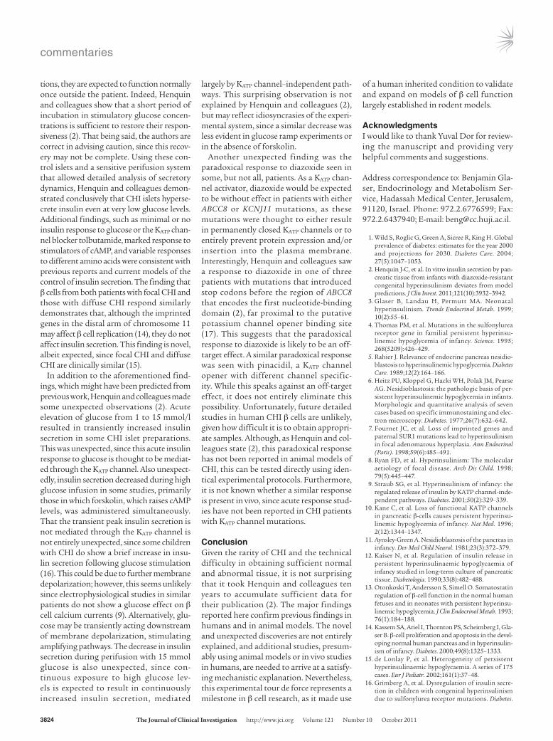
ConclusionGiven the rarity of CHI and the technical difficulty in obtaining sufficient normal and abnormal tissue, it is not surprising that it took Henquin and colleagues ten years to accumulate sufficient data for their publication (2). The major findings reported here confirm previous findings in humans and in animal models. The novel and unexpected discoveries are not entirely explained, and additional studies, presum-ably using animal models or in vivo studies in humans, are needed to arrive at a satisfy-ing mechanistic explanation. Nevertheless, this experimental tour de force represents a milestone in β cell research, as it made use
of a human inherited condition to validate and expand on models of β cell function largely established in rodent models.
AcknowledgmentsI would like to thank Yuval Dor for review-ing the manuscript and providing very helpful comments and suggestions.
Address correspondence to: Benjamin Gla-ser, Endocrinology and Metabolism Ser-vice, Hadassah Medical Center, Jerusalem, 91120, Israel. Phone: 972.2.6776599; Fax: 972.2.6437940; E-mail: [email protected].
1. Wild S, Roglic G, Green A, Sicree R, King H. Global prevalence of diabetes: estimates for the year 2000 and projections for 2030. Diabetes Care. 2004; 27(5):1047–1053.
2. Henquin J-C, et al. In vitro insulin secretion by pan-creatic tissue from infants with diazoxide-resistant congenital hyperinsulinism deviates from model predictions. J Clin Invest. 2011;121(10):3932–3942.
3. Glaser B, Landau H, Permutt MA. Neonatal hyperinsulinism. Trends Endocrinol Metab. 1999; 10(2):55–61.
4. Thomas PM, et al. Mutations in the sulfonylurea receptor gene in familial persistent hyperinsu-linemic hypoglycemia of infancy. Science. 1995; 268(5209):426–429.
5. Rahier J. Relevance of endocrine pancreas nesidio-blastosis to hyperinsulinemic hypoglycemia. Diabetes Care. 1989;12(2):164–166.
6. Heitz PU, Kloppel G, Hacki WH, Polak JM, Pearse AG. Nesidioblastosis: the pathologic basis of per-sistent hyperinsulinemic hypoglycemia in infants. Morphologic and quantitative analysis of seven cases based on specific immunostaining and elec-tron microscopy. Diabetes. 1977;26(7):632–642.
7. Fournet JC, et al. Loss of imprinted genes and paternal SUR1 mutations lead to hyperinsulinism in focal adenomatous hyperplasia. Ann Endocrinol (Paris). 1998;59(6):485–491.
8. Ryan FD, et al. Hyperinsulinism: The molecular aetiology of focal disease. Arch Dis Child. 1998; 79(5):445–447.
9. Straub SG, et al. Hyperinsulinism of infancy: the regulated release of insulin by KATP channel-inde-pendent pathways. Diabetes. 2001;50(2):329–339.
10. Kane C, et al. Loss of functional KATP channels in pancreatic β-cells causes persistent hyperinsu-linemic hypoglycemia of infancy. Nat Med. 1996; 2(12):1344–1347.
11. Aynsley-Green A. Nesidioblastosis of the pancreas in infancy. Dev Med Child Neurol. 1981;23(3):372–379.
12. Kaiser N, et al. Regulation of insulin release in persistent hyperinsulinaemic hypoglycaemia of infancy studied in long-term culture of pancreatic tissue. Diabetologia. 1990;33(8):482–488.
13. Otonkoski T, Andersson S, Simell O. Somatostatin regulation of β-cell function in the normal human fetuses and in neonates with persistent hyperinsu-linemic hypoglycemia. J Clin Endocrinol Metab. 1993; 76(1):184–188.
14. Kassem SA, Ariel I, Thornton PS, Scheimberg I, Gla-ser B. β-cell proliferation and apoptosis in the devel-oping normal human pancreas and in hyperinsulin-ism of infancy. Diabetes. 2000;49(8):1325–1333.
15. de Lonlay P, et al. Heterogeneity of persistent hyperinsulinaemic hypoglycaemia. A series of 175 cases. Eur J Pediatr. 2002;161(1):37–48.
16. Grimberg A, et al. Dysregulation of insulin secre-tion in children with congenital hyperinsulinism due to sulfonylurea receptor mutations. Diabetes.
commentaries
TheJournalofClinicalInvestigation http://www.jci.org Volume 121 Number 10 October 2011 3825
2001;50(2):322–328. 17. Uhde I, Toman A, Gross I, Schwanstecher C,
Schwanstecher M. Identification of the potassium channel opener site on sulfonylurea receptors. J Biol Chem. 1999;274(40):28079–28082.
18. Glaser B, et al. Familial hyperinsulinism caused by an activating glucokinase mutation. N Engl J Med. 1998; 338(4):226–230.
19. Stanley CA, et al. Hyperinsulinism and hyperammo-nemia in infants with regulatory mutations of the glutamate dehydrogenase gene. N Engl J Med. 1998; 338(19):1352–1357.
20. Otonkoski T, et al. Physical exercise-induced hypo-glycemia caused by failed silencing of monocarbox-ylate transporter 1 in pancreatic β cells. Am J Hum Genet. 2007;81(3):467–474.
21. Hardy OT, et al. Functional genomics of the β-cell: short-chain 3-hydroxyacyl-coenzyme A dehydroge-nase regulates insulin secretion independent of K+ currents. Mol Endocrinol. 2007;21(3):765–773.
22. Li C, et al. Mechanism of hyperinsulinism in short-chain 3-hydroxyacyl-CoA dehydrogenase deficiency involves activation of glutamate dehydrogenase. J Biol Chem. 2010;285(41):31806–31818.
ApoE controls the interface linking lipids and inflammation in atherosclerosis
Christian Weber1,2 and Oliver Soehnlein1
1Institute for Cardiovascular Prevention, Ludwig-Maximilians-University Munich, Munich, Germany. 2Munich Heart Alliance, Munich, Germany.
Atherosclerosisisachronicinflammatorydiseaseofthearterialwallsthatoftenleadstomyocardialinfarctionand/orstroke.Hypercholesterolemiaandanimbalanceofperipheralleukocytecounts,leadingtoarterialleuko-cyteinfiltration,areconsideredindependentriskfactorsforatherosclerosis.However,inthisissueoftheJCI,Murphyandcolleaguesidentifyamecha-nisticlinkbetweenhypercholesterolemia,leukocytosis,andthesubsequentdevelopmentofatheroscleroticlesionsinmice.Thesefindingscouldpavethewayforthedevelopmentofnoveltreatmentstrategiestocontrolleuko-cytehomeostasisandatherosclerosis.
Atherosclerosis is a chronic inflammatory disease of the arteries that increasingly threatens human health worldwide as a result of its debilitating, and sometimes life-threatening, consequences, which include limb ischemia, myocardial infarction, and stroke. Identification of novel therapeutic targets is needed to put the brakes on the global increase in the number of cases of atherosclerosis-related diseases.
Two major factors contributing to the pathophysiology of atherosclerosis are hyper-lipidemia and inflammation (1). Hyperlipid-emia, in particular hypercholesterolemia, is regarded as an independent risk factor in the development of ischemic heart disease, including myocardial infarction (1, 2). Epi-demiologic studies have established a strong correlation between elevated total cholester-ol levels in serum and morbidity and mortal-ity from myocardial infarction (2). Elevated numbers of circulating neutrophils and monocytes have also been shown to be pre-dictive of cardiovascular events independent of serum cholesterol levels (3, 4). Consistent with this, recent data from mouse models has provided evidence of a direct correla-tion between the number of circulating and
lesional neutrophils or monocytes and ath-erosclerotic lesion size (5, 6).
In this issue of the JCI, Murphy and col-leagues have identified ApoE on hematopoi-etic stem and multipotential progenitor cells (HSPCs) as part of an ABC transporter–medi-ated cholesterol efflux pathway and suppres-sor of proliferation, providing a mechanistic link between hypercholesterolemia, leuko-cytosis, and the subsequent development of atherosclerotic lesions in mice (ref. 7 and Figure 1). These data have the potential to open the door to the development of novel options for treating atherosclerosis.
ApoE controls HSPC proliferation and atherosclerosis developmentThe most commonly used mouse models of atherosclerosis involve feeding a high-fat diet to mice lacking either ApoE or LDLR. Mice lacking ApoA1 (a major component of HDL) do not develop atherosclerosis, but do mani-fest hypercholesterolemia. Murphy and col-leagues report that Apoe–/– mice fed a high-fat diet responded with pronounced prolifera-tion of HSPCs, neutrophilia, and monocyto-sis, whereas Ldlr–/– and Apoa1–/– mice exhib-ited modest increases in these parameters, suggestive of a specific role for ApoE in sup-pressing HSPC proliferation (7).
Subsequent analyses showed that ApoE was expressed on the surface of HSPCs and that its expression was further upregulated
after treatment with liver X receptor (LXR) activators (7), which have previously been shown to reduce both HSPC proliferation and atherosclerotic lesion formation (8, 9). Moreover, Murphy and colleagues deter-mined that HSPCs themselves secreted ApoE, which became anchored to proteo-glycans on the cell surface (7). There it was shown to interact with ABCA1, which trans-ports excessive cholesterol from membranes to nascent HDL particles, and ABCG1, which transports cholesterol to mature HDL particles, thereby controlling intracellular cholesterol levels. The authors then showed that in HSPCs lacking ApoE, cholesterol efflux pathways were disrupted and choles-terol accumulated intracellularly, resulting in increased responsiveness to the hemato-poietic growth factors IL-3 and GM-CSF (Figure 1). The net effect was an increase in HSPC proliferation, neutrophilia, and monocytosis, with accelerated atherosclero-sis. Thus, a proteoglycan-bound reservoir of ApoE on HSPCs controls their proliferation and circulating leukocyte numbers.
In a therapeutic experiment, Murphy and colleagues injected Apoe–/– mice with choles-terol-poor phospholipid/ApoA-I complexes as cholesterol acceptors, thereby restoring normal HSPC proliferation and reducing the expansion of circulating neutrophils and monocytes (7). All the data generated by Murphy and colleagues provide convinc-ing evidence of a mechanistic link between hypercholesterolemia and leukocytosis, and indicate that this contributes to the devel-opment of atherosclerotic lesions in mice.
Keeping the ill alliance of lipids and leukocytosis in checkTo fully appreciate the importance of the data provided in this study (7) and to be able to translate these data into the devel-
Conflictofinterest: Christian Weber and Oliver Soehnlein are stakeholders of Carolus Therapeutics Inc.
Citationforthisarticle: J Clin Invest. 2011; 121(10):3825–3827. doi:10.1172/JCI60457.