leptodeira frenata micrurus diastema leptodeira...
TRANSCRIPT

929 929
During ongoing nocturnal snake surveys in the Chetumal area of southern Quintana Roo, Mexico, the authors have recorded 31 species of snakes. The species treated in the following article (Leptodeira frenata, Ninia sebae, and Micrurus diastema) have been some of the most commonly encountered snakes in this study. The road-killed specimens are collected and later dissected to generate data on the diet and reproduction of the species. Pictured here is an adult female of Leptodeira frenata from between Calderitas and Ruinas de Oxtankah, Quintana Roo, Mexico (not collected), a nocturnal, arboreal snake with a diet consisting mostly of anurans and lizards.
' © Pablo M. Beutelspacher-García

The Chetumal Snake Census: generating biological data from road-killed snakes. Part 3. Leptodeira frenata,
Ninia sebae, and Micrurus diastema
Gunther Köhler1, J. roGelio Cedeño-Vázquez2, Manuela Spaeth1,
and pablo M. beutelSpaCher-GarCía3
1Senckenberg Forschungsinstitut und Naturmuseum, Senckenberganlage 25, 60325 Frankfurt am Main, Germany. E-mail: [email protected] (Corresponding author) 2Depto. Sistemática y Ecología Acuática, Grupo Académico: Sistemática, Ecología y Manejo de Recursos Acuáticos, El Colegio de la Frontera Sur, Unidad Chetumal, Av. Centenario Km. 5.5, C.P. 77014 Chetumal, Quintana Roo, Mexico. E-mail: [email protected] 342, Fracc. Caribe, C.P. 77086 Chetumal, Quintana Roo, Mexico. E-mail: [email protected]
abStraCt: We present data and observations on the snake species Leptodeira frenata, Ninia sebae, and Micrurus diastema collected during bimonthly surveys along a 39 km road transect near the city of Chetumal, Quintana Roo, Mexico, since February of 2010. For these species, we present data on their external morphology, seasonality, spatial distribution, reproduction, and diet.
Key Words: Dipsadidae, diet, Elapidae, Mexico, monitoring, population dynamics, Quintana Roo, repro-duction, road-kills, snake survey
reSuMen: Presentamos datos y observaciones sobre las especies de serpientes Leptodeira frenata, Ninia sebae y Micrurus diastema registradas durante muestreos realizados cada 15 días a lo largo de un tran-secto de carretera de 39 km cercano a la ciudad de Chetumal (Quintana Roo, Mexico) desde febrero de 2010. De cada una de estas especies presentamos datos sobre morfología externa, estacionalidad, distribu-ción espacial, reproducción y dieta.
Palabras Claves: Dipsadidae, dieta, Elapidae, Mexico, mortalidad por atropello, muestreo y monitoreo de serpientes, dinámica poblacional, Quintana Roo, reproducción
Citation: Köhler, G., J. R Cedeño-Vázquez, M. Spaeth, and P. M. Beutelspacher-García. 2016. The Chetumal Snake Census: generating biological data from road-killed snakes. Part 3. Leptodeira frenata, Ninia sebae, and Micrurus diastema. Mesoamerican Herpetology 3: 930–947.
Copyright: Köhler et al. 2016. This work is licensed under a Creative Commons Attribution-NoDerivatives 4.0 International License.
Received: 23 November 2016; Accepted: 5 December 2016; Published: 31 December 2016.
www.mesoamericanherpetology.com www.eaglemountainpublishing.com
930 Mesoamerican Herpetology December 2016 | Volume 3 | Number 4

Köhler et al. Chetumal Snake Census, Part 3
931 Mesoamerican Herpetology December 2016 | Volume 3 | Number 4
INTRODUCTION
In a previous article (Köhler et al., 2016), we introduced our long-term snake survey study based on snakes found along a 39 km road transect in southern Quintana Roo, Mexico. Here we report the data for three species of snakes (Leptodeira frenata, Ninia sebae, and Micrurus diastema), generated from road-killed specimens and supplemented by observations of living individuals found on the road during our nocturnal surveys. Since 13 February 2010, we recorded 49 specimens of Leptodeira frenata, 49 of Ninia sebae, and 25 of Micrurus diastema (see Appendix 1 for list of specimens collected). For measurements, we use the abbreviations SVL (snout–vent length) and TL (tail length).
SPECIES ACCOUNTS
Leptodeira frenata (Cope 1886)
Figs. 1–2
Material: We collected 49 specimens of Leptodeira frenata (Fig. 1), of which we identified 24 as males, 22 as females, and 3 as indeterminate.
External morphology: See Table 1 for variation in selected morphometric and scalation characters.
Table 1. Selected measurements, proportions, and scale characters of Leptodeira frenata, Ninia sebae, and Micrurus dias-tema. Range is followed by mean value and standard deviation in parentheses. See text for abbreviations.
Character
Leptodeira frenata 16
17
Ninia sebae 14
14
Micrurus diastema 13
7
SVL (mm) Males 181–452 (345.00 ± 88.10) 174–265 (228.00 ± 24.00) 357–669 (505.00 ± 108.00)
Females 237–512 (367.00 ± 84.00) 189–304 (254.00 ± 35.40) 389–690 (553.00 ± 120.00)
TL / SVL Males 0.260–0.368 (0.301 ± 0.029) 0.237–0.351 (0.287 ± 0.023) 0.159–0.249 (0.187 ± 0.024)
Females 0.247–0.336 (0.290 ± 0.029) 0.215–0.249 (0.230 ± 0.012) 0.124–0.129 (0.126 ± 0.002)
Ventrals Males 164–186 (177.10 ± 5.75) 130–142 (137.80 ± 3.28) 197–213 (205.3 ± 5.4)
Females 170–187 (179.30 ± 4.56) 133–158 (143.30 ± 6.68) 216–229 (221.5 ± 5.8)
Subcaudals Males 65–79 (74.00 ± 4.71) 49–60 (53.60 ± 2.87) 52–65 (54.9 ± 4.1)
Females 64–78 (72.10 ± 4.83) 40–49 (44.90 ± 2.30) 40–46 (42.8 ± 2.5)
Number of dorsal scales rows at midbody
17-21 (20.10 ± 1.38) 19 (19.0 ± 0.0) 15 (15.0 ± 0.0)
Number of dorsal scales rows anterior to vent
15-19 (16.20 ± 1.40) 19 (19.0 ± 0.0) 15 (15.0 ± 0.0)
Cloacal scute divided single divided
Number of loreal scales 1 (1.0 ± 0.0) 1 (1.0 ± 0.0) 0 (0.0 ± 0.0)
Number of preocular scales 1 (1.0 ± 0.0) 0 (0.0 ± 0.0) 1 (1.0 ± 0.0)
Number of postocular scales 2 (2.0 ± 0.0) 2 (2.0 ± 0.0) 2 (2.0 ± 0.0)
Number of anterior temporals 1 (1.0 ± 0.0) 1 (1.0 ± 0.0) 1 (1.0 ± 0.0)
Number of posterior temporals 2 (2.0 ± 0.0) 2 (2.0 ± 0.0) 2 (2.0 ± 0.0)
Number of supralabials 8 (8.0 ± 0.0) 7 (7.0 ± 0.0) 7 (7.0 ± 0.0)
Number of infralabials 9–10 (9.90 ± 0.26) 6–7 (6.2 ± 2.3) 7 (7.0 ± 0.0)

932 Mesoamerican Herpetology
Köhler et al. Chetumal Snake Census, Part 3
December 2016 | Volume 3 | Number 4
Diet: Three of the preserved specimens of Leptodeira frenata contained identifiable intestinal contents. SMF 100553 contained the remains (hind feet and part of tail, SMF 101254) of a Sceloporus, presumably S. chrysostic-tus. In the stomach of SMF 100552 we found the hind legs of a subadult Incilius valliceps (SMF 101256), and in the stomach of SMF 100556 we detected a partly digested subadult of Smilisca baudinii (SMF 101255).
Fig. 1. Leptodeira frenata in life. (A) An adult (ECO-CH-H 3738); and (B) a juvenile (not collected).
' © Rogelio Cedeño-Vázquez (A), Gunther Köhler (B)

933 Mesoamerican Herpetology December 2016 | Volume 3 | Number 4
Köhler et al. Chetumal Snake Census, Part 3
Fig. 2. Specimens of Leptodeira frenata dissected to study their gonads (A, B) as well as (C) a female (not collected) that produced a clutch on 7 June 2013. (A) A male (SMF 100555); and (B) a female (ECO-CH-H 3738). The testes and follicles, respectively, are indicated by arrows. Scale bar = 5 mm. ' © Gunther Köhler (A), Nidia Gabriela Blanco Campos (B), and Pablo M. Beutelspacher-García (C)
934 Mesoamerican Herpetology December 2016 | Volume 3 | Number 4
Köhler et al. Chetumal Snake Census, Part 3
Reproduction: The dissection of 3 male and 2 female specimens of Leptodeira frenata yielded data on reproduction (also see Fig. 2A,B). The relative testis size (ratio of testis length × width/SVL) in the 3 males was 0.050–0.110 (0.081 ± 0.026). We found the largest relative testis size (0.110) in a specimen collected in March, and the one with the smallest value (0.050) in a specimen preserved in September. The relative ovary size (ratio of ovary length × width/SVL) in the two females was 0.056 (SMF 100554, collected in November) and 0.054 (SMF 100558, collected in April). SMF 100554 contained four vitellogenic follicles per side, with the largest follicles measuring 2.6–3.1 mm × 1.1–1.4 mm. None of our specimens contained oviducal eggs. On 30 August 2016, we collected a female Leptodeira frenata (SVL 604 mm; ECO-CH-H 3738) that we encountered outside of our road transect, but in same the general area (Chetumal, 18.49964°N, -88.33601°W, elev. 17 m). It contained seven large vitellogenic follicles (Fig. 2B), with a size of 10.2–12.7 (11.0 ± 0.79) mm × 4.9–6.3 (5.5 ± 0.37) mm. These dissection results are supplemented by data on clutch size obtained from three gravid females that PGB found alive in the study area and later laid eggs in captivity. The clutches contained 2 eggs (14 February 2008), 4 eggs (5 January 2011), and 7 eggs (7 June 2013), respectively (see also Fig. 2C).
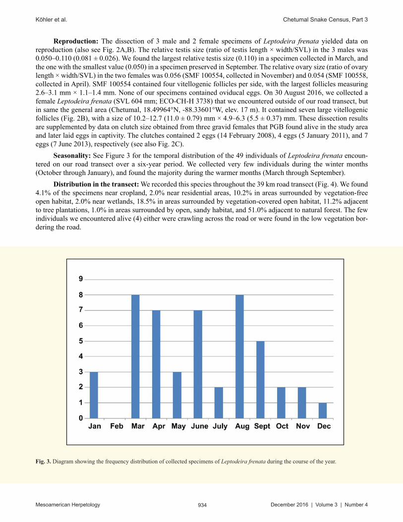
Seasonality: See Figure 3 for the temporal distribution of the 49 individuals of Leptodeira frenata encoun-tered on our road transect over a six-year period. We collected very few individuals during the winter months (October through January), and found the majority during the warmer months (March through September).
Distribution in the transect: We recorded this species throughout the 39 km road transect (Fig. 4). We found 4.1% of the specimens near cropland, 2.0% near residential areas, 10.2% in areas surrounded by vegetation-free open habitat, 2.0% near wetlands, 18.5% in areas surrounded by vegetation-covered open habitat, 11.2% adjacent to tree plantations, 1.0% in areas surrounded by open, sandy habitat, and 51.0% adjacent to natural forest. The few individuals we encountered alive (4) either were crawling across the road or were found in the low vegetation bor-dering the road.
Fig. 3. Diagram showing the frequency distribution of collected specimens of Leptodeira frenata during the course of the year.

935 Mesoamerican Herpetology December 2016 | Volume 3 | Number 4
Köhler et al. Chetumal Snake Census, Part 3
Fig. 4. Spatial distribution of the collected specimens (black dots with white centers) of Leptodeira frenata along the transect.

936 Mesoamerican Herpetology December 2016 | Volume 3 | Number 4
Köhler et al. Chetumal Snake Census, Part 3
Ninia sebae (Duméril, Bibron, & Duméril 1854)
Figs. 5–6
Material: We collected 50 specimens of Ninia sebae (Fig 5), of which we identified 27 as males, 18 as fe-males, and 5 as indeterminate.
External morphology: See Table 1 for variation in selected morphometric and scalation characters.
Fig. 5. Ninia sebae in life. (A) SMF 99640; and (B) an adult from near Chetumal (not collected). Both specimens exhibited the cobra-like defense behavior typical of this species. ' © Gunther Köhler (A) and J. Rogelio Cedeño-Vázquez (B)

937 Mesoamerican Herpetology December 2016 | Volume 3 | Number 4
Köhler et al. Chetumal Snake Census, Part 3
Diet: Several specimens of Ninia sebae (i.e., ECO-CH-H 3108, 3494, SMF 99639–40) contained earthworms or their remains in their gastrointestinal tract whereas undigested parts of arthropods were detected in quite a few individuals (i.e., ECO-CH-H 3192, 3494, 3606, SMF 99640–41, 100585); and in the gut of ECO-CH-H 3108 we also identified the remains of a snail shell.
Reproduction: The dissection of 13 male and 6 female specimens of Ninia sebae yielded data on reproduc-tion (also see Fig. 6). The relative testis size (ratio of testis length × width/SVL) in the 12 males was 0.039–0.231 (0.100 ± 0.045). We found the largest relative testis size (0.231) in a specimen collected in October, and the one with the smallest value (0.039) in a specimen preserved in June. The relative ovary size (ratio of ovary length × width/SVL) in the 6 females was 0.059–0.180 (0.107 ± 0.045). We detected the largest relative ovary size (0.180) in a female collected in October, and the lowest value (0.059) in a female collected in September. The number of vitellogenic follicles per side ranged from 4 to 10 (6.2 ± 1.94). The follicle length was 1.5–4.7 (2.66 ± 0.99), and the follicle width 0.57–2.01 (1.18 ± 0.38). We counted the highest and the lowest number of follicles, respectively, in females collected in October, and found the largest and the smallest follicles, respectively, in females collected in July. None of our specimens contained oviducal eggs.
Fig. 6. Specimens of Ninia sebae dissected to study their gonads. (A) A male (SMF 99640); and (B) a female (ECO-CH-H 2924). The testes and ovaries, respectively, are indicated by arrows. Scale bar = 5 mm. ' © Gunther Köhler

938 Mesoamerican Herpetology December 2016 | Volume 3 | Number 4
Köhler et al. Chetumal Snake Census, Part 3
Seasonality: See Figure 7 for the temporal distribution of the 50 individuals of Ninia sebae encountered on our road transect over a six-year period. The months with the highest numbers of collected individuals were during the winter (October, November, January), which is in contrast to the pattern observed in most other species we collected in this study.
Distribution in the transect: We recorded this species throughout the 39 km road transect (Fig. 8). We found 3.1% of the specimens near cropland, 6.2% near residential areas, 7.8% in areas surrounded by vegetation-free open habitat, 1.6% near wetlands, 17.0% in areas surrounded by vegetation-covered open habitat, 17.0% adjacent to tree plantations, 2.3% in areas surrounded by open, sandy habitat, and 45.0% adjacent to natural forest. The few individuals we encountered alive (10) were crawling across the road.
Fig. 7. Diagram showing the frequency distribution of collected specimens of Ninia sebae during the course of the year.

939 Mesoamerican Herpetology December 2016 | Volume 3 | Number 4
Köhler et al. Chetumal Snake Census, Part 3
Fig. 8. Spatial distribution of collected specimens (black dots with white centers) of Ninia sebae along the transect.

940 Mesoamerican Herpetology December 2016 | Volume 3 | Number 4
Köhler et al. Chetumal Snake Census, Part 3
Micrurus diastema (Duméril, Bibron, & Duméril 1854)
Figs. 9–11
Material: We collected 25 specimens of Micrurus diastema (Fig. 9), of which we identified 13 as males, 8 as females, and 4 as indeterminate.
External morphology: See Table 1 for variation in selected morphometric and scalation characters.
Diet: Several of the preserved specimens of Micrurus diastema contained identifiable intestinal contents. SMF 99635 contained the remains of a smaller conspecific (SMF 99636; Fig. 10A) and SMF 100564 (SVL 357 mm) had eaten an adult Ficimia publia (SMF 101253, body length 252 mm, head missing; Fig. 10B). The specimen ECO-CH-H 3194 contained a Blind Swamp Eel (Ophisternon infernale) in its stomach (Fig 10C). In the gastro-intestinal tract of a specimen (field number PBG 277, now lost) we found a specimen of Ninia sebae as well as a centipede.
Fig. 9. Micrurus diastema in life. (A) ECO-CH-H 2932; and (B) an adult (not collected).
' © Pablo M. Beutelspacher-García (A) and Gunther Köhler (B)

941 Mesoamerican Herpetology December 2016 | Volume 3 | Number 4
Köhler et al. Chetumal Snake Census, Part 3
Fig. 10. Micrurus diastema with prey items in the gastrointestinal tract. (A) SMF 99635 with the remains of a smaller conspecific (SMF 99636; arrow); (B) SMF 100564 with an adult individual of Ficimia publia (SMF 101253; arrow); and (C) ECO-CH-H 3194 with a Blind Swamp Eel (Ophisternon infernale) (arrow). Scale bar = 10 mm. ' © Gunther Köhler (A, B), and J. Rogelio Cedeño-Vázquez (C)

942 Mesoamerican Herpetology December 2016 | Volume 3 | Number 4
Köhler et al. Chetumal Snake Census, Part 3
Reproduction: The dissection of 3 male and 5 female specimens of Micrurus diastema yielded data on repro-duction (also see Fig. 11). The relative testis size (ratio of testis length × width/SVL) in the 3 males was 0.010–0.110 (0.047 ± 0.035). We found the largest (0.110) and the smallest (0.010) relative testis size, respectively, in males collected in August. The relative ovary size (ratio of ovary length × width/SVL) in the 5 females was 0.021–0.150 (0.077 ± 0.040). We detected the largest (0.150) and the smallest (0.021) relative ovary size, respectively, in females collected in June. The number of vitellogenic follicles per side ranged from 1 to 6 (3.4 ± 2.06). The follicle length was 2.0–3.6 (2.80 ± 0.46), and the follicle width 1.2–2.8 (1.80 ± 0.51). We counted the highest number of follicles (6) in females collected in July and October, and the lowest number in females preserved in June and November. None of our specimens contained oviducal eggs.
Seasonality: See Figure 12 for the temporal distribution of the 25 individuals of Micrurus diastema en-countered on our road transect over a six-year period. We collected very few individuals during the winter months (December through February), and found the majority during the warmer months.
Distribution in the transect: We recorded this species throughout the 39 km road transect (Fig. 13). We found 4.2% of the specimens near cropland, 6.9% near residential areas, 6.9% in areas surrounded by vegeta-tion-free open habitat, 9.7% near wetlands, 18.1% in areas surrounded by vegetation-covered open habitat, 4.2% adjacent to tree plantations, 2.8% in areas surrounded by open, sandy habitat, and 47.2% adjacent to natural forest. The few individuals we encountered alive (4) either were crawling across the road or were found in the low vege-tation bordering the road.
Fig. 11. A male specimen of Micrurus diastema (ECO-CH-H 3127) dissected to study its gonads. The testes are indicated by arrows. Scale bar = 5 mm. ' © Gunther Köhler

943 Mesoamerican Herpetology December 2016 | Volume 3 | Number 4
Köhler et al. Chetumal Snake Census, Part 3
DISCUSSION
We depict the annual number of collected specimens of the three species in Fig. 14. All three species exhibit a decrease in the numbers collected during the years 2011 and 2012, compared to other years of our study so far. Otherwise, we could not detect a clear trend during this six-year period. Regarding the distribution of these three species within the transect, we collected them along the entire transect and found the majority adjacent to natural forest.
Our data agree well with the published morphological and ecological information on these three species of snakes (e.g., Lee, 1996; Campbell, 1998; Köhler, 2008; McCranie, 2011, and references therein). Ninia sebae has been reported to feed mostly on earthworms, leeches, slugs, and snails (Greene, 1975; McCranie, 2011, and references therein), which agrees well with our data, but we also found the remains of arthropods in several of the specimens we dissected. Since this species has not been known to prey upon arthropods, it is not clear at this point whether these findings actually represent the remains of prey items of N. sebae or were ingested unintentionally (adhering to other prey such as mollusks?). Most of our specimens with very large testis size (relative testis size > 0.10) were collected in the months between September and January, which might indicate mating activity during the winter months. Gravid females and egg laying had been reported in this species in the months of March, April, July to September, and November, which suggests that N. sebae produces more than one clutch per year (Goldberg, 2008).
Leptodeira frenata has been reported to feed on frogs, toads, and lizards (Lee, 1996, and references therein), which agrees with our findings. Clutches of 3–7 eggs have been reported in the literature (Stuart, 1935; Himmelstein, 1980), which is congruent with our observations except that one of the females we studied produced a clutch of only two eggs. Including our data, egg laying has been reported in this species in the months of January, February, April, and June (Stuart, 1935; Himmelstein, 1980).
Micrurus diastema, just like all the species in this genus, is known to feed on snakes, caecilians, and eels, and occasionally also consumes lizards (Lee, 1996; Campbell, 1998; McCranie, 2011, and references therein), which is in accordance with our observations. One of our specimens had preyed upon a conspecific. Seib (1985) also re-ported an instance of cannibalism in this species. Gravid females and clutches have been reported in this species in the months of April and July (Stuart, 1948; McCranie, 2011).
Fig. 12. Diagram showing the frequency distribution of collected specimens of Micrurus diastema during the course of the year.

944 Mesoamerican Herpetology December 2016 | Volume 3 | Number 4
Köhler et al. Chetumal Snake Census, Part 3
Fig. 13. Spatial distribution of collected specimens (black dots with white centers) of Micrurus diastema along the transect.

945 Mesoamerican Herpetology December 2016 | Volume 3 | Number 4
Köhler et al. Chetumal Snake Census, Part 3
Acknowledgments.––Collecting and exportation permits (OFICIO NÚM. SGPA/DGVS/02570/15, OFICIO NÚM. SGPA/DGVS/01629/16, both issued to Fausto Méndez-de la Cruz with extentions to JRCV and GK) were issued by Martin Vargas-Prieto, and Jorge Maksabedian-de la Roquette, Dirección General de Vida Silvestre of Secretaría del Medio Ambiente y Recursos Naturales, México D.F., Mexico. We thank the numerous students who have joined PMBG during his surveys. We are especially grateful to Raymundo Mineros-Ramírez, Dulce M. Noriega-Flores, N. Gabriela Blanco-Campos (Chetumal, Mexico), and Linda Mogk (Frankfurt am Main, Germany) for their help with processing the collected specimens. We also thank Roberto Carlos Barrientos-Medina for kindly identifying the eel preyed upon by a Micrurus distema. Finally, we thank the earlier reviewers for the comments they provided for previous sections of the survey, as well as Louis Porras for helpful comments and corrections on the manuscript.
literature Cited
Fig. 14. Graph of the annual number of collected specimens of the three species treated in this paper. Leptodeira frenata (blue); Ninia sebae (red); and Micrurus diastema (green).
CAmpbell, J. A. 1998. Amphibians and Reptiles of Northern Guatemala, the Yucatán, and Belize. University of Oklahoma Press, Norman, Oklahoma, United States.
Cope, E. D. 1886. Pp. 125–199 In Ferrari-Perez: Catalogue of animals collected by the geographical and exploring commission of the Republic of Mexico [Part III. Reptiles and Amphibians]. Proceedings of the United States National Museum 9: 182–199
Duméril, A. m. C., bibron, G. & Duméril, A. H. A., 1854. Erpétologie générale ou histoire naturelle complète des reptiles. Vol. 7. Paris xii + 1,536 pp.
GolDberG, S. R. 2008 “2007”. Reproduction in the Redback Coffee Snake, Ninia sebae (Serpentes: Colubridae), from southern Mexico and Central America. Texas Journal of Science 59: 311–316.

946 Mesoamerican Herpetology December 2016 | Volume 3 | Number 4
Köhler et al. Chetumal Snake Census, Part 3
Green, H. W. 1975. Ecological observations on the Red Coffee Snake, Ninia sebae, in southern Veracruz, Mexico. American Midland Naturalist 93: 478–484.
Himmelstein, J. 1980. Observations and distributions of amphibians and reptiles in the state of Quintana Roo, Mexico. Bulletin of the New York Herpetological Society 16: 18–34.
KöHler, G. 2008. Reptiles of Central America. 2nd ed. Herpeton, Offenbach, Germany.
KöHler, G., J. r. CeDeño-Vázquez, AnD p. m. beutelspACHer-GArCíA. 2016. The Chetumal Snake Census: generating biological data from road-killed snakes. Part 1. Introduction and identification key to the snakes of southern Quintana Roo, Mexico. Mesoamerican Herpetology 3: 640–660.
lee, J. C. 1996. The Amphibians and Reptiles of the Yucatán Peninsula. Comstock Publishing Associates, Cornell University Press, Ithaca, New York, United States.
mCCrAnie, J. R. 2011. The Snakes of Honduras: Systematics, Distribution, and Conservation. Contributions to Herpetology, Volume 26, Society for the Study of Amphibians and Reptiles, Ithaca, New York, United States.
seib, R. L. 1985. Feeding Ecology and Organization of Neotropical Snake Faunas. Unpublished Ph.D. dissertation, University of California, Berkeley, California, United States.
stuArt, L. C. 1935. A contribution to a knowledge of the herpetofauna of a portion of the savanna region of central Petén, Guatemala. Miscellaneous Publications Museum of Zoology University of Michigan 29: 1–56.
stuArt, l. C. 1948. The amphibians and reptiles of Alta Verapaz, Guatemala. Miscellaneous Publications Museum of Zoology University of Michigan 69: 1–109.
Appendix 1. Specimens collected.
Leptodeira frenata.—MEXICO: QUINTANA ROO: between Calderitas and Ruinas de Oxtankah: ECO-CH-H 3685, 3697–98, 3722, SMF 100550–51; between Calderitas and turn to Laguna Guerrero: ECO-CH-H 2915–16, 3064, 3112, 3257, 3710, 3721, SMF 100557, 100559; between Laguna Guerrero and turn to Calderitas: ECO-CH-H 2946, 3062, 3067, 3109–10, 3114, 3381, 3709, 3656, 3719; between Luis Echeverría and Ruinas de Oxtankah: ECO-CH-H 3375, SMF 100553–54, between Luis Echeverría and turn to Laguna Guerrero: ECO-CH-H 3063, 3066, 3111, 3113, SMF 100552; coastal road between Calderitas and Ruinas de Oxtankah: ECO-CH-H 2917–18, SMF 100549, 100558; near entrance to Ruinas de Oxtankah: ECO-CH-H 3686; road from Chetumal to Bacalar: ECO-CH-H 3738; village of Laguna Guerrero: ECO-CH-H 3748, SMF 100555–56; between Calderitas and turn to Laguna Guerrero: ECO-CH-H 3715.
Ninia sebae.—MEXICO: QUINTANA ROO: 2 km on road from Luis Echeverría to Calderitas: ECO-CH-H 2927; between Calderitas and Ruinas de Oxtankah: ECO-CH-H 3428, SMF 100576–78; between Calderitas and turn to Chetumal: ECO-CH-H 3798; between Calderitas and turn to Laguna Guerrero: ECO-CH-H 2925, 3107, SMF 100572–75; between Laguna Guerrero and turn to Calderitas: ECO-CH-H 2924, 3104–06, 3487, 3494–5, 3737, 3196, 3799, SMF 100579–83; between Luis Echeverría and Ruinas de Oxtankah: ECO-CH-H 3606; between Luis Echeverria and turn to Laguna Guerrero: ECO-CH-H 3192, 2926, 3486, SMF 99641, 100584–85; coastal road between Calderitas and Ruinas de Oxtankah: SMF 99637–38; near entrance to Ruinas de Oxtankah: SMF 99639–40; near Ruinas de Oxtankah: SMF 101357; village of Laguna Guerrero: ECO-CH-H 3108.
Micrurus diastema.—MEXICO: QUINTANA ROO: between Calderitas and turn to Laguna Guerrero: SMF 100563, 100571; between Laguna Guerrero and turn to Calderitas: ECO-CH-H 2932, 3127, 3141–42, 3194, SMF 99634–36, 100564–66; between Luis Echeverría and Ruinas de Oxtankah: ECO-CH-H 3716, SMF 100569; between Luis Echeverría and turn to Laguna Guerrero: ECO-CH-H 2934, 3140, SMF 100567–68; coastal road between Calderitas and Ruinas de Oxtankah: ECO-CH-H 2933; village of Laguna Guerrero: SMF 100570.

947 Mesoamerican Herpetology December 2016 | Volume 3 | Number 4
Köhler et al. Chetumal Snake Census, Part 3
Gunther Köhler received a degree in Veterinary Medicine (Staatsexamen) at the University Gießen, Germany, in 1993, and a Doctoral degree at Goethe University Frankfurt am Main, Germany, in 1995; since that time, he has been the Curator of Herpetology at the Senckenberg Research Institute, Frankfurt am Main, Germany. His research focuses on the Neotropical herpetofauna, primarily that of Central America and Mexico. To date, Gunther has authored or co-authored 27 books and 210 research papers on amphibians and reptiles.
José Rogelio Cedeño-Vázquez completed his Licenciatura in biology at the Universidad Michoacana de San Nicolás de Hidalgo, Morelia, Michoacán, Mexico in 1995, and received his Master’s and Doctoral degrees at El Colegio de la Frontera Sur (ECOSUR) in 2002 and 2008, respectively. From 1996 to 2000 he collaborated in several research projects in the Yucatan Peninsula. He was a researcher and in-structor in the school of Biology at the Instituto Tecnológico de Chetumal, Quintana Roo, Mexico from 2008 to 2012, and since 2013 has been a faculty member in the Departamento de Sistemática y Ecología Acuática at ECOSUR Unidad Chetumal; he also is the Curator of Herpetology at the Museo de Zoología of ECOSUR. Rogelio is interested in the systematics, ecology, conservation, and management of amphibians and reptiles from the Yucatan Peninsula, and to date has co-authored a book, sev-eral book chapters, research notes, and scientific and popular articles. He is a mem-ber of the Sistema Nacional de Investigadores (National System of Researchers), of Mexican herpetological associations, and of the IUCN/ SSC-Amphibian and Crocodile Specialist Group.
Manuela Spaeth graduated with a B.Sc. in Biology and recently started her M.Sc. in Ecology and Evolution at the Goethe University Frankfurt, Germany. Manuela is particularly interested in biodiversity, evolution and systematics. During an internship at Gunther Köhler’s lab at Senckenberg Museum, her fascination for the field of her-petology amplified.
Pablo M. Beutelspacher-García is an independent researcher. Although Pablo did not pursue a professional career, he is a born naturalist with huge empirical knowledge on the herpetofauna of the Yucatan Peninsula. Pablo’s curiosity and passion for rep-tiles (especially snakes) arose in childhood, when he began making detailed observa-tions on their behavior in order to distinguish between facts and myths. He has collab-orated with researchers from El Colegio de la Frontera Sur, Chetumal, Quintana Roo, Mexico, in several research projects involving biodiversity inventories in Campeche, Quintana Roo, and Yucatán, Mexico, and also has co-authored technical reports, and several distribution and natural history notes on amphibians and reptiles.