lecture’l06 · lecture’l06 developmentofbcells ... figure*13:*geminalcenter....
TRANSCRIPT

Lecture L06
Development of B Cells !!!
Most of what we once thought we knew about global health has been proved wrong by the relentless advances of HIV/AIDS, tuberculosis and malaria…..There can be no more urgent cause facing us today. In Africa, the enemy is already
among us. In Asia, the enemy is at the gates.” – Richard G. A. Feachem, Houston Chronicle, 1/21/03.
!
Week 5
Outline OverviewI. B-‐Cell Maturation
A. Initial Steps (Bone Marrow)
B. Differentiation and Gene Rearrangement (bone marrow), review
C. Removing Self-‐Reactive B Cells
II. B-‐Cell Activation and Proliferation
A. Initiating Antigen Exposure
B. Activating Signals-‐ Generating signal 1
C. Role of TH cells
III. Primary Versus Secondary Response
A. The Primary Response
B. The Secondary Response: The Sadder, but Wiser, Immune System
IV. B-‐Cell Maturation in Anatomical and Histological Context
A. Lymph Nodes
B. Germinal Centers
C. Class Switching – directed by TH cells
D. To Remember or to Act: The Final Decision.
!V. Regulation
A. B-‐Cell Differentiation
B. Overall Immune Effector Response -‐Tolerance
!
I. B-‐Cell Maturation (figure 1 & 2)A. Initial Steps (Bone Marrow)
1. Stem cell commits to lymphoid line.
2. Lymphoid progenitor commits to progenitor B cell, or pro-‐B cell, first developing signaling ability using Ig/Ig transmembrane proteins with ITAMs
3. Pro-‐B cells begin gene rearrangement and differentiate into pre-‐B cells upon stimulation by the stromal cells

Figure 1: B-‐Cell Maturation
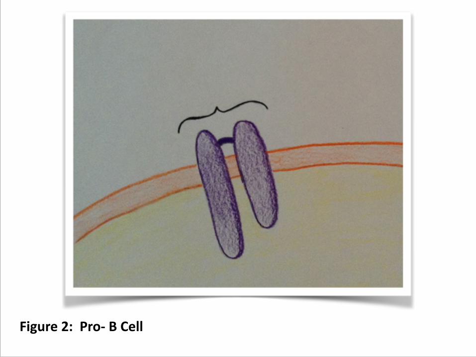
Figure 2: Pro-‐ B Cell
B. Differentiation and Gene Rearrangement (bone marrow), review
1. Pro-‐B cells bring DH and JH together
2. Then they add leader plus VH to DHJH, and do the P and N nucleotide additions.
3. If this produces a non-‐productive (frame shifted) gene rearrangement, then they try the other allele.
4. If the rearrangement is productive, then the heavy chain is put into the membrane with a surrogate light chain, composed of the products of two genes that can function without rearrangement (figure 3)
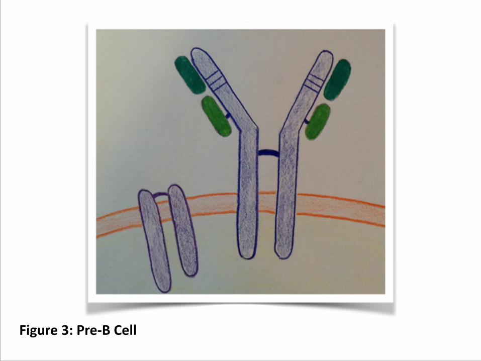
Figure 3: Pre-‐B Cell

5. The immature receptor associates with the Ig/Ig transmembrane signal. This signals allelic exclusion and initiates the light chain gene rearrangement.
6. Once there is a productive H chain gene, the cell is a pre-‐B cell. If there is no productive rearrangement, the cell apoptoses.
7. The pre-‐B cell then undergoes rearrangements of first one , then the other, and one , then the other, stopping as soon as there is a productive light chain arrangement and ultimately apoptosing if there is not.
8. Once you have two productive rearrangements, you have an immature B cell, one that has a determined antigenic specificity (CDR) and uses the CH region to produce membrane-‐bound antibody. (figure 4)
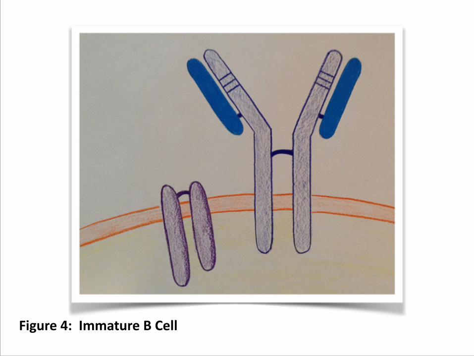
Figure 4: Immature B Cell

9. Undergoes negative selection (below).
10. Shortly thereafter, the cell also begins to express membrane-‐bound CH, and become a mature (but naïve) B cell, expressing IgM and IgD on the cell surface.
11. At this point the cells are released into the plasma and head to the peripheral (secondary) lymphoid organs: lymph nodes, spleen and mucosa.
C. Removing Self-‐Reactive B Cells. 1. 90% of the B cells produced by the above process never
make it to the plasma. At least some of the negative selection occurs as cell lines expressing antibodies to self-‐antigen are eliminated.
2. The elimination is signaled by the crosslinking of IgM.
3. Artificially crosslinking IgM will lead to apoptosis of developing B cells.
4. However, B cells can sometimes get a second chance. If cross-‐linking occurs, the cells may arrest and reactivate their RAG enzymes, and try rearranging again.
5. If they have a chain involved in the CDR for a self-‐antigen, they may try to replace it with a λ.

2. What Specifically Makes the Adaptive Immune System Unique
a. Differenfafon of B (and T) cells involves clipping DNA out of specific regions of the immunoglobulin genes.
b. The clipping is not precise within those regions.
c. The clipping takes place at different parts of the regions in different cells.
d. The clipping does not take place in regions other than those for immune proteins.
e. The clipping results in different cells (and their progeny) ulfmately having different versions of the immunoglobulin genes.
f. These site-‐specific DNA rearrangements are unique to the vertebrate adapfve immune system.

II. B-‐Cell Activation and Proliferation If a circulafng B cell does not encounter an anfgen that can bind to its surface anfbody, it will undergo apoptosis and die within a few weeks.
A. Inifafng Anfgen Exposure 1. Thymus-‐dependent activation: In most cases, when
antigen cross-‐links a B cell’s Ig receptors, this sets off a signal (signal 1) causing the B cell to seek out a TH cell. They synapse (more below) and the TH cell provides the second necessary signal to prompt development into an antibody-‐producing plasma cell.

2. Thymus independent activation; there are a few antigen (TI antigens) that can prompt B-‐cell development independent of TH cell co-‐stimulus. These antigen also simultaneously activate toll-‐like receptors.
a. Type 1 antigen -‐ lipopolysaccharide such as those found in the outer bacterial cell walls of gram negative bacteria, which also activates TLR4 (figure 5)
b. Type 2 antigen -‐ repetitive polymeric proteins, such as bacterial flagellin, can cross link the membrane-‐bound immunoglobulins and kick off proliferation if the simultaneously activate TLR 5. (figure 6)
3. However, TI activation does not induce class switching (you mostly just make IgM) and does not produce memory cells. For that you need TH cells.

Figure 5: TI-‐1 Antigen

Figure 6: TI-‐2 Antigen
B. Activating Signals-‐ Generating signal 1. 1. Review Ig receptor.
a. mIgM or mIgD molecule
b. Ig/Ig heterodimers
c. immunoreceptor tyrosine-‐based activation motif, or ITAM extend into the cytoplasm
2. When an antigen cross-‐links one antibody with the next outside the cell, it brings together the cytosolic Ig/Ig domains, activating the ITAMs. (figure 7)
3. This causes the complex to change conformation and activates src-‐like kinases. These are enzymes that add phosphate to molecules and they add them to the ITAMs.
4. Once the ITAMs have phosphate, another kinase, syk, docks and triggers several different enzymes cascades.

Figure 7: Activation
5. These lead to the up-‐regulation of transcription factors, the inflammatory transcription factor NF-‐κB being involved in this.
6. The cells begin to divide and secrete antibodies.
7. As antibodies build up, they bind to CD-‐22. the Fc or antibody stem receptor, which provides brakes on the system.
C. Role of TH cells 1. However, the BCR does not signal effectively without contact with a TH cell,
nor do the cells divide rapidly without additional stimulus from TH cell cytokines.
2. When B cells bind antigen, they bring some it inside and hydrolyze it.
3. Some of the hydrolyzed peptide winds up attached to class II MHC molecules, the genes for which are upregulated along with the one for B7.
4. Thus the B cell can present some of the antigen to a TH cell and also contact the T cell using B7 to CD28.
a. Because of its ability to gather up the antigen using the BCR, a B cell is very effective at presenting antigen, and can stimulate a TH cell at concentrations 100 to 10,000 times lower than those necessary for a macrophage or dendritic cell.
b. Of course the antigen received is different from the antigen presented. The presented antigen is a peptide derived from the overall molecule.
5. Te cells attach, forming a conjugate or immune synapse.
6. This causes the TH cell to produce CD40L, which is a juxtacrine factor that turns around and signals the B cell through CD40 receptor. (figure 8)
7. The contact reorganizes the interior of the TH cell so that cytokines are released toward the B cell.
8. The B cells begin producing receptors for the cytokines.
9. Cytokine signaling activates the B cells and they begin proliferating and differentiating.
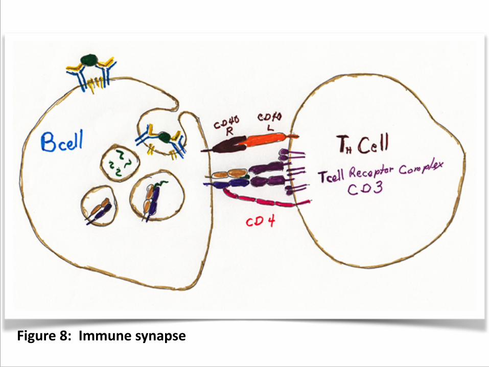
Figure 8: Immune synapse

III. Primary Versus Secondary ResponseA. The Primary Response
1. naïve lymphocytes 2. 4 to 7 day lag time 3. produces antibody secreting plasma cells and memory
cells 4. initial antibodies mostly IgM; IgG toward the end.
B. The Secondary Response: The Sadder, but Wiser, Immune System
1. Produced primarily by memory cells
2. 1 to 3 day lag time
a. The number of memory cells specific for the antigen increases.
b. These memory cells are more easily activated.
c. They have already been through affinity maturation, so they're better at binding antigen
3. more antibody secreted, and over a longer time
4. much higher proportion of IgG and other isotypes

IV. B-‐Cell Maturation in Anatomical and Histological Contex
A. Lymph Nodes (figure 9) 1. Lymph drains from tissues and passes through these.
2. Antigen enters. It can be
a. free" -‐ particle from pathogen, or the whole bacteria or viruses themselves
b. proteins or other antigens from the pathogens complexed with antibodies
c. carried in by presenting cell (dendritic or macrophages) that have picked it up elsewhere
3. Free and antibody-‐bound antigen in the plasma is likely to be picked up by
a. interdigitating dendritic cells (figure 10)
b. macrophages, (figure 11)
c. follicular dendritic cells (figure 12)
4. Naïve lymphocytes from the bone marrow enter via the lymph.
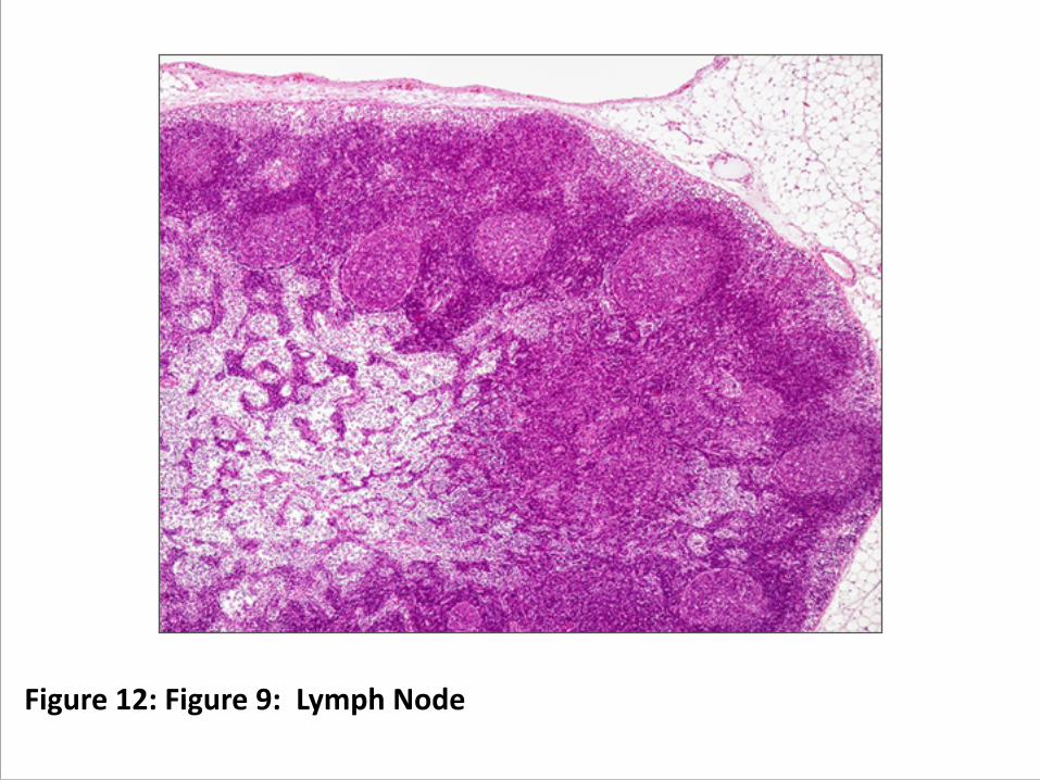
Figure 12: Figure 9: Lymph Node
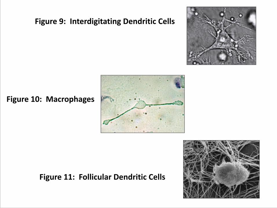
Figure 9: Interdigitating Dendritic Cells
Figure 10: Macrophages
Figure 11: Follicular Dendritic Cells

5. Activation begins in the paracortex, the layer between the outer cortex and the inner medulla, where there is a high concentration of T cells, macrophages, and dendritic cells.
6. First the macrophages and the dendritic cells activate the TH cells.
7. Naïve B cells contact the TH cells, presenting any antigen they have internalized via the class II MHC, and forming a conjugate (immune synapse).
8. The B cell begins to divide, producing a clonal cluster (focus) at the boundary between the paracortex and cortex.
9. A few activated B and TH cells migrate together from one of these foci to a primary follicle in the cortex.
10. The follicle becomes a secondary follicle, one with a germinal center where B, TH, and follicular dendritic cells interact. (figure 12)
11. A reminder about follicular dendritic cells: these are NOT regular dendritic cells. They capture antigen-‐antibody complexes in beaded structures (iccosomes) and present them to the B cells.
B. Germinal Centers. These are the sites of affinity maturation (somatic hypermutation CDR selection), the processes that refine the ability of a B cell's CDR to bind antigen effectively. (figure 13) 1. Activated B cells (centroblasts) proliferate and move to one edge
or the follicle, forming a dark zone. At this stage the centroblasts:
a. enlarge and begun to divide rapidly
b. begin somatic hypermutation-‐ mutating the regions in the heavy and light chain genes that code for the variable loops. (figure 14)
c. stop displaying the membrane Ig (recycles the original via membrane turnover)
2. The centroblasts differentiate into centrocytes which
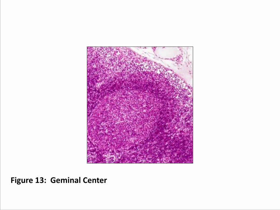
Figure 13: Geminal Center

Figure 14: Somatic Hypermutation CDR Selection
2. The centroblasts differentiate into centrocytes which
a. stop dividing
b. begin expressing membrane Ig
c. move into light zone
d. contact follicular dendritic cells
e. undergo selection B cells during which more effective receptors will survive and multiply at a greater rate
3. What happens in the follicles is a highly unusual form of accelerated natural selection. It works like evolution in general, except that the time frame is days and not centuries.
a. random mutation (dark zone)
b. excess reproduction (dark zone)
c. selection (light zone)

4. The centrocytes leave the germinal center as plasma cells, lose their surface antibody again, and begin secreting antibodies.
5. Most centrocytes do not contact an antigen that fits with their surface receptors, however, and these die by apoptosis, and are recycled by macrophages.
6. Purpose -‐ The environment of the light zone selects for those centrocytes that express the most effective antibodies. Cells with effective antibodies live, those without may return to the dark zone for more mutation, or they may die by apoptosis. (figure 15 & 16)
a. The maturing B cell that enters the germinal center and begins dividing does so if it can bind (with its surface antibody) to some degree an antigen currently arriving in the lymph node.
b. It differentiates into a centroblast that undergoes random mutational events to the very region of the gene responsible for determining the effectiveness of this antigen binding (the CDR). (figure 15)
c. The mutated centrocyte now displays the new antibody at its surface. (figure 16)
d. As with most random mutations, most of the progeny centroblast cells produced by this will bind antigen less effectively than the original cell.
e. A few however, will bind the antigen more effectively. Cells with improved receptors divide more rapidly than cells with less effective receptors.
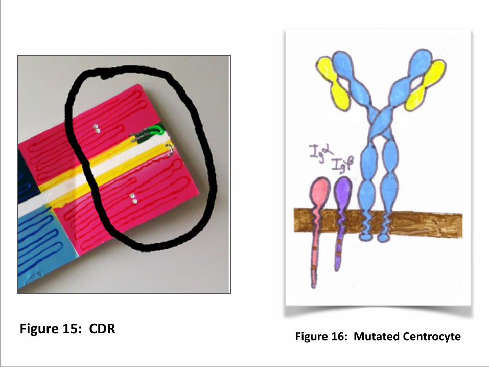
Figure 15: CDR Figure 16: Mutated Centrocyte

7. Signals
a. Follicular dendritic cells play an important role in the selection process. If the centrocyte can bind to one of the little beads with antigen-‐antibody complex, then it gets a signal necessary (but not sufficient) for its survival.
b. However, the beads are essentially a scarce resource, and the centrocytes have to compete for them.
c. Thus the more effectively the centrocyte surface antigen binds to the antibody displayed by the follicular dendritic cell, the more likely it is to live.
d. In addition, the centrocytes have to receive signals from the TH cells, especially the contact of CDC40L to the CDC40R. This doesn't work either if the B centrocyte cell does not display processed antigen back to the TH cell using its class II MHC.
C. Class Switching – directed by TH cells 1. The next decision the future plasma cell must make is
exactly what class of antibody to send out with the refined CDR region produced by affinity maturation. (figure 17)
2. Cytokine signals from the TH cells will determine this (more later).
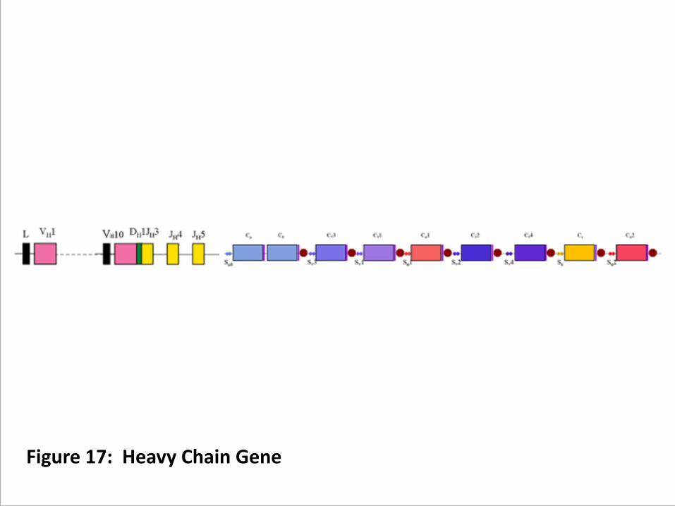
Figure 17: Heavy Chain Gene

D. To Remember or to Act: The Final Decision. 1. Centrocyte now decides whether to become a plasmoblast and
generate a plasma cell or become a memory cell and wait for a subsequent exposure to antigen.
2. Recall that plasma cells do not express membrane-‐bound antibody. This means that the sequence of differentiation in the lymph node involves:
a. dividing mature B cells with surface antigen
b. dividing centroblasts with no surface antigen (undergoing hypermutation)
c. non-‐dividing centrocytes expressing surface antibody and undergoing selection
d. dividing plasma cells not expressing surface antibody secreting soluble (humoral) antibody
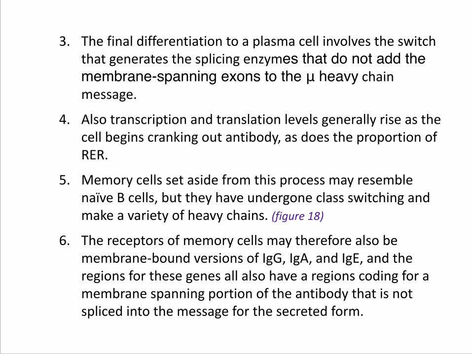
3. The final differentiation to a plasma cell involves the switch that generates the splicing enzymes that do not add the membrane-spanning exons to the μ heavy chain message.
4. Also transcription and translation levels generally rise as the cell begins cranking out antibody, as does the proportion of RER.
5. Memory cells set aside from this process may resemble naïve B cells, but they have undergone class switching and make a variety of heavy chains. (figure 18)
6. The receptors of memory cells may therefore also be membrane-‐bound versions of IgG, IgA, and IgE, and the regions for these genes all also have a regions coding for a membrane spanning portion of the antibody that is not spliced into the message for the secreted form.

Figure 18: Cross Switching Heavy Chain

IV. RegulationA. B-‐Cell Differentiation
1. B-‐Cell Specific Activator Protein (BSAP) functions as master regulator.
2. Present ONLY in members of the B cell lineage.
3. Present in ALL members of the B cell lineage EXCEPT mature plasma cells, which are done differentiating.
4. Binds to a variety of B-‐cell gene promoter regions, including those like the surrogate L chains and class switching regions that are involved in developmental decision making.
5. High levels tend to maintain a cell as a memory cell, low levels tend to promote formation of plasma cells.
B. Overall Immune Effector Response -‐ Tolerance 1. You would like NOT to make antibodies against your own proteins,
and therefore tolerate them.n.
2. On the other hand, you do NOT want to develop tolerance for foreign antigens, especially those associated with pathogenic infection.
3. Constant monitoring of your antigens by Treg cells suppresses immune responses to your own proteins and to those of benign commensal bacteria and fungi.
4. Moreover, you need to apply brakes once an infection is under control.
5. If you introduce foreign antibodies to an antigen, this will tie up the antigen and prevent it from promoting an immune response on the part of the host:
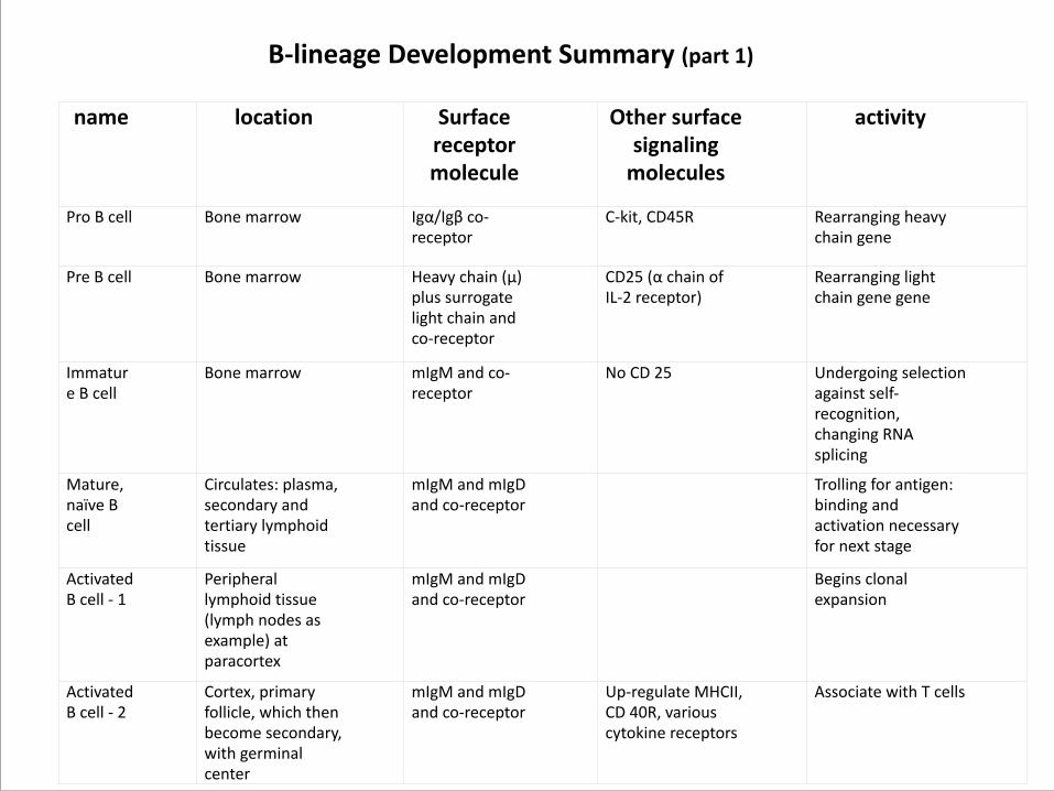
name location Surface receptor molecule
Other surface signaling molecules
activity
Pro B cell Bone marrow Igα/Igβ co-‐receptor
C-‐kit, CD45R Rearranging heavy chain gene
Pre B cell Bone marrow Heavy chain (µ) plus surrogate light chain and co-‐receptor
CD25 (α chain of IL-‐2 receptor)
Rearranging light chain gene gene
Immature B cell
Bone marrow mIgM and co-‐receptor
No CD 25 Undergoing selection against self-‐recognition, changing RNA splicing
Mature, naïve B cell
Circulates: plasma, secondary and tertiary lymphoid tissue
mIgM and mIgD and co-‐receptor
Trolling for antigen: binding and activation necessary for next stage
Activated B cell -‐ 1
Peripheral lymphoid tissue (lymph nodes as example) at paracortex
mIgM and mIgD and co-‐receptor
Begins clonal expansion
Activated B cell -‐ 2
Cortex, primary follicle, which then become secondary, with germinal center
mIgM and mIgD and co-‐receptor
Up-‐regulate MHCII, CD 40R, various cytokine receptors
Associate with T cells
B-‐lineage Development Summary (part 1)
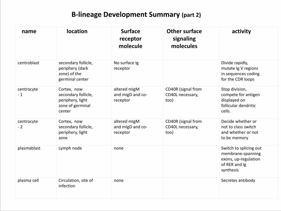
name location Surface receptor molecule
Other surface signaling molecules
activity
centroblast secondary follicle, periphery (dark zone) of the germinal center
No surface Ig receptor
Divide rapidly, mutate Ig V regions in sequences coding for the CDR loops
centrocyte -‐ 1
Cortex, now secondary follicle, periphery, light zone of germinal center
altered mIgM and mIgD and co-‐receptor
CD40R (signal from CD40L necessary, too)
Stop division, compete for antigen displayed on follicular dendritic cells.
centrocyte -‐ 2
Cortex, now secondary follicle, periphery, light zone
altered mIgM and mIgD and co-‐receptor
CD40R (signal from CD40L necessary, too)
Decide whether or not to class switch and whether or not to be memory
plasmablast Lymph node none Switch to splicing out membrane-‐spanning exons, up-‐regulation of RER and Ig synthesis
plasma cell Circulation, site of infection
none Secretes antibody
B-‐lineage Development Summary (part 2)
REFERENCES AND LINKS
Follicular Dendrifc web site hrp://home.comcast.net/~aszakal/ !!Gates Foundafon hrp://www.gatesfoundafon.org/default.htm !!Virtual Interdisciplinary Biology Educafon (VIBE) B-‐cell development: IniZaZon of anZbody response: slides 1 through 10 http://bcs.whfreeman.com/immunology6e/content/cat_070/Stanford%20VIBE/index.html
�45