lecture no.5.cvs,hs pptx
TRANSCRIPT
Lecture №5. The syndrome of an arterial hypertension and pulmonary hypertension.
Systemic hypertension and hypotonia. Pulmonary hypertension and “Cor
pulmonale”.
1
Causes
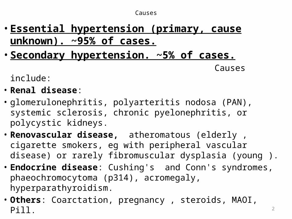
• Essential hypertension (primary, cause unknown). ~95% of cases.
• Secondary hypertension. ~5% of cases. Causes include:• Renal disease: • glomerulonephritis, polyarteritis nodosa (PAN), systemic
sclerosis, chronic pyelonephritis, or polycystic kidneys. • Renovascular disease, atheromatous (elderly , cigarette
smokers, eg with peripheral vascular disease) or rarely fibromuscular dysplasia (young ).
• Endocrine disease: Cushing's and Conn's syndromes, phaeochromocytoma (p314), acromegaly, hyperparathyroidism.
• Others: Coarctation, pregnancy , steroids, MAOI, Pill. 2
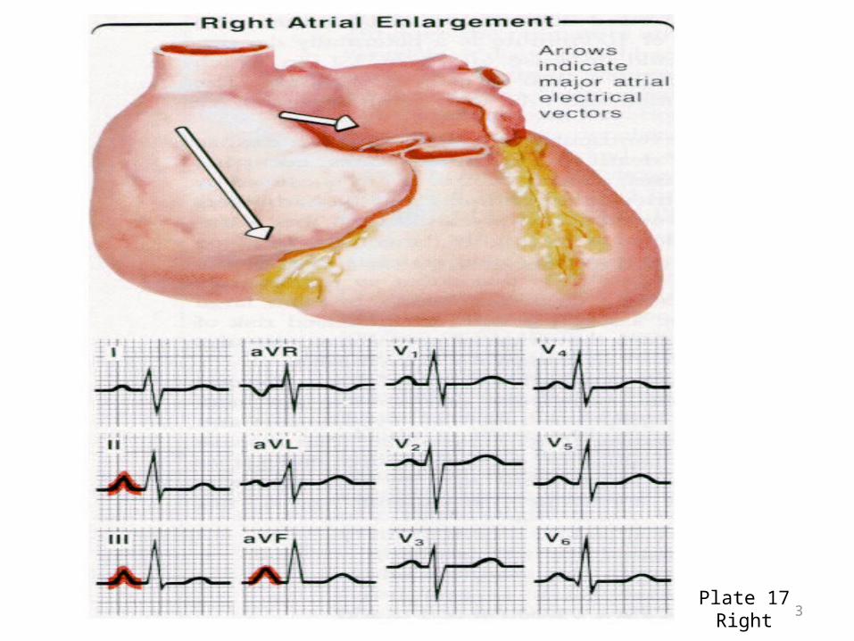
Plate 17Right 3
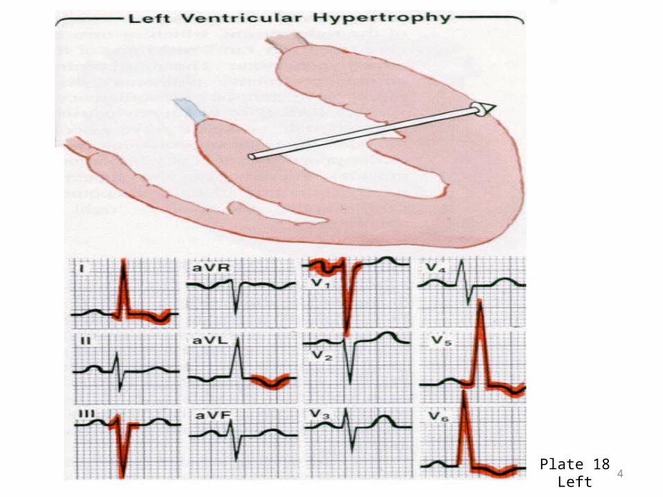
Plate 18Left 4

Plate 18Right
5
Signs & symptoms
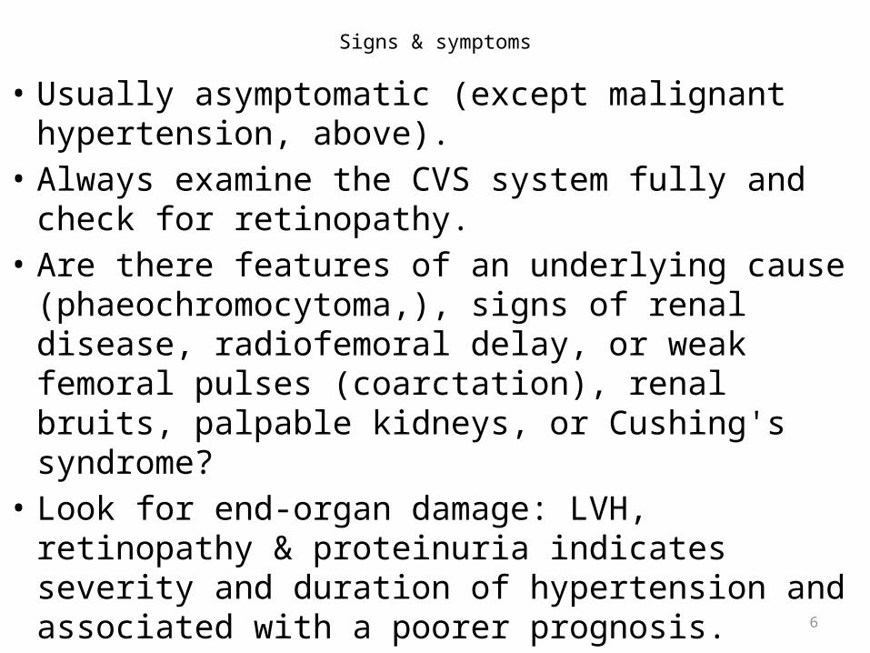
• Usually asymptomatic (except malignant hypertension, above).
• Always examine the CVS system fully and check for retinopathy.
• Are there features of an underlying cause (phaeochromocytoma,), signs of renal disease, radiofemoral delay, or weak femoral pulses (coarctation), renal bruits, palpable kidneys, or Cushing's syndrome?
• Look for end-organ damage: LVH, retinopathy & proteinuria indicates severity and duration of hypertension and associated with a poorer prognosis.6

Investigations
Basic:• U&E, • creatinine, • cholesterol, • glucose, • ECG, • urine analysis (for protein, blood).• Specific (exclude a secondary cause): renal
ultrasound, renal arteriography, 24-h urinary VMA Г— 3 (314), urinary free cortisol , renin, and aldosterone.
• ECHO and 24-h ambulatory BP monitoring may be helpful in some cases eg white coat or borderline hypertension.
7
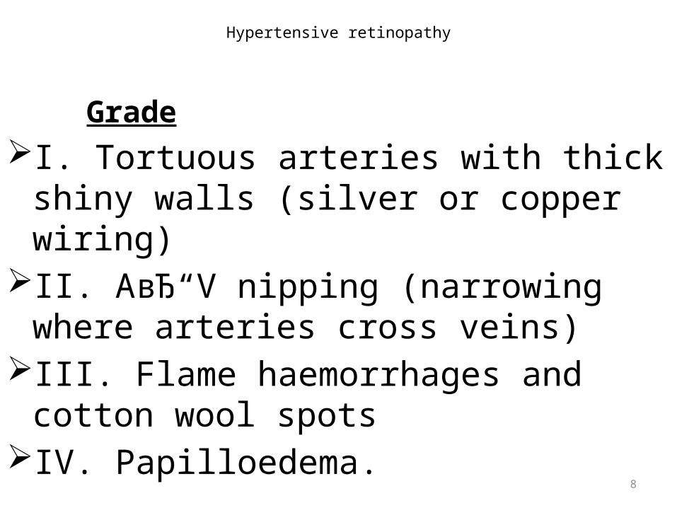
Hypertensive retinopathy
GradeI. Tortuous arteries with thick shiny walls
(silver or copper wiring)II. A–V nipping (narrowing where arteries
cross veins)III. Flame haemorrhages and cotton wool
spotsIV. Papilloedema.
8
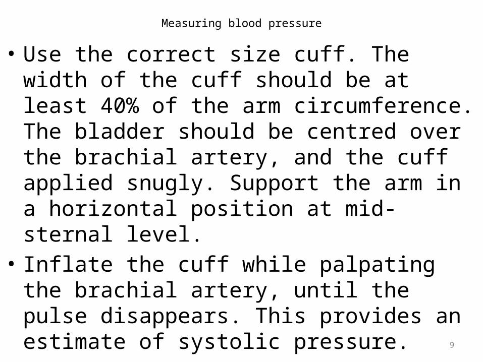
Measuring blood pressure
• Use the correct size cuff. The width of the cuff should be at least 40% of the arm circumference. The bladder should be centred over the brachial artery, and the cuff applied snugly. Support the arm in a horizontal position at mid-sternal level.
• Inflate the cuff while palpating the brachial artery, until the pulse disappears. This provides an estimate of systolic pressure.
• Inflate the cuff until 30mmHg above systolic pressure, then place stethoscope over the brachial artery. Deflate the cuff at 2mmHg/s.
9
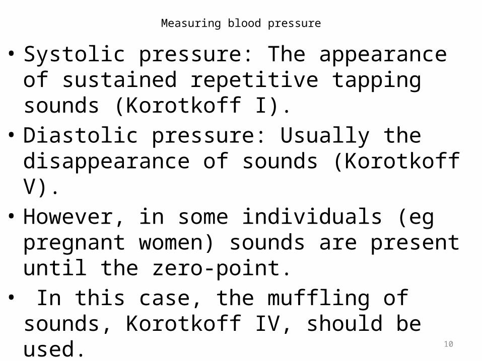
Measuring blood pressure
• Systolic pressure: The appearance of sustained repetitive tapping sounds (Korotkoff I).
• Diastolic pressure: Usually the disappearance of sounds (Korotkoff V).
• However, in some individuals (eg pregnant women) sounds are present until the zero-point.
• In this case, the muffling of sounds, Korotkoff IV, should be used.
10
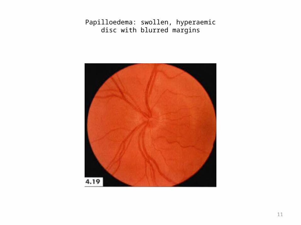
Papilloedema: swollen, hyperaemicdisc with blurred margins
11
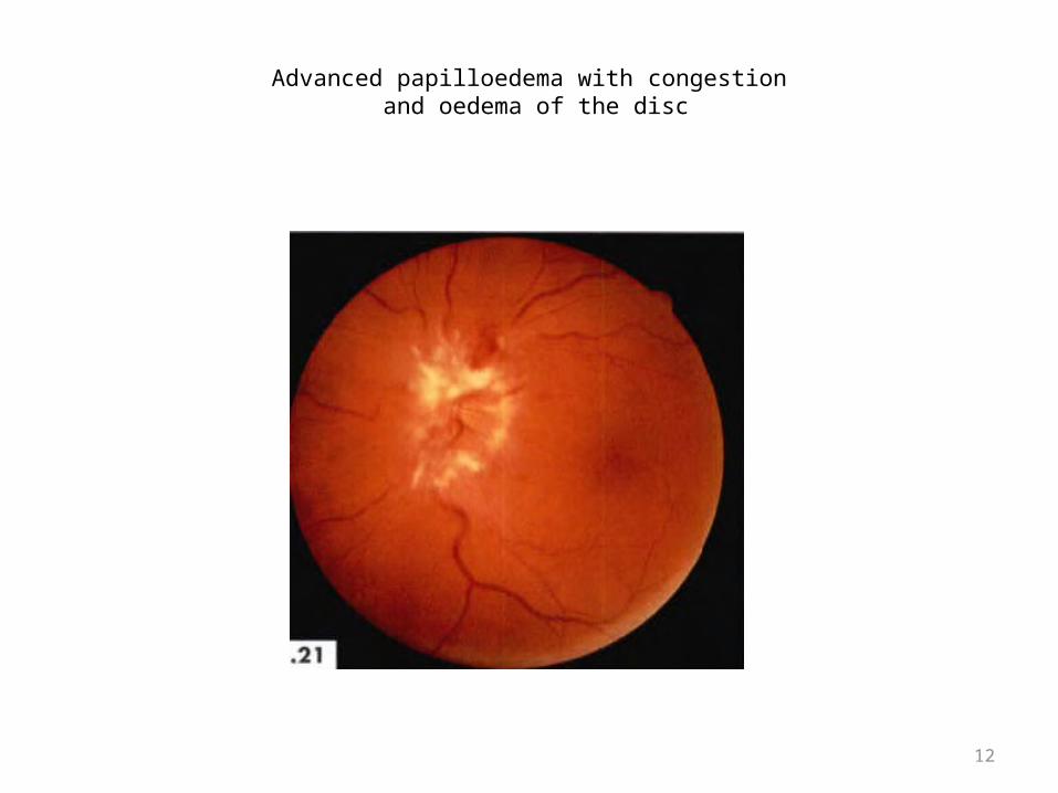
Advanced papilloedema with congestion and oedema of the disc
12
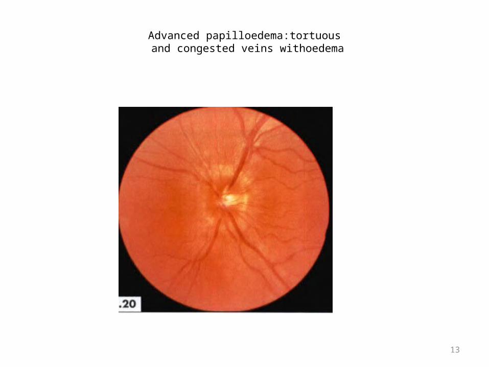
Advanced papilloedema:tortuous and congested veins withoedema
13

Papilloedema with completely obscureddisc. Note tortuous veins with dark blood columns
14
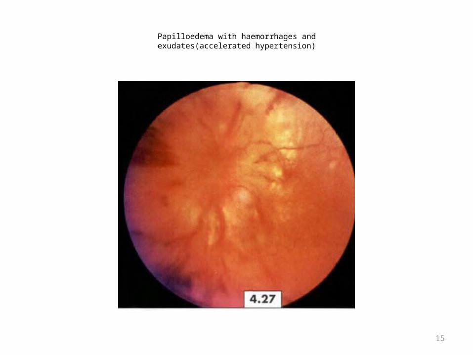
Papilloedema with haemorrhages andexudates(accelerated hypertension)
15
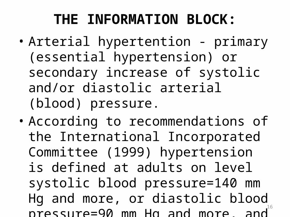
THE INFORMATION BLOCK:• Arterial hypertention - primary (essential
hypertension) or secondary increase of systolic and/or diastolic arterial (blood) pressure.
• According to recommendations of the International Incorporated Committee (1999) hypertension is defined at adults on level systolic blood pressure=140 mm Hg and more, or diastolic blood pressure=90 mm Hg and more, and are classified further on degrees:
16
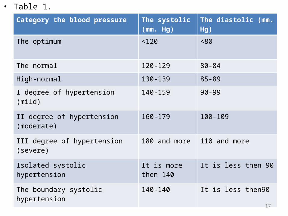
• Table 1.Category the blood pressure The systolic (mm.
Hg)The diastolic (mm. Hg)
The optimum <120 <80
The normal 120-129 80-84
High-normal 130-139 85-89
I degree of hypertension (mild) 140-159 90-99
II degree of hypertension (moderate) 160-179 100-109
III degree of hypertension (severe) 180 and more 110 and more
Isolated systolic hypertension It is more then 140 It is less then 90
The boundary systolic hypertension 140-140 It is less then90
17
The causes: the сause of the essential АH or primary АH is not known; symptomatic АH have a known etiology (parenhematous renal diseases, diseases of renal vessels, diseases of adrenal glands, anomalies of vessels, aortic regurgitation, etc.).
Undoubtedly, there is a genetic predisposition to the hypertensia; apparently, environment factors (quantity of sodium in food, character of a food and the lives, promoting adiposity, and stress, smoking, a diabetes mellitus) have the an effect only on genetically predisposed individuals.
.18
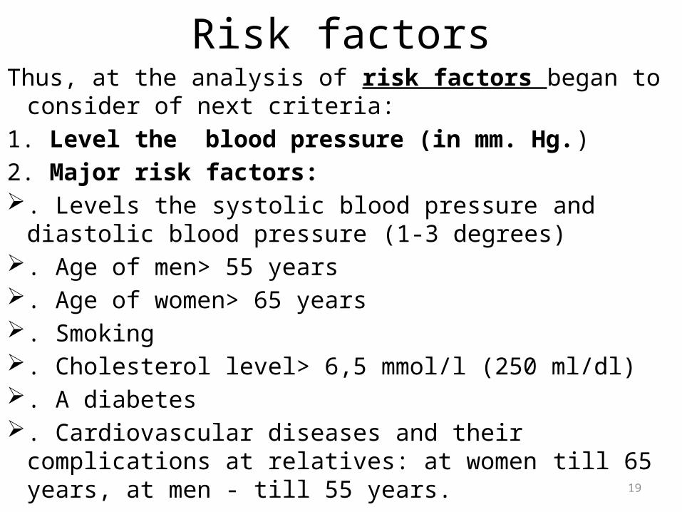
Risk factorsThus, at the analysis of risk factors began to consider of next
criteria:1. Level the blood pressure (in mm. Hg.)2. Major risk factors: . Levels the systolic blood pressure and diastolic blood
pressure (1-3 degrees) . Age of men> 55 years . Age of women> 65 years . Smoking . Cholesterol level> 6,5 mmol/l (250 ml/dl) . A diabetes . Cardiovascular diseases and their complications at
relatives: at women till 65 years, at men - till 55 years.19
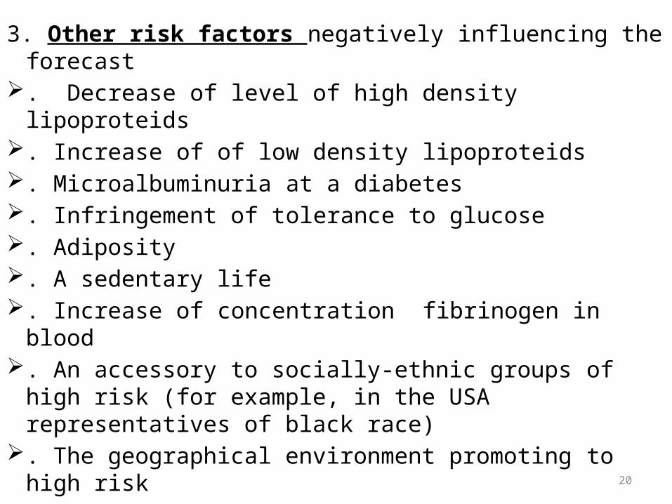
3. Other risk factors negatively influencing the forecast . Decrease of level of high density lipoproteids . Increase of of low density lipoproteids . Microalbuminuria at a diabetes . Infringement of tolerance to glucose . Adiposity . A sedentary life . Increase of concentration fibrinogen in blood . An accessory to socially-ethnic groups of high risk (for
example, in the USA representatives of black race) . The geographical environment promoting to high risk
20

4.Damage of targets-organs-
. Left ventricular hypertrophy (according to an electrocardiogram, ultrasound and-or X-ary investigation).
.Proteinuria and-or passing increase of creatinine in blood (1,2-2,0 mg/dl)
. Initial displays of an atherosclerosis of an aorta and peripheral arteries (carotid, femoral) according to ultrasonic or X-ray investigations
.General or local narrowing of arteries of a retina of an eye 21
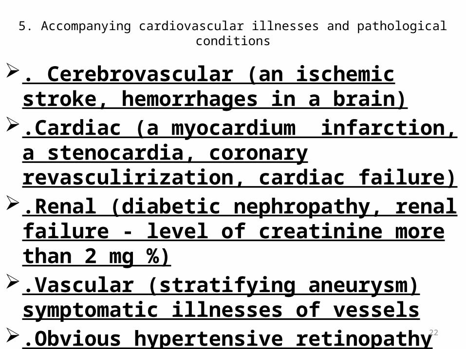
5. Accompanying cardiovascular illnesses and pathological conditions
. Cerebrovascular (an ischemic stroke, hemorrhages in a brain)
.Cardiac (a myocardium infarction, a stenocardia, coronary revasculirization, cardiac failure)
.Renal (diabetic nephropathy, renal failure - level of creatinine more than 2 mg %)
.Vascular (stratifying aneurysm) symptomatic illnesses of vessels
.Obvious hypertensive retinopathy (hemorrage or exudate in retina, a hypostasis of an optic nerve)
22
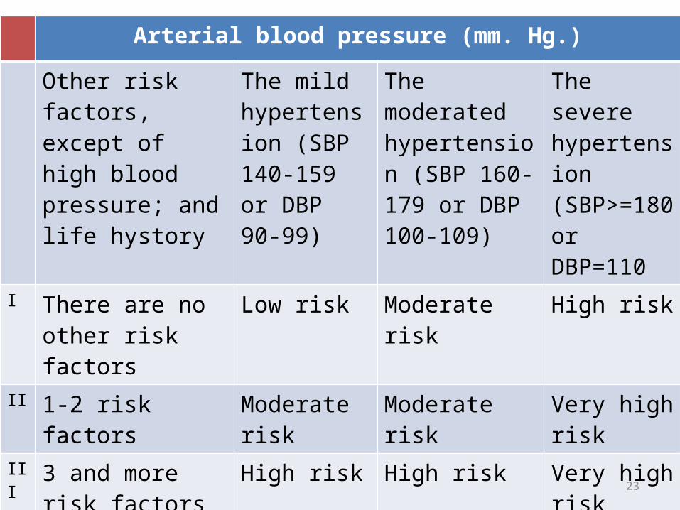
Arterial blood pressure (mm. Hg.)
Other risk factors, except of high blood pressure; and life hystory
The mild hypertension (SBP 140-159 or DBP 90-99)
The moderated hypertension (SBP 160-179 or DBP 100-109)
The severe hypertension (SBP>=180 or DBP=110
I There are no other risk factors
Low risk Moderate risk High risk
II 1-2 risk factors Moderate risk Moderate risk Very high risk
III 3 and more risk factors or damage of bodies-targets or diabetes
High risk High risk Very high risk
IV The accompanying the clinical conditions.
Very high risk Very high risk Very high risk
23

PathogenesisExact mechanism unknown. Following are the
suggestions.• B.P = Cardiac output X Peripheral resistance
In the beginning of the essential hypertension, the increase of blood pressure is due to a small increase in cardiac output. This could be due to sympathetic overactivity. Later in the disease, the cardiac output induces vascular changes (increased peripheral resistance) that increase the blood pressure.
24
• In regulation of the pressure of blood volume of participates juxtaglomerular apparatus (JGA). Formed in granulus cells of JGA the proteolytic enzyme rhenine cataliziratat the transformation of angiotenzine (one of protein of plasma) in decapeptidis angiotensin I which is split by enzyme (mainly in lungs, but also in kidneys and a brain) till ocapeptidis angiotensin II which operates as powerful vasoconstrictor, and also stimulates liberation aldosterone.
• Besides, in blood is available not containing of aspoginate acids hectapeptidis (angiotensin III) which stimulates liberation аldosterone, as well as angiotensin II, but possesses with much smaller pressor activity.
• The aldosterone conducts to a sodium delay in an organism; surplus of endocellular sodium raises sensitivity of smooth muscles of vessels to sympathetic stimulation.25
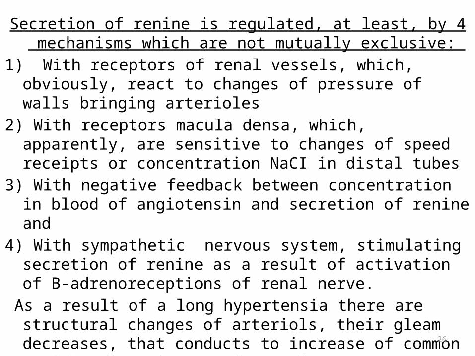
Secretion of renine is regulated, at least, by 4 mechanisms which are not mutually exclusive:
1) With receptors of renal vessels, which, obviously, react to changes of pressure of walls bringing аrterioles
2) With receptors macula densa, which, apparently, are sensitive to changes of speed receipts or concentration NaCI in distal tubes
3) With negative feedback between concentration in blood of angiotensin and secretion of renine and
4) With sympathetic nervous system, stimulating secretion of renine as a result of activation of B-adrenoreceptions of renal nerve.
As a result of a long hypertensia there are structural changes of arteriols, their gleam decreases, that conducts to increase of common peripheral resistanceof vessels.
• The hypertensia can develop as a result of deficiency of vasodilating substances. Their reduction or the absence caused by damage of ren promotes increase the arterial blood pressure.
26
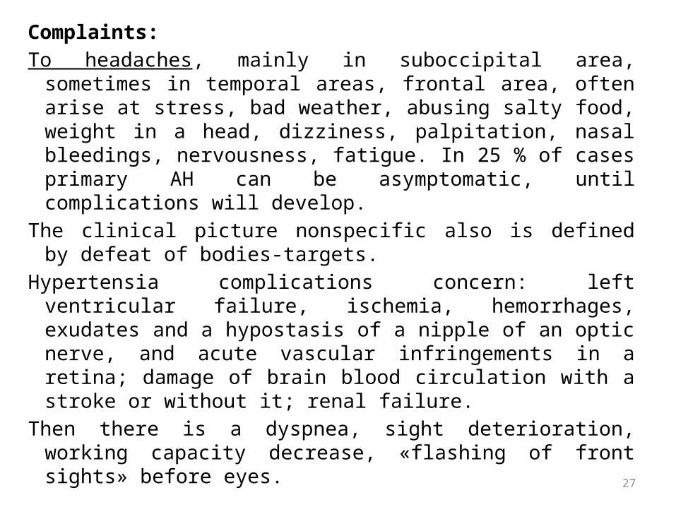
Complaints:To headaches, mainly in suboccipital area, sometimes in
temporal areas, frontal area, often arise at stress, bad weather, abusing salty food, weight in a head, dizziness, palpitation, nasal bleedings, nervousness, fatigue. In 25 % of cases primary АH can be asymptomatic, until complications will develop.
The clinical picture nonspecific also is defined by defeat of bodies-targets.
Hypertensia complications concern: left ventricular failure, ischemia, hemorrhages, exudates and a hypostasis of a nipple of an optic nerve, and acute vascular infringements in a retina; damage of brain blood circulation with a stroke or without it; renal failure.
Then there is a dyspnea, sight deterioration, working capacity decrease, «flashing of front sights» before eyes.
27

Survey:• Hyperemia of face, the expressed temporal arteries, a
symptom of "worm". Depending on causes: puffiness of the face- at renal АH, moonface- at Ikushing’s syndrome, аssymetry trunks: the developed superior part, in comparison with inferior part- at aortic coarctation, etc.
Palpation:• The apex beat is strengthened, displaced to the left, pulse -
firm, full, strained. Percussion:• Displacement to the left the left border of relative dullness of
heart.Auscultation:• Sounds are strengthened, accent II sound over an aorta in 2
intercostals space on the right, can be functional systolic murmur on an apex. 28
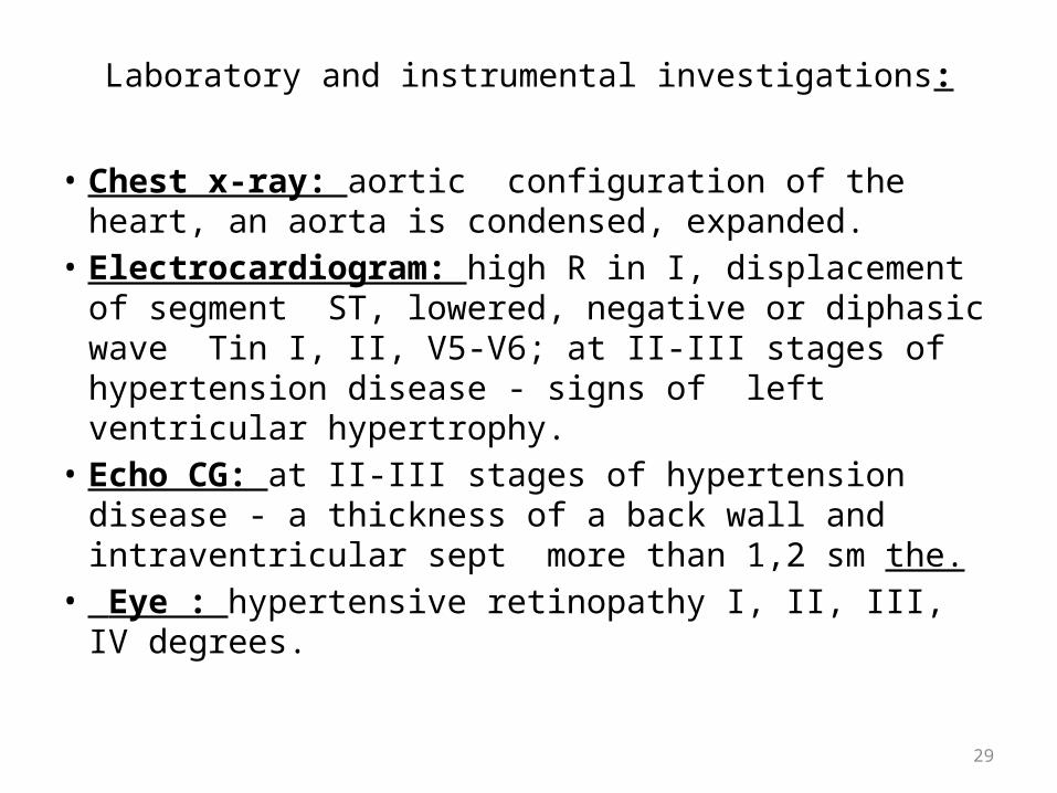
Laboratory and instrumental investigations:
• Chest x-ray: aortic configuration of the heart, an aorta is condensed, expanded.
• Electrocardiogram: high R in I, displacement of segment SТ, lowered, negative or diphasic wave Tin I, II, V5-V6; at II-III stages of hypertension disease - signs of left ventricular hypertrophy.
• Echo CG: at II-III stages of hypertension disease - a thickness of a back wall and intraventricular sept more than 1,2 sm the.
• Eye : hypertensive retinopathy I, II, III, IV degrees.29

Laboratory and biochemical investigations
Common blood count, urine analysis (at disease progressing - microalbuminuria and proteinuria), definition of sodium and calium , creatinine, blood glucose on an empty stomach, the common cholesterol (HS) and high density lipoproteids cholesterol.
Late displays of arteriolonephrosclerosis-polyuria, nicturia, reduction the concentration function of kidneys, proteiuria, microhematuria, cylindruria and a nitrogen delay.
• Symptomatic AH owing by hyperkatecholemy, the caused tumour of chromaffine cells of adrenal glands (pheochromacytoma), besides increase the blood pressure usually causes symptoms (the headaches, the expressed palpitation, arrhytmias a tachycardia, эectopic beats, paroxysmal tachycardia, increase of sweating , tremor and pallor of skin), allowing to suspect this АH.
• The diagnosis leans against revealing in urine or in plasma of blood the raised concentration in urine their metabolytes- methanephrines and vaninilmindal acids. 30
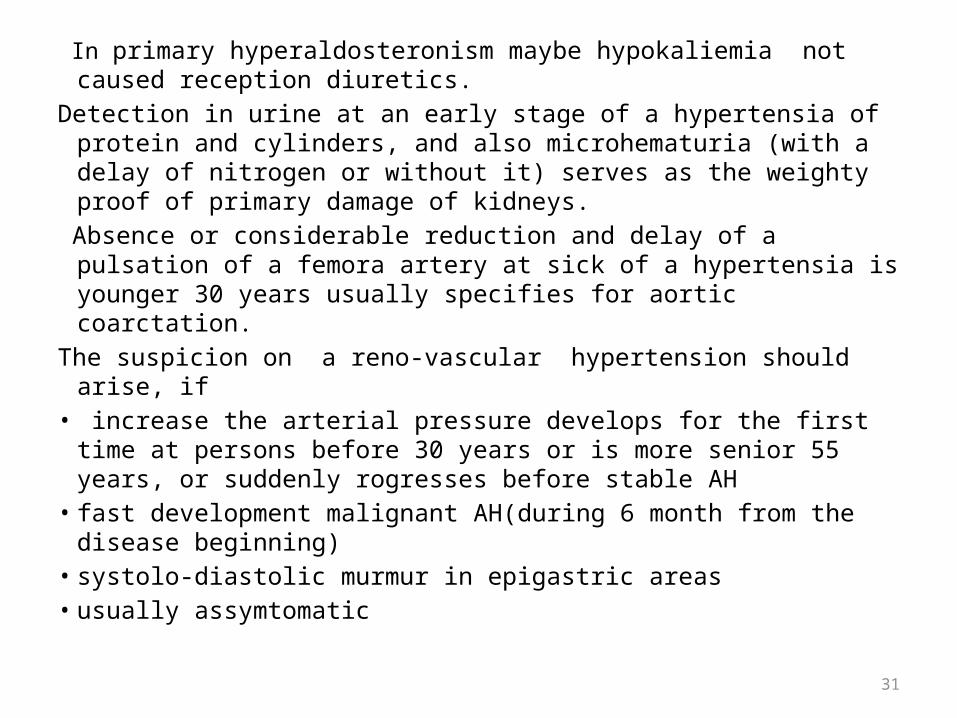
In primary hyperaldosteronism maybe hypokaliemia not caused reception diuretics.
Detection in urine at an early stage of a hypertensia of protein and cylinders, and also microhematuria (with a delay of nitrogen or without it) serves as the weighty proof of primary damage of kidneys.
Absence or considerable reduction and delay of a pulsation of a femora artery at sick of a hypertensia is younger 30 years usually specifies for aortic coarctation.
The suspicion on a reno-vascular hypertension should arise, if• increase the arterial pressure develops for the first time at persons
before 30 years or is more senior 55 years, or suddenly rogresses before stable АH
• fast development malignant АH(during 6 month from the disease beginning)
• systolo-diastolic murmur in epigastric areas• usually assymtomatic
31
Hypertensive crisis• The hypertensive crisis is the syndrome characterised by acute lifting the
arterial pressure (as a rule, diastolic blood pressure exceeds 120 mm hg) and symptoms of infringement of regional blood circulation, more often the brain.
The causes of a hypertensive crisis:• Inadequately treatment of essential hypertension• Renovascular hypertension• Diseases of kidneys, acute glomerulonephritis• Pheochromocytoma• Sclerodermia and other diseases of a connecting tissues• Use sympathomimethic means (cocaine, etc.)• A cancellation syndrome (Clonidine, beta-blockers)• Eklampsya• Head traumas• renin- and aldosteron secreting tumours• vasculitis
32
Now allocate crises of 2 types: I type (emergency) - demands emergency treatment, when it is necessary to achieve decrease the arterial pressure during 1 hour to reduce danger of development of irreversible infringements and death of the patient; hypertensive crises of this type concern:– A hypertensive encephalopathy– An intracranial hemorrhage– A stroke– A hypostasis of lungs– A myocardial infarction– Pheochromosytomic crisis– Stratifying an aorta aneurysm– eklampsy (pregnancy nephropathy)
II types (uгgency) - demand urgent treatment. The arterial pressure should be lowered during 12-24 hours; them concern:– A malignant hypertensia– An unstable angina pectoris– Left ventricular failure (cardiac asthma)– A hypertensia in pre-and the postoperational period– Preeklampsy. 33
• CLINICAt a hypertensive encephalopathy the acute
headache, a nausea, vomiting, visual frustration is marked. Usually the blood pressure very high, symptoms accrue imperceptibly during 48-72 частов (unlike an intracranial hemorrhage).
34
Complications of hypertensionThe adverse effects of hypertension principally involve the
CNS, retina, heart and kidneys. • CNSStroke: It results from cerebral hemorrhage or
infarction mostly as a complication ofhypertension.
Hypertensive encephalopathy:It is characterized by severe hypertension with neurological symptoms e.g. transient disturbance of speech or vision, disorientation, fits and unconsciousness.
Subarachnoid hemorrhage: It is also more common in hypertensive patients.
Multi-infarct dementia. 35

• RETINA Retinal changes are graded as following:
Grade I: tortuosity of the retinal arteries with increased reflectiveness (silver wiring).
Grade II: Grade I plus appearance of arteriovenous nipping produced when thickened retinal arteries pass over the retinal vein.
Grade III: grade II plus flame-haped hemorrhages and soft "cotton wool" exudates due to small infarcts.
Grade IV: Grade III plus papilledema (blurring of the margins of the optic disc).
• HEARTLeft ventricujar hypertrophy and ultimately left ventricular failure.Ischemic heart disease. Aortic dissection • KIDNEYS• Long stending hypertension may cause nephrosclerosis
(hypertensive nephropathy – proteinuria and progressive renal feilure). 36
Hypotonia• Arterial hypotonia characterized by decreases the blood pressure less
then 100/60 mm.Hg. for the persons till 25 age and less then 105/65 mm.Hg. for the persons more then 30 age.
Physiological arterial hypotonia: causes-the constitutional and genetic factors, are found at healthy persons and no accompanied with pathological chandes (adaptationaly hypotonia).
Patological hypotonia may be primary and secondary ( symptomatic). Secondary may be acute and chronic. Causes: primary hypotonic disease due to infrigement central and vegetative neurological system with infrigement of regulation of the vessel tone. Causes of secondary hypotonia – acute and chronic failure of renal gland, hypothyreosis.
At an acute vessels failure ( collapse, syncope) can be sharp collapse of vessels tone and develop decrease of inflow of the blood to the heart, decrease of cardiac output and of volume of blood circulation.
37
• Clinically: fatuge, confusion, syncope, vomiting, palpitation, dyspnea, pallor of skin, sweating, decreases systolic and diastolic blood pressure. Pulse are threadly.
• Survey: hyperhydrosis, tremor of fingers.• Border of the heart is normal, cardiac sound is
weakened.• ECG: decrease of voltage.• Laboratory and instrumental investigation is
normal.
38
Pulmonary arterial hypertension and “cor” pulmonale.
• PH is characterized by decrease if moderate pressure in pulmonary arteries in rest more then 25 mm.Hg.( normal-9-16 mm. Hg.) PH decreased loading right part of heart, lead to develop of the pulmonary heart9 cor pulmonale). Cor pulmonale its RVH due to disease, which damage of function and structure of lungs, pulmonary vessels.
• Causes: disease of broncho-pulmonary apparats and intrapulmonary airways, kyphoscolyosis, neuro-muscular diseases, primary pulmonary hypertensia. thrombosis., emboli, arteriitis and ect.
• Clinically: symptoms of basic diseases, pulmonary failure, pulmonary arterial hypertension, HRV, right ventricular cardiac failure.
• Complaints: dyspnea, cough with sputum and another symptoms of basic diseases. May be syncope, chestpaine due to anlarged of the truncus of pulmonary artery and increased of pulmonary pressure, palpitation, edema, olygouria.
39

• Survey: tachypnea, diffuse cyanosis, swelling of neck veins at expirate, pulsation in left on the II intercostal space, accent II sound over pulmonary artery, HRV ( increase of border of the heart to right, precardial pulsation, epigastrical pulsation). The signs of right ventricular cardiac failure consist swelling of neck veins in breath, increase of right border of the heart, weakned I sound, systolic murmur over pulmonary artery, diastolic Grehem- Still’s noise, arrhytmias, hepatomegaly, acytes, edema of feet.
• X-ray: strenfthening of pulmonary roots, pulsation of vessels and increase of diameter of the right pulmonary artery ( norma-14-16 mm), RVH.
• ECG: axis deviation to right, P-pulmonale, HRV.• EchoCG: increase pulmonary arterial pressure (N-diastolic less then 12mm.Hg
systolic more then 30 mm. Hg., moderate less then 25 mm. Hg.), increase of anterio-posterior size of right ventricule and tickening of anterior wall of right ventricule ( more then 5mm).
• Peak expiratory flow rate: sharp expressed infringement by obstruction type (forced inspiratory volume more then 1Iitre), restriction type ( vital capacity less then 50%).
• Arterial blood- gas analysis: P O2 less then 87%(N- more then 95%).• Heart catheterization: level of pulmonary pressure.• May be investigation-pulmonary angyography, byopsy and morphological
investigation.40