lecture 37 competent cells - nptel · lecture 37 competent cells. introduction: the delivery of dna...
TRANSCRIPT
NPTEL – Biotechnology – Experimental Biotechnology
Joint initiative of IITs and IISc – Funded by MHRD Page 1 of 23
Lecture 37 Competent Cells
Introduction: The delivery of DNA into the host is required for generation of
genetically modified organism. DNA delivery to host is a 3 stage process, DNA
sticking to the host cell, internalization and release into the host cell. As a result, it
depends on 2 parameters-
Surface chemistry of host cell-Host cell surface charges either will attract or repell
DNA as a result of opposite or similar charges. Presence of cell wall (in the case of
bacteria, fungus and plant) causes additional physical barrier to the up-take and entry
of DNA.
Charges on DNA-Negative charge on DNA modulates interaction with the host cell
especially cell surface.
Modulation of these two properties is achieved in different methods to deliver
DNA into the host cell and it is the topic of the discussion of today’s lecture.
Lab experiment 37.1: Preparation of chemically (CaCl2) treated E.coli competent
cells.
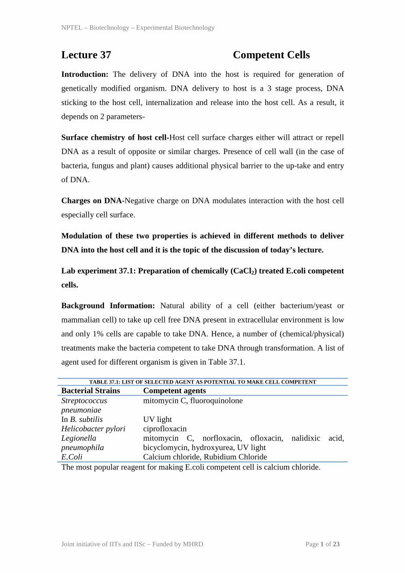
Background Information: Natural ability of a cell (either bacterium/yeast or
mammalian cell) to take up cell free DNA present in extracellular environment is low
and only 1% cells are capable to take DNA. Hence, a number of (chemical/physical)
treatments make the bacteria competent to take DNA through transformation. A list of
agent used for different organism is given in Table 37.1.
TABLE 37.1: LIST OF SELECTED AGENT AS POTENTIAL TO MAKE CELL COMPETENT
Bacterial Strains Competent agents Streptococcus pneumoniae
mitomycin C, fluoroquinolone
In B. subtilis UV light Helicobacter pylori ciprofloxacin Legionella pneumophila
mitomycin C, norfloxacin, ofloxacin, nalidixic acid, bicyclomycin, hydroxyurea, UV light
E.Coli Calcium chloride, Rubidium Chloride The most popular reagent for making E.coli competent cell is calcium chloride.
NPTEL – Biotechnology – Experimental Biotechnology
Joint initiative of IITs and IISc – Funded by MHRD Page 2 of 23
Material Required:
1. DH5α Host cells stock
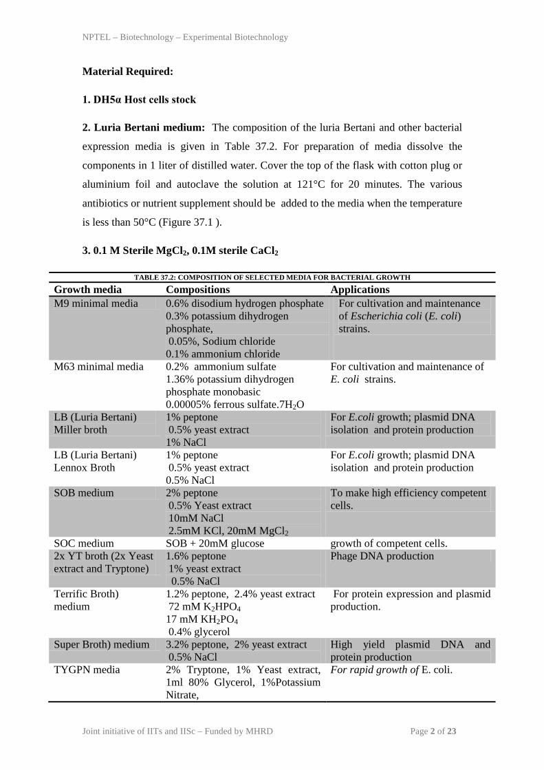
2. Luria Bertani medium: The composition of the luria Bertani and other bacterial
expression media is given in Table 37.2. For preparation of media dissolve the
components in 1 liter of distilled water. Cover the top of the flask with cotton plug or
aluminium foil and autoclave the solution at 121°C for 20 minutes. The various
antibiotics or nutrient supplement should be added to the media when the temperature
is less than 50°C (Figure 37.1 ).
3. 0.1 M Sterile MgCl2, 0.1M sterile CaCl2
TABLE 37.2: COMPOSITION OF SELECTED MEDIA FOR BACTERIAL GROWTH
Growth media Compositions Applications M9 minimal media 0.6% disodium hydrogen phosphate
0.3% potassium dihydrogen phosphate, 0.05%, Sodium chloride 0.1% ammonium chloride
For cultivation and maintenance of Escherichia coli (E. coli) strains.
M63 minimal media 0.2% ammonium sulfate 1.36% potassium dihydrogen phosphate monobasic 0.00005% ferrous sulfate.7H2O
For cultivation and maintenance of E. coli strains.
LB (Luria Bertani) Miller broth
1% peptone 0.5% yeast extract 1% NaCl
For E.coli growth; plasmid DNA isolation and protein production
LB (Luria Bertani) Lennox Broth
1% peptone 0.5% yeast extract 0.5% NaCl
For E.coli growth; plasmid DNA isolation and protein production
SOB medium 2% peptone 0.5% Yeast extract 10mM NaCl 2.5mM KCl, 20mM MgCl2
To make high efficiency competent cells.
SOC medium SOB + 20mM glucose growth of competent cells. 2x YT broth (2x Yeast extract and Tryptone)
1.6% peptone 1% yeast extract 0.5% NaCl
Phage DNA production
Terrific Broth) medium
1.2% peptone, 2.4% yeast extract 72 mM K2HPO4 17 mM KH2PO4 0.4% glycerol
For protein expression and plasmid production.
Super Broth) medium 3.2% peptone, 2% yeast extract 0.5% NaCl
High yield plasmid DNA and protein production
TYGPN media 2% Tryptone, 1% Yeast extract, 1ml 80% Glycerol, 1%Potassium Nitrate,
For rapid growth of E. coli.

NPTEL – Biotechnology – Experimental Biotechnology
Joint initiative of IITs and IISc – Funded by MHRD Page 3 of 23
0.5% Sodium Phosphate dibasic
Figure 37.1:Equipments and media required for sterilization and growth of bacterial expression system. (A)autoclave
(B)autoclaved LB broth (C) E.coli grown in LB broth.
Methods :
1. Bacterial Culture- The growth stage of the bacteria has a significant impact for its
ability to take up foreign DNA. The bacterium at log phase is more active and
efficient to perform DNA damage and repair than stationery phase. As a result, it is
preferred to use a bacteria of log phase for making competent cells for transformation.
2. Preparation of Competent Cell-Bacteria is incubated with divalent cation
(Calcium chloride,Manganese chrloride or Rubidium chloride) for 30mins at 40C.
During this process, cell wall of treated bacteria is swell and it gather factors required
for intake of DNA docked on the plasma membrane. The whole process of E.coli
competent cells preparation is as follows:
1. Inoculate single colony into the 100ml LB media and allow the cells to grow at
370C, 180rpm until
OD600 nm reaches to the 0.4-0.6.
2. Centrifuge the bacterial culture at 4000 rpm, at 4°C 10 min. Discard the
supernatant.
3. Resuspend the cell pellet gently first in 1-2 ml and then in 10 ml of ice-cold 0.1 M
MgCl2.
4. Centrifuge the bacterial suspension at 4000 rpm, at 4°C 10 min. Discard the
supernatant.
5. Resuspend cells gently in 3.0 ml of ice-cold 0.1 M CaCl2.
6. Incubate the cell suspension on ice for additional 2hrs.

NPTEL – Biotechnology – Experimental Biotechnology
Joint initiative of IITs and IISc – Funded by MHRD Page 4 of 23
7. Centrifuge the bacterial suspension at 4000 rpm, at 4°C 10 min. Discard the
supernatant.
8. Resuspend cells gently in 3.0 ml of ice-cold 0.1 M CaCl2 containing 10% glycerol
and store in small aliquot (100µl) at -800C. The cells can be used for transformation
(discussed in later lectures).
Lab Experiment 37.2 : Preparation of E.coli competent cells for Electroporation.
Background Information: The permeability of the cell membrane can be increased in
the presence of high external electric field. This approach is used to deliver the
molecular probe, drugs or DNA into the different types of cells. Several hundred volts
across plasma membrane is applied in this process. The deliver process is ~10 times
more efficient than calcium chloride mediated transformation. Electroporation is
performed with the help of electroporator, which can create very high electric field in
a specialized cuvette (Figure 37.2).
Figure 37.2:Equipments required for electroporation. (A) electroporator (B) Cuvette.
Material:
1. DH5α Host cells stock
2. Triple distilled water.
Preparation of Competent Cell- The whole process of E.coli competent cells
preparation is as follows:
1. Inoculate single colony into the 100ml LB media and allow the cells to grow at
370C, 180rpm until
OD600 nm reaches to the 0.4-0.6.
NPTEL – Biotechnology – Experimental Biotechnology
Joint initiative of IITs and IISc – Funded by MHRD Page 5 of 23
2. Centrifuge the bacterial culture at 4000 rpm, at 4°C 10 min. Discard the
supernatant.
3. Resuspend the cell pellet gently in 5-10ml triple distilled water (multiple times).
4. Centrifuge the bacterial suspension at 4000 rpm, at 4°C 10 min. Discard the
supernatant.
5. Resuspend cells gently in 3.0 ml of ice-cold distilled water.
6. Centrifuge the bacterial suspension at 4000 rpm, at 4°C 10 min. Discard the
supernatant.
7. Resuspend cells gently in 3.0 ml of ice-cold triple distilled water containing 10%
glycerol and store in small aliquot (100µl) at -800C. The cells can be used for
transformation (discussed in later lectures).
NPTEL – Biotechnology – Experimental Biotechnology
Joint initiative of IITs and IISc – Funded by MHRD Page 6 of 23
Lecture 38 Transformation
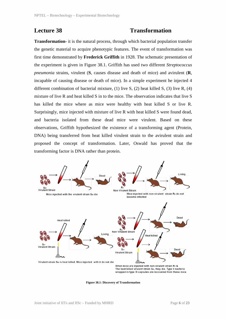
Transformation- it is the natural process, through which bacterial population transfer
the genetic material to acquire phenotypic features. The event of transformation was
first time demonstrated by Frederick Griffith in 1928. The schematic presentation of
the experiment is given in Figure 38.1. Griffith has used two different Streptococcus
pneumonia strains, virulent (S, causes disease and death of mice) and avirulent (R,
incapable of causing disease or death of mice). In a simple experiment he injected 4
different combination of bacterial mixture, (1) live S, (2) heat killed S, (3) live R, (4)
mixture of live R and heat killed S in to the mice. The observation indicates that live S
has killed the mice where as mice were healthy with heat killed S or live R.
Surprisingly, mice injected with mixture of live R with heat killed S were found dead,
and bacteria isolated from these dead mice were virulent. Based on these
observations, Griffith hypothesized the existence of a transforming agent (Protein,
DNA) being transferred from heat killed virulent strain to the avirulent strain and
proposed the concept of transformation. Later, Oswald has proved that the
transforming factor is DNA rather than protein.
Figure 38.1: Discovery of Transformation
NPTEL – Biotechnology – Experimental Biotechnology
Joint initiative of IITs and IISc – Funded by MHRD Page 7 of 23
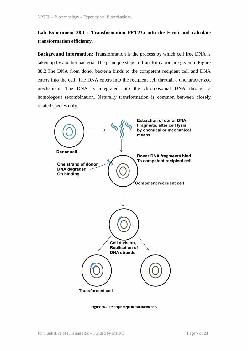
Lab Experiment 38.1 : Transformation PET23a into the E.coli and calculate
transformation efficiency.
Background Information: Transformation is the process by which cell free DNA is
taken up by another bacteria. The principle steps of transformation are given in Figure
38.2.The DNA from donor bacteria binds to the competent recipient cell and DNA
enters into the cell. The DNA enters into the recipient cell through a uncharacterized
mechanism. The DNA is integrated into the chromosomal DNA through a
homologous recombination. Naturally transformation is common between closely
related species only.
Figure 38.2: Principle steps in transformation.
NPTEL – Biotechnology – Experimental Biotechnology
Joint initiative of IITs and IISc – Funded by MHRD Page 8 of 23
Material and Equipments:
1. E.Coli Competent Cells
2. Water Bath
3. Luria Bertani Media
4. LB-Agar Plate
5. Incubator
Procedure : The outline of the procedure involved in calcium chloride mediated
transformation is given in the Figure 38.3.
1. On the day of transformation, competent cells are incubated with DNA or circular
plasmid containing appropriate resistance gene such as ampicillin resistance gene for
30mins on ice.
2. Heat Shock-Competent cells are given a brief heat shock (420C for 90 sec) to relax
the cell wall and high temperature stress causes upregulation of the factor responsible
for DNA recombination and repair.
3. A chilled bacterial media is added for faster recovery of transformed cells.
4. it is plated on the solid media with appropriate antibiotics such as ampicillin and
allowed to grow for another 18-24 hrs.
5. Transformed cells with appropriate resistance will grow and give colony (Figure
38.4).
Observation: The transformed LB agar plate in Figure 38.4 has 1488 colonies.
Calculation of the transformation efficiency:
If 10ng of plasmid gives ~1488 colonies.
Then 1µg of plasmid transform 1488 x102=1.488x 10⁵ colonies
Therefore the transformation efficiency was found to be -1.4 x 10⁶ colonies.

NPTEL – Biotechnology – Experimental Biotechnology
Joint initiative of IITs and IISc – Funded by MHRD Page 9 of 23
Figure 38.3: Steps in bacterial transformation by CaCl2 method.
Figure 38.4: Transformation by CaCl2 method.
Lab Experiment 38.2 : Transformation the vector into the yeast.
Background Information: The surface chemistry of yeast cells are different from the
E.coli and as a result different methods to deliver DNA.
1. Lithium Acetate/ssDNA/PEG Method: In this method, yeast cells are incubated
with a transformation mixture of lithium acetate, PEG 3500, single stranded carrier

NPTEL – Biotechnology – Experimental Biotechnology
Joint initiative of IITs and IISc – Funded by MHRD Page 10 of 23
DNA and foreign plasmid at 420C for 40mins. The purpose of adding carrier DNA is
to block the non-specific sites on cell wall and made plasmid available for uptake.
Post-transformation, cells are pelleted to remove transformation mixture and re-
suspended in 1ml water. It is plated on a solid media with an appropriate selection
pressure such as antibiotics.
2. Spheroplast Transformation Method: In this method, yeast cell wall is removed
partially to produce spheroplast. Spheroplasts are very fragile for osmotic shock but
are competent to takes up free DNA at high rate. In addition, polyethyl glycol (PEG)
is used to facilitate deposition of plasmid and carrier DNA on cell wall for easier
uptake. The mechanism of DNA uptake in yeast is not very clear. A schematic of
spheroplast method is given in Figure 38.5. (1) In the spheroplast method, yeast cells
are incubated with zymolyase to partially remove cell wall to produce spheroplast. (2)
They are collected by centrifugation and incubated with carrier DNA and plasmid
DNA for 10mins at room temperature. (3) It is now treated with PEG and calcium for
10mins with gentle shaking. (4) Transformed spheroplast are plated on selective solid
media and incubated on 300C for 4 days.
Figure 38.5: Steps in yeast transformation by sphereplast method.
NPTEL – Biotechnology – Experimental Biotechnology
Joint initiative of IITs and IISc – Funded by MHRD Page 11 of 23
Lab Experiment 38.3 : Transfect GFP into the COS-7 Cells using ion for
Mammalian cells.
Background Information: mammalian cell membrane surface chemistry,
intracellular comparatmentization and uptake mechanism is different from the
prokaryotic cells or yeast. Hence specialized methods have been developed to suit
mammalian cells. There are 4 major strategies to deliver the DNA in mammalian
cells:
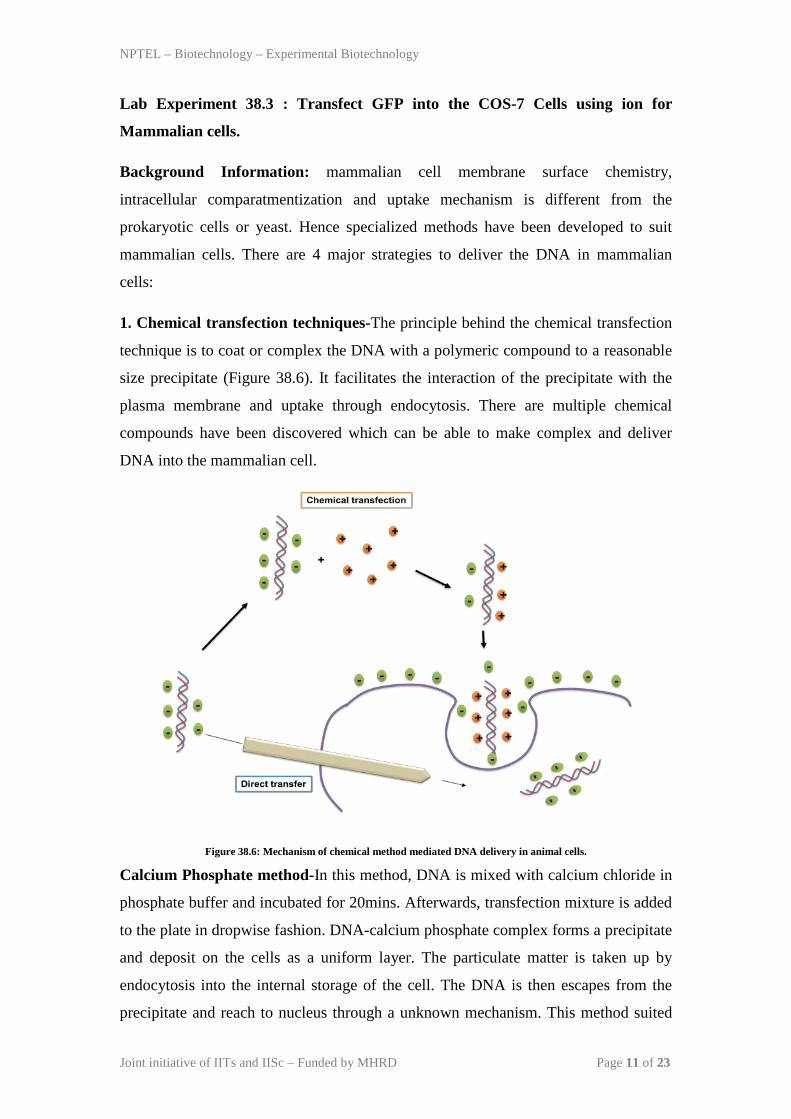
1. Chemical transfection techniques-The principle behind the chemical transfection
technique is to coat or complex the DNA with a polymeric compound to a reasonable
size precipitate (Figure 38.6). It facilitates the interaction of the precipitate with the
plasma membrane and uptake through endocytosis. There are multiple chemical
compounds have been discovered which can be able to make complex and deliver
DNA into the mammalian cell.
Figure 38.6: Mechanism of chemical method mediated DNA delivery in animal cells.
Calcium Phosphate method-In this method, DNA is mixed with calcium chloride in
phosphate buffer and incubated for 20mins. Afterwards, transfection mixture is added
to the plate in dropwise fashion. DNA-calcium phosphate complex forms a precipitate
and deposit on the cells as a uniform layer. The particulate matter is taken up by
endocytosis into the internal storage of the cell. The DNA is then escapes from the
precipitate and reach to nucleus through a unknown mechanism. This method suited
NPTEL – Biotechnology – Experimental Biotechnology
Joint initiative of IITs and IISc – Funded by MHRD Page 12 of 23
to the cell growing in monolayer or in suspension but not for cells growing in clumps.
But the technique is inconsistent and the successful transfection depends on DNA-
phosphate complex particle size and which is very difficult to control.
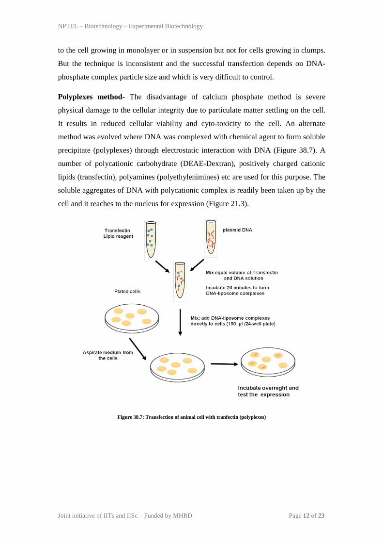
Polyplexes method- The disadvantage of calcium phosphate method is severe
physical damage to the cellular integrity due to particulate matter settling on the cell.
It results in reduced cellular viability and cyto-toxicity to the cell. An alternate
method was evolved where DNA was complexed with chemical agent to form soluble
precipitate (polyplexes) through electrostatic interaction with DNA (Figure 38.7). A
number of polycationic carbohydrate (DEAE-Dextran), positively charged cationic
lipids (transfectin), polyamines (polyethylenimines) etc are used for this purpose. The
soluble aggregates of DNA with polycationic complex is readily been taken up by the
cell and it reaches to the nucleus for expression (Figure 21.3).
Figure 38.7: Transfection of animal cell with tranfectin (polyplexes)
NPTEL – Biotechnology – Experimental Biotechnology
Joint initiative of IITs and IISc – Funded by MHRD Page 13 of 23
Liposome and lipoplex method-Another approach of DNA transfection in animal
cell is to pack the DNA in a lipid vesicle or liposome. In this approach, DNA
containing vesicle will be fused with the cell membrane and deliver the DNA to the
target cell. Preparation of liposome and encapsulating DNA was a crucial step to
achieve good transfection efficiency. Liposome prepared with the cationic or neutral
lipid facilitates DNA binding to form complex (lipoplex) and allow uptake of these
complexes by endocytosis. The lipoplex method was applicable to a wide variety of
cells, and found to transfect large size DNA as well. Another advantage of
liposome/lipoplexes is that with the addition of ligand in the lipid bilayer, it can be
used to target specific organ in the animal or a site within an organ.
3. Transduction (Virus mediated)- Viral particle has a natural tendency to attack
and deliver the DNA into the eukaryotic cells. As discussed previously, cloning gene
of interest in to the viral vectors is a innovative way to deliver the DNA into the host
cell. If the recombination sequences are available, the delivered DNA is integrated
into the host and replicate. Virus has essential components for expression of proteins
required for DNA replication, RNA polymerase and other ligand for attachment onto
the host cell. In addition, it has additional structural components to regulate infection
cycle. The virus vector contains cassettes to perform all these functions then it is fully
sufficient to propagate independently. Few virus strains may cause disease if their
propagation will be uncontrolled. A mechanism has been devised to keep a check on
the uncontrolled propagation of virus in cell. Few crucial structural blocks are placed
on another helper plasmid, in this case virus propagate only if helper plasmid has been
supplied along with the viral vector. This particular arrangement is made with the
virus strains which can cause disease after integrating into the genome such as
lentivirus.

NPTEL – Biotechnology – Experimental Biotechnology
Joint initiative of IITs and IISc – Funded by MHRD Page 14 of 23
Lecture 39 Isolation of recombinant clones.
Lab Experiment 39.1 : Isolation of plasmid DNA
Background Information: Plasmid are widely been used for cloning of foreign DNA
into the bacteria as host strain. Before getting into the details of discussing isolation of
bacterial plasmid we will discuss the basic properties of plasmids.
Different forms of plasmids: Bacterial plasmid is a double stranded circular DNA
exists in 3 different forms (Figure 39.1). If the both strands of circular double strands
are intact then it is called as covalently closed circles (CCC) where as if one of the
strand has nick, then it acquire the conformation of open circle DNA (OC, DNA).
During the isolation of plasmid DNA from bacteria, covalently closed circular DNA
losses few number of turns and as a result it acquire supercoiled configuration. The
interchange between these different forms are possible under the in-vitro or in-vivo
conditions, such as DNA gyrase produces additional turn into the circular DNA to
adopt supercoiled conformation.
Figure 39.1: Different forms of plasmids
Features of different plasmids: There are minimum molecular components to assemble bacterial plasmid to perform the function of vector are as follows-
1. Origin of replication-Like any other replicating DNA, plasmid DNA needs its
own independent origin of replication to provide replication start site to make more
copies. It decides the range of bacterial host strain can be use with the particular
plasmid vector. The plasmids containing ori region from Col E1 can be able to grow
in limited bacterial species such as E.Coli etc. In contrast, plasmid containing ori from
RP4 or RSF1010 can be able to grow in gram (-) bacteria and gram (+) bacteria.

NPTEL – Biotechnology – Experimental Biotechnology
Joint initiative of IITs and IISc – Funded by MHRD Page 15 of 23
2. Selection marker- Selection marker in the form of either antibiotic resistance gene
or enzymatic gene is essential to give phenotypic changes in host after entry of the
plasmid.
3. Promoter- Plasmid replication in host is performed by the host provided proteins
such as DNA gyrase, helicase, polymerase and DNA ligase. But proteins required for
conferring antibiotic resistance or enzyme use for selecting transformed host cells is
present on plasmid and a promoter adjacent is required to express genes present on
plasmid DNA. In addition, promoter is also needed to express gene present on foreign
DNA.
Material and Instrument:
1. E. coli cells culture (~1.5ml) containing plasmid of interest.
2. Solution I (50 mM glucose, 25 mM TrisHCl pH 8.0, 10 mM EDTA pH 8.0): Prepare the solution of 50 mM TrisHCl pH 8.0, 50 mM EDTA pH 8.0 and sterile by autoclave. Prepare the 100mM Glucose and sterile by filter through 0.2µm. Solution-I is prepared by mixing both solution to prepare solution-I.
3. Solution II (0.2 N NaOH and 1% SDS): This solution has to prepare fresh by mixing stock solution of 2N NaOH and 10% SDS in sterile molecular biolog grade water. Don’t vortex this solution and discard any left-over solution.
4. Solution III (potassium acetate, glacial acetic acid.):
5. Phenol : Chloroform: Isoamyl alchol: Mix Phenol (equliberated at pH 8.0), chloroform and Isoamyl alchol in the ratio of 25:24:1,mix by vortexing.
6. Absolute Ethanol:
7. Luria Bertani medium: The composition of the luria Bertani and other bacterial
expression media is given in Table 37.2. For preparation of media dissolve the
components in 1 liter of distilled water. Cover the top of the flask with cotton plug or
aluminium foil and autoclave the solution at 121°C for 20 minutes. The various
antibiotics or nutrient supplement should be added to the media when the temperature
is less than 50°C (Figure 37.1 ).
8. Autoclaved molecular biology grade water

NPTEL – Biotechnology – Experimental Biotechnology
Joint initiative of IITs and IISc – Funded by MHRD Page 16 of 23
9. Agarose and Buffers for Electrophoresis
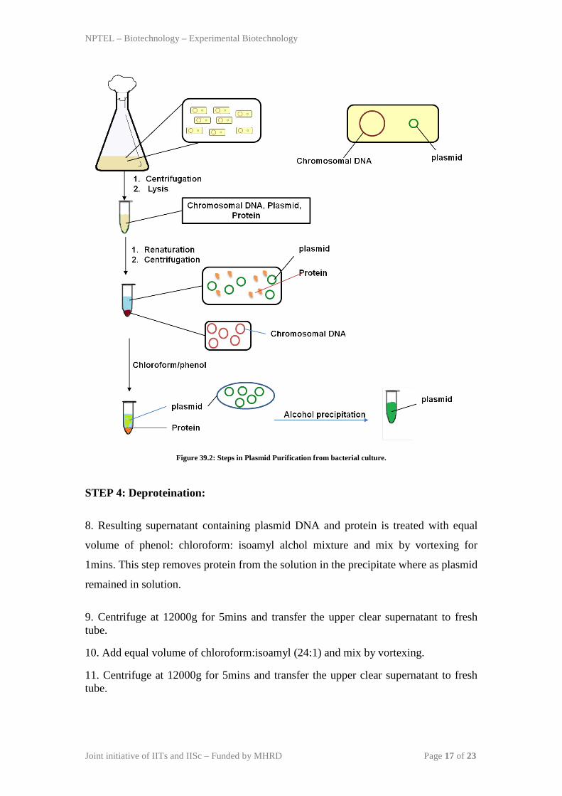
Methods: The different steps in isolation in bacterial plasmid is given in Figure 39.2.
STEP 1 (resuspension and lysis of bacterial cells): The bacteria containing plasmid
was grown in suitable culture media in high density (~0.8 optical density). Each
Bacterial cell contains chromosomal DNA, plasmid DNA and cellular proteins.
1. 1.5 ml bacterial culture is collected by centrifugation at the bottom and discard the
supernatant and invert the tube on a dry paper towel to dry the pellet.
2. Resuspended the bacterial pellet in 0.1 ml solution I containing 50 mM glucose, 25
mM TrisHCl pH 8.0, 10 mM EDTA pH 8.0. Break the cell pellet by vortexing for
2mins.
3. Incubate the Bacterial pellet on ice for 5mins. This step will partially lyse the
bacteria by hypotonic osmosis and releases cellular content.
STEP 2 (Alkaline Lysis):
4. Bacterial cells are treated with 0.2ml solution II containing 0.2 N NaOH and 1%
SDS. Don’t vortex but mix by inversion. Incubate the tube on ice for 5mins. This step
will completely lyse the cells and denature DNA (both chromosomal and plasmid
DNA) and protein.
STEP 3: Renaturation:
5. Add 0.15ml ice cold Solution III. Mix by inversion and incubate on ice for 5mins.
In this step, denatured DNA is renatured with solution III containing potassium
acetate, glacial acetic acid. In this step small DNA (plasmid) renature back quickly
whereas chromosomal DNA remained denatured.
6. Centrifuge at 12000g for 5mins and transfer the clear supernatant to fresh tube.
Avoid contamination of white precipitate, even if you needs to leave 10-20µl
supernatant.
7. Add RNase A to the supernatant at a final concentration of 20µg/ml. Incubate the
tube at 370C for 30min. This step will degrades RNA present in the sample.
NPTEL – Biotechnology – Experimental Biotechnology
Joint initiative of IITs and IISc – Funded by MHRD Page 17 of 23
Figure 39.2: Steps in Plasmid Purification from bacterial culture.
STEP 4: Deproteination:
8. Resulting supernatant containing plasmid DNA and protein is treated with equal
volume of phenol: chloroform: isoamyl alchol mixture and mix by vortexing for
1mins. This step removes protein from the solution in the precipitate where as plasmid
remained in solution.
9. Centrifuge at 12000g for 5mins and transfer the upper clear supernatant to fresh tube.
10. Add equal volume of chloroform:isoamyl (24:1) and mix by vortexing.
11. Centrifuge at 12000g for 5mins and transfer the upper clear supernatant to fresh tube.

NPTEL – Biotechnology – Experimental Biotechnology
Joint initiative of IITs and IISc – Funded by MHRD Page 18 of 23
STEP 5: Precipitation :
12. Add 2 volume of absolute alchol and mix by vortexing to precipitate the plasmid DNA.
13. Centrifuge at 12000g for 5mins and discard the supernatant. Wash the DNA pellet with the 70% ethanol.
14. Centrifuge at 12000g for 5mins and discard the supernatant.
15. Air dry the pellet and dissolve the pellet in the sterile molecular biology grade water or TE Buffer (Tris pH 8.0 containing 10mM EDTA).
16. Analyze the DNA on the 0.8% agarose gel.
Observation: Analysis of plasmid DNA on the agarose gel gives 3 bands corresponding to the 3 different forms of DNA (Figure 39.3).
Plasmid DNA
RNA
Figure 39.3: Analysis of plasmid analyzed on the agarose gel.
NPTEL – Biotechnology – Experimental Biotechnology
Joint initiative of IITs and IISc – Funded by MHRD Page 19 of 23
Lecture 40 Screening of Recombinant Clone
Lab Experiment 40.3 : Verification of recombinant DNA (Colony PCR)
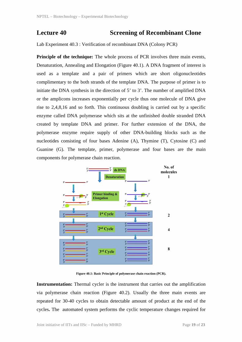
Principle of the technique: The whole process of PCR involves three main events,
Denaturation, Annealing and Elongation (Figure 40.1). A DNA fragment of interest is
used as a template and a pair of primers which are short oligonucleotides
complimentary to the both strands of the template DNA. The purpose of primer is to
initiate the DNA synthesis in the direction of 5’ to 3’. The number of amplified DNA
or the amplicons increases exponentially per cycle thus one molecule of DNA give
rise to 2,4,8,16 and so forth. This continuous doubling is carried out by a specific
enzyme called DNA polymerase which sits at the unfinished double stranded DNA
created by template DNA and primer. For further extension of the DNA, the
polymerase enzyme require supply of other DNA-building blocks such as the
nucleotides consisting of four bases Adenine (A), Thymine (T), Cytosine (C) and
Guanine (G). The template, primer, polymerase and four bases are the main
components for polymerase chain reaction.
Figure 40.1: Basic Principle of polymerase chain reaction (PCR).
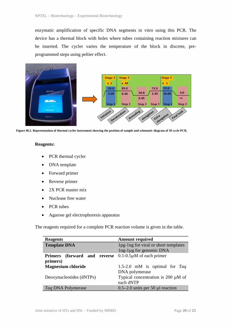
Instrumentation: Thermal cycler is the instrument that carries out the amplification
via polymerase chain reaction (Figure 40.2). Usually the three main events are
repeated for 30-40 cycles to obtain detectable amount of product at the end of the
cycles. The automated system performs the cyclic temperature changes required for
NPTEL – Biotechnology – Experimental Biotechnology
Joint initiative of IITs and IISc – Funded by MHRD Page 20 of 23
enzymatic amplification of specific DNA segments in vitro using this PCR. The
device has a thermal block with holes where tubes containing reaction mixtures can
be inserted. The cycler varies the temperature of the block in discrete, pre-
programmed steps using peltier effect.
Figure 40.2. Representation of thermal cycler instrument showing the position of sample and schematic diagram of 30 cycle PCR.
Reagents:
• PCR thermal cycler
• DNA template
• Forward primer
• Reverse primer
• 2X PCR master mix
• Nuclease free water
• PCR tubes
• Agarose gel electrophoresis apparatus
The reagents required for a complete PCR reaction volume is given in the table.
Reagents Amount required Template DNA 1pg-1ng for viral or short templates
1ng-1µg for genomic DNA Primers (forward and reverse primers)
0.1-0.5µM of each primer
Magnesium chloride 1.5-2.0 mM is optimal for Taq DNA polymerase
Deoxynucleotides (dNTPs) Typical concentration is 200 µM of each dNTP
Taq DNA Polymerase 0.5–2.0 units per 50 µl reaction

NPTEL – Biotechnology – Experimental Biotechnology
Joint initiative of IITs and IISc – Funded by MHRD Page 21 of 23
Primers: A primer is a short oligonucleotide that serves as a starting point for DNA
synthesis. In PCR, two primers are required to bind to each of the single stranded
DNA (obtained after denaturation) flanking the target sequence. These are called
Forward and Reverse primers. They primers are designed in such a way that they have
a sequence complimentary to the sequence in the template DNA. Two restriction
enzymes sites are added at the 5’ end of each of the primer to facilitate cloning. The
chosen restriction enzymes will not cut DNA fragment (non-cutters). Typically 3 to 4
nucleotides are added at the end of the restriction sites to allow efficient cutting by
restriction enzymes.
Methodology: Colony PCR has three major events (Denaturation, Annealing and
Elongation) to complete the amplification process (Figure 40.1). The complete
process of colony PCR is as follows-
1. Inoculate single colony into the 100ml LB media and allow the cells to grow at
370C, 180rpm until
OD600 nm reaches to the 0.4-0.6.
2. Isolate plasmid by alkalike lysis method. Alternatively 2-3 µl of bacterial culture
can be added.
3. Perform the PCR reaction as outlined.
1. Initial denaturation: Heating the PCR mixture at 94°C to 96°C for 10min to
ensure complete denaturation of template DNA. This step wil lyse bacteria in the
culture and released plasmid DNA. It is followed by the cyclic events which has
different steps as described below:
A. Denaturation: This is the first step in which the double stranded DNA template is
denatured to form two single strand by heating at 95°C for 15-30 secs.
B. Annealing: This is the annealing step where at lower temperature (usually 50-
650C) primers are allowed to bind to template DNA, annealing time is 15-30 secs and
it depends on the length and bases of the primers. Generally annealing temperature is
about 3-5°C below the melting temperature (Tm) of the pair of the primers is used.
C. Elongation: This is the synthesis step where the polymerase perform synthesis of
new strand in the 5’ to 3’ direction using primer and deoxyribonucleoside
triphosphates (dNTPs). An average DNA polymerase adds about 1,000 bp/minute.
Step 1,2,3 makes one cycle and in general 35-40 such cycles are performed in a
typical PCR amplification.
NPTEL – Biotechnology – Experimental Biotechnology
Joint initiative of IITs and IISc – Funded by MHRD Page 22 of 23
2. After the cycles are completed, the reaction is held at 70-74°C for several minutes
to allow final extension of the remaining DNA to be fully extended.
3. The reaction is complete and the resulting amplified nucleic acids are held at a low
temperature (~4°C) until analysis.
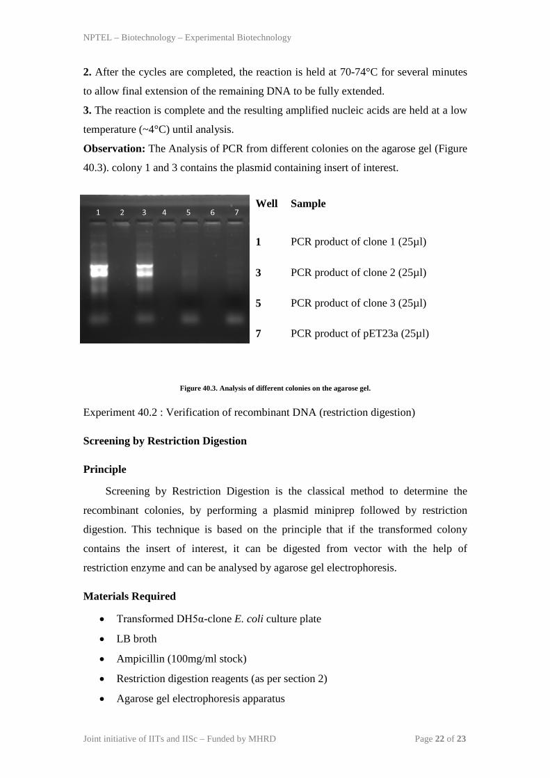
Observation: The Analysis of PCR from different colonies on the agarose gel (Figure
40.3). colony 1 and 3 contains the plasmid containing insert of interest.
Well
Sample
1 PCR product of clone 1 (25µl)
3 PCR product of clone 2 (25µl)
5 PCR product of clone 3 (25µl)
7 PCR product of pET23a (25µl)
Figure 40.3. Analysis of different colonies on the agarose gel.
Experiment 40.2 : Verification of recombinant DNA (restriction digestion)
Screening by Restriction Digestion
Principle
Screening by Restriction Digestion is the classical method to determine the
recombinant colonies, by performing a plasmid miniprep followed by restriction
digestion. This technique is based on the principle that if the transformed colony
contains the insert of interest, it can be digested from vector with the help of
restriction enzyme and can be analysed by agarose gel electrophoresis.
Materials Required
• Transformed DH5α-clone E. coli culture plate
• LB broth
• Ampicillin (100mg/ml stock)
• Restriction digestion reagents (as per section 2)
• Agarose gel electrophoresis apparatus

NPTEL – Biotechnology – Experimental Biotechnology
Joint initiative of IITs and IISc – Funded by MHRD Page 23 of 23
Methodology
• All the 10 transformed DH5α-clond E. coli colonies were picked up from the
culture plate and inoculated in LB amp+ broth.
• Plasmid was isolated by TELT method from all grown culture.
• Restriction digestion was setup for the plasmid isolated by one colony of 50µl
reaction volume.
• 50µl of restriction digested sample was loaded on 0.8% agarose gel and
electrophoresed for analysis.
Restriction digestion done for clone and plasmid pET23a. The digested products were
run on agarose gel. After electrophoresis the result is as below.
Well Sample
1 1kb DNA marker (5µl)
2 RD product of clone plasmid (50µl)
4 RD product of pET23a (50µl)
Figure 40.4. Analysis of restriction digestion of different colonies on the agarose gel.
Conclusion: Insert is present in lane 2 and digested vector (pET23a) is present in lane
4.