(lec 2) embryology - embryologic derivation of oral and dental structures 2
TRANSCRIPT
Embryologic derivation Of Oral and Dental StructuresLubna Mohammad Abu Al-Rub
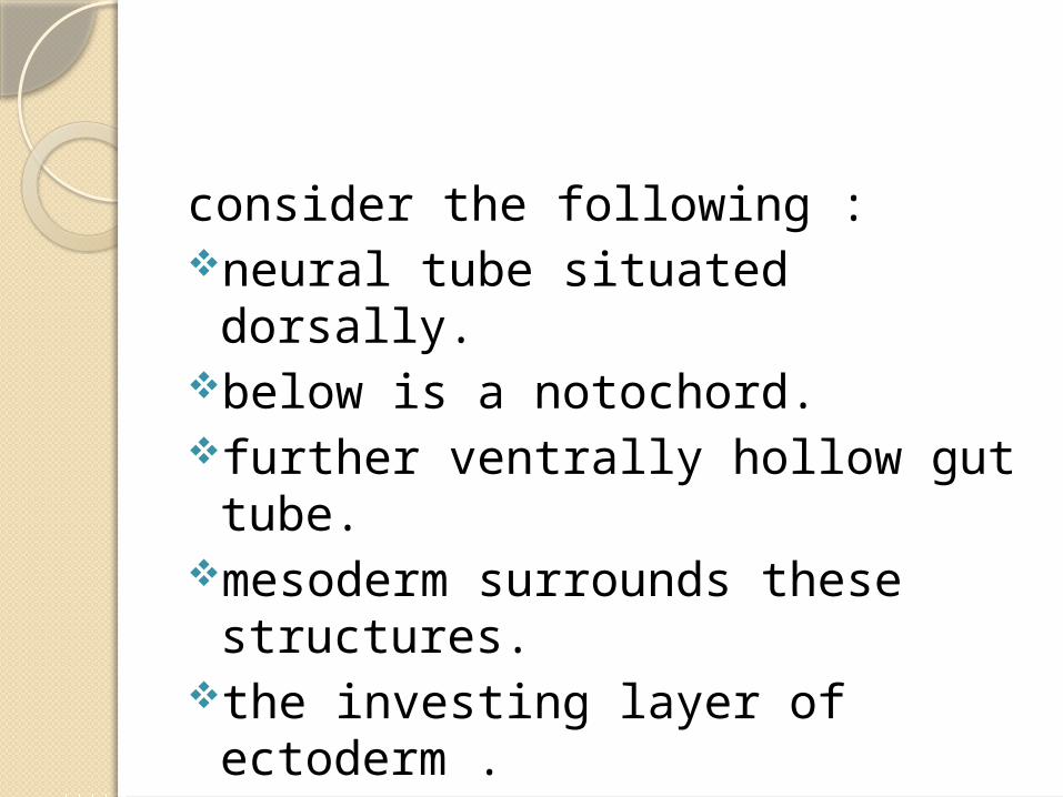
consider the following :neural tube situated dorsally.below is a notochord.further ventrally hollow gut tube.mesoderm surrounds these
structures.the investing layer of ectoderm .

Neural tube

formation of the somites by the middle of the third week
the embryo consists of three layers :
1. ectoderm which is continuous with the wall of the amnion above.
2. the endoderm - which is continuous with the yolk sac below
3. the mesoderm - continuous with the extraembryonic mesoderm peripherally.

3 weeks Embryo

the mesoderm separates the endoderm and the ectoderm except centrally where it is interrupted by the notochord.
the buccopharengeal membrane laterally and the cloaclal membrane coadally.
Mesoderm initially forms a thin sheet of tissues on either
side of the midbrain.around day 17 , mesoderm on either side thickens
to form a mass called PARAXIAL MESODERM
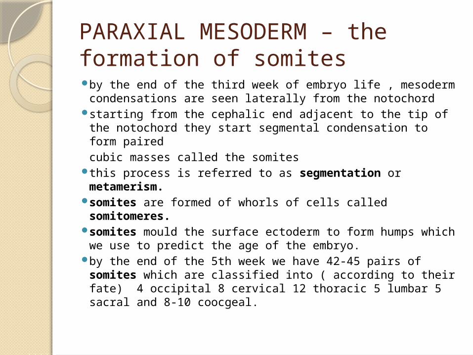
PARAXIAL MESODERM – the formation of somites by the end of the third week of embryo life , mesoderm
condensations are seen laterally from the notochord starting from the cephalic end adjacent to the tip of the
notochord they start segmental condensation to form pairedcubic masses called the somites
this process is referred to as segmentation or metamerism.
somites are formed of whorls of cells called somitomeres.
somites mould the surface ectoderm to form humps which we use to predict the age of the embryo.
by the end of the 5th week we have 42-45 pairs of somites which are classified into ( according to their fate) 4 occipital 8 cervical 12 thoracic 5 lumbar 5 sacral and 8-10 coocgeal.

The formation Of Somites

Further differentiation of the somites by the end of the fourth week each somite undergoes more
diffrentiaiton and has a myoceol - slit like cavity contains condensation of cells
which divides and by this way somites grow dermatome - beneath the myoceal- will eventually spread
beneath the ectoderm to form the dermis of the skin.
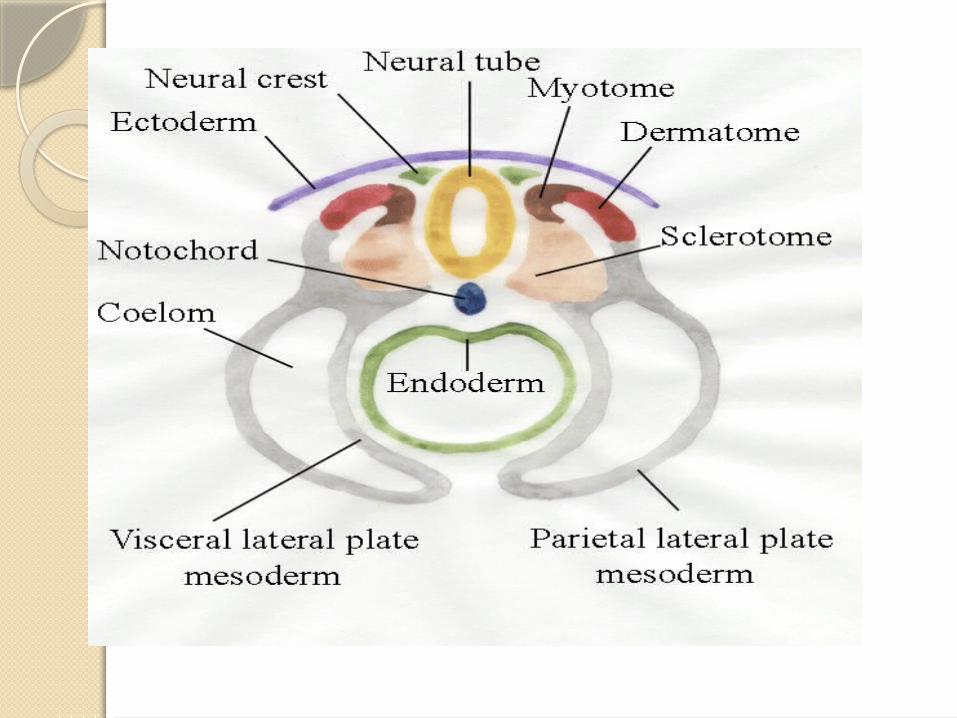
medial to the myoceol we have 2 divisions : 1.medially sclerotome : differentiates into connective
tissues MESENCHYME which migrates and condenses around the neural tube to form vertebral column the ribs and the sternumthe sclerotomes of the metotic somites are believed to contribute to the scull formation.
2. laterally myotome : forms the flexor and the extensor muscles of the vertebral column the intercostal muscles and part of the limb muscles .

Somites
paraxial mesoderm undergoes metameric segmentation .
the resulting somites become divided into dermotome , sclerotome, and myotome .
in the head region the formation of myotomes can be described as " atypical" , a number of somitomeres can reach to seven ( but this number varies according to species studied) are found within the mesoderm alongside the forebrain , midbrain and the cranial half of the hindbrain . and these do not progress to form somites.
those somitomeres give rise to the striated muscles of the face jaw and throat.
the most cephalic fully formed somites are four in number and are called the occipital or metotic behind the ottic capsule ) somites.
an older and more classic interpretation of head somites describes three pairs of prootic somites ( anterior to the ottic capsule ). prootic somites do not form seperate and identifiable structures in modern species.
their myotomes give rise to extraoccular muscles.
the nerves supplying their myotomes ( occlumotor trochlear and abducent) are equivilant to the ventral roots of the spinal nerves through which the muscles of the trunk are supplied .

dermatomes of prootic somites are supplied by cranial nerves equivalent to dorsal roots of the spinal nerves.
these cranial nerves are the ophthalmic of the trigiminal and the combined maxillary and mandibular of the trigeminal.
the dermatomes of the first and second prootic somites are enlarged thus the corresponding trigiminal divisions are enlarged and carry many sensations
the dermatome of the third prootis somite is small and the cutanous content of the facial nerve supplying it thus is small


occipital somites there are controversy regarding their number. according to the CLASICAL view they are 6 on each side but the
first has disappeared and the second is a transient structure . so usually we describe four instead of 6 ( the original number) the mesoderm of the occipital myotome migrates to the floor of
the mouth where it forms the musculature of the tongue .___> the nerves supplying theose myotomes are ventral root of spinal nerves.
in higher vertebrates they are fused to form the hypoglossal nerve
corresponding dorsal nerves are fused to form the vagues nerve. the glossopharengeal nerve is the nerve of the first missing
occipital somite according to the classical view. the sclerotomes of the occipital somites become incorporated in
the posterior part of the scull base -parachordal their number is still disputed .

unlike the spinal dorsal nerves , the dorsal nerves of the head region fail to fuse with the corresponding ventral nerves .the contain :
1. sensory fibers2. visceral motor fibers3. motor fibers which supply the striated
muscles associated with the pharengeal (brachial )arches , thats why they are sometimes called the brachial nerves .