learning, techniques, and complications of endoscopic ... · learning, techniques, and...
TRANSCRIPT

Learning, techniques, and complications ofendoscopic ultrasound (EUS)-guided samplingin gastroenterology: European Society of Gastro-intestinal Endoscopy (ESGE) Technical Guideline
Authors M. Polkowski1, A. Larghi2, B. Weynand3, C. Boustière4, M. Giovannini5, B. Pujol6, J.-M. Dumonceau7
Institutions Institutions are listed at the end of article.
submitted 10. May 2011accepted after revision10. October 2011
BibliographyDOI http://dx.doi.org/10.1055/s-0031-1291543Published online: 16.12.2011Endoscopy 2012; 44: 190–205© Georg Thieme Verlag KGStuttgart · New YorkISSN 0013-726X
Corresponding authorM. Polkowski, MDDepartment ofGastroenterology,The M. Sklodowska-CurieMemorial Cancer Center andInstitute of Oncology,Roentgena 502–781 WarsawPolandFax: [email protected]
Guideline190
1. Introduction!
The current Technical Guideline discusses issuesrelated to the learning, techniques, and complica-tions of endoscopic ultrasound (EUS)-guidedsampling and to processing of specimens obtain-ed with EUS-guided fine needle aspiration (EUS-FNA) or EUS-guided Trucut biopsy (EUS-TCB).The results of EUS-guided sampling in variousclinical indications, the role of this technique inpatient management, and recommendations onits use are discussed in the associated ClinicalGuideline from the European Society of Gastroin-testinal Endoscopy (ESGE) [1].
2.Methods!
The ESGE commissioned and funded thisGuideline. The method for guideline developmentwas similar to that used for other ESGE Guidelines[2,3]. Briefly, subgroups were formed, eachcharged with a series of clearly defined key ques-tions (see●" Appendix e1, available online). Thecommittee chair worked with subgroup leadersto identify pertinent search terms that always in-
cluded, as a minimum, “endoscopic ultrasonogra-phy” and words pertinent to specific key ques-tions. Evidence tables were generated for eachkey question based on meta-analyses or random-ized controlled trials (RCTs) if these were avail-able; otherwise, case–control studies, retrospec-tive analyses, and case series were included. Thenumber of articles retrieved and selected foreach task force is indicated in the Evidence table(see●" Appendix e2, available online). Evidencelevels and recommendation grades used in theseguidelines were those recommended by theamended Scottish Intercollegiate Guidelines Net-work (SIGN) (●" Table1) [4]. Subgroups agreedelectronically on draft proposals that were pres-ented to the entire group for general discussionduring two meetings held in 2010 and 2011.Thesubsequent Guideline version was discussedusing electronic mail until unanimous agreementwas reached. Searches were re-run in February2011 (this date should be taken into account forfuture updates). The final draft was approved byall members of the Guideline development group;it was sent to all individual ESGE members inApril 2011 and, after incorporation of their com-ments, it was endorsed by the ESGE Governing
Polkowski M et al. Learning, techniques, and complications of EUS-guided sampling… Endoscopy 2012; 44: 190–205
This article is the second of a two-part publicationthat expresses the current view of the EuropeanSociety of Gastrointestinal Endoscopy (ESGE)about endoscopic ultrasound (EUS)-guided sam-pling, including EUS-guided fine needle aspira-tion (EUS-FNA) and EUS-guided Trucut biopsy.The first part (the Clinical Guideline) focused onthe results obtained with EUS-guided sampling,and the role of this technique in patient manage-ment, and made recommendations on circum-stances that warrant its use. The current Techni-cal Guideline discusses issues related to learning,techniques, and complications of EUS-guidedsampling, and to processing of specimens. Techni-cal issues related to maximizing the diagnostic
yield (e.g., rapid on-site cytopathological evalua-tion, needle diameter, microcore isolation for his-topathological examination, and adequate num-ber of needle passes) are discussed and recom-mendations are made for various settings, includ-ing solid and cystic pancreatic lesions, submuco-sal tumors, and lymph nodes. The target reader-ship for the Clinical Guideline mostly includesgastroenterologists, oncologists, internists, andsurgeons while the Technical Guideline shouldbe most useful to endoscopists who performEUS-guided sampling. A two-page executive sum-mary of evidence statements and recommenda-tions is provided.
Board prior to submission to Endoscopy for international peer re-view. The final revised version was approved by all members ofthe Guideline development group before publication.Evidence statements and recommendations are stated in italics,key evidence statements and recommendations are in bold. ThisGuideline will be considered for review in 2014, or sooner if im-portant new evidence becomes available. Any updates to theGuideline in the interim period will be noted on the ESGE web-site: http://www.esge.com/esge-guidelines.html.
3.Summary of statements and recommendations!
Learning EUS-FNAEUS-FNA is an extension of EUS; all endoscopists who reported theirlearning curve for EUS-FNA had prior experience in EUS.Materialavailable for learning EUS-FNA includes common didactic material(e.g., books, videos), various types of simulators, and live pigs.Among models available for “hands-on” training, live pigs are themost realistic and could allow the improvement of EUS-FNA skillsbut are not widely available. The learning process of EUS-FNA hasbeen studied for solid pancreatic lesions only; it showed a learningcurve with increasing sensitivity for the cytopathological diagnosisof cancer (reaching 80% after 20–30 EUS-FNA), decreasing number
of passes needed to obtain adequate results (reaching a median of 3after 150 EUS-FNA), but no variation in severe morbidity. In all re-ported studies, rapid on-site cytopathological examination (ROSE)was used to guide the number of FNA passes needed (Evidence level2+).Trainees should demonstrate competence in linear EUS beforeundertaking EUS-FNA. We discourage self-learning of EUS-FNA.We recommend combination of the use of different simulatorsand, if available, live pigs, during training in EUS-FNA. We recom-mend that a minimum of 20 and 30 supervised EUS-FNAs of non-pancreatic and pancreatic lesions, respectively, be performedwith ROSE before assessment of competency in these techniques(Recommendation grade C). ROSE is preferable although directsupervision by an endosonographer experienced in EUS-FNAcan be another option. Close collaboration with a cytopathologistexperienced in evaluation of EUS-FNA samples is recommended(Recommendation grade D).
Techniques of EUS-FNAFor EUS-FNA of pancreatic lesions the 19G, 22G and 25G needles arecharacterized by similar diagnostic yields (Evidence level 1+) andsafety profiles (Evidence level 1– ). Although 19G needles provide ahigher amount of cellular material than do thinner needles, and, iftechnically successful, offer better diagnostic yield, these advanta-ges are offset by a higher rate of technical failures in the case of le-sions that need to be punctured from the duodenum (Evidence level1– ). Studies comparing EUS-FNA needles of different sizes in indi-cations other than pancreatic masses are lacking. We recommendagainst using 19G needles for transduodenal biopsy (Recommenda-tion grade C).Applying continuous suction with a syringe during EUS-FNA improvesthe sensitivity for the diagnosis of malignancy in patients with solidmasses but not in patients with lymphadenopathy (Evidence level1– ).We recommend using suction for EUS-FNA of solid masses/cys-tic lesions and not using suction for EUS-FNA of lymph nodes (Re-commendation grade C).Using the needle stylet does not seem to impact EUS-FNA samplequality and results (Evidence level 1– ). There is insufficient evidenceto recommend for or against using the stylet and the decision in thisregard should be left to the discretion of the endosonographer per-forming the procedure (Recommendation grade C).Diagnostic accuracy of EUS-FNA does not differ depending on wheth-er the sampling is performed from the edge of a lymph node or fromits center (Evidence level 1– ). No data on this topic are available forlesions other than lymph nodes. We recommend sampling all partsof solid lesions or lymph nodes (Recommendation grade C) andsampling any solid component inside pancreatic cysts and thewall of the cyst (Recommendation grade D).Gross visual inspection is unreliable in assessing the adequacy ofEUS-FNA specimens for cytopathological examination. ROSE pro-vides a highly reliable diagnosis with an excellent agreement withthe final cytopathological diagnosis (Evidence level 2+). There islimited evidence to suggest that ROSE increases the diagnostic yieldof EUS-FNA (Evidence level 2– ). The diagnostic yield of EUS-FNAwith ROSE in most studies exceeds 90%; however, similarly good re-sults have been reported from selected studies without ROSE. (Evi-dence level 2+). Data on cost–effectiveness of ROSE are very lim-ited. In view of these data, it is felt that implementation of ROSEshould be considered especially during the learning phase of EUS-FNA and at centers in which specimen adequacy rates are below90% (Recommendation grade D).
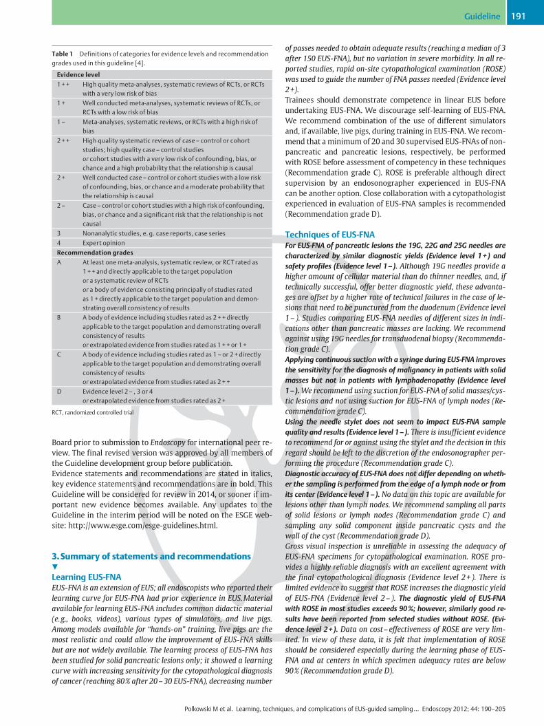
Table 1 Definitions of categories for evidence levels and recommendationgrades used in this guideline [4].
Evidence level
1++ High quality meta-analyses, systematic reviews of RCTs, or RCTswith a very low risk of bias
1 + Well conducted meta-analyses, systematic reviews of RCTs, orRCTs with a low risk of bias
1– Meta-analyses, systematic reviews, or RCTs with a high risk ofbias
2 ++ High quality systematic reviews of case– control or cohortstudies; high quality case– control studiesor cohort studies with a very low risk of confounding, bias, orchance and a high probability that the relationship is causal
2 + Well conducted case – control or cohort studies with a low riskof confounding, bias, or chance and a moderate probability thatthe relationship is causal
2– Case– control or cohort studies with a high risk of confounding,bias, or chance and a significant risk that the relationship is notcausal
3 Nonanalytic studies, e. g. case reports, case series
4 Expert opinion
Recommendation grades
A At least one meta-analysis, systematic review, or RCT rated as1 ++ and directly applicable to the target populationor a systematic review of RCTsor a body of evidence consisting principally of studies ratedas 1 + directly applicable to the target population and demon-strating overall consistency of results
B A body of evidence including studies rated as 2 + +directlyapplicable to the target population and demonstrating overallconsistency of resultsor extrapolated evidence from studies rated as 1 ++or 1+
C A body of evidence including studies rated as 1 – or 2+ directlyapplicable to the target population and demonstrating overallconsistency of resultsor extrapolated evidence from studies rated as 2 ++
D Evidence level 2– , 3 or 4or extrapolated evidence from studies rated as 2 +
RCT, randomized controlled trial
Polkowski M et al. Learning, techniques, and complications of EUS-guided sampling… Endoscopy 2012; 44: 190–205
Guideline 191

Various studies have investigated the adequate number of needlepasses that should be performed if ROSE is not used. Discordantconclusions have been reached for solid masses, while more concor-dant results have been reported for lymph nodes, liver lesions, andpancreatic cysts. We recommend performing 3 needle passes forlymph nodes and liver lesions, at least 5 needle passes for solid pan-creatic masses, and a single pass for pancreatic cysts (Recommen-dation grade C).
Techniques to obtain tissue for histopathologicalevaluationEUS-FNAwith standard needles can provide tissue adequate for histo-pathological evaluation frommost pancreatic tumors (Evidence level2+). Combining EUS-FNA histology and EUS-FNA cytology seems toincrease EUS-FNA diagnostic yield (Evidence level 2– ) and sensitiv-ity for pancreatic cancer detection (Evidence level 2+). Other po-tential advantages of EUS-FNA histology consist of facilitated im-munostaining and better capability to diagnose specific tumortypes (Evidence level 2– ). We suggest implementation of this tech-nique into routine practice (Recommendation grade D).Transduodenal EUS-TCB is characterized by a very high failure rate(Evidence level 2+ ). For non-transduodenal routes, the failure rate islow and the accuracy for the detection of malignancy is similar tothat of EUS-FNA (Evidence level 2+ ). The accuracy of dual sampling(EUS-TCB+EUS-FNA) is superior to either technique alone (Evi-dence level 2+). Sequential sampling (EUS-TCB with EUS-FNA res-cue) has similar accuracy to that of dual sampling (Evidence level2– ). EUS-TCB is superior to EUS-FNA in establishing some specificdiagnoses, especially of benign tumors or if immunostaining is re-quired (Evidence level 2– ). In most instances EUS-TCB does not of-fer advantages over EUS-FNA; however, EUS-TCB should be consid-ered when tissue architectural details and immunostaining are re-quired to establish a specific diagnosis (Recommendation grade C).
Specimen processingNo adequate study has compared direct smear cytology vs. liquid-based cytology (LBC) for processing specimens collected with EUS-FNA. Similarly, no study has evaluated which of the methods de-scribed for collecting tissue fragments for histopathological exami-nation is better. In the case of suspected tuberculosis or lymphoma,polymerase chain reaction (suspected tuberculosis) on histopatholo-gical specimens and flow cytometry (suspected lymphoma), afterplacement of the collected specimen in an adequate transport medi-um have, been shown to significantly increase the diagnostic yield(Evidence level 2+). The specific method to be used for both cytopa-thological processing and collection of histopathological specimensshould be left to the discretion of each center, depending on theirconfidence with available methods (Recommendation grade D).Cell blocks can be used as a complement to rather than a replace-ment for smears or LBC (Recommendation grade D). If tuberculosisis suspected, then polymerase chain reaction should be used; iflymphoma is suspected then flow cytometry should be used (Re-commendation grade C).
Complications of EUS-FNA and their preventionEUS-FNA is a safe procedure with a complication rate of approxi-mately 1% (Evidence level 2++). Complications include infection,bleeding, and acute pancreatitis; they are more frequent for EUS-FNA of cystic compared with solid lesions (Evidence level 2– ). Bac-teremia is rare after EUS-FNA, including that of perirectal and rectallesions (Evidence level 2++). The 19G, 22G, and 25G EUS-FNA needlespresent similar complication rates. Transesophageal and transgas-
tric EUS-TCB have similar safety profiles compared with EUS-FNA, atleast in experienced hands (Evidence level 1– ). Aspirin/nonsteroidalanti-inflammatory drugs (NSAIDs) do not seem to increase the riskof bleeding following EUS-FNA (Evidence level 2– ).Antibiotic prophylaxis is recommended before EUS-guided sam-pling of cystic lesions (Recommendation grade C) but not of solidlesions (Recommendation grade B). Antibiotic prophylaxis of infec-tive endocarditis is not recommended (Recommendation grade B).Coagulation check-up is recommended before EUS-FNA only in pa-tients with a personal or family history suggesting bleeding disor-der or with a clear clinical indication (Recommendation grade C).EUS-guided sampling should not be performed in patients treatedwith oral anticoagulants (Recommendation grade C) or thienopyr-idines (Recommendation grade D). In addition, treatment with as-pirin or NSAIDs is a contraindication for EUS-guided sampling ofcystic lesions (Recommendation grade C).EUS-TCB is contraindicated for lesions requiring a transduodenalapproach, lesions <20mm or of cystic appearance, and when theoperator has limited experience with standard EUS-FNA (Recom-mendation grade D).
4.Learning EUS-FNA!
EUS-FNA is an extension of EUS; all endoscopists who reported theirlearning curve for EUS-FNA had prior experience in EUS.Materialavailable for learning EUS-FNA includes common didactic material(e.g., books, videos), various types of simulators, and live pigs.Among models available for “hands-on” training, live pigs are themost realistic and could allow the improvement of EUS-FNA skillsbut are not widely available. The learning process of EUS-FNA hasbeen studied for solid pancreatic lesions only; it showed a learningcurve with increasing sensitivity for the cytopathological diagnosisof cancer (reaching 80% after 20–30 EUS-FNA), decreasing numberof passes needed to obtain adequate results (reaching a median of 3after 150 EUS-FNA), but no variation in severe morbidity. In all re-ported studies ROSE was used to guide the number of FNA passesneeded (Evidence level 2+).Trainees should demonstrate competence in linear EUS before un-dertaking EUS-FNA. We discourage self-learning of EUS-FNA. Werecommend combination of the use of different simulators and, ifavailable, live pigs, during training in EUS-FNA. We recommendthat a minimum of 20 and 30 supervised EUS-FNA of non-pancre-atic and pancreatic lesions, respectively, be performed with ROSEbefore assessment of competency in these techniques (Recommen-dation grade C). ROSE is preferable although direct supervision byan endosonographer experienced in EUS-FNA can be another op-tion. Close collaboration with a cytopathologist experienced inevaluation of EUS-FNA samples is recommended (Recommendationgrade D).
4.1.Training in EUS-FNAThe results of two methods for learning EUS-FNA have been re-ported, i. e. formal training, consisting of fellowship in a dedica-ted training center for 6–24 months, and informal training, con-sisting of short repeated exposures to various didactic situationsthat usually included short ‘‘hands-on’’ experiences [5]. Formaltraining programs are scarce in Europe and even in countrieswhere they are most developed (e.g., France), they allow trainingof only a small number of endoscopists per year [6–8]. In addi-tion, the long duration of formal training programs is impracticalfor the practicing, experienced endoscopist. The proportion of
Polkowski M et al. Learning, techniques, and complications of EUS-guided sampling… Endoscopy 2012; 44: 190–205
Guideline192

endosonographers who report that they are self-taught variesbetween 8% and 50% [9–11]. Although unalloyed self-educationis feasible for simple endoscopic procedures while maintaininghigh quality and safety standards [12], it has not been reportedfor more complex procedures such as EUS-FNA [5].It seems reasonable to assume that any training has to be foun-ded on theoretical and clinical knowledge [7]. Furthermore, allof the endoscopists who reported their learning curve for EUS-FNA had performed diagnostic EUS before performing supervisedEUS-FNA [13–15]. Competence in percutaneous abdominal ul-trasound is not a prerequisite for EUS or EUS-FNA because no evi-dence was found in the literature that it improves competence inEUS.Criteria useful for assessing whether competence in EUS hasbeen reached are available [16].The use of textbooks and videos is recommended in most con-sensus statements as a basis for EUS training. Hands-on trainingin EUS-FNA has used: (i) phantoms devoid of animal material(Olympus, Tokyo, Japan; self-made phantoms constructed withcommonly available materials); (ii) models using porcine organs(upper and lower digestive EUS-FNA) [17–19]; and (iii) live pigs[20]. Simulators (www.simbionix.com) do not currently offertraining in FNA. All of these models have been subjected to feasi-bility studies only, except for the live pig model. The latter wasevaluated during a 4-week EUS course: a significant improve-ment was noted in terms of duration and precision of the proce-dure between the first and second attempt at FNA of lymphnodes at the liver hilum [6]. This model was judged by eight EUSexperts as the most realistic and useful for teaching EUS-FNA, butthe least easy to incorporate into fellowship training [18]. Theseexperts recommended using different learning tools at differenttime periods during the learning curve for EUS and EUS-FNA. Amodel using porcine organs was preferred over both live pigsand phantoms devoid of animal material.
4.2.Learning curve of EUS-FNAFive endosonographers have reported their learning curve forEUS-FNA of solid pancreatic lesions, the procedure considered tobe the most complex (●" Table2) [13–16]. All endosonographershad performed a minimum of 132–300 diagnostic EUS prior toEUS-FNA, and had participated in a formal or informal trainingprogram, and they used ROSE to guide the number of FNA passes.For the cytopathological diagnosis of pancreatic cancer, sensitiv-ity increased with the operator’s experience and reached 80%after 20 to 30 EUS-FNA (including operator’s experience prior to
the study if applicable). ROSE may be useful to guide the numberof FNA passes, learn which parts of the lesion may be targeted forincreased diagnostic yield, and correct technical errors (e.g.,bloody or paucicellular material) [21,22].The American Society of Gastrointestinal Endoscopy has reap-proved in November 2008 its recommendations that competencyshould be assessed separately for pancreatic and non-pancreaticEUS-FNA, after at least 25 supervised procedures of each type[16]. For all endoscopy procedures, substantial variations existbetween individuals with regard to the speed of learning [23],so that this number should be considered to be the minimum be-fore evaluating the trainee. In addition, as shown by Eloubeidi etal., the learning curve continues long after EUS fellowship. In aprospective study evaluating 300 EUS-FNA performed by a singleendosonographer, who had performed 45 supervised proceduresduring a training period before the study period, the proportionof EUS-FNA that required ≥5 passes significantly decreased after100 additional procedures and the complication rate decreasedafter 200 additional procedures (most of these complicationswere graded as minor) [13].
5.Techniques of EUS-FNA!
Needles for sampling under EUS guidance are available from fourmanufacturers (●" Table3). Most models are intended for aspirat-ing cellular material for cytopathological examination. Tissuefragments suitable for histopathological examination can be ob-tained using standard 19G or 22G FNA needles as well as withdedicated histopathological needles (e.g. Trucut, ProCore). Thefollowing sections discuss various technical issues related toEUS-FNA and EUS-TCB. For detailed expert instruction on howto perform step-by-step EUS-FNA and EUS-TCB, readers are refer-red to other sources [24–26].
5.1.Does the diameter of EUS-FNA needle matter?For EUS-FNA of pancreatic lesions, the 19G, 22G and 25G needlesare characterized by similar diagnostic yields (Evidence level 1+)and safety profiles (Evidence level 1– ). Although 19G needles pro-vide a higher amount of cellular material than do thinner needles,and, if technically successful, offer a better diagnostic yield, theseadvantages are offset by a higher rate of technical failures in thecase of lesions that need to be punctured from the duodenum (Evi-
Table 2 Series reporting the learning curve for endoscopic ultrasound (EUS)-guided fine needle aspiration (FNA) of solid pancreatic lesions.
First author,
year
Patients/
operators, n/n
Operator
experience prior
to study period
Training: type, duration Sensitivity for
cancer diagnosis:
First vs. last FNA
series compared
Complications FNAs needed to
reach 80%
sensitivity for
cancer diagnosis,
n
Harewood,2002 [15]
65 /3 > 300 EUS< 10 EUS-FNA
Informal1, 2 months 44% vs. 91% No data 20
Mertz, 2004[14]
57 /1 132 EUSNo EUS-FNA
Informal 1, no data 50% vs. 80% 0 30
Eloubeidi, 2005[13]
300 /1 316 EUS45 EUS-FNA
Formal, 1 year 92% vs. 95% 13%2 No data
1 Mentoring during the performance of 2 to 10 pancreatic EUS-FNAs by an experienced endosonographer2 Including major complications (oversedation that required the administration of a reversal agent or hospitalization or emergency department visit) in 2% of patients
Polkowski M et al. Learning, techniques, and complications of EUS-guided sampling… Endoscopy 2012; 44: 190–205
Guideline 193

dence level 1– ). Studies comparing EUS-FNA needles of different si-zes in indications other than pancreatic masses are lacking.We recommend against using 19G needles for transduodenal biop-sy (Recommendation grade C).Most of the studies on EUS–FNA have been conducted using 22Gneedles. Data on thinner (25G) or larger (19G) needles are lim-ited. A number of recent studies, including two RCTs, comparedresults obtained with needles of various diameters. All thesestudies were performed in the setting of pancreatic masses [27–31].It has been suggested that although thinner needles provide lesscellular material than do larger needles, the specimens from theformer are less contaminated by blood, and thus easier to inter-pret. In addition, thinner needles may be easier to use becauseof greater flexibility, particularly for locations requiring impor-tant scope bending [30,31]. This preliminary evidence was onlypartly confirmed in further research.In a small prospective, non-randomized study in 24 patients, thetechnical success rate for the 25G needle was significantly higherthan for the 22G needle, but only for tumors located in the unci-nate process [29].An RCT in 131 patients found no significant differences between22G and 25G needles in terms of diagnostic yield for malignancy,number of needle passes needed to obtain a diagnosis, ease ofneedle passage into the mass, and rates of needle malfunctionand of complications [27]. ROSEwas used in this study.Another RCT compared EUS-FNAwithout ROSE using 19G or 22Gneedles in 117 patients [28]. In the intention-to-treat analysis, di-agnostic accuracy was similar for both needles. However, if tech-nical failures were excluded (per-protocol analysis), diagnosticaccuracy was higher with the 19G compared to the 22G needle(95% vs. 79%, respectively; P=0.015). Technical failures were re-ported only for 19G needles in patients with pancreatic headmasses (in 19% of cases). The 19G needle provided a higheramount of cellular material with fewer passes (2.4 vs. 2.8; respec-tively; P=0.01). No complications were observed in either group.
5.2.Should suction be applied during EUS-FNA?Applying continuous suction with a syringe during EUS-FNA im-proves the sensitivity for the diagnosis of malignancy in patientswith solid masses but not in patients with lymphadenopathy (Evi-dence level 1– ). We recommend using suction for EUS-FNA of solidmasses/cystic lesions and not using suction for EUS-FNA of lymphnodes (Recommendation grade C).Traditionally, suction is applied during EUS-FNA using a syringe[32]. EUS-FNA without suction has been tested in an attempt todecrease sample bloodiness and to improve accuracy of micro-scopic examination. Two RCTs have compared EUS-FNA with orwithout suction, in a total of 95 patients with suspected malig-nant lymph nodes, pancreatic masses or submucosal tumors(SMTs) [33,34]. In a study on 46 patients with lymphadenopathy,applying suction did not improve diagnostic accuracy and wors-ened specimen bloodiness compared with EUS-FNAwithout suc-tion [33]. In the other study, however, using suction during EUS-FNA of solid masses was associated with a significantly highersensitivity for cancer diagnosis (86% vs. 67%; P=0.05) [34]. A pi-lot trial suggested that applying continuous high pressure suc-tion (using a balloon inflation device) allowed retrieval of tissuesamples for histopathological examination in most cases [35].
5.3.With or without needle stylet?Using the needle stylet does not seem to impact EUS-FNA samplequality and results (Evidence level 1– ). There is insufficient evi-dence to recommend for or against using the stylet and the decisionin this regard should be left to the discretion of the endosonogra-pher performing the procedure (Recommendation grade C).For years the standard approach has been to reinsert the styletinto the needle before every pass to prevent sample contamina-tion by cells from the digestive wall as well as blockage of theneedle that would hinder sample aspiration. Recently the valueof this measure has been questioned by the results of three stud-ies, including one RCT [36–38]. While these studies found no ad-vantages of using the stylet with regard to the quality of sampleobtained or the diagnostic yield of malignancy, they also did notdemonstrate any disadvantages of this approach. In addition, twoof these studies suffered from significant methodological limita-tions [37–39].
Table 3 Needles forendoscopic ultrasound (EUS)-guided sampling.
Manufacturer Model Needle type Needle diameter Single-use vs. reusable
Boston Scientific
Expect Aspiration needle 19G, 22G, 25G Single-use
Expect Flex Aspiration needle 19G Single-use
Cook
Echotip Aspiration needle 22G Single-use
Echotip Ultra Aspiration needle 19G, 22G, 25G Single-use
Echotip ProCore1 Aspiration needle with a core trap 19G, 22G Single-use
QuickCore Core biopsy needle 19G Single-use
EchoBrush2 Needle with cytology brush 19G Single-use
Mediglobe
Sonotip Pro Control Aspiration needle 19G, 22G, 25G Single-use
Olympus
Power-Shot3 Aspiration needle 22G Reusable
EZ-Shot3 Aspiration needle 22G Single-use
EZ-Shot 2 Aspiration needle 19G, 22G, 25G Single-use
EZ-Shot 2 with sideport4 Aspiration needle with sideport 22G Single-use
1 A newly marketed needle designed with a core trap and reverse bevel technology to increase sampling yield and promote collection of histopathological samples.2 A modified stylet with a 1 ×5-mm brush at its end; it is designed to pass through a 19G EUS-FNA needle, for brushing the cyst wall.3 Compatible exclusively with Olympus endoscopes.4 A newly marketed needle designed with a sideport to draw tissue from both the tip and side of the needle.
Polkowski M et al. Learning, techniques, and complications of EUS-guided sampling… Endoscopy 2012; 44: 190–205
Guideline194

5.4.Which part of the lesion should be puncturedto maximize the diagnostic yield?Diagnostic accuracy of EUS-FNA does not differ depending onwhether the sampling is performed from the edge of a lymph nodeor from its center (Evidence level 1– ). No data on this topic areavailable for lesions other than lymph nodes.We recommend sampling all parts of solid lesions or lymph nodes(Recommendation grade C) and sampling any solid component in-side pancreatic cysts and the wall of the cyst (Recommendationgrade D).Because malignant masses and lymph nodes may undergo cen-tral necrosis, it has been assumed that FNA of the edges of the le-sion rather than of the center would increase the diagnosticyield. The study by Wallace et al. in which 46 lymph nodes werepunctured found that aspiration from the edge of the lymph nodedid not increase the likelihood of a correct diagnosis when com-pared to aspiration from the lymph node center [33]. This issuehas not been studied for lesions other than lymph nodes. In prac-tice, the needle is usually “fanned” throughout the lesion to sam-ple all its parts.Recent research indicates that new techniques such as contrast-enhanced EUS and elastography may potentially be useful to se-lect themost suspicious area of a lymph node/tumor for EUS-FNA[40,41].According to expert opinion, the diagnostic yield of EUS-FNA ofpancreatic cysts may be improved by aspirating cells from thecyst wall after having aspirated cyst fluid. Using this method, Ro-gart et al. collected cellular material adequate for cytopathologi-cal assessment in 82 (76.6%) of 107 cysts [42]. If a cyst wall thick-ening is present (or solid nodules or a solid component inside thecyst), it is advised to sample these targets before aspirating cystfluid (this would become more difficult once the cyst has col-lapsed). A cytology brush can also be introduced into the cystthrough a 19G needle to scrape the cyst wall [43–45]. This tech-nique has been shown to increase the cellular and diagnosticyields; however, serious concerns exist about its complication,in particular a high risk of intracystic bleeding [43,45,46].
5.5.What is the role of ROSE?Gross visual inspection is unreliable in assessing the adequacy ofEUS-FNA specimens for cytopathological examination. ROSE pro-vides a highly reliable diagnosis with an excellent agreement withthe final cytopathological diagnosis (Evidence level 2+). There islimited evidence to suggest that ROSE increases the diagnostic yieldof EUS-FNA and accuracy for malignancy detection (Evidence level2– ). The diagnostic yield of EUS-FNA with ROSE in most studies ex-ceeds 90%; however, similarly good results have been reported fromselected studies without ROSE. (Evidence level 2+). Data on cost–effectiveness of ROSE are very limited.In view of these data, it is felt that implementation of ROSE shouldbe considered especially during the learning phase of EUS-FNA andat centers in which specimen adequacy rates are below 90% (Re-commendation grade D).
5.5.1.Gross visual inspection of the specimenIn a prospective, double-blind, study that included 37 patientswith a solid pancreatic mass, neither trained EUS technologistsnor cytotechnologists were able to provide a reliable assessmentof specimen adequacy by using gross visual inspection. Theagreement between their assessment and the final microscopicassessment by a cytopathologist was only fair, with kappa valuesof about 0.2. False-positive assessments occurred for about 30% of
the slides, with the potential consequence of premature proce-dure termination [47].
5.5.2.ROSE by a cytopathologistROSE has been evaluated mostly in studies of percutaneous FNA:it is generally accepted that ROSE diagnosis is highly reliable andROSE is the critical procedure for reducing the number of inade-quate diagnoses. In addition, ROSE may reduce costs by decreas-ing the number of repeat procedures [48–50].Data on ROSE of EUS-FNA specimens are limited. Based on earlyreports that suggested that ROSE may increase adequacy rates ofEUS-FNA specimens by 10%–29% [51, 52], ROSE has been imple-mented at many EUS centers, especially in the United States [53],and has been used in many important studies on EUS-FNA [54–57]. The very high specimen adequacy rates consistently report-ed in these studies (>90%–95%) have been assumed to be linkedto ROSE. However, EUS-FNA with vs. without ROSE has neverbeen compared in an RCT. In addition, as discussed below, thereare data to suggest that neither does ROSE guarantee, nor is it es-sential to achieve high adequacy rates.Evaluating ROSE accuracy or impact has been the primary focusof a few studies only:▶ In a prospective study evaluating 607 EUS-FNA procedures
(mostly of pancreatic masses and lymph nodes), the agree-ment between ROSE and final cytopathological diagnosis wasexcellent (kappa=0.84) [58]. Compared with the true final di-agnosis, accuracies of ROSE and final cytopathological exami-nation were not statistically different (93.9% and 95.8%,respectively).
▶ In a retrospective comparison of EUS-FNA results obtained byone endosonographer in two university hospital centers (withROSE available in only one of them), unequivocal cytopatholo-gical diagnosis was obtained significantly more frequently(78% vs. 52%; odds ratio [OR], 2.94; P=0.001) with a lower rateof unsatisfactory specimens (9% vs. 20%; OR, 0.36; P=0.035) atthe center where ROSEwas available [21]. Because patient po-pulations and indications for EUS-FNA significantly differedbetween the compared centers, no definite conclusions can bedrawn from this study.
▶ In a prospective multicenter study that evaluated 409 patients,two centers used ROSEwhereas the other two did not [59]. Theresults obtained in these two settings were not significantlydifferent (except for a higher negative predictive value in thesubgroup of patients with extraintestinal mass lesions whenROSEwas used).
▶ In a retrospective analysis of risk factors for inadequate EUS-FNA specimens in 247 pancreatic tumors and 276 lymphnodes, cytopathological adequacy was significantly higher forlymph nodes (96% vs. 84%, P=0.008) but not for pancreatictumors (99% vs. 100%; P=1) when an on-site cytotechnologistwas present [60].
▶ A recent retrospective analysis of data from a prospectivelymaintained database showed that in patients with solid pan-creatic masses, ROSE reduced the number of inadequate FNAsamples (1% vs. 12.6%; P=0.002) and improved the sensitivity(96.2% vs. 78.2%; P=0.002) and overall accuracy (96.8% vs.86.2%; P=0.013) of EUS-guided FNA for the diagnosis of ma-lignancy. In addition, a significantly lower number of needlepasses was required when ROSEwas used [61]. An importantlimitation of this study was that patient allocation to studygroups (95 patients biopsied with and 87 without ROSE) was
Polkowski M et al. Learning, techniques, and complications of EUS-guided sampling… Endoscopy 2012; 44: 190–205
Guideline 195
not random but based on whether on-site cytopathology ser-vice was available on a given day of the week.
Of note, many recent studies in which ROSEwas not used report-ed adequacy rates of >90%, indicating that at high volume centersROSE is not indispensable to achieve excellent results [60,62–64]. On the other hand, the use of ROSE does not unconditionallyguarantee EUS-FNA success. In a survey of 21 EUS centers in theUnited States, the diagnostic rate for malignancy in patients withpancreatic tumors varied widely from center to center, despitethe fact that ROSEwas used at almost all of them [53].Little is known on the impact of ROSE on EUS-FNA proceduraltime and it remains unclear whether using ROSE prolongs theprocedure or makes it less time-consuming by reducing thenumber of needle passes. It is assumed that an average time forobtaining the specimen and performing on-site examination is15min per sample [65]. Average time expenditure by the cytopa-thologist for ROSE of computed tomography (CT)-guided and ul-trasound-guided FNA specimens is relatively high (48.7 and 44.4minutes, respectively; measured from the time the pathologistleft the office to the time the pathologist returned to the officeafter the aspiration procedure and interpretation) [66].
5.5.3.ROSE by endosonographersA prospective double-blind study showed that even endosono-graphers with special training and extensive experience at re-viewing cytopathological material alongside a cytopathologistare less accurate than a cytotechnician in the assessment of speci-men adequacy (68%–76% for three endosonographers vs. 82%for a cytotechnician; P=0.004) and in the diagnosis of malignancy(69%–72% for three endosonographers vs. 89% for a cytotechni-cian; P<0.001) [67].Another study did not find significant differences in specimenadequacy rates, number of needle passes or EUS-FNA perform-ance characteristics in two subsequent 2-year periods in whichROSEwas performed by endosonographers (first period) or cyto-pathologists (second period). The study evaluated only a total of73 EUS-FNA procedures [68].
5.6.Howmany passes should be performed if ROSEis not used?Various studies have investigated the adequate number of needlepasses that should be performed if ROSE is not used. Discordantconclusions have been reached for solid masses, while more concor-dant results have been reported for lymph nodes, liver lesions, andpancreatic cysts. We recommend performing 3 needle passes forlymph nodes and liver lesions, at least 5 needle passes for solid pan-creatic masses, and a single pass for pancreatic cysts (Recommen-dation grade C).The knowledge of the adequate number of needle passes to beperformed to reach a good diagnostic accuracy is of paramountimportance in centers where ROSE is not used. Differences existbased on the nature of the target lesion.
5.6.1.Pancreatic massesErickson et al. found in a large study (95 patients) that a mean of3.4±2.2 needle passes (range, 1–10) were required to make a di-agnosis [52]. Well-differentiated pancreatic adenocarcinomas re-quired a higher number of passes (5.5 ± 2.7) as compared tomod-erately (2.7 ± 1.2) and poorly (2.3±1.1) differentiated tumors. Theauthors recommended performing 5–6 needle passes for pan-creatic masses. In another study of 33 patients (9 with cystic le-sions), Leblanc et al. found that the sensitivity gradually in-
creased from 16.7% for the first pass to 86.7% when more than 7passes were performed [69]. Pellisé Urquiza et al. found in a studyin 102 patients that the accuracy of EUS-FNA for pancreatic mas-ses reached a plateau at the 4th needle pass [65]. More recently,Turner et al. reported in a large cohort of 559 patients with a pan-creatic mass that a diagnostic accuracy of about 80% could be ob-tained with only 2 to 3 needle passes [70]. A high yield with amean of 1.88 needle passes was also found in another study, inwhich the material gathered with a 22-gauge EUS-FNA needlewas first evaluated for the presence of small tissue core samplesthat were placed in formalin for histopathological examinationand the rest of the material was sent for cytopathological analysis[63].
5.6.2.Lymph nodesLymph nodes generally require a lower number of needle passesto obtain an adequate diagnostic accuracy. Apart from the studyby Leblanc et al. who recommended performance of at least 5needle passes, other studies agreed that 3 needle passes weresufficient [33,52,65,69].
5.6.3.Submucosal tumorsIn the study by Pellisé Urquiza et al., the accuracy of EUS-FNA forintramural lesions in 11 patients increased gradually with eachsubsequent pass to reach a plateau at the 45% level after thefourth pass [65]. In another study, conducted in 112 patients, amean of 5.3 needle passes (range 3–9) was done, with a diagnos-tic accuracy of 83.9% when both diagnostic and suspicious sam-ples were considered to be positive [71]. Differently, in a studyfrom Japan on 141 patients, a mean of 2.5 ± 0.7 (range 1–5) pas-ses were performed with an overall rate of sample adequacy of83% that was significantly better for lesions greater than 2cmthan for those with a smaller diameter [72]. In the latter twostudies, multivariate analysis did not show the number of needlepasses to be associated with the adequacy of the collected speci-mens.
5.6.4.Miscellaneous and liver lesionsThe numbers of needle passes recommended for miscellaneousand liver lesions are similar to those recommended by differentauthors for pancreatic masses and lymph nodes, respectively. Inparticular, Leblanc et al. found that for miscellaneous lesions thesensitivity of EUS-FNA increased from 33% up to 92% after 7 pas-ses and did not change with additional passes [69]. For liver le-sions, Erickson et al. suggested a good diagnostic accuracy with2–3 needle passes, a number which is in agreement with otherstudies [52,73,74].
6.Techniques to obtain tissue for histopathologicalevaluation!
Although cytopathological examination of EUS-FNA specimensallows detection of malignancy, it often cannot provide morespecific information that may be necessary for patient manage-ment. Potential advantages of tissue specimens include informa-tion about tissue architecture andmore reliable immunostaining.
Polkowski M et al. Learning, techniques, and complications of EUS-guided sampling… Endoscopy 2012; 44: 190–205
Guideline196

6.1.Should tissue fragments be isolated from EUS-FNAspecimens and processed for histology?EUS-FNA with standard needles can provide tissue adequate forhistopathological evaluation from most pancreatic tumors (Evi-dence level 2+). Combining EUS-FNA histology and EUS-FNA cytol-ogy seems to increase EUS-FNA diagnostic yield (Evidence level 2– )and sensitivity for pancreatic cancer detection (Evidence level 2+).Other potential advantages of EUS-FNA histology consist of facilita-ted immunostaining and better capability to diagnose specific tu-mor types (Evidence level 2– ).We suggest implementation of this technique into routine practice(Recommendation grade D).There is accumulating evidence that tissue adequate for histopa-thological assessment can be obtained using standard EUS-FNAin a significant proportion of cases [63,75–79]. This technique(EUS-FNA histology) involves gross visual inspection of the sam-ple to collect minute tissue fragments that are subsequently pro-cessed for histopathological examination (see section 7.3, below).EUS-FNA histology has been evaluated mostly in the setting ofpancreatic tumors, submucosal tumors (SMTs) and lymphadeno-pathy of unknown origin (●" Table4).
6.1.1.Pancreatic massTissue adequate for histopathological evaluation can be obtainedfrom 67%–86.5% of pancreatic masses using a single pass or fewneedle passes with a standard 22G needle [63,76,77,79,80].Combining EUS-FNA cytology and histology significantly increas-es the sensitivity for malignancy diagnosis compared to cytologyor histology alone (82.9% vs. 68.1% for cytology [P=0.007], and60% for histology [P<0.0001]) [63]. FNA histology also showed a
trend towards higher accuracy in diagnosing specific tumortypes other than adenocarcinoma.
6.1.2.Submucosal tumorsComparative data are lacking, but studies that used EUS-FNA his-tology alone or combined with EUS-FNA cytology reported ahigher diagnostic yield than studies that relied only on cytopa-thological preparations (cell blocks and especially smears) [75,81–83].
6.1.3.Lymphadenopathy of unknown originIn a series of 104 patients with mediastinal or/and abdominallymphadenopathy of unknown origin, a specimen adequate forhistopathological evaluation was obtained in all cases using a19G needle [78]. Among 50 patients with a diagnosis of lympho-ma, subtyping was possible in 88% of cases.
6.2.What is the role of EUS-guided Trucut biopsy?Transduodenal EUS-TCB is characterized by a very high failure rate(Evidence level 2+). For non-transduodenal routes, the failure rateis low and the accuracy for the detection of malignancy is similar tothat of EUS-FNA (Evidence level 2+). The accuracy of dual sampling(EUS-TCB+EUS-FNA) is superior to either technique alone (Evi-dence level 2+). Sequential sampling (EUS-TCB with EUS-FNA res-cue) has similar accuracy to that of dual sampling (Evidence level2– ). EUS-TCB is superior to EUS-FNA in establishing some specificdiagnoses, especially benign tumors or if immunostaining is requir-ed (Evidence level 2– ).
Table 4 Studies in which endoscopic ultrasound-guided fine needle aspiration (EUS-FNA) with standard needles was used to obtain tissue for histopathologicalevaluation (EUS-FNA histology).
First author, year (design) Indication for
EUS-FNA
Patients,
n
Needle size
Number of
passes, n
Specimen adequate for:
Cytology Histology Either histology or
cytology
Moller, 2009 (retrospective) [63] Pancreatic mass 192 22G1.88 (mean)
93% 87% 99%
Iglesias-Garcia, 2007 (prospective)[77]
Pancreatic mass 62 22G2+11
82% 84% 90.3%
Voss, 2000 (retrospective) [79] Pancreatic mass 99 22G2.7 (mean)
– 74% –
Papanikolaou, 2008 (prospective) [76] Various2 42 22G2 (median)
62% 67% 74%
Larghi, 2005 (prospective) [35] Various2 27 22GSingle pass3
– 96% –
Turhan, 2010 (prospective) [81] Upper gastrointes-tinal SMTs
49 22G3 (median)
– 43% –
6 19G2 (median)
100%
Yoshida, 2009 (unclear whetherprospective or retrospective) [75]
GIST 49 22GNot reported
71% 63% 82%
Ando, 2002 (prospective) [82] GIST 23 22G2.8 (mean)
– 100% –
Akahoshi, 2007 (prospective) [83] SMT 53 22G2.4 (mean)
– – 79%
Yasuda, 2006 (prospective) [78] Lymphadenopathy 104 19G2 median
– 100% –
GIST, gastrointestinal stromal tumor; SMT, submucosal tumor.1 Two passes for cytology plus third pass for histology.2 Mostly pancreatic mass (30 cases, 71% [76]; and 17 cases, 63% [35]).3 Single pass using continuous high negative pressure suction.
Polkowski M et al. Learning, techniques, and complications of EUS-guided sampling… Endoscopy 2012; 44: 190–205
Guideline 197

In most instances EUS-TCB does not offer advantages over EUS-FNA; however, EUS-TCB should be considered when tissue architec-tural details and immunostaining are required to establish a specif-ic diagnosis (Recommendation grade C).There is much less experience with EUS-TCB than with EUS-FNA,with around 1250 EUS-TCB procedures reported to date from afew centers. The 19G Quick Core needle, the only needle availablefor EUS-TCB, is relatively stiff and prone to malfunction whenbiopsy is attempted in positions that require scope flexion, espe-cially from the duodenum. For that reason most studies excludedpatients in whom a transduodenal approach was required. Thefew studies in which transduodenal EUS-TCB was attemptedreported consistently very low technical success rates, rangingbetween 8% and 40% in consecutive patients [29,84,85]. Inaddition, most studies included only patients with target lesions≥20mm [54,86–89].Adequacy rates reported for non-transduodenal EUS-TCB aremuch higher (83%–100% in prospective studies) [29,64,84–86,90–93]. In the largest series published an adequate sample wasobtained in 215 of 239 patients (90%), with a median of 3 needlepasses [93].In an RCT that compared EUS-TCB vs. EUS-FNA using high nega-tive pressure, the former method provided core specimens morefrequently (95.3% vs. 27.8%; P<0.0001); however, this fact didnot translate into better diagnostic accuracy (88.3% vs. 77.8%; P=0.24) [90]. Also other studies that directly compared EUS-TCBand EUS-FNA found no significant difference between thesemethods in the accuracy for malignancy detection (●" Table 5)[29,64,84,86–88,90,94,95]. Dual sampling (EUS-FNA+EUS-TCB) was consistently shown to improve accuracy when compar-ed to either technique alone. Because using two needles in onepatient is impractical and costly, a sequential approach has beenevaluated that involved EUS-TCB with EUS-FNA rescue. The accu-
racy of such an approach was similar to that obtained with dualsampling [84]. Rescue EUS-FNA was necessary only in 10%–11%of cases in which EUS-TCB failed [84,91]. A reverse approach(EUS-FNAwith EUS-TCB rescue) has not been evaluated.Although not superior to EUS-FNA in detecting malignancy, EUS-TCB offers advantages in establishing some specific diagnoses, inparticular of benign diseases [86,87,93]. Limited evidence sug-gests that immunostaining studies can be performed more reli-ably on EUS-TCB than on EUS-FNA samples [89]. Promising re-sults have been reported from studies evaluating EUS-TCB in con-ditions inwhich the diagnosis relies mostly on tissue architectur-al details and EUS-FNA with cytopathological examination haslimited value (autoimmune pancreatitis, non-focal chronic pan-creatitis, tuberculosis and sarcoidosis, liver parenchymal disease,lymphoma) [96–100].The feasibility and yield of sampling with a new histology needle(Echotip ProCore 19G;●" Table3) were recently evaluated in amulticenter study involving 109 consecutive patients with 114lesions (pancreatic masses, lymph nodes and other indications)[101]. Biopsy was successful in all but two cases (98%). A sampleadequate for histopathological evaluationwas obtained from 89%lesions. In the remaining 9% of cases the sample was adequate forcytopathological evaluation (cell blocks). Sensitivity, specificity,positive predictive value, negative predictive value, and overallaccuracy for diagnosis of malignancy were 90.2%, 100%, 100%,78.9%, and 92.9%, respectively. No complications were observed.Of note, transduodenal biopsy was successful in 33 of 35 conse-cutive cases (94%) which seems to be an important advantageover EUS-TCB. However, direct comparisons of the ProCore nee-dle with other needle types are lacking.
Table 5 Studies directly comparing endoscopic ultrasound (EUS)-guided Trucut biopsy (TCB) with EUS-guided fine needle aspiration (FNA) for detection ofmalignancy.
First author, year (design) Indications for biopsy
(Sampling route)
Patients, n Diagnostic accuracy for malignancy
EUS-TCB EUS-FNA1 EUS-FNA+EUS-TCB
Gerke, 2010 (prospective RCT) [90] Various(Transesophageal, transgastric,transrectal)
44 /362 88% 78% –
Sakamoto, 2009 (prospective) [29] Pancreatic mass(Transgastric, transduodenal)
24 50% 79% –
Kipp, 2009 (retrospective) [94] Mediastinal/abdominal lesion(Not reported)
86 77% 70% 87%
Storch, 2008 (retrospective) [87] Mediastinal/thoracic lesions(Transesophageal)
48 79% 79% 98%
Shah, 2008 (retrospective) [95] Pancreatic mass(Transgastric)
1233 – 89% 96%
Aithal, 2007 (prospective) [84] Various(Transesophageal, transgastric)
95 89% 82% 93%
Saftoiu, 2007 (prospective) [86] Mediastinal masses(Transesophageal)
30 68% 74% –
Wittmann, 2006 (prospective) [64] Various(Transesophageal, transgastric)
1594 73% 77% 91%
Storch, 2006 (retrospective) [88] Various(Transesophageal, transgastric)
41 76% 76% 95%
RCT, randomized controlled trial1 EUS-FNA was performed using 22G needles in all studies. Rapid on-site cytopathological evaluation was used only in the study by Kipp et al. In the study by Gerke et al. a single passusing high-negative suction was performed for EUS-FNA.
2 44 and 36 patients in EUS-TCB and EUS-FNA groups, respectively.3 72 patients had only EUS-FNA and 51 patients had both EUS-FNA and EUS-TCB.4 63 patients with lesions <2cm had only EUS-FNA and 96 patients with lesions ≥2cm had both EUS-FNA and EUS-TCB.
Polkowski M et al. Learning, techniques, and complications of EUS-guided sampling… Endoscopy 2012; 44: 190–205
Guideline198

7.Specimen processing!
No adequate study has compared direct smear cytology vs. liquid-based cytology (LBC) for processing specimens collected with EUS-FNA. Similarly, no study has evaluated which of the methods de-scribed for collecting tissue fragments for histopathological exami-nation is better. In the case of suspected tuberculosis or lymphoma,polymerase chain reaction (suspected tuberculosis) on histopatho-logical specimens and flow cytometry (suspected lymphoma), afterplacement of the collected specimen in an adequate transportmedium, have been shown to significantly increase the diagnosticyield (Evidence level 2+). The specific method to be used for bothcytopathological processing and collection of histopathological spe-cimens should be left to the discretion of each center, depending ontheir confidence with available methods (Recommendation gradeD). Cell blocks can be used as a complement to rather than a repla-cement for smears or LBC (Recommendation grade D). If tuberculo-sis is suspected, then polymerase chain reaction should be used; iflymphoma is suspected, then flow cytometry should be used (Re-commendation grade C).Because EUS-FNA accuracy may be compromised by inadequatespecimens, appropriate processing of samples is crucial. Al-though a direct smear has traditionally been used for preparingEUS-FNA specimens, other methods are available including LBC,cell block preparation, and EUS-FNA histology.
7.1.SmearsSmears may be prepared using the conventional direct smearmethod or the LBCmethod. Direct smears are prepared in the en-doscopy suite by extruding the needle content onto a glass slideand spreading the material in an evenly thin way. This techniquerequires a certain level of skill and practice to avoid common pit-falls, including a smear that is too thick (cells obscured withinclusters) and air-drying artifacts [102–104]. For detailed instruc-tion, readers are referred to recent guidelines [48].Smears may be allowed to dry or be fixed immediately by sprayfixation or immersion into 95% alcohol. Unfixed smears are a po-tential biohazard and should be handled accordingly [48]. Air-dried and alcohol-fixed direct smears are usually stained usingGiemsa and Papanicolaou methods, respectively. Needle wash-ings, preserved in a liquid transport medium, provide additionalmaterial for further studies including special stains, immunocy-tochemistry, microbiological investigations, flow cytometry, ormolecular testing [48].For LBC, the aspirate is transferred into a vial containing a fixativeor a transport medium. Smears are then prepared in the labora-tory. Importantly, the remainder of the samples should be storedso that it is available for additional preparation that may proveuseful after initial cytopathological examination. The variousavailable LBCmethods have not been compared in EUS-FNA stud-ies. Therefore, the specific method used should be at the discre-tion of each pathology laboratory. Thin-layer LBC is an automatedprocess designed to overcome the problems associated withmanual preparation of smears described above. This techniquehas mostly been evaluated in cervical cytology and shown to beequivalent or superior to conventional smear methods in this set-ting [105]. Data on the use of thin-layer LBC preparations for EUS-FNA aspirates are limited and contradictory [22,104,106–108].
7.2.Cell blockCell block is a preparation in which the specimen is centrifugedinto a pellet, formalin-fixed, paraffin-embedded, and sectionedfor standard staining or ancillary tests such as immunocytochem-istry and genetic analysis. Cell blocks can be prepared from left-over material rinsed from the needle after preparation of smearsor frommaterial especially obtained for this purpose (by alternat-ing drops of the aspirate for smears and for cell blocks or by per-forming additional needle passes) [102]. Cell blocks are used as acomplement to rather than a replacement for smears.
7.3.Specimen processing for histologyMethods described for collecting tissue fragments for histopatho-logical examination from specimens obtained with standardEUS-FNA needles include injection of 2ml saline through theneedle to expel the specimen directly into a fixative [77,79], orexpelling the specimen with the needle stylet onto a glass slideor into saline and picking up tissue fragments to immerse theminto a fixative [63,75]. Tumor tissue is usually whitish; however,red coagula may also contain tumor tissue [63,75,78]. Gross vis-ual inspection seems to be a relatively reliable way to confirmthat the specimen is adequate for histology; however, false-posi-tive misinterpretation occurs in about 13.5%–33% of cases[63, 76]. Mean length of the core specimens obtained with EUS-FNA is 6.5 ± 5.3mm (range 1–22mm) [77]. Collecting tissue frag-ments for EUS-FNA histology does not seem to interfere with fur-ther cytopathological evaluation of the remaining specimen [63,76,80].Tissue obtainedwith EUS-TCB is usually carefully retrievedwith athin injection needle from the specimen notch of the Trucut nee-dle and then placed in buffered formalin and processed in thesame way as forceps biopsy specimens. Median length of corespecimens obtained with EUS-TCB is 10mm (range 2–18); onethird of samples are fragmented [93]. Special care should be tak-en not to lose those tiny specimens during processing.
7.4.Special handlingIn cases of suspected mycobacterial infection, microbiologicalconfirmation should be obtained before treatment. Therefore,the material from one needle pass should be reserved for specificanalysis and adequately fixed according to local protocols. Poly-merase chain reaction can be performed on paraffin-embeddedmaterial obtained with EUS-guided biopsy to detect mycobacter-ia if the diagnosis was not suspected initially [109,110]. Similarly,in the case of suspected lymphoma, a specimen should be placedin a transport medium adequate for flow cytometry, which hasbeen reported to significantly increase the yield for the diagnosisof lymphoma [111,112].
8.Complications of EUS-FNA and their prevention!
EUS-FNA is a safe procedure with a complication rate of approxi-mately 1% (Evidence level 2++). Complications include infection,bleeding, and acute pancreatitis; they are more frequent for EUS-FNA of cystic compared with solid lesions (Evidence level 2– ). Bac-teremia is rare after EUS-FNA, including that of perirectal and rec-tal lesions (Evidence level 2++). The 19G, 22G, and 25G EUS-FNAneedles present similar complication rates. Transesophageal andtransgastric EUS-TCB have similar safety profiles compared withEUS-FNA, at least in experienced hands (Evidence level 1– ). Aspir-in/non-steroidal anti-inflammatory drugs (NSAIDs) do not seem to
Polkowski M et al. Learning, techniques, and complications of EUS-guided sampling… Endoscopy 2012; 44: 190–205
Guideline 199

increase the risk of bleeding following EUS-FNA (Evidence level2– ).Antibiotic prophylaxis is recommended before EUS-guided sam-pling of cystic lesions (Recommendation grade C) but not of solidlesions (Recommendation grade B). Antibiotic prophylaxis of infec-tive endocarditis is not recommended (Recommendation grade B).Coagulation check-up is recommended before EUS-FNA only in pa-tients with a personal or family history suggesting bleeding disor-der or with a clear clinical indication (Recommendation grade C).EUS-guided sampling should not be performed in patients treatedwith oral anticoagulants (Recommendation grade C) or thienopyr-idines (Recommendation grade D). In addition, treatment with as-pirin or NSAIDs is a contraindication for EUS-guided sampling ofcystic lesions (Recommendation grade C).EUS-TCB is contraindicated for lesions requiring a transduodenalapproach, and lesions <20mm or of cystic appearance, and whenthe operator has limited experience with standard EUS-FNA (Re-commendation grade D).
8.1.What is the overall risk of complications associatedwith EUS-FNA?EUS-FNA morbidity reported in the prospective series listed in●" Table 6 ranged between 0 and 2.5% (1.2% in pooled materialfrom all studies); there was one death among 2468 patients(0.04%) [57,59 ,113–117]. Complications mostly included infec-tion, bleeding, and acute pancreatitis. Retrospective series tendto under-report complications [118].
8.2.What are the risk factors for EUS-FNA complications?Because complications associated with EUS–FNA are very rare,studies would require very large numbers of patients to be ade-quately powered to evaluate risk factors for complications.Wiersema et al. reported a significantly higher incidence of com-plications for EUS-FNA of pancreatic fluid collections than forpancreatic solid lesions (3/22 [14%] vs. 2 /452 [0.5%], respective-ly; P<0.001). The complications observed after FNA of fluid col-lections included two febrile episodes and one pseudocyst he-morrhage; two of these patients required surgery [59]. In all sub-sequent studies (●" Table 6), antibiotic prophylaxis was adminis-tered before EUS-FNA of pancreatic fluid collections, but in spiteof that the overall morbidity remained higher than for solid mas-ses (5 /210 [2.4%] vs. 10 /1386 [0.7%], respectively). Based onthese data, a cystic character of the lesion is considered a risk fac-tor for complications, both of infection and bleeding.A larger needle size does not seem to be associated with a highercomplication risk. Two RCTs comparing needles of different sizes
(22G vs. 25G, and 19G vs. 25G) recorded no complications at all[27, 28]. In another study, the number of needle passes was notassociated with the risk of complications [119]. Given the verylow risk of EUS-FNA complications, these studies were clearly un-derpowered to detect a significant difference.EUS-TCB presents a safety profile similar to that of EUS-FNA, atleast in experienced hands. It has to be noted, however, that be-cause of the limited flexibility of the Trucut needle, in most stud-ies EUS-TCB was not performed transduodenally. In addition,only lesions ≥2cmwere usually punctured. An RCTof 77 patientswho underwent either EUS-TCB or EUS-FNA (using a 22G needleand high suction pressure) found similar complication rates inboth groups (2.3% vs. 2.8%) [90]. Similar complication rates(1%–2.4%) were reported in two large prospective series ofEUS-TCB including 96 and 247 patients, respectively [64, 93].The latter study suggested that operator experience might be animportant factor because all complications were observed amongthe first 100 patients [93]. Selected small studies have reportedhigher morbidity rates (4%–12.5%, including complications re-quiring surgery) [26, 96,120].
8.3.Specific complications and their prevention
8.3.1. InfectionThe incidence of bacteremia following EUS-FNA, including EUS-FNA of rectal and perirectal lesions, is low (0%–6%) and similarto that observed after EUS without FNA [121–124]. According torecent guidelines, antibiotic prophylaxis is not recommended forthe prevention of infective endocarditis in patients with cardiacrisk factors who undergo EUS-FNA [125,126].Clinical infectious complications after EUS-FNA of solid lesions(including rectal and perirectal lesions) are very rare, with inci-dences of 0% to 0.6% in large prospective series [57,59,113–115,117,123]. As discussed above it is generally accepted thatthe higher risk of EUS-FNA in fluid collections warrants antibioticprophylaxis. Fluoroquinolones administered intravenously be-fore the procedure and orally for 3–5 days thereafter are prob-ably the regimen used most commonly [114,125]; betalactamantibiotics have also been used in this setting [115, 119]. The in-cidence of infectious complications in prospective studies thatused prophylaxis was low (0% to 1.4%) [57,113–115]. In a largeretrospective analysis of 603 patients with 651 pancreatic cysts, asingle patient (0.2%) developed infection [127].EUS-FNA of mediastinal cysts may be complicated by infection,including life-threatening mediastinitis [26,128,129]. For thatreason, and because of limited clinical impact, EUS-FNA of simple
Table 6 Complications of endoscopic ultrasound (EUS)-guided fine needle aspiration (FNA) in selected prospective series.
First author, year Patients, n Follow-up,
days
Lost to
follow-up, %
Morbidity,
%
Complications after EUS-FNA of: Procedure-related
mortality, %Fluid collec-
tions
Solid masses
Al-Haddad, 2008 [114] 483 30 14 1.4 3 /831 4 /400 0
Bournet, 2006 [115] 2132 1 0 2.2 1 /741 4 /139 0
Eloubeidi, 2006 [117] 355 3 1 2.5 0 /0 9 /355 0
Mortensen, 2005 [113] 5672 No data No data 0.4 0 /33 1 2 /534 0.2
Williams, 1999 [57] 333 No data No data 0.3 1 /20 1 0 /313 0
Bentz, 1998 [116] 60 No data No data 0 No data No data 0
Wiersema, 1997 [59] 457 30 0 1.1 3 /22 2 /435 0
1 Antibiotic prophylaxis administered.2 Patients with EUS-guided interventions other than fine needle aspiration were not included in the table.
Polkowski M et al. Learning, techniques, and complications of EUS-guided sampling… Endoscopy 2012; 44: 190–205
Guideline200

mediastinal cystic lesions is usually considered to be contraindi-cated [130,131]. On the other hand, EUS-FNAmight be of value inatypical/complexmediastinal cystic lesions to rule out malignan-cy. In such cases antibiotic prophylaxis should be administered[132].
8.3.2.BleedingClinically significant bleeding is a possible, but very rare compli-cation of EUS-FNA. Only single cases have been reported, includ-ing one that was fatal [113]. The incidence reported in large pro-spective series ranged between 0% and 0.5% [57,59,113–115,117]. Self-limited intraprocedural bleeding with no clinical con-sequences is more common. Extraluminal bleeding (visible as anexpanding echopoor region adjacent to the sampled lesion) hasbeen reported to occur during 1.3%–2.6% of procedures [133,134], and intracystic bleeding (gradually expanding hyperechoicarea within the cyst) during 6% of EUS-FNA of pancreatic cysts[135]. In both instances the management consisted in cessationof further needle passes, observation by EUS, and a short courseof antibiotics to prevent infection [133,135]. The clinical coursehas been uneventful in all cases. Rare cases of intraprocedural lu-minal bleeding requiring intervention (adrenaline injection andhemostatic clips) have been described [134]. The true effective-ness of the above measures used in the management of extra-luminal or intraluminal bleeding related to EUS-FNA has notbeen investigated.Although not evidence-based, platelet count and coagulationcheck-up are performed before EUS-FNA in most centers, withplatelet count <50000/mm3 and international normalized ratio>1.5 considered to be contraindications to EUS-guided sampling[43,114,130,136]. Criticisms of this approach include poor sensi-tivity and specificity for the prediction of postintervention bleed-ing, costs, and the risk that it might indeed increase rather thandecrease the risk of litigation [137]. Recent guidelines recom-mend the taking of a bleeding history including detail of familyhistory, previous excessive post-traumatic or postsurgical bleed-ing and use of antithrombotic drugs, and performance of coagu-lation testing only in patients with a positive history or a clearclinical indication (e.g., liver disease) [137].Data on EUS-guided sampling in patients undergoing treatmentwith antithrombotic agents are limited. A prospective controlledstudy found no increased bleeding risk following EUS-FNA in 26patients taking aspirin or NSAIDS when compared to 190 con-trols (overall bleeding rates of 0% and 3.7% respectively) [134].Two of six patients (33%) on a prophylactic dose of low-molecu-lar weight heparin (LMWH) had clinically non-significant bleed-ing episodes. Based on these data, and according to publishedguidelines, the authors recommended that if EUS-guided sam-pling has to be performed in a patient on LMWH, the procedureshould be performed 8 hours or more after administration of thedrug [134,138]. No data on the risk of EUS-guided sampling inpatients treated with thienopyridines are available.EUS-guided sampling should not be performed in patients takingoral anticoagulants [139]. According to a recently issued ESGEguideline on endoscopy and antiplatelet agents (APA), EUS-FNAof solid masses can be performed in patients taking aspirin orNSAIDS, but not in patients taking thienopyridines (e.g. clopido-grel). EUS-FNA of cystic lesions should not be performed in pa-tients taking APA of any kind [140]. If a change in antithrombotictherapy is required for performance of EUS-FNA, the throm-boembolic risk in a given patient and the risk-to-benefit ratioshould be considered (for details on the management of antith-
rombotic agents for endoscopic procedures, readers are referredto specific guidelines) [139, 140].
8.3.3.Acute pancreatitisThe reported incidence of acute pancreatitis after EUS-FNA ofpancreatic lesions ranged from 0.26% in a large multicenter sur-vey study to 2% in a prospective study that specifically searchedfor this complication in 100 consecutive patients [117,118,134,141]. Among the 14 cases analyzed in the multicenter survey,pancreatitis was mild, moderate, and severe in 10 (71%), 3 (21%), and 1 (7%) case, respectively. The median duration of hospita-lization for treatment of pancreatitis was 3 days (range 1–21days). One patient (7%) with multiple comorbid conditions diedas a result of pancreatitis [118]. Factors suggested to predisposeto post-EUS-FNA pancreatitis included a history of recent pan-creatitis and puncture of a benign pancreatic lesion; however, asignificant relationship was not demonstrated [118,141].
8.3.4.Other complicationsLess frequent complications include esophageal or duodenal per-foration [59,113,115], bile peritonitis after FNA of obstructed bileducts or the gallbladder [142], and seeding of tumorous cellsalong the needle tract [143,144]. The rate of cervical esophagusperforation at the time of intubation with a curvilinear echoen-doscope is 0.06%, as assessed in a large prospective study [145].The perforation risk is higher in stenotic tumors and aggressiveattempts at passing the stenosis with the endoscope should be a-voided [113]. Bile peritonitis frequently requires surgery and hasbeen reported after inadvertent biliary puncture during EUS-FNAaggravated by subsequent endoscopic retrograde cholangiopan-creatography [142]. Three cases of tumor seeding followingEUS-FNA have been reported to date [144,146,147]; a retrospec-tive study suggests that peritoneal carcinomatosis related to pan-creas cancer may occur more frequently after percutaneous com-pared to EUS-guided FNA [148].
Note!
ESGE guidelines represent a consensus of best practice based onthe available evidence at the time of preparation. They may notapply in all situations and should be interpreted in the light ofspecific clinical situations and resource availability. Further con-trolled clinical studies may be needed to clarify aspects of thesestatements, and revision may be necessary as new data appear.Clinical consideration may justify a course of action at varianceto these recommendations. ESGE guidelines are intended to bean educational device to provide information that may assist en-doscopists in providing care to patients. They are not rules andshould not be construed as establishing a legal standard of careor as encouraging, advocating, requiring, or discouraging anyparticular treatment.
Alberto Larghi and Marc Giovannini have received research sup-port from Cook Endoscopy Inc., Limerick, Ireland.
Polkowski M et al. Learning, techniques, and complications of EUS-guided sampling… Endoscopy 2012; 44: 190–205
Guideline 201

Institutions1 Department of Gastroenterology and Hepatology, Medical Centre for Post-graduate Education and Department of Gastroenterology, The M. Sklodows-ka-Curie Memorial Cancer Center and Institute of Oncology, Warsaw, Poland
2 Digestive Endoscopy Unit, Universita’ Cattolica del Sacro Cuore, Rome, Italy3 CHU Mont-Godinne, Université catholique de Louvain, Yvoir, Belgium4 Department of Digestive Endoscopy, Hôpital Saint Joseph, Marseille, France5 Endoscopic Unit, Paoli-Calmettes Institut, Marseille, France6 Department of Gastroenterology, Hôpital Privé Jean Mermoz, Lyon, France7 Service of Gastroenterology and Hepatology, Geneva University Hospitals,Geneva, Switzerland
Acknowledgment!
The authors thank Dr. Geneviève Ranchin-Monges for helpfulcomments.
References1 Dumonceau JM, Polkowski M, Larghi A et al. Indications, results and
clinical impact of endoscopic ultrasound (EUS)-guided sampling ingastroenterology: European Society of Gastrointestinal Endoscopy(ESGE) Clinical Guideline. Endoscopy 2011; 43: 897–912
2 Dumonceau JM, Riphaus A, Aparicio JR et al. European Society of Gas-trointestinal Endoscopy, European Society of Gastroenterology andEndoscopy Nurses and Associates, and the European Society of Anaes-thesiology Guideline: Non-anesthesiologist administration of propofolfor GI endoscopy. Endoscopy 2010; 42: 960–974
3 Dumonceau JM, Andriulli A, Deviere J et al. European Society of Gastro-intestinal Endoscopy (ESGE) Guideline: prophylaxis of post-ERCP pan-creatitis. Endoscopy 2010; 42: 503–515
4 Harbour R,Miller J. A new system for grading recommendations in evi-dence based guidelines. BMJ 2001; 323: 334–336
5 Chang KJ. EUS-guided FNA: the training is moving. Gastrointest Endosc2004; 59: 69–73
6 Barthet M, Gasmi M, Boustière C et al. EUS training in a live pig model:does it improve echo endoscope hands-on and trainee competence?Endoscopy 2007; 39: 535–539
7 Rösch T. State of the art lecture: endoscopic ultrasonography: trainingand competence. Endoscopy 2006; 38: 69–72
8 Savides TJ, Fisher AH, Gress FG et al. 1999 ASGE endoscopic ultrasoundsurvey. Gastrointest Endosc 2000; 52: 745–750
9 Wasan SM, Kapadia AS, Adler DG. EUS training and practice patternsamong gastroenterologists completing training since 1993. Gastroin-test Endosc 2005; 62: 914–920
10 Ho KY. Survey of endoscopic ultrasonographic practice and training inthe Asia-Pacific region. J Gastroenterol Hepatol 2006; 21: 1231–1235
11 Das A,Mourad W, Lightdale CJ et al. An international survey of the clin-ical practice of EUS. Gastrointest Endosc 2004; 60: 765–770
12 Maffei M, Dumortier J, Dumonceau JM. Self-training in unsedatedtransnasal EGD by endoscopists competent in standard peroral EGD:prospective assessment of the learning curve. Gastrointest Endosc2008; 67: 410–418
13 Eloubeidi MA, Tamhane A. EUS-guided FNA of solid pancreatic masses:a learning curvewith 300 consecutive procedures. Gastrointest Endosc2005; 61: 700–708
14 Mertz H, Gautam S. The learning curve for EUS-guided FNA of pancre-atic cancer. Gastrointest Endosc 2004; 59: 33–37
15 Harewood GC,Wiersema LM, Halling AC et al. Influence of EUS trainingand pathology interpretation on accuracy of EUS-guided fine needleaspiration of pancreatic masses. Gastrointest Endosc 2002; 55: 669–673
16 Eisen GM, Dominitz JA, Faigel DO et al. Guidelines for credentialing andgranting privileges for endoscopic ultrasound. Gastrointest Endosc2001; 54: 811–814
17 Sorbi D, Vazquez-Sequeiros E, Wiersema MJ. A simple phantom forlearning EUS-guided FNA. Gastrointest Endosc 2003; 57: 580–583
18 Matsuda K, Tajiri H, Hawes RH. How shall we experience EUS and EUS-FNA before the first procedure? The development of learning tools.Dig Endosc 2004; 16: 236–239
19 Bussen D, Sailer M, Fuchs KH et al. A teaching model for endorectal ul-trasound-guided biopsy and drainage of pararectal tumors. Endoscopy2004; 36: 217–219
20 Bhutani MS, Aveyard M, Stills HF. Improved model for teaching inter-ventional EUS. Gastrointest Endosc 2000; 52: 400–403
21 Klapman JB, Logrono R, Dye CE et al. Clinical impact of on-site cytopa-thology interpretation on endoscopic ultrasound-guided fine needleaspiration. Am J Gastroenterol 2003; 98: 1289–1294
22 LeBlanc JK, Emerson RE, Dewitt J et al. A prospective study comparingrapid assessment of smears and ThinPrep for endoscopic ultrasound-guided fine-needle aspirates. Endoscopy 2010; 42: 389–394
23 Sarker SK, Albrani T, Zaman A et al. Procedural performance in gastro-intestinal endoscopy: live and simulated. World J Surg 2010; 34:1764–1770
24 Vilmann P, Saftoiu A. Endoscopic ultrasound-guided fine needle aspira-tion biopsy: equipment and technique. J Gastroenterol Hepatol 2006;21: 1646–1655
25 Wiersema MJ, Levy MJ, Harewood GC et al. Initial experience with EUS-guided trucut needle biopsies of perigastric organs. Gastrointest En-dosc 2002; 56: 275–278
26 Varadarajulu S, Fraig M, Schmulewitz N et al. Comparison of EUS-guid-ed 19-gauge Trucut needle biopsy with EUS-guided fine-needle as-piration. Endoscopy 2004; 36: 397–401
27 Siddiqui UD, Rossi F, Rosenthal LS et al. EUS-guided FNA of solid pancre-atic masses: a prospective, randomized trial comparing 22-gauge and25-gauge needles. Gastrointest Endosc 2009; 70: 1093–1097
28 Song TJ, Kim JH, Lee SS et al. The prospective randomized, controlledtrial of endoscopic ultrasound-guided fine-needle aspiration using22G and 19G aspiration needles for solid pancreatic or peripancreaticmasses. Am J Gastroenterol 2010; 105: 1739–1745
29 Sakamoto H, Kitano M, Komaki T et al. Prospective comparative studyof the EUS guided 25-gauge FNA needle with the 19-gauge Trucut nee-dle and 22-gauge FNA needle in patients with solid pancreatic masses.J Gastroenterol Hepatol 2009; 24: 384–390
30 Yusuf TE,Ho S, Pavey DA et al. Retrospective analysis of the utility of en-doscopic ultrasound-guided fine-needle aspiration (EUS-FNA) in pan-creatic masses, using a 22-gauge or 25-gauge needle system: a multi-center experience. Endoscopy 2009; 41: 445–448
31 Itoi T, Itokawa F, Kurihara T et al. Experimental endoscopy: objectiveevaluation of EUS needles. Gastrointest Endosc 2009; 69: 509–516
32 Bhutani MS, Suryaprasad S, Moezzi J et al. Improved technique for per-forming endoscopic ultrasound guided fine needle aspiration of lymphnodes. Endoscopy 1999; 31: 550–553
33 Wallace MB, Kennedy T, Durkalski V et al. Randomized controlled trialof EUS-guided fine needle aspiration techniques for the detection ofmalignant lymphadenopathy. Gastrointest Endosc 2001; 54: 441–447
34 Puri R, Vilmann P, Săftoiu A et al. Randomized controlled trial of endo-scopic ultrasound-guided fine-needle sampling with or without suc-tion for better cytological diagnosis. Scand J Gastroenterol 2009; 44:499–504
35 Larghi A, Noffsinger A, Dye C et al. EUS-guided fine needle tissue acqui-sition by using high negative pressure suction for the evaluation of so-lid masses: a pilot study. Gastrointest Endosc 2005; 62: 768–774
36 Rastogi A, Wani S, Gupta N et al. A prospective, single-blind, random-ized, controlled trial of EUS-guided FNA with and without a stylet.Gastrointest Endosc 2011; 74: 58–64
37 Wani S, Gupta N, Gaddam S et al. A comparative study of endoscopic ul-trasound guided fine needle aspiration with and without a stylet. DigDis Sci 2011; 56: 2409–2414
38 Sahai AV, Paquin SC, Gariépy G. A prospective comparison of endo-scopic ultrasound-guided fine needle aspiration results obtained inthe same lesion, with and without the needle stylet. Endoscopy 2010;42: 900–903
39 Dumonceau JM. Should we discard the needle stylet during EUS-FNA?Endoscopy 2011; 43: 167
40 Napoleon B, Alvarez-Sanchez MV, Gincoul R et al. Contrast-enhancedharmonic endoscopic ultrasound in solid lesions of the pancreas: re-sults of a pilot study. Endoscopy 2010; 42: 564–570
41 Giovannini M, Thomas B, Erwan B et al. Endoscopic ultrasound elasto-graphy for evaluation of lymph nodes and pancreatic masses: a multi-center study. World J Gastroenterol 2009; 15: 1587–1593
42 Rogart JN, Loren DE, Singu BS et al. Cyst wall puncture and aspirationduring EUS-guided fine needle aspiration may increase the diagnosticyield of mucinous cysts of the pancreas. J Clin Gastroenterol 2011; 45:164–169
43 Al-Haddad M, Gill KR, Raimondo M et al. Safety and efficacy of cytologybrushings versus standard fine-needle aspiration in evaluating cysticpancreatic lesions: a controlled study. Endoscopy 2010; 42: 127–132
Polkowski M et al. Learning, techniques, and complications of EUS-guided sampling… Endoscopy 2012; 44: 190–205
Guideline202

44 Thomas T, Bebb J,Mannath J et al. EUS-guided pancreatic cyst brushing:a comparative study in a tertiary referral centre. JOP 2010; 11: 163–169
45 Sendino O, Fernández-Esparrach G, Solé M et al. Endoscopic ultrasono-graphy-guided brushing increases cellular diagnosis of pancreaticcysts: A prospective study. Dig Liver Dis 2010; 42: 877–881
46 Al-Haddad M, Raimondo M,Woodward T et al. Safety and efficacy of cy-tology brushings versus standard FNA in evaluating cystic lesions ofthe pancreas: a pilot study. Gastrointest Endosc 2007; 65: 894–898
47 Nguyen YP, Maple JT, Zhang Q et al. Reliability of gross visual assess-ment of specimen adequacy during EUS-guided FNA of pancreaticmasses. Gastrointest Endosc 2009; 69: 1264–1270
48 Kocjan G, Chandra A, Cross P et al. BSCC Code of Practice – fine needleaspiration cytology. Cytopathology 2009; 20: 283–296
49 Nasuti JF, Gupta PK, Baloch ZW. Diagnostic value and cost-effectivenessof on-site evaluation of fine-needle aspiration specimens: review of5,688 cases. Diagn Cytopathol 2002; 27: 1–4
50 Woon C, Bardales RH, StanleyMW et al. Rapid assessment of fine needleaspiration and the final diagnosis – how often and why the diagnosesare changed. Cytojournal 2006; 3: 25
51 Chang KJ, Katz KD, Durbin TE et al. Endoscopic ultrasound-guided fine-needle aspiration. Gastrointest Endosc 1994; 40: 694–699
52 Erickson RA, Sayage-Rabie L, Beissner RS. Factors predicting the numberof EUS-guided fine-needle passes for diagnosis of pancreatic malig-nancies. Gastrointest Endosc 2000; 51: 184–190
53 Savides TJ,DonohueM,Hunt G et al. EUS-guided FNA diagnostic yield ofmalignancy in solid pancreatic masses: a benchmark for quality per-formance measurement. Gastrointest Endosc 2007; 66: 277–282
54 Eloubeidi MA, Jhala D, Chhieng DC et al. Yield of endoscopic ultrasound-guided fine-needle aspiration biopsy in patients with suspected pan-creatic carcinoma. Cancer 2003; 99: 285–292
55 Gress FG, Hawes RH, Savides TJ et al. Endoscopic ultrasound-guidedfine-needle aspiration biopsy using linear array and radial scanningendosonography. Gastrointest Endosc 1997; 45: 243–250
56 Jhala NC, Jhala D, Eltoum I et al. Endoscopic ultrasound-guided fine-needle aspiration biopsy: a powerful tool to obtain samples from smalllesions. Cancer 2004; 102: 239–246
57 Williams DB, Sahai AV, Aabakken L et al. Endoscopic ultrasound guidedfine needle aspiration biopsy: a large single centre experience. Gut1999; 44: 720–726
58 Eloubeidi MA, Tamhane A, Jhala N et al. Agreement between rapid on-site and final cytologic interpretations of EUS-guided FNA specimens:implications for the endosonographer and patient management. Am JGastroenterol 2006; 101: 2841–2847
59 Wiersema MJ, Vilmann P, Giovannini M et al. Endosonography-guidedfine-needle aspiration biopsy: diagnostic accuracy and complicationassessment. Gastroenterology 1997; 112: 1087–1095
60 Cleveland P, Gill KRS, Coe SG et al. An evaluation of risk factors for inade-quate cytology in EUS-guided FNA of pancreatic tumors and lymphnodes. Gastrointest Endosc 2010; 71: 1194–1199
61 Iglesias-Garcia J, Dominguez-Munoz JE, Abdulkader I et al. Influence ofon-site cytopathology evaluation on the diagnostic accuracy of endo-scopic ultrasound-guided fine needle aspiration (EUS-FNA) of solidpancreatic masses. Am J Gastroenterol 2011; 106: 1705–1710
62 Cherian PT, Mohan P, Douiri A et al. Role of endoscopic ultrasound-guided fine-needle aspiration in the diagnosis of solid pancreatic andperipancreatic lesions: is onsite cytopathology necessary? HPB (Ox-ford) 2010; 12: 389–395
63 Moller K, Papanikolaou IS, Toermer T et al. EUS-guided FNA of solid pan-creatic masses: high yield of 2 passes with combined histologic-cytolo-gic analysis. Gastrointest Endosc 2009; 70: 60–69
64 Wittmann J, Kocjan G, Sgouros SN et al. Endoscopic ultrasound-guidedtissue sampling by combined fine needle aspiration and trucut needlebiopsy: a prospective study. Cytopathology 2006; 17: 27–33
65 Pellisé Urquiza M, Fernández-Esparrach G, Solé M et al. Endoscopic ul-trasound-guided fine needle aspiration: predictive factors of accuratediagnosis and cost-minimization analysis of on-site pathologist. Gas-troenterol Hepatol 2007; 30: 319–324
66 Layfield LJ, Bentz JS, Gopez EV. Immediate on-site interpretation of fine-needle aspiration smears: a cost and compensation analysis. Cancer2001; 93: 319–322
67 Savoy AD, Raimondo M, Woodward TA et al. Can endosonographersevaluate on-site cytologic adequacy? A comparison with cytotechnol-ogists. Gastrointest Endosc 2007; 65: 953–957
68 Hikichi T, Irisawa A, Bhutani MS et al. Endoscopic ultrasound-guidedfine-needle aspiration of solid pancreatic masses with rapid on-sitecytological evaluation by endosonographers without attendance of cy-topathologists. J Gastroenterol 2009; 44: 322–328
69 LeBlanc JK, Ciaccia D, Al-Assi MT et al. Optimal number of EUS-guidedfine needle passes needed to obtain a correct diagnosis. GastrointestEndosc 2004; 59: 475–481
70 Turner BG, Cizginer S, Agarwal D et al. Diagnosis of pancreatic neoplasiawith EUS and FNA: a report of accuracy. Gastrointest Endosc 2010; 71:91–98
71 Hoda KM, Rodriguez SA, Faigel DO. EUS-guided sampling of suspectedGI stromal tumors. Gastrointest Endosc 2009; 69: 1218–1223
72 Mekky MA, Yamao K, Sawaki A et al. Diagnostic utility of EUS-guidedFNA in patients with gastric submucosal tumors. Gastrointest Endosc2010; 71: 913–919
73 Nguyen P, Feng JC, Chang KJ. Endoscopic ultrasound (EUS) and EUS-guided fine-needle aspiration (FNA) of liver lesions. Gastrointest En-dosc 1999; 50: 357–361
74 Singh P, Mukhopadhyay P, Bhatt B et al. Endoscopic ultrasound versusCT scan for detection of the metastases to the liver: results of a pro-spective comparative study. J Clin Gastroenterol 2009; 43: 367–373
75 Yoshida S, Yamashita K, Yokozawa M et al. Diagnostic findings of ultra-sound-guided fine-needle aspiration cytology for gastrointestinalstromal tumors: proposal of a combined cytology with newly definedfeatures and histology diagnosis. Pathol Int 2009; 59: 712–719
76 Papanikolaou IS, Adler A, Wegener K et al. Prospective pilot evaluationof a new needle prototype for endoscopic ultrasonography-guidedfine-needle aspiration: comparison of cytology and histology yield.Eur J Gastroenterol Hepatol 2008; 20: 342–348
77 Iglesias-Garcia J, Dominguez-Munoz E, Lozano-Leon A et al. Impact ofendoscopic ultrasound-guided fine needle biopsy for diagnosis of pan-creatic masses. World J Gastroenterol 2007; 13: 289–293
78 Yasuda I, Tsurumi H, Omar S et al. Endoscopic ultrasound-guided fine-needle aspiration biopsy for lymphadenopathy of unknown origin. En-doscopy 2006; 38: 919–924
79 Voss M,Hammel P,Molas G et al. Value of endoscopic ultrasound guidedfine needle aspiration biopsy in the diagnosis of solid pancreatic mas-ses. Gut 2000; 46: 244–249
80 Ardengh JC, Paulo GA, Nakao FS et al. Endoscopic ultrasound guidedfine-needle aspiration core biopsy: comparison between an automaticbiopsy device and two conventional needle systems. Acta Gastroenter-ol Latinoam 2008; 38: 105–115
81 Turhan N, Aydog G, Ozin Y et al. Endoscopic ultrasonography-guidedfine-needle aspiration for diagnosing upper gastrointestinal submuco-sal lesions: A prospective study of 50 cases. Diagn Cytopathol 2011;39: 808–817
82 Ando N, Goto H, Niwa Y et al. The diagnosis of GI stromal tumors withEUS-guided fine needle aspiration with immunohistochemical analy-sis. Gastrointest Endosc 2002; 55: 37–43
83 Akahoshi K, Sumida Y, Matsui N et al. Preoperative diagnosis of gastro-intestinal stromal tumor by endoscopic ultrasound-guided fine needleaspiration. World J Gastroenterol 2007; 13: 2077–2082
84 Aithal GP, Anagnostopoulos GK, Tam W et al. EUS-guided tissue sam-pling: comparison of “dual sampling” (Trucut biopsy plus FNA) with“sequential sampling” (Trucut biopsy and then FNA as required). En-doscopy 2007; 39: 725–730
85 Larghi A, Verna EC, Stavropoulos SN et al. EUS-guided trucut needlebiopsies in patients with solid pancreatic masses: a prospective study.Gastrointest Endosc 2004; 59: 185–190
86 Saftoiu A, Vilmann P, Guldhammer Skov B et al. Endoscopic ultrasound(EUS)-guided Trucut biopsy adds significant information to EUS-guid-ed fine-needle aspiration in selected patients: a prospective study.Scand J Gastroenterol 2007; 42: 117–125
87 Storch I, Shah M, Thurer R et al. Endoscopic ultrasound-guided fine-needle aspiration and Trucut biopsy in thoracic lesions: when tissueis the issue. Surg Endosc 2008; 22: 86–90
88 Storch I, Jorda M, Thurer R et al. Advantage of EUS Trucut biopsy com-bined with fine-needle aspiration without immediate on-site cytopa-thologic examination. Gastrointest Endosc 2006; 64: 505–511
89 Fernández-Esparrach G, Sendino O, Solé M et al. Endoscopic ultrasound-guided fine-needle aspiration and trucut biopsy in the diagnosis ofgastric stromal tumors: a randomized crossover study. Endoscopy2010; 42: 292–299
Polkowski M et al. Learning, techniques, and complications of EUS-guided sampling… Endoscopy 2012; 44: 190–205
Guideline 203

90 Gerke H, Rizk MK, Vanderheyden AD et al. Randomized study compar-ing endoscopic ultrasound-guided Trucut biopsy and fine needle as-piration with high suction. Cytopathology 2010; 21: 44–51
91 Gines A, Wiersema MJ, Clain JE et al. Prospective study of a Trucut nee-dle for performing EUS-guided biopsy with EUS-guided FNA rescue.Gastrointest Endosc 2005; 62: 597–601
92 Thomas T, Kaye PV, Ragunath K et al. Endoscopic-ultrasound-guidedmural trucut biopsy in the investigation of unexplained thickening ofesophagogastric wall. Endoscopy 2009; 41: 335–339
93 Thomas T, Kaye PV, Ragunath K et al. Efficacy, safety, and predictive fac-tors for a positive yield of EUS-guided Trucut biopsy: a large tertiaryreferral center experience. Am J Gastroenterol 2009; 104: 584–591
94 Kipp BR, Pereira TC, Souza PC et al. Comparison of EUS-guided FNA andTrucut biopsy for diagnosing and staging abdominal and mediastinalneoplasms. Diagnostic cytopathology 2009; 37: 549–556
95 Shah SM, Ribeiro A, Levi J et al. EUS-guided fine needle aspiration withandwithout trucut biopsy of pancreatic masses. JOP 2008; 9: 422–430
96 Dewitt J, McGreevy K, Leblanc J et al. EUS-guided Trucut biopsy of sus-pected nonfocal chronic pancreatitis. Gastrointest Endosc 2005; 62:76–84
97 Gleeson FC, Clayton AC, Zhang L et al. Adequacy of endoscopic ultra-sound core needle biopsy specimen of nonmalignant hepatic parench-ymal disease. Clin Gastroenterol Hepatol 2008; 6: 1437–1440
98 Mizuno N, Bhatia V, Hosoda W et al. Histological diagnosis of autoim-mune pancreatitis using EUS-guided trucut biopsy: a comparisonstudy with EUS-FNA. J Gastroenterol 2009; 44: 742–750
99 Song HJ, Park YS, Seo DW et al. Diagnosis of mediastinal tuberculosis byusing EUS-guided needle sampling in a geographic region with an in-termediate tuberculosis burden. Gastrointest Endosc 2010; 71: 1307–1313
100 Eloubeidi MA,Mehra M, Bean SM. EUS-guided 19-gauge trucut needlebiopsy for diagnosis of lymphoma missed by EUS-guided FNA. Gas-trointest Endosc 2007; 65: 937–939
101 Iglesias-Garcia J, Poley J-W, Larghi A et al. Feasibility and yield of anew EUS histology needle: results from a multicenter, pooled, cohortstudy. Gastrointest Endosc 2011; 73: 1189–1196
102 Behling C. A cytology primer for endosonographers. In: Hawes RH,Fockens P eds. Endosonography. Philadelphia: Saunders Elsevier;2006: 273–291
103 de Luna R, Eloubeidi MA, Sheffield MV et al. Comparison of ThinPrepand conventional preparations in pancreatic fine-needle aspirationbiopsy. Diagn Cytopathol 2004; 30: 71–76
104 Wallace WA, Monaghan HM, Salter DM et al. Endobronchial ultra-sound-guided fine-needle aspiration and liquid-based thin-layer cy-tology. J Clin Pathol 2007; 60: 388–391
105 Arbyn M, Bergeron C, Klinkhamer P et al. Liquid compared with con-ventional cervical cytology: a systematic review and meta-analysis.Obstet Gynecol 2008; 111: 167–177
106 Fabre M, Ben-Lagha N, Palazzo L. La cytologie en milieu liquide peut-elle remplacer la cytologie conventionnelle pour le diagnostic desponctions a l’aiguille fine sous échoendoscopie? Résultats d’uneétude prospective multicentrique [abstract]. Gastroenterol Clin Biol2003; 27: A66
107 Meara RS, Jhala D, Eloubeidi MA et al. Endoscopic ultrasound-guidedFNA biopsy of bile duct and gallbladder: analysis of 53 cases. Cytopa-thology 2006; 17: 42–49
108 Rossi ED, Larghi A, Verna EC et al. Endoscopic ultrasound-guided fine-needle aspiration with liquid-based cytologic preparation in the di-agnosis of primary pancreatic lymphoma. Pancreas 2010; 39: 1299–1302
109 Berzosa M, Tsukayama DT, Davies SF et al. Endoscopic ultrasound-guided fine-needle aspiration for the diagnosis of extra-pulmonarytuberculosis. Int J Tuberc Lung Dis 2010; 14: 578–584
110 Michael H, Ho S, Pollack B et al. Diagnosis of intra-abdominal andmediastinal sarcoidosis with EUS-guided FNA. Gastrointest Endosc2008; 67: 28–34
111 Khashab M, Mokadem M, Dewitt J et al. Endoscopic ultrasound-guid-ed fine-needle aspiration with or without flow cytometry for the di-agnosis of primary pancreatic lymphoma – a case series. Endoscopy2010; 42: 228–231
112 Ribeiro A, Pereira D, Escalón MP et al. EUS-guided biopsy for the diag-nosis and classification of lymphoma. Gastrointest Endosc 2010; 71:851–855
113 Mortensen MB, Fristrup C, Holm FS et al. Prospective evaluation of pa-tient tolerability, satisfaction with patient information, and compli-
cations in endoscopic ultrasonography. Endoscopy 2005; 37: 146–153
114 Al-Haddad M,Wallace MB,Woodward TA et al. The safety of fine-nee-dle aspiration guided by endoscopic ultrasound: a prospective study.Endoscopy 2008; 40: 204–208
115 Bournet B,Migueres I, Delacroix M et al. Early morbidity of endoscopicultrasound: 13 years' experience at a referral center. Endoscopy2006; 38: 349–354
116 Bentz JS, Kochman ML, Faigel DO et al. Endoscopic ultrasound-guidedreal-time fine-needle aspiration: clinicopathologic features of 60 pa-tients. Diagn Cytopathol 1998; 18: 98–109
117 Eloubeidi MA, Tamhane A, Varadarajulu S et al. Frequency of majorcomplications after EUS-guided FNA of solid pancreatic masses: aprospective evaluation. Gastrointest Endosc 2006; 63: 622–629
118 Eloubeidi MA, Gress FG, Savides TJ et al. Acute pancreatitis after EUS-guided FNA of solid pancreatic masses: a pooled analysis from EUScenters in the United States. Gastrointest Endosc 2004; 60: 385–389
119 O’Toole D, Palazzo L, Arotçarena R et al. Assessment of complicationsof EUS-guided fine-needle aspiration. Gastrointest Endosc 2001; 53:470–474
120 Polkowski M, GerkeW, Jarosz D et al. Diagnostic yield and safety of en-doscopic ultrasound-guided trucut biopsy in patients with gastricsubmucosal tumors: a prospective study. Endoscopy 2009; 41: 329–334
121 Janssen J, König K, Knop-Hammad V et al. Frequency of bacteremiaafter linear EUS of the upper GI tract with and without FNA. Gastro-intest Endosc 2004; 59: 339–344
122 Barawi M, Gottlieb K, Cunha B et al. A prospective evaluation of theincidence of bacteremia associated with EUS-guided fine-needle as-piration. Gastrointest Endosc 2001; 53: 189–192
123 Levy MJ, Norton ID, Clain JE et al. Prospective study of bacteremia andcomplications with EUS FNA of rectal and perirectal lesions. Clin Gas-troenterol Hepatol 2007; 5: 684–689
124 Levy MJ, Norton ID, Wiersema MJ et al. Prospective risk assessment ofbacteremia and other infectious complications in patients undergo-ing EUS-guided FNA. Gastrointest Endosc 2003; 57: 672–678
125 Banerjee S, Shen B, Baron TH et al. Antibiotic prophylaxis for GI endos-copy. Gastrointest Endosc 2008; 67: 791–798
126 Allison MC, Sandoe JAT, Tighe R et al. Antibiotic prophylaxis in gastro-intestinal endoscopy. Gut 2009; 58: 869–880
127 Lee LS, Saltzman JR, Bounds BC et al. EUS-guided fine needle aspira-tion of pancreatic cysts: a retrospective analysis of complicationsand their predictors. Clin Gastroenterol Hepatol 2005; 3: 231–236
128 Aerts JGJV, Kloover J, Los J et al. EUS-FNA of enlarged necrotic lymphnodes may cause infectious mediastinitis. J Thorac Oncol 2008; 3:1191–1193
129 Annema JT, Veselic M, Versteegh MI et al. Mediastinitis caused by EUS-FNA of a bronchogenic cyst. Endoscopy 2003; 35: 791–793
130 Jenssen C, Dietrich CF. Endoscopic ultrasound-guided fine-needle as-piration biopsy and trucut biopsy in gastroenterology – An overview.Best Pract Res Clin Gastroenterol 2009; 23: 743–759
131 Erickson RA. EUS-guided FNA. Gastrointest Endosc 2004; 60: 267–279
132 Fazel A,Moezardalan K, Varadarajulu S et al. The utility and the safetyof EUS-guided FNA in the evaluation of duplication cysts. Gastroin-test Endosc 2005; 62: 575–580
133 Affi A, Vazquez-Sequeiros E, Norton ID et al. Acute extraluminal he-morrhage associated with EUS-guided fine needle aspiration: fre-quency and clinical significance. Gastrointest Endosc 2001; 53:221–225
134 Kien-Fong VuC, Chang F,Doig L et al. A prospective control study of thesafety and cellular yield of EUS-guided FNA or Trucut biopsy in pa-tients taking aspirin, nonsteroidal anti-inflammatory drugs, or pro-phylactic low molecular weight heparin. Gastrointest Endosc 2006;63: 808–813
135 Varadarajulu S, Eloubeidi MA. Frequency and significance of acute in-tracystic hemorrhage during EUS-FNA of cystic lesions of the pan-creas. Gastrointest Endosc 2004; 60: 631–635
136 Brugge WR, Lewandrowski K, Lee-Lewandrowski E et al. Diagnosis ofpancreatic cystic neoplasms: a report of the cooperative pancreaticcyst study. Gastroenterology 2004; 126: 1330–1336
137 Chee Y, Crawford J, Watson H et al. Guidelines on the assessment ofbleeding risk prior to surgery or invasive procedures. Br J Haematol2008; 140: 496–504
Polkowski M et al. Learning, techniques, and complications of EUS-guided sampling… Endoscopy 2012; 44: 190–205
Guideline204

138 Zuckerman MJ, Hirota WK, Adler DG et al. ASGE guideline: the man-agement of low-molecular-weight heparin and nonaspirin antiplate-let agents for endoscopic procedures. Gastrointest Endosc 2005; 61:189–194
139 Anderson MA, Ben-Menachem T, Gan SI et al. Management of antith-rombotic agents for endoscopic procedures. Gastrointest Endosc2009; 70: 1060–1070
140 Boustière C, Veitch A, Vanbiervliet G et al. Endoscopy and antiplateletagents. European Society of Gastrointestinal Endoscopy (ESGE)Guideline. . Endoscopy 2011; 43: 445–461
141 Gress F, Michael H, Gelrud D et al. EUS-guided fine-needle aspirationof the pancreas: evaluation of pancreatitis as a complication. Gastro-intest Endosc 2002; 56: 864–867
142 Di Matteo F, Shimpi L, Gabbrielli A et al. Same-day endoscopic retro-grade cholangiopancreatography after transduodenal endoscopic ul-trasound-guided needle aspiration: do we need to be cautious? En-doscopy 2006; 38: 1149–1151
143 Hirooka Y, Goto H, Itoh A et al. Case of intraductal papillary mucinoustumor in which endosonography-guided fine-needle aspiration
biopsy caused dissemination. J Gastroenterol Hepatol 2003; 18:1323–1324
144 Paquin SC, Gariépy G, Lepanto L et al. A first report of tumor seedingbecause of EUS-guided FNA of a pancreatic adenocarcinoma. Gastro-intest Endosc 2005; 61: 610–611
145 Eloubeidi MA, Tamhane A, Lopes TL et al. Cervical esophageal perfora-tions at the time of endoscopic ultrasound: a prospective evaluationof frequency, outcomes, and patient management. Am J Gastroenter-ol 2009; 104: 53–56
146 Doi S, Yasuda I, Iwashita T et al. Needle tract implantation on theesophageal wall after EUS-guided FNA of metastatic mediastinal lym-phadenopathy. Gastrointest Endosc 2008; 67: 988–990
147 Shah JN, Fraker D, Guerry D et al. Melanoma seeding of an EUS-guidedfine needle track. Gastrointest Endosc 2004; 59: 923–924
148 Micames C, Jowell PS,White R et al. Lower frequency of peritoneal car-cinomatosis in patients with pancreatic cancer diagnosed by EUS-guided FNA vs. percutaneous FNA. . Gastrointest Endosc 2003; 58:690–695
Appendix 1 and 2 are available online:
online content viewable at:www.thieme-connect.de/ejournals/abstract/endoscopy/doi/10.1055/s-0031-1291543
Polkowski M et al. Learning, techniques, and complications of EUS-guided sampling… Endoscopy 2012; 44: 190–205
Guideline 205