layer-by-layer films composed of poly(allylamine) and insulin for ph-triggered release of insulin
TRANSCRIPT

Lr
KJa
b
a
ARRAA
KLDIpP
1
vppicm[tpfipehauotfipa
0d
Colloids and Surfaces B: Biointerfaces 91 (2012) 274– 279
Contents lists available at SciVerse ScienceDirect
Colloids and Surfaces B: Biointerfaces
j our na l ho me p age: www.elsev ier .com/ locate /co lsur fb
ayer-by-layer films composed of poly(allylamine) and insulin for pH-triggeredelease of insulin
entaro Yoshidaa,b, Ryosuke Hashidea, Tsuyoshi Ishii a, Shigehiro Takahashia, Katsuhiko Satoa,un-ichi Anzaia,∗
Graduate School of Pharmaceutical Sciences, Tohoku University, Aramaki, Aoba-ku, Sendai 980-8578, JapanSchool of Pharmaceutical Sciences, Ohu University, 31-1 Misumido, Tomita-machi, Koriyama, Fukushima 963-8611, Japan
r t i c l e i n f o
rticle history:eceived 15 October 2011eceived in revised form 9 November 2011ccepted 9 November 2011vailable online 18 November 2011
a b s t r a c t
Layer-by-layer (LbL) thin films containing insulin were prepared by alternately depositing insulin andpoly(allylamine hydrochloride) (PAH) onto a solid surface at pH 7.4. The deposition behavior of theLbL films was studied using a quartz crystal microbalance, UV–vis absorption spectrometer, �-potentialanalyzer, and an atomic force microscope. The insulin-containing LbL films were stable in neutral pHmedia, but the films decomposed in solutions at pH 5.0 or lower. The decomposition of the LbL films can
eywords:ayer-by-layer filmrug delivery
nsulinH-response
be rationalized on the basis of the loss of electrostatic interactions between insulin and PAH due to a shiftin the net electric charge of insulin from negative to positive in acidic media. The films also decomposedto some extent in media at pH 9.0 and 10. A circular dichroism analysis showed that insulin retained itsoriginal conformation when released from LbL film into acidic solutions.
© 2011 Elsevier B.V. All rights reserved.
oly(allylamine). Introduction
Layer-by-layer (LbL) thin films are attracting much attention inarious diverse fields of science and technology. LbL thin films arerepared from polymeric compounds such as synthetic polymers,olysaccharides, and proteins, which interact through electrostatic
nteractions, hydrogen bonding, and biological affinity [1–7]. Appli-ation of LbL thin films to the selective transport of ions andolecules [8–10], electronic and optical devices [11–14], catalysis
15], pharmaceutics [16], molecular recognition [17], and con-rolled release [18,19] has been studied. There has recently beenarticular emphasis on the development of stimuli-sensitive LbLlms that disintegrate in response to environmental stimuli such asH [20–26], ionic strength [27], biological molecules [28–30], andlectric signals [31–38]. For example, it has been demonstrated thatydrogen bond-mediated LbL films composed of poly(carboxyliccid), polyphenol, and carboxyl-terminated dendrimer decomposender neutral and basic pH conditions, due to the deprotonationf phenolic and carboxylic acid moieties in the film [20–22], andhat pH-sensitive poly(carboxybetaine) and poly(sulfobetaine) LbL
lms disintegrate in response to pH changes [23,24]. LbL films com-osed of proteins such as avidin and lectin are sensitive to biotinnd sugars, respectively, and decompose in the presence of these∗ Corresponding author. Fax: +81 22 795 6840.E-mail address: [email protected] (J.-i. Anzai).
927-7765/$ – see front matter © 2011 Elsevier B.V. All rights reserved.oi:10.1016/j.colsurfb.2011.11.017
biomolecules [28–30]. In addition, it was recently demonstratedthat LbL films can be electrochemically removed from the surfaceof an electrode by local pH changes at the electrode surface arisingfrom the electrochemical oxidation of water [31,35]. It has also beendemonstrated that LbL films composed of heparin or DNA depositedon an electrode surface decompose upon application of an electrodepotential higher than +1.8 V.
Drug delivery systems for insulin based on LbL films havebeen studied. For example, LbL films composed of insulin-loadedthermally sensitive microgels have been studied for temperature-controlled on-off release of insulin [39,40]. In another approach,insulin nanoaggregates and nanocrystals were coated with LbLfilms, allowing the kinetics of insulin release to be controlled[41–43] and demonstrating significantly enhanced insulin load-ing in nanoparticles. In this context, we recently reported thepH-triggered release of insulin from LbL films in which insulin isreleased at neutral pH, protecting it from acidic and enzymaticdegradation under strongly acidic conditions (such as in the stom-ach) [44]. It was also suggested that this insulin-containing LbL filmcould be developed for the oral administration of insulin. In con-trast, the present paper describes insulin-containing LbL films thatare stable at neutral pH but decompose in acidic media. These films,constructed by alternately depositing insulin and poly(allylamine
hydrochloride) onto a solid surface at pH 7.4 are stabilized throughelectrostatic interactions but decompose at low pH to release thebound insulin. A possible use of the LbL films for the controlleddelivery of insulin is discussed.
aces B
2
2
Cmm(ga
2
Jfiati3e2�sriefi
2
apcH
2
aoswtpNrrpfLr
fiaactT3tad
K. Yoshida et al. / Colloids and Surf
. Experimental
.1. Materials
Insulin (human recombinant) was purchased from Wako Purehemical Ind. (Osaka, Japan). Poly(allylamine hydrochloride) (PAH;olecular weight: 70,000; Nittobo Co., Tokyo, Japan) is a com-ercially available product. Poly(lactic acid) (PLA) microbeads
2 �m diameter) were obtained from Micromod Partikeltechnolo-ie GmbH (Germany). All reagents were of the highest gradevailable and were used without further purification.
.2. Apparatus
A quartz crystal microbalance (QCM; 440E QCM, BAS Co., Tokyo,apan) was used for gravimetric analysis of insulin-containing LbLlms. The probe was an 8-MHz AT-cut quartz resonator coated with
thin gold (Au) layer (geometric surface area, 0.20 cm2); adsorp-ion of 1 ng of substrate induces an approximate change of −0.75 Hzn resonance frequency. A UV–vis absorption spectrometer (UV-100PC, Shimadzu Co., Kyoto, Japan) was used for the spectroscopicvaluation of the LbL films. A �-potential analyzer (Zeecom/ZC-000, Microtec Co., Funabashi, Japan) was used to measure the-potential of LbL film-coated microspheres. An atomic force micro-cope (AFM; SPM-9600, Shimadzu Co., Kyoto, Japan) was used forecording the surface morphology of LbL films. A circular dichro-sm (CD) spectrometer (J720, Jasco Co., Tokyo, Japan) was used tovaluate the structural integrity of insulin immobilized in the LbLlm as well as insulin released from the film.
.3. Turbidity of mixed insulin–PAH solutions
The optical density of aqueous mixtures of insulin (0.2 mg mL−1)nd PAH (0.1 mg mL−1) was recorded at 600 nm using a 10 mmathlength quartz cuvette to evaluate the formation of insulin/PAHomplexes at pH 2.0–9.0. The pH of the solutions was adjusted withCl and NaOH.
.4. Preparation of insulin-containing LbL films
The Au surface layer of the quartz resonator was cleaned using mixture of H2O2 and H2SO4 (piranha solution; 1:3 v/v) and thor-ughly rinsed in pure water before use (Note that piranha solutionhould be handled with extreme care). The cleaned quartz resonatoras mounted in a flow-through cell, and the surface was exposed
o a solution of 0.1 mg mL−1 PAH in 10 mM 4-(2-hydroxyethyl)-1-iperazineethanesulfonic acid (HEPES) buffer containing 150 mMaCl (pH 7.4) for 15 min to deposit the first PAH layer and was then
insed with the working buffer for 5 min. The PAH-deposited quartzesonator was then exposed to 0.5 mg mL−1 insulin prepared in theH 7.4 HEPES buffer for 15 min and was then rinsed with the bufferor 5 min. Deposition of PAH and insulin was repeated to preparebL films, and the resonance frequency of the quartz resonator wasecorded during the deposition and rinsing procedures.
To spectrophotometrically determine insulin loading in the LbLlms, the films were deposited in a similar manner on the surface of
quartz slide (50 mm × 9 mm × 1 mm) that had been cleaned with mixture of sulfuric acid and chromic acid. However, the insulin-ontaining LbL films were found to be slightly turbid and werehus not suitable for the quantitative analysis of insulin loading.herefore, these films were dissolved in a 10 mM citrate buffer (pH
.0) and the UV absorbance of the solution was recorded to assesshe insulin loading. The film was immersed in the buffer until UVbsorption of the quartz slide became nearly zero to completelyissolve the LbL films.: Biointerfaces 91 (2012) 274– 279 275
2.5. �-Potential of LbL film-coated microspheres
The surface of PLA microspheres was coated with insulin-containing LbL film by immersing the PLA microspheres alternatelyin 0.1 mg mL−1 PAH and 0.5 mg mL−1 insulin in 10 mM HEPES buffercontaining 150 mM NaCl (pH 7.4). After each deposition, the PLAmicrospheres were rinsed with the working buffer. Then, the coatedmicrospheres were dispersed in 150 mM NaCl solution (pH 7.4) andplaced in the measuring cell of the �-potential analyzer to recordthe �-potential.
2.6. Atomic force microscope images
The surface morphology of dried LbL films was studied using anatomic force microscope (AFM). Measurements were performed inair at room temperature (approx. 20 ◦C) using a SPM-9600 operat-ing in dynamic (tapping) mode.
2.7. pH-induced decomposition of insulin-containing LbL films
A QCM quartz resonator coated with insulin-containing LbL filmwas exposed to acidic or basic solutions containing 150 mM NaCl.The pH of the solution was regulated by adding HCl or NaOH. Thechange in the resonance frequency of the QCM was recorded tomonitor the decomposition of the LbL film.
The pH-induced decomposition of insulin-containing LbL filmswas also evaluated by UV–vis absorption spectroscopy. To evalu-ate the decomposition kinetics of the films, one side of the quartzslide was covered with LbL film, and the slide was placed in aquartz cuvette (light pathlength, 10 mm) filled with a solution ofa different pH stirred gently with a magnetic stirrer (the slide wasplaced near the wall of the cuvette parallel to the light path sothat incident light was not blocked). Absorbance of the solutionwas recorded at 277 nm for approximately 15 min to monitor therelease of insulin from the LbL film. To evaluate the percent decom-position of the films, individual LbL film-coated quartz slides wereimmersed in solutions of different pH for 30 min, and the amount ofinsulin released was calculated from the absorbance of each solu-tion. Experiments were carried out at 20 ◦C and 37 ◦C.
2.8. Circular dichroism
The insulin-containing LbL film was deposited on the surface ofa quartz slide, and the CD spectrum of the sample in the dry statewas recorded. The insulin-containing film was then immersed in a10 mM citrate buffer containing 150 mM NaCl (pH 3.0), and the CDspectrum of the released insulin was measured.
3. Results and discussion
3.1. Complexation of insulin and PAH
Prior to preparing the LbL films, the complexation of insulinand PAH was evaluated by measuring the turbidity of aqueousinsulin–PAH mixtures. Fig. 1 shows typical results for the opticaldensity of insulin–PAH solutions at 600 nm as a function of pH. Themixtures became turbid at pH 5.0 and higher, but remained essen-tially transparent at acidic pH. It was separately confirmed thatinsulin remained fully soluble over the tested pH range under theexperimental conditions. These results suggest that insoluble com-plexes formed between insulin and PAH at pH 5.0 and higher. Since
the reported isoelectric point of insulin is approximately 5.4 [45], itis likely that negatively charged insulin and PAH formed complexesthrough electrostatic interaction at pH 5 and higher, while no com-plex formed in acidic media due to the positive charge of insulin.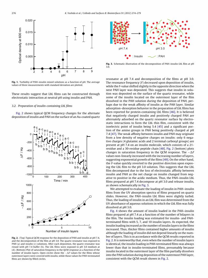
276 K. Yoshida et al. / Colloids and Surfaces B: Biointerfaces 91 (2012) 274– 279
0
0.0 10
0.0 15
0.02 0
0.00 5
5432 6 87 9
pH
Op
tica
l d
en
sity a
t 6
00
nm
Fv
Te
3
d
FaPr(nofi
: Insul in : PAH
pH 7.4 pH 3.0
ig. 1. Turbidity of PAH–insulin mixed solutions as a function of pH. The averagealues of three measurements with standard deviation are plotted.
hese results suggest that LbL films can be constructed throughlectrostatic interaction at neutral pH using insulin and PAH.
.2. Preparation of insulin-containing LbL films
Fig. 2 shows typical QCM frequency changes for the alternateeposition of insulin and PAH on the surface of an Au-coated quartz
0
-200
-400
-600
-80 0
-10000 100 200 300 400
Time / min
ΔF
/ H
z
d
bb b
b bb b b b
b bb b
b b
b b
b
b
aa a a
aa
a
a
a
a
cc
ccc
cc
c
c
c
b
0 5 100
200
400
600
-Δ
F / H
z
800
1000
Number of insulin layers
ig. 2. (Top) Typical QCM response for the deposition of PAH and insulin at pH 7.4,nd the decomposition of the film at pH 3.0. The quartz resonator was exposed toAH (a) and insulin (c) solutions. After each deposition, the quartz resonator wasinsed with pH 7.4 buffer (b). The LbL films were exposed to pH 3.0 medium atd). (Bottom) Plot of saturation frequency in the QCM response as a function of theumber of insulin layers. Open circles show the −�F values for the films whoseutermost layers were covered with insulin, while those values for PAH-terminatedlms are shown by filled circles.
Fig. 3. Schematic illustration of the decomposition of PAH–insulin LbL film at pH3.0.
resonator at pH 7.4 and decomposition of the films at pH 3.0.The resonance frequency (F) decreased upon deposition of insulin,while the F-value shifted slightly in the opposite direction when thenext PAH layer was deposited. This suggests that insulin in solu-tion was deposited on the surface of the quartz resonator, whilesome of the insulin located on the outermost layer of the filmdissolved in the PAH solution during the deposition of PAH, per-haps due to the weak affinity of insulin as the PAH layer. Similaradsorption–desorption behavior in the preparation of LbL films hasbeen reported for protein-containing LbL films [46]. It is believedthat negatively charged insulin and positively charged PAH arealternately adsorbed on the quartz resonator surface by electro-static interactions to form the LbL thin film, consistent with theisoelectric point of insulin being 5.4 [45] and a significant por-tion of the amino groups in PAH being positively charged at pH7.4 [47]. The weak affinity between insulin and PAH may originatefrom a low density of negative charges on insulin: only 6 nega-tive charges (4 glutamic acids and 2 terminal carboxyl groups) arepresent at pH 7.4 on an insulin molecule, which consists of a 21-residue and a 30-residue peptide chain [48]. Fig. 2 (bottom) plotschanges in saturation frequency in the QCM response. The −�Fvalues non-linearly increased with the increasing number of layers,suggesting exponential growth of the films [49]. On the other hand,the F-value quickly reverted to the positive direction upon expos-ing the LbL film to the pH 3.0 solution. This suggests that the LbLfilm decomposed due to the loss of electrostatic affinity betweeninsulin and PAH as the net charge on insulin changed from neg-ative to positive in the acidic medium. Thus, the PAH–insulin LbLfilms prepared at pH 7.4 decompose at pH 3.0 and release insulin,as shown schematically in Fig. 3.
We attempted to evaluate the loading of insulin in PAH–insulinfilms from the UV absorption spectra of films prepared on quartzslides. However, the PAH–insulin LbL films were slightly turbid.Thus, the loading of insulin in an LbL film was determined from theUV absorbance of aqueous solutions in which the LbL film was fullydissolved at pH 3.0.
Fig. 4 shows the amount of insulin loaded in the PAH–insulinfilms prepared at pH 7.4 as a function of the number of bilayers inthe film. The insulin loading was estimated for insulin- and PAH-terminated films with 5, 7, and 10 insulin layers. As expected, theinsulin loading increased as the number of insulin layers in the filmsincreased. Thus, thicker films contained higher amounts of insulinalthough the loading of insulin did not depend linearly on the num-ber of layers. This is in accordance with the QCM results reported inFig. 2. It is noteworthy that, even when the number of insulin layersis identical, the insulin loading in PAH-terminated films was alwayslower than that in insulin-terminated films, presumably because
insulin located in the outermost layer of the film partially dissolvesinto the PAH solution during deposition of the outermost PAH layer,consistent with the QCM result shown in Fig. 2.
K. Yoshida et al. / Colloids and Surfaces B
0
2
4
6
8
10
12
10.5107.575.55
Numbe r of bil aye rs
Loa
din
g o
f in
su
lin /
10
-6 g
cm
-2
Fig. 4. Loading of insulin in the (PAH–insulin)n and (PAH–insulin)nPAH films. Thebca
u[fieafsccashastmiifid
3
d
Fmim
ilayer numbers, 5.5, 7.5, and 10.5, show the LbL films whose outermost surface wasovered with PAH. The average values of three preparations with standard deviationre shown.
The loading of closely packed insulin molecules in a monomolec-lar layer on a flat surface has been reported to be 1.1 × 10⁻⁷ g cm−2
50]. Therefore, the observed loading of insulin in the PAH–insulinlms is higher than that expected for a monomolecular deposit inach layer. It is probable that insulin is deposited in the films in anggregated form, resulting in a higher loading. Insulin is known toorm dimers, hexamers, and higher aggregates in solution and onolid surfaces, and the size of the aggregates is dependent on theomposition of the solution [50,51]. It is noteworthy that negativelyharged proteins such as human serum albumin and �-lactalbumindsorb to form an aggregated thick layer on the oppositely chargedurface of PAH-terminated LbL films at pH 7.4 [52,53]. Thus, theigher loading of insulin is likely due to the formation of insulinggregates in the LbL films. The coiled conformations of PAH on theurface may also contribute to the higher loading of insulin, sincehe apparent surface area of the PAH-terminated LbL films may be
uch higher than the geometric surface area. Higher loading ofnsulin was also reported for LbL films constructed with polyan-on and insulin at acidic pH [44]. High loading of insulin in the LbLlms may be desirable for future applications of the films in insulinelivery devices.
.3. �-Potential of PAH–insulin and PLL–insulin films
Fig. 5 shows a plot of typical �-potentials for PAH–insulin filmseposited on the surface of PLA microspheres. Positive values in
Number of bilayers
ζ-p
ote
ntia
l /
mV
543210
0
-10
-20
-30
40
30
20
10
ig. 5. Typical �-potentials of PAH–insulin films deposited on the surface of PLAicrospheres at pH 7.4. The outermost surface of the LbL films whose bilayer number
s indicated by the integers was covered with insulin. The average values for threeeasurements with standard deviation are plotted.
: Biointerfaces 91 (2012) 274– 279 277
the �-potentials were observed when the outermost surface of thefilm was covered with PAH, while the values were negative for theinsulin-terminated films. These results suggest that electric chargeson the outermost surface of the LbL films were reversed alternatelyfrom positive to negative and vice versa during the deposition of theLbL film. Thus, electrostatic interactions are primarily responsiblefor the LbL deposition of insulin and PAH.
3.4. Surface morphology of PAH–insulin films
The surface morphology of the (PAH/insulin)5 and(PAH/insulin)5PAH films was studied using AFM. Fig. 6 showsAFM images of the surface of the films. The surface is rather rough,with the root mean square (rms) roughness being 21 and 14 nmfor the (PAH/insulin)5 and (PAH/insulin)5PAH films, respectively,over a scan area of 5�m × 5 �m (Fig. 6a and b). The rough surfaceof the films may relate to the formation of insulin aggregates onthe surface. In addition, one cannot exclude the possibility thatthe surface roughness of the dry films was induced in part upondrying. Fig. 6c and d shows the surface images of relatively smoothareas of the films (1�m × 1 �m), where the rms roughness is 4.7and 4.0 nm, respectively. For both images, the rms roughness forthe PAH-terminated (PAH/insulin)5PAH film is lower than thatfor the (PAH/insulin)5 film. This may originate from the fact thatthe insulin aggregates were removed in part from the outermostsurface of the (PAH/insulin)5 film during deposition of the nextPAH layer, as suggested by QCM results in Fig. 2.
3.5. pH-induced decomposition of PAH–insulin films
As shown in Fig. 2, PAH–insulin LbL films decompose at pH 3.0due to loss of the net negative charge on insulin. UV spectroscopywas used to monitor the insulin released from LbL films depositedon the surface of a quartz slide in order to evaluate the kineticsof film decomposition. Fig. 7 shows typical results for changes inthe UV absorbance of solutions in which the (PAH/insulin)10 filmwas immersed at 20 ◦C (A) and 37 ◦C (B). At both temperatures,the absorbance increased rapidly at pH 3.0, suggesting that theLbL film was decomposed at pH 3.0, while the lack of significantincrease in absorbance at pH 7.4 indicated that the film was ratherstable at this pH, in line with the QCM result shown in Fig. 2. Theeffect of temperature on film stability was not significant, althoughthe initial decomposition of the LbL film in the acidic medium at37 ◦C was slightly more rapid than that at 20 ◦C. The rapid pH-induced decomposition of the LbL film suggests that such filmsmight be useful for insulin delivery. This rapid response is an advan-tage of LbL films compared with the slower response reported forinsulin release from hydrogels [54], polymer films [55], and cap-sules [56].
Fig. 8 shows the pH threshold for the release of insulin from(PAH/insulin)10 and (PAH/insulin)10PAH films at 20 ◦C (A) and 37 ◦C(B). The percent of insulin released after immersing the films for30 min in the solutions was plotted as a function of the pH ofthe solution. The LbL films were rather stable in neutral pH solu-tions, while insulin was released at low and high pH as a resultof film decomposition. The pH threshold for insulin release (orfilm decomposition) in acidic media was pH 4.7–5.7 for both the(PAH/insulin)10 and (PAH/insulin)10PAH films, which qualitativelycorresponds to the isoelectric point of insulin (5.4). The effect oftemperature on the pH threshold for film decomposition was small.These results further support the view that the pH-dependentdecomposition of the films is caused by the loss of electrostatic
affinity between insulin and PAH in the film, originating from ashift in the net electric charge of insulin from negative to positive.The LbL films also somewhat decomposed at pH 9.0–10, probablydue to weakened electrostatic binding between PAH and insulin
278 K. Yoshida et al. / Colloids and Surfaces B: Biointerfaces 91 (2012) 274– 279
Fig. 6. AFM three-dimensional images of (PAH/insulin)5 (a and c) and (PAH/insulin)5PAH films (b and d).
0
0.01
0.02
0.03
0 5 10 15 20
Time / min
mn
77
2t
ae
cn
abr
os
bA
(A) 20 oC
(a)
(b)
0
0.01
0.02
0.03
0 5 10 15 20
Time / min
mn
77
2t
ae
cn
abr
os
bA
(a)
(b)
(B) 37 oC
Fig. 7. pH-induced decomposition of the (PAH–insulin)10 film monitored by UVabsorption at 20 ◦C (A) and 37 ◦C (B). The absorbance of the solution in which eachLbL film was immersed was recorded. After 5-min of exposure of the film to pH 7.4buffer, the pH of the medium was changed to pH 3.0 (a) or the buffer was replacedwith a new one at pH 7.4 (b).
100
80
60
40
20
02 4 6 8 10
(A) 20 oC
pH
Insulin
rele
ased / %
100
80
60
40
20
02 4 6 8 10
(B) 37 oC
pH
Insulin
rele
ased / %
Fig. 8. pH-dependent release of insulin from (PAH/insulin)10 (�) and(PAH/insulin)10PAH (©) films at 20 ◦C (A) and 37 ◦C (B). The average valuesfor three measurements with standard deviation are plotted.

K. Yoshida et al. / Colloids and Surfaces B
-10
0
10
200 22 0 24 0 26 0 280 30 0
wave lengt h / nm
Elli
ptic
ity / m
de
g
Fig. 9. CD spectra of the (PAH–insulin)5 film (broken line) and that of the insulinrao
icwtTi
3
wtfirncOfismfifiaft[
4
aPtfiwtfii
cieafvt[
[[[
[[[
[
[[[[
[[[
[[[[[
[[[[[
[[
[
[
[[[[[
[[[[[
[[
[
[[
[
[[[[
eleased from the film (solid line). The CD spectrum of the LbL film was recorded inir, then the film was immersed in a pH 3.0 buffer solution to record the spectrumf the released insulin.
n the films originating from the decreased number of positiveharges in PAH in basic solution. The protonation degree of PAHas reported to be approximately 0.7 at pH 7.4, while it decreased
o approximately 0.4 and 0.2 at pH 9.0 and 10.0, respectively [47].he instability of the films at pH 9.0–10 is unlikely to have a negativempact on medical applications of the films.
.6. CD spectra of insulin released from PAH–insulin film
The structural integrity of insulin released from the LbL filmsas evaluated using CD spectra of insulin-containing LbL film and
he released insulin. Fig. 9 shows the CD spectra of (PAH/insulin)5lm and insulin released from the film. The CD spectrum of theeleased insulin exhibited typical �-helical characteristics, withegative ellipticity at 208 and 222 nm, suggesting that the originalonformation is almost preserved in the released insulin [50,56,57].n the other hand, the CD spectrum of insulin in (PAH/insulin)5lm is slightly different from that of native insulin. These resultsuggest that insulin released from LbL film has a native-like confor-ation, although its conformation may be slightly modified in the
lm. We previously observed similar CD spectral features for LbLlms composed of poly(vinyl sulfate) and insulin [44]. Middaughnd coworkers also observed reduced ellipticity at 208 and 222 nmor insulin entrapped in polymer nanoparticles and ascribedhis to an enhanced �-sheet fraction in the entrapped insulin57].
. Conclusions
We have demonstrated that LbL thin films composed of insulinnd PAH can be prepared by the alternate deposition of insulin andAH at pH 7.4. The loading of insulin in the LbL film increased withhe increasing number of layers in the films. Insulin-containing LbLlms are stable at neutral pH, but decompose to release insulinhen exposed to acidic solutions (pH 5.0 or lower) due to a shift in
he net electrical charge of insulin from negative to positive. The LbLlm also decomposed to some extent at pH 9.0–10. Insulin retained
ts native structure when released from the LbL films.These results suggest potential applications of insulin-
ontaining LbL films for insulin delivery systems. For example,t may be possible to develop electrically triggered insulin deliv-ry systems by coating the insulin-containing LbL films onton electrode. Insulin would be released from the electrode sur-
ace in response to an electric signal because the pH in theicinity of the electrode surface can be acidified through elec-rolysis of H2O by the application of positive electrode potential58–60].[
[[
: Biointerfaces 91 (2012) 274– 279 279
References
[1] G. Decher, Science 277 (1997) 1232.[2] Z. Tang, Y. Wang, P. Podasiadlo, N.A. Kotov, Adv. Mater. 18 (2006) 3203.[3] J.F. Quinn, A.P.R. Johnston, G.K. Such, A.N. Zelikin, F. Caruso, Chem. Soc. Rev. 36
(2007) 707.[4] K. Ariga, J.P. Hill, Q. Ji, Phys. Chem. Chem. Phys. 9 (2007) 2319.[5] B.G. De Geest, N.N. Sanders, G.B. Sukhorukov, J. Demeester, S.C. De Smedt, Chem.
Soc. Rev. 36 (2007) 639.[6] K. Ariga, J.P. Hill, M.V. Lee, A. Vinu, R. Charvet, S. Acharya, Sci. Technol. Adv.
Mater. 9 (2008) 014109.[7] L.L. der Mercato, P. Rivera-Gil, A.Z. Abbasi, M. Ochs, C. Ganas, I. Zins, C. Sonnich-
sen, W.L. Parak, Nanoscale 2 (2010) 458.[8] L. Krasemann, B. Tieke, Langmuir 16 (2000) 287.[9] W. Jin, A. Toutianoush, B. Tieke, Appl. Surf. Sci. 245 (2005) 444.10] M.D. Miller, M.L. Bruening, Chem. Mater. 17 (2005) 5375.11] A. Liu, J. Anzai, Langmuir 19 (2003) 4043.12] J. Park, J. Kim, S.L. Lee, J. Bang, B.J. Kim, Y.S. Kim, J. Cho, J. Mater. Chem. 19 (2009)
4488.13] T. Noguchi, J. Anzai, Langmuir 22 (2006) 2870.14] B. Wang, J. Anzai, Langmuir 23 (2007) 7378.15] S. Disawal, J. Qiu, B.B. Elmore, Y.M. Lvov, Colloids Surf. B: Biointerfaces 32 (2003)
145.16] S.J. Strydom, D.P. Otto, W. Liebenberg, Y.M. Lvov, M.M. de Villiers, Int. J. Pharm.
404 (2011) 57.17] K. Sato, I. Suzuki, J. Anzai, Langmuir 19 (2003) 7406.18] S.A. Sukhishvili, Curr. Opin. Colloid Interface Sci. 10 (2005) 37.19] D.M. Lynn, Adv. Mater. 19 (2007) 4118.20] E. Kharlampieva, V. Izumrudov, J. Macromol. Sci. Part C: Polym. Rev. 46 (2006)
377.21] I. Erel-Unal, S.A. Sukhishvili, Macromolecules 41 (2008) 3962.22] S. Tomita, K. Sato, J. Anzai, J. Colloid Interface Sci. 326 (2008) 35.23] Z. Gui, J. Qian, Y. He, Q. An, X. Wang, C. Tian, W. Sun, J. Colloid Interface Sci. 361
(2011) 122.24] K. Sato, K. Yoshida, S. Takahashi, J. Anzai, Adv. Drug Deliv. Rev. 63 (2011) 809.25] J. Niu, F. Shi, Z. Liu, Z. Wang, X. Zhang, Langmuir 23 (2007) 6377.26] X. Liu, J. Zhang, D.M. Lynn, Soft Matter 4 (2008) 1688.27] S.T. Dubas, T.R. Farhat, J.B. Schlenoff, J. Am. Chem. Soc. 123 (2001) 5368.28] K. Sato, Y. Imoto, J. Sugama, S. Seki, H. Inoue, T. Odagiri, T. Hoshi, J. Anzai,
Langmuir 21 (2005) 797.29] H. Inoue, K. Sato, J. Anzai, Biomacromolecules 6 (2005) 27.30] H. Inoue, J. Anzai, Langmuir 21 (2005) 8354.31] F. Boulmedais, C.S. Tang, B. Keller, J. Vörös, Adv. Funct. Mater. 16 (2006) 63.32] K. Sato, D. Kodama, Y. Naka, J. Anzai, Biomacromolecules 7 (2006) 3302.33] K.C. Wood, N.S. Zacharie, D.J. Schmidt, S.N. Wrightman, B.J. Andaya, P.T. Ham-
mond, Proc. Natl. Acad. Sci. U.S.A. 105 (2008) 2280.34] F. Wang, D. Li, G. Li, X. Liu, S. Dong, Biomacromolecules 9 (2008) 2645.35] L. Dieguez, N. Darwish, N. Graf, J. Vörös, T. Zambelli, Soft Matter 5 (2009)
2415.36] Y. Ma, W. Dong, M.A. Hempenius, H. Möhwald, G.J. Vancso, Nat. Mater. 5 (2006)
724.37] D.J. Schmidt, F.C. Cebeci, Z.I. Kaicioglu, S.G. Wyman, C. Ortiz, K.J. Van Vliet, P.T.
Hammond, ACS Nano 3 (2009) 2207.38] D.J. Schmidt, P.T. Hammond, Chem. Commun. 46 (2010) 7358.39] C.M. Nolan, M.J. Serpe, L.A. Lyon, Biomacromolecules 5 (2004) 1940.40] C.M. Nolan, M.J. Serpe, L.A. Lyon, Macromol. Symp. 227 (2005) 285.41] Y.F. Fan, Y.N. Wang, Y.G. Fan, J.B. Ma, Int. J. Pharm. 324 (2006) 158.42] Z. Dai, A. Heilig, H. Zastrow, E. Donath, H. Möhwald, Chem. Eur. J. 10 (2004)
6369.43] J. Zheng, X. Yue, Z. Dai, Y. Wang, S. Liu, X. Yan, Acta Biomater. 5 (2009) 1499.44] K. Yoshida, K. Sato, J. Anzai, J. Mater. Chem. 20 (2010) 1546.45] F. Cui, K. Shi, L. Zhang, Y. Kawashima, J. Control. Release 114 (2006) 242.46] Y. Lvov, K. Ariga, I. Ichinose, T. Kunitake, J. Am. Chem. Soc. 117 (1995) 6117.47] T. Mauser, C. Dejugnat, G.B. Sukhorukov, Macromol. Rapid Commun. 25 (2004)
1781.48] N.A. Peppas, N.J. Kavimandan, Eur. J. Pharm. Sci. 29 (2006) 183.49] C. Picart, J. Mutterer, L. Richrert, Y. Luo, G.D. Prestwich, P. Schaaf, J.-C. Voegel,
P. Lavalle, Proc. Natl. Acad. Sci. U.S.A. 99 (2002) 12531.50] S.H. Mollmann, L. Jorgensen, J.T. Bukrinsky, U. Elofsson, W. Norde, S. Frokjaer,
Eur. J. Pharm. Sci. 27 (2006) 194.51] T. Arnebrant, T. Nylander, J. Colloid Interface Sci. 122 (1988) 557.52] G. Ladam, C. Gergely, B. Senger, G. Decher, J.-C. Voegel, P. Schaaf, F.J.G. Cuisinier,
Biomacromolecules 1 (2000) 674.53] G. Ladam, P. Schaaf, F.J.G. Cuisinier, G. Decher, J.-C. Voegel, Langmuir 17 (2001)
878.54] B. Kim, N.A. Peppas, Int. J. Pharm. 266 (2003) 29.55] S. Ye, C. Wang, X. Liu, Z. Tong, B. Ren, F. Zeng, J. Control. Release 112 (2006) 79.56] Y. Pocker, S.B. Biswas, Biochemistry 19 (1980) 5043.57] W. Tiyaboonchai, J. Woiszwillo, R.C. Sims, C.R. Middaugh, Int. J. Pharm. 255
(2003) 139.
58] M. Nagasaka, K. Yoshida, S. Takahashi, K. Sato, J. Anzai, Mater. Sci. Eng. C 31(2011) 258.59] I.C. Kwon, Y.H. Bae, S.W. Kim, Nature 354 (1991) 291.60] J.W. Choi, I.H. Lim, H.H. Kim, J. Min, W.H. Lee, Biosens. Bioelectron. 16 (2001)
141.