lateral flow assays and point of care devices - semi.org ge_lenigk... · current hospital workflows...
TRANSCRIPT

Imagination at work.
Nano Bio Manufacturing Consortium WorkshopBlood, Sweat and Tears III
Ralf LenigkSenior Microfluidics Researcher, MicroSystems Lab
GE Global Research Center, Niskayuna, NY USA
November 2, 2016
Lateral Flow Assays and Point of Care Devices –Detecting Infectious Diseases in Low Resource Settings

See tutorial regarding confidentiality disclosures. Delete if not needed.
GE Global Research: Market-focused R&DThe cornerstone of GE’s commitment to technology
• First U.S. industrial lab.
• ~2000 scientists/engineers, nearly two-thirds PhDs.
• ~3615 U.S. patents filed by GE in 2011 alone.
• One of the world’s most diversified industrial research organizations, providing innovative technology for all of GE’s businesses.
© 2013, General Electric Company

See tutorial regarding confidentiality disclosures. Delete if not needed.
Expanding our global presence
1
4
76
53
2
Global Software Center San Ramon, CA
1
Brazil Technology Center Rio de Janeiro, Brazil
4
China Technology Center Shanghai, China
7
India Technology Center Bangalore, India
6
Global Research Europe Munich, Germany
5
Global Research Headquarters Niskayuna, NY
3
AMSTCAnn Arbor, MI
2

@#GECON&*4
November 2, 2016
Agenda
1. Lateral flow assays - membranes and papers
2. Towards Precision Diagnostics - Tailoring Component Properties
3. Modifying Membrane & Paper Surfaces & Transport Characteristics
4. Materials Applications into Advanced IVD Devices for POC detection
5. Photothermal spectroscopy reader for enhanced sensitivity read-out

@#GECON&*5
November 2, 2016
Introduction
• LFA is low-cost test of choice for immuno-assays, protein detection
• Lessons learned from LFA & POC development are applicable to Wearables
• Fluidics and volumes are comparable to Wearable devices
• Efficient supply chain and manufacturing processes
• Many materials, such as membranes, are usable for Wearables
Membranes & Papers

@#GECON&*7
November 2, 2016
Polymeric: most commonInorganic: more stable
What is the Difference: Membrane vs Paper?
Cellulular
e.g. Lungs
Biological
Glassy Rubbery
Organic
(polymeric)
Glass
Ceramic
Metallic
Zeolitic
Inorganic
Synthetic
Membrane
Materials
Membrane: Layer(s) of material which act as a selective barrier between components
hybrid
Paper: Semi-permeable material, typically derived from mineral fibers or lignocellulosic pulps
Lignocellulosic
Glass Fiber
Quartz
Mineral Fibers
Paper
Materials

@#GECON&*8
November 2, 2016
Micrometers(Log scale) Ionic Range Molecular Range Macro Molecular Range Particle Range
Membranes & Papers
0.001 0.01 0.1 1
Membranes & Paper: Across the Separation Range
Membranes
Paper
10
Relative Size of Common Materials
Separation Processes
Reverse Osmosis(Hyperfiltration)
Nanofiltration (NF)
Ultrafiltration (UF)
Microfiltration (MF)
particleFiltration
BacteriaVirusAqueous Salt
SugarAlgae
Albumin ProteinMetal IonPaint Pigment
Markets
Brackish water
Sea water
Tough-to-Treat Produced Waters – oily waste, high suspended solids
Boiler feed – especially power
Cooling tower - blow down
Industrial process fluid separations
Surface water treatment
Dairy, food , pharma
Diagnostics - Lateral flow assays Biopharmaceutical processing
Non-porous
Porous

@#GECON&*9
November 2, 2016
1 - Sample Pad2 - Conjugate Release Pad3 – Test Line4 – Reaction Membrane5 – Control Line6 – Absorbent Pad (Wick)7 – Backing Layer
Reverse Osmosis
Pervaporation
Nanofiltration
Ultrafiltration
Microfiltration
Dense Thin Film
Gross Filtration
Membranes & PapersAcross the Separation Range
Depth FiltersLab Filtration
Forensics/Human IDDried Blood Spots
Biospecimen Stabilization
Diagnostics Components
Glass Fiber
NitrocelluloseParticle Size(m)
0.0001
0.001
0.01
0.1
1
10
100

@#GECON&*10
November 2, 2016
Membrane vs. Paper: Morphology
Composite Membrane
NanofibersTrack Etched
Nonwoven fabric
Expanded film
Pore Filled
Phase Inversion
Membrane Paper
Lignocellulosics
FTA™ – DNA storage
Mineral Fibers

@#GECON&*11
November 2, 2016
Membranes & Papers: Substrate to DeviceM
em
bra
ne
Pa
pe
r
Substrate Format
Module/ Cartridge
System/ Device
Hollow Fiber
Flat Sheet
EasiCollect™

Towards Precision Diagnostics: Tailoring Component Properties

@#GECON&*13
November 2, 2016
Membranes/Papers: Discovery to Manufacturing
Paper/Membrane Development
Advanced Materials Design & Membrane Fabrication Surface Activation & Modification
Material Characterization & TestingStructure-Property-Performance Evaluation
Substrate Modified
substrate
Membrane Pilot Manufacturing Process Optimization & LifingMaterials and Process Cost Analysis
Transfer Function for Performance Balance
Biological Performance
Biomolecule Stabilization (DNA, RNA, proteins)Dried Blood Spot (Small Molecule Collection & Testing)Nucleic Acid Amplification & TransportDetection - Immunoassay/Nucleic Acid Testing

Chemically Modifying Membrane & Paper Surfaces

@#GECON&*15
November 2, 2016
Platform Approach to Modifying Membrane/Paper
Membrane/Paper
Activated Substrate
1) Activation 2) Modification
ModifiedSubstrate
Modified Membranes - Nitrocellulose Substrates:• Activated Nitrocellulose• Hydrophilic Nitrocellulose
Bing Li
Cathryn Olsen
Bill Alberts
Li, B.; Moore, D. R.; Olsen, C. O.; “Porous Membranes Having a Polymeric Coating and Methods for Their Preparation and Use.” U.S. Pat. Appl. 13/339,960; 13/339,996; 13/340,052 .
Li, B.; Moore, D. R.; Olsen, C. O.; “Porous Membranes Having a Hydrophilic Coating and Methods for Their Preparation and Use.” U.S. Pat. Appl. 13/340,793; 13/362,793.
Substrate Breadth – Cellulosics to Synthetics

@#GECON&*16
November 2, 2016
Materials Enabling Diagnostic PerformanceReducing Non-Specific Binding
[DARPA contract HR0011-11-2-0007]
Isothermal NA Amplification in Porous Substrates
NCHydrophilic
NC
Improved Biomolecule Transport in Porous Matrices
HydrophilicNC
UnmodifiedNC
Time
Running buffer: 40 nm gold nanoparticle coated bovine serum
albumin (BSA)

Controlling Membrane/Paper Transport - Hydrophilicity

@#GECON&*18
November 2, 2016
Physical Properties… HydrophilicityResults for qcf of (un)modified membranes
J. Nichols
J. Davis
Sample qcf
° (s)*Cap. Rise
s*
Nitrocellulose-1 34(2) 101
Nitrocellulose-2 51 134
Nitrocellulose-3 67(1) 2000
Studies performed on membranes from GE Healthcare or modified membranes; *Average of >3 replicates
Membrane Contact Angle via Capillary Flow Porometry
Hydrophilicity/contact angle is a critical (& often oversimplified) control parameter to enable precision diagnostics
𝒅𝑽 =𝜸𝝅𝑫𝟑
𝟑𝟐𝝁𝑳𝒄𝒐𝒔𝜽𝒅𝒕

Materials Applications into Advanced IVD Devices

@#GECON&*20
November 2, 2016
http://www.fujifilmusa.com/
http://www.biodot.com/
Printing Methods at GE GRC-Lab scale at GE GRC
Gravure
Printing
Screen
Printing
Inkjet
Printing
BioDot XYZ-
3000
Printing FormEngraved
cylinder
Stencil and
meshDigital Digital
Ink Viscosity (Pas) 0.01-0.2 0.1-50 0.002-0.1 0.01-1
SubstratesPaper,
polymersAll All
Paper,
polymers
Line Width (μm) 10-50 50-150 1-20 500
Registration (μm) >10 >25 <5 <15
Throughput
(m²/sec)10 <10 0.01-0.1 <1
Adapted and updated from Cagler 2009
http://www.dek.com/

@#GECON&*21
November 2, 2016
Patterned Membranes and Fluid Paths
Flow demonstration
Time
Patterned Membrane
Pattern Printed on to Nitrocellulose
Create Fluidic Structures in Membranes

@#GECON&*22
November 2, 2016
DARPA Diagnostics on Demand Limited Resource Settings“Multiplexable Autonomous Disposables for Nucleic Acid Amplification Test (MAD NAAT)”
Overview
• Instrument‐free, single-use, paper-baseddisposable device
• Unitized, self-contained nucleic acid test
• Administered by untrained user (CLIA waived) w/ sample to read-out in < 1 hr
• Low Cost to Enable Broad Use
Potential end-use scenarios
• Nurse or physician in hospital, ER, ICU
• Physician’s office
• Retail clinics
• Home use (prescribed by physicians)
• Telemedicine
PI = Paul Yager
The Team:
Image provided by Paul Yager,
U. Wash

@#GECON&*23
November 2, 2016
Sample
Flow
1 - Sample Pad2 - Conjugate Release Pad3 – Test Line4 – Reaction Membrane5 – Control Line6 – Absorbent Pad (Wick)7 – Backing Layer
Overview
Sample Prep
Nucleic Acid Amplification & Detection - PCR
Immunoassay - Lateral Flow IVD Test
Instrumented Workflow
MAD NAAT Device:Sample Collection (Swab/Blood)
Sample PrepIsothermal NA Amplification
DetectionSample Archiving
The Vision: A platform device as simple as a pregnancy test with capability for detecting multiple infectious diseases
within a single biospecimen
Paul Yager, U. Wash
@#GECON&*24
November 2, 2016
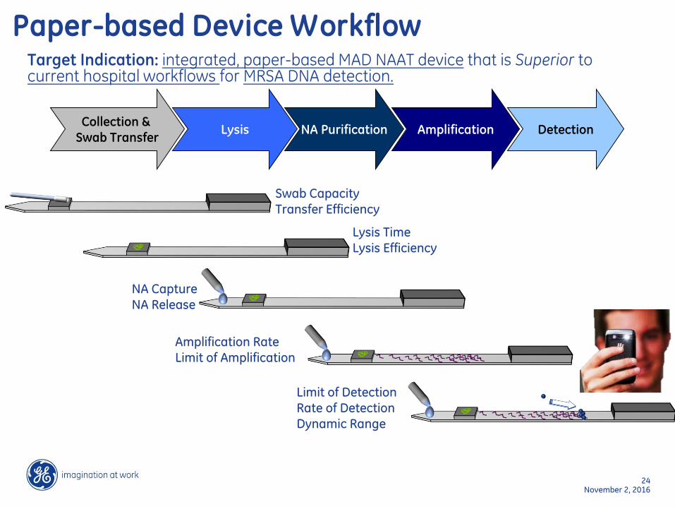
Target Indication: integrated, paper-based MAD NAAT device that is Superior to current hospital workflows for MRSA DNA detection.
Paper-based Device Workflow
DetectionCollection &
Swab TransferLysis NA Purification Amplification
Swab CapacityTransfer Efficiency
Lysis TimeLysis Efficiency
NA CaptureNA Release
Amplification RateLimit of Amplification
Limit of DetectionRate of DetectionDynamic Range

@#GECON&*25
November 2, 2016
IVD Development Across Specimen Testing Workflow:Materials That Matter
Chemical & Biological Reagents
IVD Materials
Fluidics
System Integration
Device
+
+
DetectionCollection &
Swab TransferLysis NA Purification Amplification
Cellulose (FTATM) Glass fiber Nitrocellulose
• Excellent wicking power
• Supports efficient lysis methods
• Compatibility with amplification, lysis and reagent storage
• Fastest flow• High fluid capacity
• Compatibility with amplification (when chemically modified)
• Predictable flow • High binding capacity
(for detection)

@#GECON&*26
November 2, 2016
Point-of-Care In Vitro Diagnostics
Typical assembly of a lateral
flow assay strip.
1 = Sample Pad
2 = Conjugate Release Pad
3 = Test Line
4 = Reaction Membrane
5 = Control Line
6 = Absorbent Pad (wick)
7 = Backing Layer
GE Healthcare WhatmanTM catalogue. “Whatman A Guide to Diagnostic Rapid Test Development.”
Sample Pad
Sample
Flow
Conjugate Release Pad
Test Line
Backing Layer
Absorbent Pad (Wick)
Reaction MembraneControl Line
Lateral Flow Assay Strip Components
How does a Pregnancy Test Work?
Analyte / antigen
Bioconjugate with reporting tag
Capture antibody
Control Line antibody
Positive
Negative

Phase I Objectives: producing a TRL-5 prototype of the complete MRSA device
2/5/2014 MAD NAAT Project Open Talk 27
Integrated device that performs DNA test for MRSA (atTRL-5 level)Nasal swab sample captureBacterial lysis(DNA purification)Isothermal amplificationVisible lateral flow readoutCell-phone image capture and data transmission of result Staged expansion of targets to include:Broader bacterial panel (Gram + and –species using DNA)Viruses (RSV by RNA using RT step)Blood sample capabilitySample archiving capability for sequencing
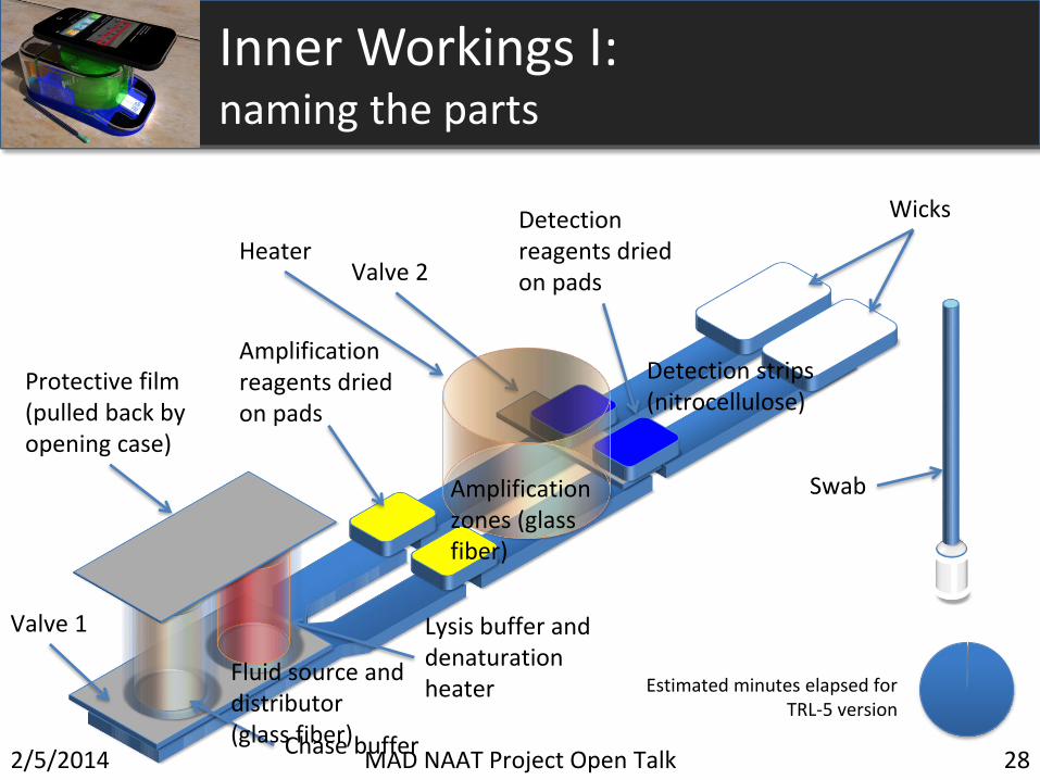
Valve 2
Valve 1
Inner Workings I: naming the parts
Detection reagents dried on pads
Amplification reagents dried on pads
Chase buffer
Swab
Wicks
Lysis buffer and denaturation heater
Heater
Protective film (pulled back by opening case)
Fluid source and distributor (glass fiber)
Amplification zones (glass fiber)
Detection strips (nitrocellulose)
2/5/2014 MAD NAAT Project Open Talk 28
Estimated minutes elapsed for TRL-5 version

Sa Sa IC 5000 copiescopies
• Perform Sa/IC biplex amplification in a tube• Followed by lateral flow detection•Not yet optimized
Lateral Flow Detection of a S. aureus gene -Internal Control-biplex iSA Amplicon Mixture
2/5/2014 MAD NAAT Project Open Talk 29

Photothermal SpectroscopyEnhanced Sensitivity LFA Reader for POC
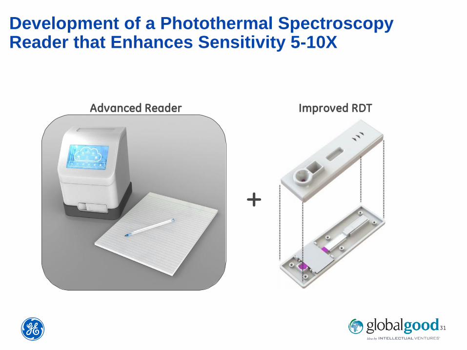
31
Development of a Photothermal Spectroscopy Reader that Enhances Sensitivity 5-10X
Improved RDT
+
Advanced Reader

32
1. Focused light
2. Conjugated Anti-Ag
3. Ag
4. Anti-Ag
5. Nitrocellulose
Photothermal Spectroscopy Response

33
1. Focused light
2. Conjugated Anti-Ag
3. Ag
4. Anti-Ag
5. Nitrocellulose
Photothermal Spectroscopy Response

@#GECON&*34
November 2, 2016
Conclusions & Outlook
• Complex fluidics functions can be achieved in paper-based devices
• Paper & membranes used for LFA, POC can be employed in Wearables
• Material properties can be fine-tuned for parameters like flow speed etc.
• Sensitivity of LFAs can be significantly enhanced with new technologies

GE, imagination at work, and GE monogram are trademarks of General Electric Company.
FTA, EasiCollect, and Whatman are trademarks of General Electric Company or one of its subsidiaries.
© 2014 General Electric Company – All rights reserved. First published May 2014.
All goods and services are sold subject to the terms and conditions of sale of the company within GE
Healthcare which supplies them. A copy of these terms and conditions is available on request. Contact your
local GE Healthcare representative for the most current information.
GE Healthcare Bio-Sciences Corp.
800 Centennial Avenue, P.O. Box 1327
Piscataway, NJ 08855-1327
USA
35