lateral epicondylitis at the 2011 concepts in orthopaedic care
TRANSCRIPT

9/26/2011
1
Concepts in Orthopaedic Care:Lateral Epicondylitis
Scott M Wein MDRaleigh Orthopaedic Clinic
9/24/11
Lateral Epicondylitis
Originally described in Lancet, 1882 as “Lawn Tennis Elbow”

9/26/2011
2
Epidemiology
Overall incidence 1-2% over course of lifetime Tennis players account for less than 5% of overall cases 4th to 5th decade of life10-50% of tennis players will suffer from lateral epicondylitisCommon in construction, gardening
Symptoms
Tenderness and pain localized to the lateral epicondylePain with activities involving wrist extensionDifficulty with grasping objects

9/26/2011
3
Symptoms
Nirschl’s Phases of Lateral EpicondylitisI. Mild pain after exercise <24hII. Pain after exercise >48hIII. Pain with exerciseIV. Pain that alters ability to exerciseV. Pain caused by heavy ADLsVI. Pain caused by light ADLs, intermittent rest painVII. Constant pain at rest, interferes with sleep
Physical Exam
Maximal tenderness 2-5 mm distal and anterior to the midpoint of the lateral epicondyleResisted wrist extension should worsen painObjective measurement– Diminished grip strength with the elbow
extended vs flexed

9/26/2011
4
Imaging Studies
16% may have radiographic evidence of calcification in the soft tissue about the lateral epicondyleUltrasound imaging– calcification of the extensor tendon, presence of
hypoechoic fluid adjacent to the tendon, thickening, decreased echogenicity, and tearing of the tendon
Imaging Studies
UltrasoundSensitivity 64-82%Specificity 67-100%
MRISensitivity 90-100%Specificity 83-100%

9/26/2011
5
Imaging Studies
Normal Signal change in ECRB tendon
Differential Diagnosis
LCL sprain/insufficiencyRadial tunnel syndromeBursitisArthritis or other intraarticular pathologyFractureTriceps tendonitisReferred pain

9/26/2011
6
Anatomy
Pathophysiology
ECRB microtearsTendon is grayish, friable, edematousFibrillation of tendonSome with gross tendon rupture

9/26/2011
7
Pathophysiology
Nirschl, et al. JBJS Am (1979)– “The lesion that was consistently identified at
surgery was immature fibroblastic and vascularinfiltration of the origin of the extensor carpiradialis brevis. “
– Angiofibroblastic dysplasia
Pathophysiology
Krauschaar, BS and Nirschl, RP. Tendinosis of the Elbow. Clinical features and findings ofhistological , immunohistochemical, and electron microscopy studies. JBJS, 1999. Vol 81.

9/26/2011
8
Treatment Options
Treatment Options
RestNSAIDs (Oral vs. Topical)Physical TherapyBracing (CF or wrist brace)Injection

9/26/2011
9
Bracing
Meyer et al. JHS, (2002) – Counterforce brace may result in force
reduction of at the ECRB origin of approximately 13-15% throughout a range of activity levels
Topical NSAIDS
Burnham, et al. Clin J Sport Med. (1998):– Compared 2% topical dicofenac to placebo in
14 symptomatic patients.Randomized, double blinded
– Improved pain on VAS at 3 and 14 days– No difference in scores at 1 month
Burnham, et al. The effectiveness of topical diclofenac for lateral epicondylitis. Clin J Sport Med. 1998; 8: 78-81.

9/26/2011
10
Physical Therapy
3 phase home exercise programPhase 1: massagePhase 2: active stretchingPhase 3: passive stretching
May include ultrasound, iontophoresis, PEMF, electrical stimulation
Injections

9/26/2011
11
Steroid Injections
Complications– Subcutaneous atrophy,
skin lesions, depigmentation
– Crystal deposition– Permanent tendon
changes
Iontophoresis
Nirschl, et al. AJSM (2003):– Iontophoretic administration of steroids to 199
patientsRandomized, double blinded, placebo controlled
– Improved VAS by patient and physician– Side effects included skin reactions (n=12 in
treatment, n=11 in placebo)

9/26/2011
12
Shock Wave TherapyExact mechanism unclear“shock waves can provoke a painful level of stimulation that leads to pain relief or analgesia through hyperstimulation and increased vascularity.”Short term success 58-73% at 12 weeks
Shock Wave Therapy
Haake, et al. JBJS-Am (2002):– Randomized, multicenter trial: – SWT vs. placebo (272 patients)
Pain relief: 25.8 %(SWT) 25.4% (placebo)

9/26/2011
13
Shock Wave Therapy
2005 Cochrane review concluded that included 9 trials concluded that ESWT provided “little or no benefit in terms of pain and function.”
Surgical Indications
5-10% of patients will require surgical managementFailure of non-operative measures for AT LEAST 6-12 months

9/26/2011
14
Surgical Procedures
Release of the extensor originExcision of the pathologic tendon +/-reattachmentLengthening the ECRB tendonArthroscopic excision of the synovial fringe and portion of the tendonAnconeus rotation
V-Y advancement
Rayan, et al. JHS (2002):– 22 patients, 16 followed up at 3 ½ years– VAS improved from 9 to 1– Grip strength 57 to 99 lbs– 95% no limitations with normal activities– 32% limited with high demand activities
Rayan, et al. V-Y Slide of the common extensor origin for lateral elbow tendinopathy.JHS 2001; 26A: 1138-45.

9/26/2011
15
V-Y advancement
Rayan, et al. V-Y Slide of the common extensor origin for lateral elbow tendinopathy.JHS 2001; 26A: 1138-45.
V-Y advancement
Rayan, et al. V-Y Slide of the common extensor origin for lateral elbow tendinopathy.JHS 2001; 26A: 1138-45.

9/26/2011
16
V-Y advancement
Rayan, et al. V-Y Slide of the common extensor origin for lateral elbow tendinopathy.JHS 2001; 26A: 1138-45.
Debridement and Reattachment

9/26/2011
17
Debridement and Reattachment
Results (Kerlan-Jobe, 1997):– 1200 patients, 60 operative (95%)– 94% dramatic improvement 2-10 yrs follow-up– 36% with limitations with heavy lifting– 15% with grip strength weakness
Debridement

9/26/2011
18
Debridement
Verhaar, et al. JBJS (1993):– 57 patients, 5 year follow-up
91% no painAll had improvement compared to 1 year after surgeryNo association between preoperative findings and outcome were found
Verhaar, et al. Lateral extensor release for tennis elbow: A prospective long-termfollow up study. JBJS-Am 1993; 75:1034-43.
Percutaneous Release
JBJS-Br, (2004):– Prospective, randomized trial of 45 patients
Significant improvements for patient satisfaction (p = 0.012), Time to return to work (p = 0.0001),Improvements in DASH score (p = 0.001) Improvement in sporting activities (p = 0.046)Quicker return to work (3 weeks)
Dunkow, et al. A comparison of open and percutaneous techniques in surgicaltreatment of tennis elbow. JBJS-Br. 2004. 86:701-4.
9/26/2011
19
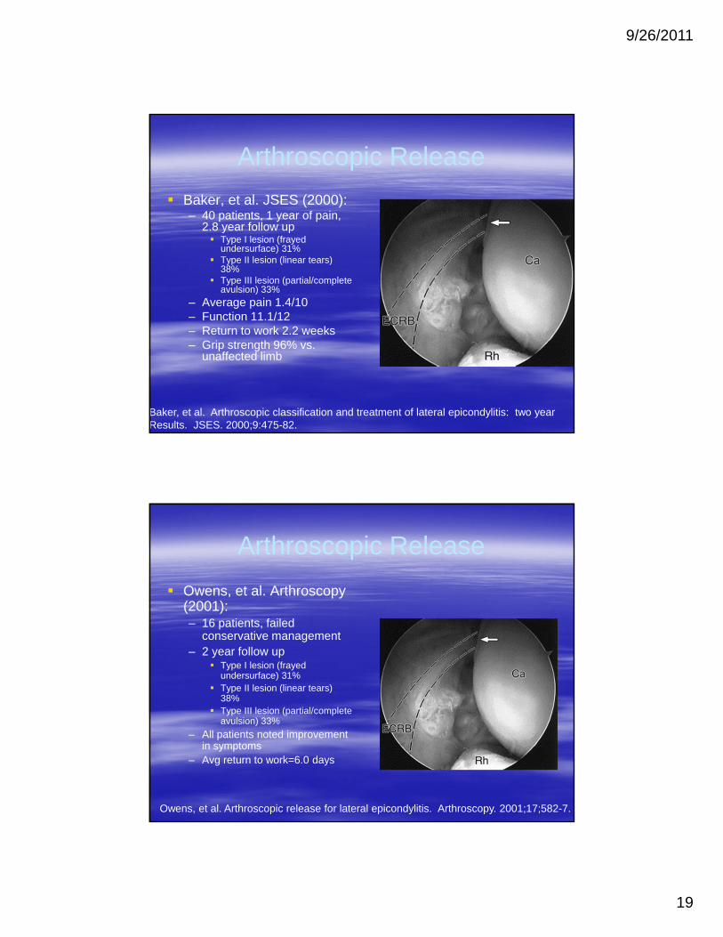
Arthroscopic ReleaseBaker, et al. JSES (2000):– 40 patients, 1 year of pain,
2.8 year follow upType I lesion (frayed undersurface) 31%Type II lesion (linear tears) 38%Type III lesion (partial/complete avulsion) 33%
– Average pain 1.4/10– Function 11.1/12– Return to work 2.2 weeks– Grip strength 96% vs.
unaffected limb
Baker, et al. Arthroscopic classification and treatment of lateral epicondylitis: two year Results. JSES. 2000;9:475-82.
Arthroscopic ReleaseOwens, et al. Arthroscopy (2001):– 16 patients, failed
conservative management– 2 year follow up
Type I lesion (frayed undersurface) 31%Type II lesion (linear tears) 38%Type III lesion (partial/complete avulsion) 33%
– All patients noted improvement in symptoms
– Avg return to work=6.0 days
Owens, et al. Arthroscopic release for lateral epicondylitis. Arthroscopy. 2001;17;582-7.

9/26/2011
20
Szabo et al, JSES, (2006).
Retrospective review of 109 patients treated surgically over 7 years– 41 open, 24 percutaneous, 44 arthroscopic– Consistent improvement in all patients preop vs
postop, but no significant difference among the groups
My approach
Counterforce brace, NSAIDs, HEP, pt education first line treatmentInjection, wrist brace, therapy Surgery if symptomatic AT LEAST 6 months and failed conservative txECRB tendon debridement, drilling of epicondyle– 5 days in LAS, then therapy. Goal back to
normal activities at 6-8 weeks.

9/26/2011
21
Conclusions
Lateral elbow pain with point tenderness95% success with conservative treatment90% success with surgical treatments for patients failing conservative treatment
Thank You