laser-induced fluorescence and ft-raman spectroscopy for characterizing patinas on stone substrates
TRANSCRIPT

ORIGINAL PAPER
Laser-induced fluorescence and FT-Raman spectroscopyfor characterizing patinas on stone substrates
M. Oujja & C. Vázquez-Calvo & M. Sanz &
M. Álvarez de Buergo & R. Fort & M. Castillejo
Received: 13 May 2011 /Revised: 29 July 2011 /Accepted: 4 August 2011 /Published online: 25 August 2011# Springer-Verlag 2011
Abstract This article reports on a compositional investi-gation of stone patinas: thin colored layers applied forprotective and/or aesthetic purposes on architectural orsculptural substrates of cultural heritage. The analysis andclassification of patinas provide important information ofhistoric and artistic interest, as their composition reflectslocal practices, the availabilities of different materials, andthe development of technological knowledge during spe-cific historical periods. Model patinas fabricated accordingto traditional procedures and applied onto limestone, and ahistoric patina sample from the main façade of the San BlasMonastery in Lerma (a village in the province of Burgos,Spain), were analyzed by laser-induced fluorescence andFourier transform Raman spectroscopy. The resultsobtained demonstrate the ability of these two analyticaltechniques to identify the key components of eachformulation and those of the reaction products which resultfrom the chemical and mineralogical transformations thatoccur during aging, as well as to provide information thatcan aid the classification of different types of patinas.
Keywords Laser-induced fluorescence . FT-Ramanspectroscopy . Patinas . Stone substrates
Introduction
Patina is a term with several meanings [1–3]. In this case itrefers to a particular type of surface treatment often foundon stone substrates of cultural heritage. This treatmentinvolves applying a thin layer for protective purposes and/or to modify or unify the colored appearance of the stonesurface. When speaking of patinas, it is useful to distin-guish between historic patinas based on calcium oxalateand calcium phosphate (mainly studied and found in Italy,Greece, and Spain) and other kinds of treatments that aresometimes also called patinas, such as plasters, lime putties,etc. The main components of historic patinas, together withcalcium oxalate and phosphate, are calcite, iron oxides orhydroxides, and clay minerals, as stated by different authors[1, 2, 4, 5]. Some organic materials such as oil, egg, milk,etc. were also traditionally employed in the preparation ofcalcium oxalate and calcium phosphate based patinas [1, 3],although the mineralogical changes that occur with agingcomplicate the identification of the original components. Inparticular, regarding calcium oxalate, various mechanismshave been proposed to explain its formation from oxalicacid [6], involving weathering processes and biologicalactivity. In turn, the presence of phosphates has been relatedto the components used, particulates deposited from theatmosphere, residues of past conservation treatments, andthe mineralization of organic protective treatments contain-ing dairy products such as milk [4, 6, 10]. However, thematerials mentioned above are not exclusive to patinas. Oneexample is the calcium oxalate found in stone monuments,which results from the reaction of calcite with ammoniumoxalate, a compound commonly used in restoration toconsolidate stone [7].
The analysis and classification of patinas or other kindof treatments on stone monuments provide important
Published in the special issue Analytical Techniques in Art,Archaeology and Conservation Science with guest editor Oliver Hahn.
M. Oujja (*) :M. Sanz :M. CastillejoInstituto de Química Física Rocasolano, CSIC,C/ Serrano 119,28006 Madrid, Spaine-mail: [email protected]
C. Vázquez-Calvo :M. Á. de Buergo : R. FortInstituto de Geociencias, CSIC-UCM,C/ José Antonio Nováis 2,28040 Madrid, Spain
Anal Bioanal Chem (2012) 402:1433–1441DOI 10.1007/s00216-011-5319-2

information of historic and artistic interest; therefore,restoration works undertaken on artistic stone façadesaim to ensure their preservation [8]. The compositions ofpatinas and other surface treatments reflect local practices
to identify the ingredients and substances used in theirpreparation, as well as subsequent chemical and mineral-ogical transformations. Techniques such as X-ray diffrac-tion, optical and fluorescence microscopies, scanningelectron microscopy with energy-dispersive X-ray spec-trometry, and electron microprobe analyzers have beenused in previous studies to characterize patina layers, andhave provided useful information on their compositions,textures, and structures [2, 9, 10]. Other spectroscopictechniques, such as Fourier transform infrared spectroscopyand gas chromatography–mass spectrometry (GC-MS), havebeen used to investigate the origins of oxalate films and tocharacterize the presence of organic materials on marblepatinas [6].
Laser-based spectroscopic techniques constitute anadvantageous approach to the study of patinas, based ontheir noninvasive character and the possibility ofperforming in situ measurements with portable systems.Vazquez-Calvo et al. [11] analyzed historic patinas onSpanish buildings from the sixteenth and seventeenthcenturies by means of laser-induced breakdown spectros-copy (LIBS), and showed that this technique can lead tofast and easy characterization and classification. Othernondestructive laser spectroscopies, such as laser-inducedfluorescence (LIF) and Raman, are widely used in theanalysis of cultural heritage substrates. LIF is a sensitivetechnique that has been shown on various occasions to beuseful for identifying differences in organic and inorganicsubstrates based on their characteristic molecular emissionbands [12–18]. Svanberg et al. [19] have used a portableLIF system to detect surface treatments and biodeteriorationlayers on the stone façades of historical buildings. Due tothe difficulties associated with fully identifying materials byLIF, this technique is often used in combination with otherspectroscopies, such as Raman, as the latter can providehighly specific information on molecular composition thatis complementary to the data obtained by LIF [13–15, 18].In particular, Fourier transform Raman spectroscopy (FT-Raman) overcomes the difficulties associated with thesometimes intense background fluorescence that accompaniesthe Raman signal [20]. Together with the advantages ofperforming complementary analyses using multianalyticalapproaches, other authors have also stressed the need forstatistical data treatment methods that can optimize andquantify the gathered information [21].
In this work, LIF and FT-Raman spectroscopies wereused to analyze model patinas with different compositions.These patinas were fabricated according to traditionalprocedures using recipes obtained through a recent studyon the historical surface treatments of stone present inSpanish cultural heritage, and which reflect the availabilityof local products [3]. The patinas studied herein wereselected from a total of 33 different combinations of variouscomponents prepared in the framework of a broader studyaiming at the identification of mixtures used to obtainartificial oxalate- and phosphate-based treatments [22]. Forthis work, two sets of model patinas (out of a total of fivesamples) that contained milk and oxalic acid as maincomponents were analyzed. Oxalic acid was chosen in thisstudy because this compound is considered to be the sourceof the calcium oxalate films present in patinas; these filmsare produced through either metabolic biological activity orchemical processes [6]. A historic oxalate and phosphatepatina that was sampled from the main façade of the SanBlas Monastery in Lerma (a village in the province ofBurgos, Spain) and has previously been characterized byother techniques [2] was also analyzed.
Although the investigation presented here was carriedout with laboratory LIF and FT-Raman equipment, thesetwo noninvasive techniques were chosen due to thepossibility of performing measurements in situ withportable systems. The information gathered by combiningthe results provided by these two laser-based analyticaltechniques allows the main components of each formula-tion to be identified, as well as the reaction products thatresult from chemical and mineralogical transformationsover the course of time.
Experimental
The model samples used for this investigation consist oflimestone substrates onto which a mixture of componentswas applied in order to produce a coating of around400 μm thickness covering a surface area of 3×5 cm2
(Fig. 1). Limestone is a sedimentary rock that is largelycomposed of calcite (CaCO3); here, blocks of the Páramogeological formation (Upper Miocene, Madrid Basin),specifically the Colmenar de Oreja variety, were used [23].The mixture of components was applied to the stonesurface by brushing. Table 1 lists the materials used toprepare the model patinas and the weight percent of eachcomponent. The samples were naturally aged for a periodof one year prior to the present experiments, under openair conditions. Another set of samples was also preparedby applying a coating of each individual component usedin the formulation of the patinas onto limestone blocks.These components were: lime paste [calcium hydroxide,
1434 M. Oujja et al.
and the availabilities of the different materials used in theirpreparation. Appropriate mineralogical and chemicalanalysis and suitable technological knowledge can beemployed to infer the original compositions of the patinas,

Ca(OH)2, provided by Emilio Quilez, Spain], unpasteur-ized goat’s milk, linseed oil (CTS, Spain), ochre pigment[iron oxyhydroxide, FeO(OH), CTS, Spain] mixed withwater, and oxalic acid 2-hydrate [(COOH)2.2H2O, Panreac,Spain], which was also mixed with water. A sample of ahistoric patina from the main façade of San BlasMonastery (Baroque style, seventeenth century, Lerma,Burgos, Spain), which was previously characterized byXRD and microscopic techniques [2], was also analyzedby means of laser-induced fluorescence (LIF) and Fouriertransform Raman spectroscopy (FT-Raman), together withthe model samples.
LIF was excited at 266 nm using the unfocused output ofthe fourth harmonic of a Q-switched Nd:YAG laser(Quantel, Brilliant B, pulse width 5 ns, repetition rate10 Hz) [12]. The beam illuminated the surface of thesample at 45° with a beam spot diameter of 1 mm and an
energy per pulse of around 0.1 mJ. The emitted fluores-cence was collected at right angles with respect to theincident laser and imaged with a lens onto the entrance slitof a 0.30 m spectrograph (TMc300 Bentham, 300 grooves/mm, 500 nm blaze). The spectrograph was coupled to atime-gated ICCD camera (2151 Andor Technologies) thatwas placed at its exit slit. The spectra were recorded, atintervals of 300 nm, in the 300–650 nm wavelength range,with a resolution of 5 nm. The light emitted uponirradiation with each laser pulse was collected within atemporal gate of 3 μs at zero delay with respect to thearrival of the laser pulse at the sample surface. For theresults presented here, a 300 nm cutoff filter was set in frontof the spectrograph to avoid second-order emissions oflower wavelengths.
FT-Raman spectra were recorded on a Bruker MultiRAMspectrometer (Bruker Optics) with a liquid-nitrogen cooledGe diode used as a detector. A cw Nd:YAG laser at1064 nm was used as the excitation source for Ramanscattering. The spectra were recorded with a laser power
The peak heights of the resulting average spectra wereobtained after vector normalization using the operatingsoftware OPUS v.6.5 (Bruker Optics).
Results and discussion
Laser-induced fluorescence spectra of model patinas
LIF spectra were collected on the bare limestone surface foreach of the five different patina components and the fiveprepared model patinas (Table 1). These are shown inFigs. 2 and 3. Before discussing the assignment of the LIFspectral features, it should be noted that the peak at 532 nmobserved in Fig. 2b and c and in Fig. 3b and c is due to aresidual second harmonic of the Nd:YAG laser used toexcite the fluorescence.
Figure 2a shows the spectrum of the bare limestonesurface, which consists of two broad bands centered at 350
Fig. 1 a Model patina sample S4. b Cross-section observed undermicroscope—the dashed line shows the contact between the modelpatina layer (top) and the stony substrate (bottom)—of the patina layeron a limestone substrate
Table 1 Compositions of modelpatinas applied onto limestonesubstrates, with the weight per-cent of each component indicatedin parentheses
Model patina Components
S1 Lime paste (54%), unpasteurized goat’s milk (46%)
S2 Lime paste (50%), unpasteurized goat’s milk (43%), linseed oil (7%)
S3 Lime paste (50%), unpasteurized goat’s milk (43%), linseed oil (7%), ochre (1%)
S4 Lime paste (28%), oxalic acid 2-hydrate (36%), distilled water (36%)
S5 Lime paste (28%), oxalic acid 2-hydrate (36%), distilled water (36%), ochre (2%)
LIF and FT-Raman spectroscopy for characterizing patinas 1435
area <0.01 cm2 of the sample was collected in thebackscattering (or 180°) geometry. Double analysis with atotal scan number of 200 was executed for each sample.
output of 60 mW over the range 3500–100 cm−1 with aspectral resolution of 4 cm−1. The light scattered from an

and 500 nm. These emissions were assigned, in agreementwith reported studies [24, 25], to acid-extractable organicsand bitumen, which are usually present in the stone as anorganic carbon fraction [25] and are the main limestonefluorophores. It is important to note that the contribution ofthe underlying limestone to the measured LIF spectrumshould be negligible due to the strong UVabsorptions of theindividual patina components and the large thicknesses ofthe patina layers. The LIF spectrum of lime paste (Fig. 2b)consists of a broad band ranging from 300 to 650 nm aswell as some superimposed structures with local maxima at370, 400, and 430 nm [26]. These fluorescent emissionscan be assigned to the organic residues of lime paste, whichis fabricated (according to the manufacturer; see the“Experimental” section) by mixing calcinated limestone,water, and some animal fat. As shown below, the presenceof peaks of organic origin in the FT-Raman spectrum of thismaterial supports such assignment.
The spectrum recorded on goat’s milk coated limestone(Fig. 2c) presents two characteristic bands centered at 350and 430 nm, along with shoulders at around 395, 480, and520 nm. The main fluorophores in milk are aromatic amino
acids and nucleic acids, lipids, vitamin A, and riboflavin.The three protein amino acids that contribute to UVfluorescence are tyrosine (2.8%), phenylalanine (3.9%),and tryptophan (1.2%) [15]. In neutral aqueous solution, thequantum yields of tyrosine and tryptophan are 0.14 and0.20, respectively [27], while the quantum yield ofphenylalanine is small (typically about 0.03), so theemission from this residue is rarely observed. Uponexcitation at 266 nm, tyrosine and tryptophan showmaximum emission intensities at 315 and 350 nm, respec-tively [15]. On the other hand, products of the oxidation,combination, and modification of amino acids—such asdityrosine, 3,4-dihydroxyphenylalanine (DOPA), N-formylkynurenine (NFK), and kynurenine—display emis-sions in the 400–500 nm region [16, 28], while phospho-lipids give rise to fluorescence emissions in the 520–570nm range [28]. Based on this information, the fluorescencerecorded from the goat’s milk coated limestone that wascentered at 350 nm was assigned to tryptophan, while thelonger broad wavelength band was ascribed to degradationand oxidation products (dityrosine, DOPA, NFK, andkynurenine), and phospholipids are mainly responsible for
350 450 550 6500.0
0.5
1.0
350 450 550 6500.0
0.5
1.0
350 450 550 6500.0
0.5
1.0
350 450 550 6500.0
0.5
1.0
Wavelength/ nm
Nor
mal
ized
LIF
inte
nsity
d)
a)
c)
b)
e)
350 450 550 6500.0
0.5
1.0
Fig. 2 a–e LIF spectraupon excitation of a limestoneat 266 nm, and of individualpatina components applied ontolimestone: b lime paste; c goat’smilk; d linseed oil; e oxalic acid2-hydrate. Spectral resolution is5 nm
1436 M. Oujja et al.

the shoulder observed at 520 nm. Riboflavin and othercrosslinking products that result from the reaction betweenamino acids and sugar or lipids in milk also contribute tothe observed wide emission [17].
The LIF spectrum of the linseed oil applied ontolimestone is presented in Fig. 2d. As reported in theliterature, a broad band in the 400–600 nm region is adistinctive feature of this oil [29]. While the LIF emissionof the ochre pigment coating (not shown) is characteristi-cally weak [30], the spectrum of oxalic acid 2-hydrate(Fig. 2e) consists of a strong band extending from 300 to450 nm. This band is due to the emission resulting from theπ*→ n transition in free oxalic acid [31]. The observed
peaks at around 340 and 355 nm are due to hydroxylgroups [12].
Figure 3 displays the LIF spectra of the model patinas.Shown in Fig. 3a is the spectrum of patina S1, a mixture oflime paste and goat’s milk. The overall fluorescenceintensity is higher than those of the individual components(Fig. 2b, c), and the relative increase in the 460 nm featurecompared to the shorter wavelength band at around 350 nmis explained by the presence of milk oxidation productsresulting from the oxidative effect of the lime paste on theconstituent proteins of the milk; emissions from these milkoxidation products are predominant in this region. The LIFspectra of patinas S2 and S3 (Fig. 3a) have a similarappearance and show a more intense short wavelength bandin the region of 350 nm than that seen in the spectrum ofpatina S1. This effect is related to the presence of linseedoil in S2 and linseed oil and ochre in S3, which restrict theoxidative effect of lime paste on the milk proteins.Figure 3a indicates that the addition of linseed oil to thecomposition of patina S1 does not induce any changes inthe fluorescence between 450 and 650 nm.
The patinas S4 and S5 are characterized by broadfluorescence emission bands ranging from 300 to 650 nm(Fig. 3b). This emission differs from that of lime paste(also displayed—taken from Fig. 2b—for comparison) inthat the 450–600 nm spectral region is slightly wider andmore intense. This difference is ascribed to the presence ofcalcium oxalate compounds formed by the coordination ofthe free C2O4
2− ligands of oxalic acid to the calciumhydroxide in the lime paste [31]. The addition of ochre—apigment of negligible fluorescence—to yield the formula-tion of patina S5 does not introduce any spectralmodification.
Figure 3c shows a comparison of the LIF spectra ofpatina S1, lime paste, and goat’s milk. The emission in theregion between 450 and 550 nm was attributed tofluorophores different from those present in the composi-tions of lime paste and goat’s milk. Hence, the emission inthis region is assigned to products of the oxidation and thecombination of amino acids in goat’s milk, as induced bythe effect of the lime paste.
FT-Raman spectra of model patinas
FT-Raman spectra of each of the different patina compo-nents as well as the five model patinas are shown in Figs. 4and 5, respectively. Table 2 lists the bands observed alongwith the corresponding assignments.
The spectrum of limestone (Fig. 4) displays characteristicbands of calcium carbonate that correspond to the vibrationmodes of the free CO3
2− ion. These bands are attributed tolattice vibrations, in-plane bending, and symmetric stretchmodes. Bands of organic residues assigned to δa(C–H) of
Nor
mal
ized
LIF
inte
nsity
b)
350 450 550 6500.0
0.5
1.0
S5 S4Lime paste
a)
350 450 550 6500.0
0.5
1.0
S3S2 S1
Wavelength/ nm
c)
350 450 550 6500.0
0.5
1.0
S1Goat milkLimepaste
Fig. 3a–c LIF spectra upon excitation at 266 nm of: a model patinas:S1, S2, and S3; b lime paste, S4 and S5; c lime paste, goat’s milk andpatina S1. Spectral resolution is 5 nm
LIF and FT-Raman spectroscopy for characterizing patinas 1437

methylene groups (α-CH3 and O–CH3), lipids and aminoacids, as well as to ν(C=O) stretching of fatty acid esters[33], are also observed. Lime paste, which mostly consistedof portlandite (calcium hydroxide), quickly transforms intocalcite, which explains the presence of the above bands. Inaddition to the calcium carbonate bands, the FT-Ramanspectrum of lime paste also displays bands assigned toorganic residues and hydrated lime [Ca(OH)2] [32]. It couldbe argued that these bands are produced by the underlyinglimestone substrate; however, as mentioned before, the limepaste layer is thick enough—over 400 μm—to avoid theRaman excitation of the limestone material. The FT-Ramanspectrum of goat’s milk on limestone substrate (Fig. 4)shows bands assigned to tryptophan, to amide III of theprotein backbone, to in-phase methylene twisting, and toν(C–C) aromatic ring chain vibrations. The bands observedin the region 2750–3100 cm−1 are attributed to the ν(C–H)mode of the aliphatic compounds present in milk [17, 18].Bands of calcite are also observed in the FT-Raman spectrumof goat milk, and are due to the limestone substrate. The FT-Raman spectrum of linseed oil on limestone is similar to thecorresponding spectrum for limestone, except for an addi-tional band at 1302 cm−1 corresponding to in-phasemethylene twisting. Finally, the FT-Raman spectrum ofoxalic acid shows bands corresponding to this compound
[34] and the calcite band at 1085 cm−1 due to the limestonesubstrate.
FT-Raman spectra of the five model patinas are pre-sented in Fig. 5. They are grouped into two sets withsimilar appearances. The spectra of patinas containing milk(S1–S3) appear in Fig. 5a, and the spectra of those based onoxalic acid (S4, S5) are shown in Fig. 5b. A common set ofbands at 154, 281, 711, 780, 1085, 1445, and 1744 cm−1 areobserved in all five spectra. As indicated before, thesebands are due to lime paste, a compound present in all ofthe studied model patina formulations.
Additionally, the spectra of patinas S1, S2, and S3(Fig. 5a) show the presence of bands assigned to trypto-phan, amide III of the protein backbone, in-phase methy-lene twisting, ν(C–C) aromatic ring chain vibrations, andthe ν(C–H) mode of the aliphatic compounds of milk.These bands are less intense than they are for goat’s milk onlimestone, due to the oxidation of components of the milkin the presence of lime paste.
Figure 5b shows the FT-Raman spectra of patinas S4and S5, based on mixtures of lime paste and oxalic acid 2-hydrate. The bands related with the lime paste component
b)
a)
Wavenumber/ cm-1
Nor
mal
ized
Ram
an in
tens
ity0
1
500 1000 1500 20000
1
281
1397
141
22419
4-20
915
4
499
517
93989
586
4
1744
1445
1463
1491
592 16
30
1728
385 S571
1
1085
S4
0
1
500 1500 2500 35000
10
1
S1
711
154 28
1
1445
780
1744
1085
S3
S21580
-160
016
56
1240
13
02
2750-3100
Fig. 5 FT-Raman spectra of a the model patinas S1, S2 and S3, and bthe model patinas S4 and S5
Wavenumber/ cm-1
Nor
mal
ized
Ram
an in
tens
ity
500 1500 2500 35000
10
10
10
10
1
1495
857
560
482
Oxalic acid
Goat milk
Linsed oil
1302
1656
1560
1240
873
2750-3100
1085
Lime paste
Limestone36
0
1744
144571
1281
154
780
Fig. 4 FT-Raman spectra of limestone and individual patina compo-nents applied onto limestone: lime paste, goat’s milk, linseed oil, andoxalic acid 2-hydrate
1438 M. Oujja et al.
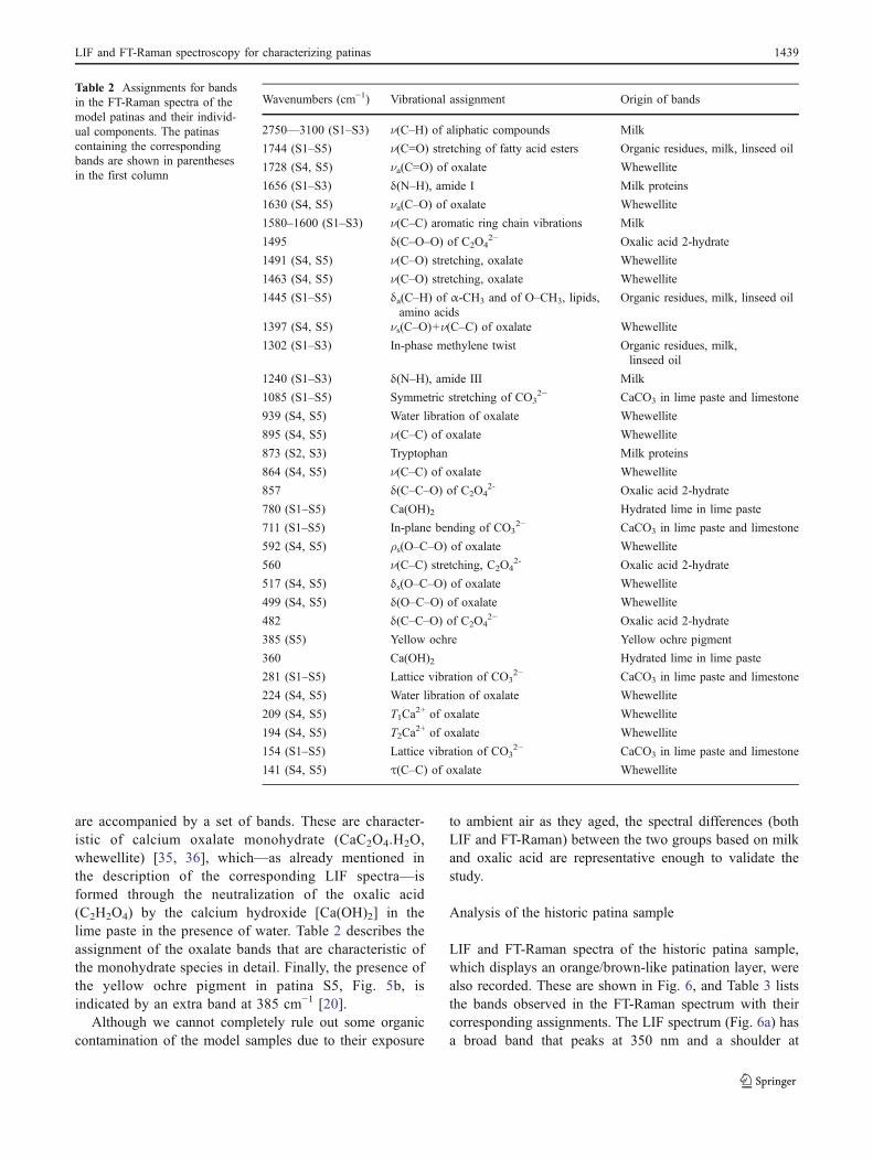
are accompanied by a set of bands. These are character-istic of calcium oxalate monohydrate (CaC2O4.H2O,whewellite) [35, 36], which—as already mentioned inthe description of the corresponding LIF spectra—isformed through the neutralization of the oxalic acid(C2H2O4) by the calcium hydroxide [Ca(OH)2] in thelime paste in the presence of water. Table 2 describes theassignment of the oxalate bands that are characteristic ofthe monohydrate species in detail. Finally, the presence ofthe yellow ochre pigment in patina S5, Fig. 5b, isindicated by an extra band at 385 cm−1 [20].
Although we cannot completely rule out some organiccontamination of the model samples due to their exposure
to ambient air as they aged, the spectral differences (bothLIF and FT-Raman) between the two groups based on milkand oxalic acid are representative enough to validate thestudy.
Analysis of the historic patina sample
LIF and FT-Raman spectra of the historic patina sample,which displays an orange/brown-like patination layer, werealso recorded. These are shown in Fig. 6, and Table 3 liststhe bands observed in the FT-Raman spectrum with theircorresponding assignments. The LIF spectrum (Fig. 6a) hasa broad band that peaks at 350 nm and a shoulder at
Table 2 Assignments for bandsin the FT-Raman spectra of themodel patinas and their individ-ual components. The patinascontaining the correspondingbands are shown in parenthesesin the first column
Wavenumbers (cm−1) Vibrational assignment Origin of bands
2750––3100 (S1–S3) ν(C–H) of aliphatic compounds Milk
1744 (S1–S5) ν(C=O) stretching of fatty acid esters Organic residues, milk, linseed oil
1728 (S4, S5) νa(C=O) of oxalate Whewellite
1656 (S1–S3) δ(N–H), amide I Milk proteins
1630 (S4, S5) νa(C–O) of oxalate Whewellite
1580–1600 (S1–S3) ν(C–C) aromatic ring chain vibrations Milk
1495 δ(C–O–O) of C2O42− Oxalic acid 2-hydrate
1491 (S4, S5) ν(C–O) stretching, oxalate Whewellite
1463 (S4, S5) ν(C–O) stretching, oxalate Whewellite
1445 (S1–S5) δa(C–H) of α-CH3 and of O–CH3, lipids,amino acids
Organic residues, milk, linseed oil
1397 (S4, S5) νs(C–O)+ν(C–C) of oxalate Whewellite
1302 (S1–S3) In-phase methylene twist Organic residues, milk,linseed oil
1240 (S1–S3) δ(N–H), amide III Milk
1085 (S1–S5) Symmetric stretching of CO32− CaCO3 in lime paste and limestone
939 (S4, S5) Water libration of oxalate Whewellite
895 (S4, S5) ν(C–C) of oxalate Whewellite
873 (S2, S3) Tryptophan Milk proteins
864 (S4, S5) ν(C–C) of oxalate Whewellite
857 δ(C–C–O) of C2O42- Oxalic acid 2-hydrate
780 (S1–S5) Ca(OH)2 Hydrated lime in lime paste
711 (S1–S5) In-plane bending of CO32− CaCO3 in lime paste and limestone
592 (S4, S5) ρs(O–C–O) of oxalate Whewellite
560 ν(C–C) stretching, C2O42- Oxalic acid 2-hydrate
517 (S4, S5) δs(O–C–O) of oxalate Whewellite
499 (S4, S5) δ(O–C–O) of oxalate Whewellite
482 δ(C–C–O) of C2O42− Oxalic acid 2-hydrate
385 (S5) Yellow ochre Yellow ochre pigment
360 Ca(OH)2 Hydrated lime in lime paste
281 (S1–S5) Lattice vibration of CO32− CaCO3 in lime paste and limestone
224 (S4, S5) Water libration of oxalate Whewellite
209 (S4, S5) T1Ca2+ of oxalate Whewellite
194 (S4, S5) T2Ca2+ of oxalate Whewellite
154 (S1–S5) Lattice vibration of CO32− CaCO3 in lime paste and limestone
141 (S4, S5) τ(C–C) of oxalate Whewellite
LIF and FT-Raman spectroscopy for characterizing patinas 1439

470 nm. A comparison between the spectra in Figs. 2 and3b (from individual patina components and oxalate-basedpatinas, respectively) suggests that the historic patina isbased on oxalates rather than on proteinaceous components.More specific information on its composition was obtainedvia FT-Raman analysis (Fig. 6b).
The FT-Raman spectrum of the substrate of historicpatina (Fig. 6b) reveals the presence of a mixture of calciteand gypsum. On the other hand, the spectrum of the historicpatina contains bands attributed to CaCO3 as calcite,gypsum, hydroxyapatite (calcium phosphate), ivory black,and calcium oxalates. The presence of gypsum is consid-ered to be a result of weathering processes; it is not anoriginal component of the patina, due to its less intensebands. Although the broad band observed at 780 cm−1 formodel patinas was previously attributed to hydrated lime[Ca(OH)2, portlandite] (Fig. 4), in this case the band is dueto the characteristic luminescent emission of hydroxyapatiteand related minerals [37]. The presence of hydroxyapatite isrelated to the ivory black based on bones that contain a highconcentration of phosphates (PO4
3−) [20]. No iron oxides,which could be responsible for the orange/brown colorationof the patina, were detected. The FT-Raman spectrum of thehistoric patina does not contain any band that could bedirectly related to proteins and lipids, indicating that theseorganic products—if they are present in the patina—occurin quantities below the detection limit of the technique. Thefull characterization of these trace elements would requiresample extraction and the use of more sensitive techniques,such as GC-MS.
Nor
mal
ized
LIF
inte
nsity
Wavelength/ nm
a)
350 450 550 6500.0
0.5
1.0
LimepasteHistoric patinaOxalic acid
Wavenumber/ cm -1
Nor
mal
ized
Ram
an in
tens
ity
b)
0
1
500 1000 1500 20000
1
181
1007
711
1085
281
154
1464
-149
0
1580
1325
780
961
Historic patina
493
619
670
414
1744
1445
1132
Substrate
Fig. 6 a LIF and b FT-Raman spectra of the historic patina samplefrom the façade of San Blas Monastery, Lerma, Burgos, Spain. Thespectra from lime paste and oxalic acid 2-hydrate and the Ramanspectrum from the stone substrate are shown for comparison in a andb, respectively
Table 3 Assignments for bandsin the FT-Raman spectra of thehistoric patina and its substratefrom the façade of San BlasMonastery, Lerma, Burgos,Spain
Wavenumbers (cm−1) Vibrational assignment Origin of bands
1580 Carbon Ivory black
1490 ν(C–O) stretching of C2O42− Calcium oxalate
1464 ν(C–O) stretching of C2O42− Calcium oxalate
1325 Carbon Ivory black
1132 ν3(S–O) mode of SO42− Gypsum
1085 Symmetric stretching of CO32− CaCO3
1007 ν1(S–O) mode of SO42− Gypsum
961 ν1(a1) symmetric streching of PO43− Ivory black
780 Luminescence emission Hydroxyapatite
711 In-plane bending of CO32− CaCO3
670 ν4(S–O) mode of SO42− Gypsum
619 ν4(S–O) mode of SO42− Gypsum
493 ν2(S–O) mode of SO42− Gypsum
414 ν2(S–O) mode of SO42− Gypsum
281 Lattice vibration of CO32− CaCO3
181 ν1(S–O) mode of SO42− Gypsum
154 Lattice vibration of CO32− CaCO3
1440 M. Oujja et al.

Conclusions
The combined application of two laser-based noninvasivetechniques, laser-induced fluorescence and FT-Raman spec-troscopy, to the study of model patinas on limestone substrateshas allowed the identification of the main patina compoundsand the products of some of the chemical and mineralogicaltransformations that these compounds have undergone overthe course of time. LIF analysis allowed the initial identifica-tion of characteristic fluorophores, such as the emission fromamino acids in the region around 350 nm. Products derivedfrom the reactions of individual components also show theirdistinctive broad band emissions (i.e., calcium oxalatesformed by the reaction of oxalic acid and calcium hydroxide).FT-Raman results supply distinctive molecular signatures ofindividual components and the products of chemical andmineralogical transformations, in particular calcium oxalatemonohydrate. The application of this methodology to ahistoric patina allowed the identification of its components,which were mainly based on calcite, calcium oxalates, andphosphates. Although it was not possible to determine theorganic content of the historic patina due to the characteristicsof the sample, using the model patinas we were able to showthat these techniques are useful for examining patinas orrenderings, especially in cases where organic materials havenot been completely chemically or mineralogically trans-formed into inorganic compounds. As portable systems forperforming LIF and FT-Raman measurements in situ areavailable, the information obtained in the present workdemonstrates that the combined application of these twospectroscopic techniques can assist in the practice ofconservation, such as in the classification of patinas used onthe stone found in historic cultural heritage. Work is inprogress to analyze a larger number of historical patinas inorder to validate the conclusions of this initial investigation.
Acknowledgements This work has been funded by the MadridRegional Government project Geomateriales (S2009/Mat-1629) andby the Ministerio de Ciencia e Innovación under projects CTQ2010-15680 and CONSOLIDER CSD2007-00058. The authors also thankthe research group from Universidad Complutense de Madrid:“Alteración y Conservación de los Materiales Pétreos del Patrimonio.”
References
1. Álvarez de Buergo M, Fort R (2003) Constr Build Mater 17:832. Vázquez-Calvo C, Álvarez de Buergo M, Fort R (2006) In: Fort R,
Álvarez de Buergo M, Gómez-Heras M, Vázquez-Calvo C (eds)Heritage weathering and conservation 969–974. Taylor & Francis/Balkema, Leiden
3. Vázquez-Calvo C, Álvarez de Buergo M, Fort R (2007) In:Prikryl R, Smith B (eds) Building stone decay: from diagnosis toconservation (Special Publication 271). The Geological Society ofLondon, London, p 295
4. Kouzeli K, Lazari C, Economopoulos A, Pavelis C (1996) In: RealiniM, Toniolo L (eds) Proc 2nd Int Symp on The Oxalate Films in theConservation of Works of Art. EDITEAM s.a.s., Castello d’Argile
5. Polikreti K, Maniatis Y (2003) Sci Total Environ 308(1–3):1116. Rampazzi L, Andreotti A, Bonaduce I, Colombini MP, Colombo
C, Toniolo L (2004) Talanta 63:9677. Taniguchi Y, Shimadzu Y, Kakoulli I (2003) In: Proc 25th Annu
Conf Jpn Soc Conserv Cultural Property, Kyoto, Japan, June 20038. Cooper M, Larson J (1996) The Conservator 20:289. Vázquez-Calvo C, Gomez Tubio B, Álvarez de Buergo M, Ortega
Feliu I, Fort R, Respaldiza MA (2008) X-Ray Spectrom 37:39910. Vázquez-Calvo C, Álvarez de Buergo M, Fort R, Varas MJ (2007)
Mater Charact 58(11–12):111911. Vázquez-Calvo C, Giakoumaki A, Anglos D, Álvarez de Buergo
M, Fort R (2007) In: Nimmrichter J, Kautek W, Schreiner M (eds)Lasers in the conservation of artworks (Springer Proceedings inPhysics 116). Springer, Vienna, pp 415–420
12. Oujja M, García A, Romero C, Vázquez de Aldana JR, Moreno P,Castillejo M (2011) Phys Chem Chem Phys 13:4625
13. Castillejo M, Martín M, Oujja M, Silva D, Torres R, ManousakiA, Zafiropulos V, Van den Brink OF, Heeren RMA, Teule R, SilvaA, Gouveia H (2002) Anal Chem 74:4662
14. Gaspard S, Oujja M, Moreno P, Méndez C, García A, Domingo C,Castillejo C (2008) Appl Surf Sci 255:2675
15. Gaspard S, Oujja M, Abrusci C, Catalina F, Lazare S, DesvergneJP, Castillejo M (2008) J Photochem Photobiol A 193:187
16. Wisniewski M, Sionkowska A, Kaczmarek H, Lazare S, TokarevV, Belin C (2007) J Photochem Photobiol A: Chem 188:192
17. Nevin A, Osticioli I, Anglos D, Burnstock A, Cather S,Castellucci E (2007) Anal Chem 79:6143
18. Oujja M, Pouli P, Fotakis C, Domingo C, Castillejo M (2010)Appl Spectrosc 64:528
19. Weibring P, Johansson T, Edner H, Svanberg S, Sundnér B,Raimondi V, Cecchi G, Pantani L (2001) Appl Opt 40:6111
20. Bell IM, Clark RJH, Gibbs JP (1997) Spectrochim Acta Part A53:2159
21. Raimondi V, Cecchi G, Lognoli D, Palombi L, Grönlund R,Johansson A, Svanberg S, Barup K, Hällström J (2009) Int BiodetBiodeg 63:823
22. Vázquez-Calvo C, Álvarez de Buergo M, Fort R (2009) Methodof preparation of patinas or films for stone surfaces andapplications thereof (patent: WO 2009024642). WIPO, Geneva
23. Bustillo A (1980) Bol Geol Minero XCI-III:50324. Wang J, Wu X, Mullins C (1997) Appl Spectrosc 51:189025. Bezouska JR, Wang J, Mullins OC (1998) Appl Spectrosc
52:160626. Aminzadeh A (1997) Spectrochim Acta Part A 53:69327. Teale FWJ, Weber G (1957) Biochem J 65:47628. Palumbo G, Pratesi R (2004) Lasers and current optical techniques
in biology (Comprehensive Series in Photochemistry and Photo-biology). Royal Society of Chemistry, Cambridge
29. Nevin A, Comelli D, Valentini G, Cubeddu R (2009) Anal Chem81:1784
30. Athanassia A, Hill AE, Fourrier T, Burgio L, Clark RJH (2000) JCult Herit 1:S209
31. Lu J, Li Y, Zhao K, Xu JQ, Yu JH, Li GH, Zhang X, Bie HY, WangTG (2004) Inorg Chem Commun 7:1154
32. Taddei P, Tinti A, Gandolfi MG, Rossi PL, Prati C (2009) J MolStruct 924–926:548
33. Nevin A, Comelli D, Osticioli I, Filippidis G, Melessanaki K,Valentini G, Cubeddu R, Fotakis C (2010) Appl Phys A 100:599
34. Jeziorowski H, Moser B (1985) Chem Phys Lett 120:4135. Frost RL (2004) Anal Chim Acta 517:20736. Shippey TA (1980) J Mol Struct 63:15737. Campos-Suñol MJ, Domínguez-Vidal A, Ayora-Cañada MJ, De la
Torre-López MJ (2008) Anal Bioanal Chem 391:1039
LIF and FT-Raman spectroscopy for characterizing patinas 1441