large gastric bezoar
TRANSCRIPT

LARGE GASTRIC BEZOAR
Report of a Case Treated Medically
CHARLES H. BROWN, M.D. Department of Gastroenterology
and
ROBERT W. SCHNEIDER, M.D. Department of Endocrinology
GASTRIC bezoars are rare medical curiosities. They are considered of historical interest because animal bezoars were thought, in the Middle
Ages, to possess certain curative powers. Among the crown jewels of Queen Elizabeth were two large bezoar stones. The bezoar was listed in the London pharmacopeias until the mid-eighteenth century. DeBakey and Ochsner1
state, " I t neutralized poisons, reanimated old age, destroyed venoms, and counteracted attacks of vertigo, epilepsy, dysentery, plague, leprosy, etc."
Bezoars are unusual in humans. DeBakey and Ochsner1 in a careful review of the literature were able to find only 303 collected cases, to which they added 8 of their own in 1938. These foreign bodies occur most frequently in the psychiatric patient who has formed the habit of eating hair, string, and other indigestible materials. The incidence of bezoars outside psychiatric institutions is extremely rare, and the case we are reporting is the only one ever observed at the Cleveland Clinic.
Bezoars may be composed of several different materials, the most common the trichobezoar or hair ball. Next in incidence is the phytobezoar, of vege-table composition. While the most ordinary type originates from persimmon seeds,2 it may be caused by other types of vegetable and plant substance. The third type of bezoar is the concretion, which may result from the ingestion of furniture polish for its alcoholic content —the shellac being precipitated and forming a gummy bezoar. Other concretions have been caused by bismuth, magnesium, calcium and sodium carbonates, and by salol.3
The symptoms and clinical manifestations in 131 collected cases of tri-chobezoar and 94 cases of phytobezoar were analyzed by DeBakey and Oshs-ner1 whose observations are presented:
Manifestation Trichobezoar (Per cent)
Phytobezoar (Per cent)
Abdominal mass Pain and tenderness . . . Nausea and vomiting . . . Weakness and loss of weight Constipation and diarrhea . Hematemesis
87.7 70.2 64.9 38.1 32.0
6.1
57.4 85.1 74.4 32.9 31.9 1 4 . 8
2 0 3
only. All other uses require permission. on December 22, 2021. For personal usewww.ccjm.orgDownloaded from

B R O W N A N D S C H N E I D E R
As may readily be seen from the table, the symptoms are similar to those often observed in advanced carcinoma of the stomach. If there is no history of ingestion, the diagnosis may be confused with carcinoma. By their irritative action bezoars may cause a superficial gastritis, or even a fairly large gastric ulcer, which may further complicate the problem of diagnosis and treatment. The diagnosis is usually made by roentgen examination, the bezoar having formed a movable mass within the stomach. Patterson and Rouse4 have re-ported 5 cases observed on gastroscopic examination, which may assist in diagnoses.
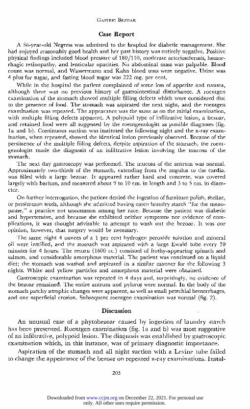
(a) (b) FIG. 1. (a) Roentgenogram of stomach showing filling defect extending from angulus to cardia. Gastroscopically, this was one large mass instead of multiple defects as suggested by
x-ray. (b) Spot film showing same filling defect.
With few exceptions the treatment of bezoars has necessarily been surgical. No successful method of dissolving the sticky gums and hair which make up their bulk has been reported, although Mills and Simpson (quoted by Hart2), succeeded in removing one bezoar by means of hydrochloric acid and Weit-zen5 was able to remove a trichophytobezoar by gastric lavage. If attempted medical treatment is not immediately effective, early surgical intervention is indicated; this should not be delayed until the patient develops a gastric ulcer and hematemesis, as these complications increase the risk of operation.
The following report of a bezoar caused by vegetable starch is the only case on file at the Cleveland Clinic, and an instance in which successful removal was accomplished without recourse to surgery.
2 0 4
only. All other uses require permission. on December 22, 2021. For personal usewww.ccjm.orgDownloaded from
G A S T R I C B E Z O A R
Case Report
A 56-year-old Negress was admitted to the hospital for diabetic management . She had enjoyed reasonably good health and her past history was entirely negative. Positive physical findings included blood pressure of 180/110, moderate arteriosclerosis, hemor-rhagic retinopathy, and lenticular opacities. No abdominal mass was palpable. Blood count was normal, and Wassermann and K a h n blood tests were negative. Urine was 4 plus for sugar, and fasting blood sugar was 222 mg. per cent.
While in the hospital the patient complained of some loss of appetite and nausea, although there was no previous history of gastrointestinal disturbance. A roentgen examination of the stomach showed multiple filling defects which were considered due to the presence of food. The stomach was aspirated the next night, and the roentgen examination was repeated. The appearance was the same as on the initial examination, with multiple filling defects apparent. A polypoid type of infiltrative lesion, a bezoar, and retained food were all suggested by the roentgenologist as possible diagnoses (fig. l a and b). Continuous suction was instituted the following night and the x-ray exam-ination, when repeated, showed the identical lesion previously observed. Because of the persistence of the multiple filling defects, despite aspiration of the stomach, the roent-genologist made the diagnosis of an infiltrative lesion involving the mucosa of the stomach.
The next day gastroscopy was performed. The mucosa of the an t rum was normal. Approximately two-thirds of the stomach, extending from the angulus to the cardia, was filled with a large bezoar. I t appeared rather hard and concrete, was covered largely with barium, and measured about 9 to 10 cm. in length and 3 to 5 cm. in diam-eter.
On further interrogation, the patient denied the ingestion of furniture polish, shellac, or persimmon seeds, although she admitted having eaten laundry starch "for the meno-pause," a practice not uncommon among her race. Because the patient was diabetic and hypertensive, and because she exhibited neither symptoms nor evidence of com-plications, it was thought advisable to at tempt to wash out the bezoar. It was our opinion, however, tha t surgery would be necessary.
The same night 4 ounces of a 1 per cent hydrogen peroxide solution and mineral oil were instilled, and the stomach was aspirated with a large Ewald tube every 10 minutes for 4 hours. The return (1600 cc.) consisted of frothy-appearing spinach and salmon, and considerable amorphous material. The patient was continued on a liquid diet; the stomach was washed and aspirated in a similar manner for the following 3 nights. White and yellow particles and amorphous material were obtained.
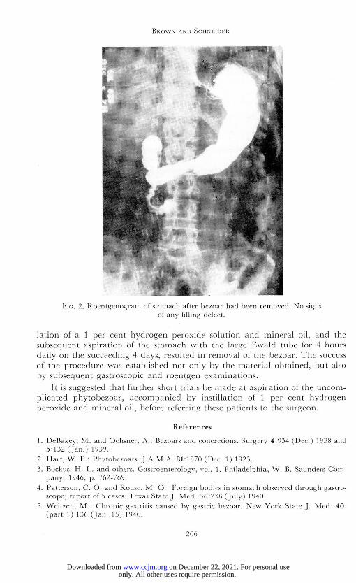
Gastroscopic examination was repeated in 4 days and, surprisingly, no evidence of the bezoar remained. The entire an t rum and pylorus were normal. In the body of the stomach patchy atrophic changes were apparent , as well as small petechial hemorrhages, and one superficial erosion. Subsequent roentgen examination was normal (fig. 2).
Discussion
An unusual case of a phytobezoar caused by ingestion of laundry starch has been presented. Roentgen examination (fig. l a and b) was most suggestive of an infiltrative, polypoid lesion. The diagnosis was established by gastroscopic examination which, in this instance, was of primary diagnostic importance.
Aspiration of the stomach and all night suction with a Levine tube failed to change the appearance of the bezoar on repeated x-ray examinations. Instal-
2 0 5
only. All other uses require permission. on December 22, 2021. For personal usewww.ccjm.orgDownloaded from
B R O W N A N D S C H N E I D E R
FIG. 2. Roentgenogram of stomach after bezoar had been removed. No signs of any filling defect.
lation of a 1 per cent hydrogen peroxide solution and mineral oil, and the subsequent aspiration of the stomach with the large Ewald tube for 4 hours daily on the succeeding 4 days, resulted in removal of the bezoar. The success of the procedure was established not only by the material obtained, but also by subsequent gastroscopic and roentgen examinations.
It is suggested that further short trials be made at aspiration of the uncom-plicated phytobezoar, accompanied by instillation of 1 per cent hydrogen peroxide and mineral oil, before referring these patients to the surgeon.
References
1. DeBakey, M. and Ochsner, A.: Bezoars and concretions. Surgery 4:934 (Dec.) 1938 and 5:132 (Jan.) 1939.
2. Hart , W. E.: Phytobezoars. J .A.M.A. 81:1870 (Dec. 1) 1923. 3. Bockus, H. L. and others. Gastroenterology, vol. 1. Philadelphia, W. B. Saunders Com-
pany, 1946, p. 762-769. 4. Patterson, C. O. and Rouse, M. O.: Foreign bodies in stomach observed through gastro-
scope; report of 5 cases. Texas State J . Med. 36:238 (July) 1940. 5. Weitzen, M. : Chronic gastritis caused by gastric bezoar. New York State J. Med. 40:
(part 1) 136 (Jan. 15) 1940.
2 0 6
only. All other uses require permission. on December 22, 2021. For personal usewww.ccjm.orgDownloaded from