l'application de la tms dans l'étude des fonctions...
TRANSCRIPT

Chotiga Pattamadilok
Laboratoire Parole et Langage
Aix-en-Provence
L'application de la TMS dans
l'étude des fonctions cognitives
RMN- Oct. 2016

Magnetic stimulation
Arsène d’Arsonval (on the right) and two of his assistants are shown demonstrating the effects of the flow of alternating current (1911). “an alternating magnetic field with an intensity of 110 volts, 30 amperes and a frequency of 42 cycles per second, gives rise to, when one places the head into the coil, phosphenes and vertigo, and in some persons, syncope.” (d’Arsonval, 1896)
Silvanus P. Thompson: on inserting the head into the interior of the coil, in the dark, or with the eyes closed, there is perceived over the whole region of vision a faint flickering illumination, colourless or of a slightly bluish tint” (1910)
The magnetic coils used by Magnusson and Stevens in 1911,. Additional sections of coils could be energized to increase the magnetic field.

Faraday’s principle of electromagnetic induction “Faraday showed that an electrical current passed through one coil could induce a current in a nearby coil. The current in the first coil produces a magnetic field that in turn causes current to flow in the second coil. In TMS that second coil is replaced by brain tissue and the induced electric field elicits neuronal activity.” (Walsh & Cowey, 2000)

TMS and study of cognitive functions
1) Causal relation between cortical activity and behavior
Complement the correlational approaches (fMRI, EEG, MEG)
Virtual lesion or neural noise: change in cortical excitability
Mostly inhibitory but sometimes facilitatory effect
Underlying mechanism is still unclear but the most critical factors are:
Stimulation parameters:
intensity, frequency, timing, coil type/orientation, protocol…
The initial state of the activated brain region:
rest, active, task-demands…

2) Timing at which activity in a particular cortical region contributes to a given task
Single-pulse (high temporal resolution…but need to know where & when!)
Begin with repetitive TMS (low temporal resolution but stronger effect -> explore
space dimension)
Double-pulse; triple-pulse
TMS temporal resolution depends on: Duration of the TMS pulse effects
Duration of the area’s involvement in the task
Adapted from Walsh & Coway, 2000
TMS pulse (neural noise)
Inte
nsi
ty
Timing (ms)
Time
Probability of the area contributing to the task
x

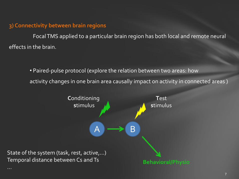
3) Connectivity between brain regions
Focal TMS applied to a particular brain region has both local and remote neural
effects in the brain.
• Paired-pulse protocol (explore the relation between two areas: how
activity changes in one brain area causally impact on activity in connected areas )
B
Behavioral/Physio
A
Conditioning stimulus
Test stimulus
State of the system (task, rest, active,…) Temporal distance between Cs and Ts …

• TMS combined with neuro-imaging (EEG, PET, fMRI)
3) Connectivity between brain regions
Massimini et al. (2005)

Is the causal inference between ROI and function still valid?
Can behavioural TMS studies without imaging still be considered as valid
empirical tools for revealing the functional necessity of the stimulated
brain region?
YES… because remote neural effects of TMS is not necessarily functionally relevant!
A Behav
B
A Behav
B
Need TMS interference protocol to prove the causality of the connected regions
A B Bahav
?

4) State-dependent TMS paradigm/TMS-adaptation paradigm
TMS affects the less active neurons (Silvanto et al., 2008)
One can control which neural populations are preferentially affected by TMS
Silvanto, Muggleton, Walsh (2008)
Reveal some degree of specificity in a region that contains functionally overlapping populations of neurons
TMS effect State of the system Neurons’ activation level
Reduce the excitability of
Increase the (facilita- tory) TMS effect on
TMS adaptaion paradigm: Induce habituation

How to choose the most appropriate TMS protocol? Pitfalls? How to control for non-specific TMS effects (artefacts)?

1Laboratoire Parole et Langage, 2Cognitive Neuroscience Experiment and Consulting, 3Institut de Neurosciences des Systèmes
The Contribution of writing to reading
A neuronavigated TMS Study
Chotiga Pattamadilok1, Aurélie Ponz2, Samuel Planton1
& Mireille Bonnard3
Pattamadilok, C., Ponz, A., Planton, S., & Bonnard, M. (2016). Contribution of writing to reading: Dissociation between cognitive and motor process in the left dorsal premotor cortex. Human Brain Mapping, 37, 1531–1543.

Reading and writing are closely related
Already at the first stage of literacy acquisition, children learn to reproduce the form of written characters that they read.
Writing practice can facilitate reading acquisition
Bara et al., 2004 : Reading training in 5 yrs-old children. Classic Visual
training was less efficient than Haptic + Visual training.
Longcamp et al., 2005 : In 3-5 yrs-old children, handwriting training
gave rise to a better letter recognition than the typing training.
…

In expert readers, knowledge of how letters are written influences the way
in which they are perceived :
Orliaguet et al., 1997: seeing the writing movement of a letter helps
to anticipate the identity of a forthcoming letter (while seeing the form does
not) The preparation of the second letter is partly carried out during the
production of the first letter.
Bartolomero et al., 2002: tracing out the form of the letter facilitates
letter recognition in alexic patients.
James et al., 2009: Interference of hand movement on letter
recognition.
(experimenter) (participant)

Reading and writing share central cognitive processes
/t/, /r/, /i:/ => /triː/
T, R, EE => TREE
Auditory output/input
Abstract Phono
Abstract Ortho
tree Visual input/output
Semantic
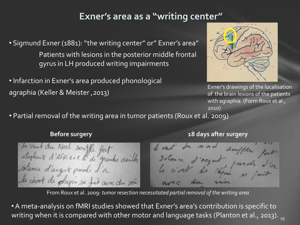
Exner’s area as a “writing center”
• Infarction in Exner's area produced phonological
agraphia (Keller & Meister ,2013)
• Partial removal of the writing area in tumor patients (Roux et al. 2009)
Before surgery 18 days after surgery
From Roux et al. 2009: tumor resection necessitated partial removal of the writing area
• Sigmund Exner (1881): “the writing center” or” Exner’s area”
Patients with lesions in the posterior middle frontal gyrus in LH produced writing impairments
Exner’s drawings of the localisation of the brain lesions of the patients with agraphia. (Form Roux et al., 2010)
• A meta-analysis on fMRI studies showed that Exner’s area’s contribution is specific to writing when it is compared with other motor and language tasks (Planton et al., 2013).

Exner’s Area and reading
• Reading difficulties observed in dyslexic children lead to a reduced activity in visual word form area and a greater reliance on Exner’s area, suggesting partial compensation through the gesture system (Monzalvo et al., 2012)
• In normal readers, an activation of Exner’s Area was found during reading tasks (Dehaene et al., 2010; Nakamura et al., 2012, Rapp & Lipka, 2011; Xu et al., 2005; Planton et al., 2013).
Activations produced by reading (words > checkerboards) in blue and spelling (spell > case) in green. Indicated with red circles are the regions of overlap between reading and spelling in the left mid-fusiform and the left IFG/IFJ
Rapp & Lipka, 2011
Reading Writing (Motor representation) Main interpretation:

1) Epiphenomenal (co-activation of the reading and writing systems) or real functional role ?
Interpretation of brain imaging data
2) If functional role? Motor: implicit evocation of writing motor processes Cognitive: shared cognitive components between writing and reading
3) How early?
Reading Writing (Exner’s area)
?
fMRI

The Contribution of Exner’s Area to reading
Task: Lexical decision
Stimuli: Words vs. Pseudowords
(global vs. sequential process)
Characters: Handwritten vs. Printed characters
(handwritten character is more related to motor knowledge => embodiment of the perception of handwritten letters)
TMS stimulation: double-pulse TMS in 3 time-windows
0/40 ms (baseline) 60/100 ms 120/160 ms
Aim: Disruptive effect of transcranial magnetic stimulation applied on Exner’s area
“Cognitive” hypothesis
“Motor” hypothesis

Participants: 15 Right-handed French speakers Responded with the left hand to avoid interference with the right hand RT & ACC were collected
Double-pulse TMS applied in one of the three time-windows: 0-40; 60-100; 120-160 ms

Localization: Individual MRI (Sack et al. 2009) Posterior middle frontal gyrus at the junction between the precentral sulcus and the superior frontal sulcus in LH
Visualization: image-guided frameless stereotaxic neuronavigation system
• 10–20 EEG (e.g., P4 : +/- right parietal sulcus)
• Individual MRI-guided TMS neuronavigation
• Group functional Talairach coordinates
• fMRI-guided TMS neuronavigation
• Individual TMS-guided (localizer task)
Different methods can be combined. The choice of the method depends on: time, budget, precision required, number of subjects, equipment, security, …
Sack et al. 2009

Intensity: Adjusted Motor Threshold* ( primary motor cortex = ) *(MT: the lowest stimulation intensity capable of producing changes of MEP or overt muscle twitch)
• At least two factors influence the susceptibility of a brain area to stimulation: magnetic field strength and excitability of the cortex
• Solutions: Motor Threshold; Adjusted MT; Constant intensity (50%-70%).
The distance between the center of the coil and the cortex
Unknown for most areas Depend on the state of the system (e.g., task, active vs. rest, …)

Control for artifacts non-specific TMS effects due to click sounds and muscle twitches (online protocol)
• Sham coil (same click sound but no scalp sensation), placebo coil (more satisfactory?)…but need naive subjects! • Control site (vertex? homologous area? …) • Control task or control condition within the same task • Double dissociation (sites * tasks) • Control time-window (here, baseline = 0/40ms) • Combination of different methods
Double-pulse TMS applied in one of the three time-windows: 0-40; 60-100; 120-160 ms

550
570
590
610
630
650
670
690
710
Baseline 60/100ms. 120/160ms.
Control task
• Sham coil (same click sound but no scalp sensation), placebo coil (more satisfactory?)…but need naive subjects! • Control site (vertex? homologous area? …) • Control task or control condition within the same task • Double dissociation (sites * tasks) • Control time-window (here, baseline = 0/40ms) • Combination of different methods
Control for artifacts non-specific TMS effects due to click sounds and muscle twitches (online protocol)

Handwritten
Printed
Main task: Reaction times
Baseline
60/100ms
120/160ms
Words Pseudowords
Lexicality x TMS
Character x TMS Character x Lexicality x TMS
TMS effect on pseudowords only
ANOVA: Character * Lexicality * TMS

Same tendency but no significant result
Main task: Accuracy

Does Exner’s area contribute to reading? Yes How early? Already within the first 100-150 ms Does it depend on the type of stimulus or character? Only for pseudoword decision regardless of the type of character. rule out the “motor” hypothesis? BUT…Did handwritten characters elicit motor representations as expected?
Baseline
60/100ms
120/160ms

TMS as a tool to investigate the causal role of a given cognitive task on the activation state of the motor cortex.
Stimulation protocol
TMS stimulation: single-pulse in 3 time-windows 0 ms (baseline), 60 ms, 120 ms Site of stimulation: Motor cortex (first dorsal interosseous) Intensity: 110% of the resting motor threshold EMG recording: First dorsal interosseous muscles of the right hand (participants responded with their left hand)
Single-pulse TMS At 0, 60 or 120 ms
Why single-pulse?
• Probe a modulation of cortico-spinal excitability of digit muscles during reading handwriting vs. printed character • Minimize artifacts on electric muscle responses • No need to disrupt the performance

Handwritten Printed
Character x TMS
Lexicality x TMS Character x Lexicality x TMS
Printed: MEP amplitudes remained stable across the different time-points Hand: MEP amplitudes decreases from T= 0 to T = 160
Handwritten but not printed character induced changes in cortico-spinal excitability of the hand muscles involved in writing gestures

Discussion
The TMS finding show the contribution of the Exner’s area during reading. Coherent with
lesion studies (e.g., Anderson et al., 1990: Lesion in the left premotor cortex (BA6) led to
pure agraphia and alexia)
Dissociation between the “motor” and “cognitive” hypothesis.
• Reading handwritten characters induced changes in cortico-spinal
excitability of the hand muscles involved in writing gestures
• But the contribution of the Exner’s area in reading seems to be explained by
the shared cognitive processes between reading and writing, i.e., sequential or
sublexical process