labmanual:! ramanspectroscopy!physics.gu.se/~starn/tif060/raman/raman_manual2014.pdf · raman...
TRANSCRIPT

CHALMERS UNIVERSITY OF TECHNOLOGY September 2014
Lab Manual: RAMAN SPECTROSCOPY
Laura Mazzei

RAMAN SPECTROSCOPY 2014
1
TABLE OF CONTENT LIST OF SYMBOLS 2
INTRODUCTION 3
SHORT THEORY ABOUT RAMAN AND INFRARED SPRECTROSCOPY 4
Raman scattering 4
Raman spectroscopy 6
Infrared spectroscopy 6
VIBRATIONAL MODES 8
Classic harmonic oscillator 8
EXPERIMENTAL 10
LAB SAFETY 13
REFERENCE 14
QUESTIONS 15

RAMAN SPECTROSCOPY 2014
2
LIST OF SIMBOLS 𝜐! Laser frequency 𝜐!,! frequency associated to the transition (1 à 2 or 2à1) between two different
vibrational levels (1)
𝜇 Molecular dipole moment 𝛼 Molecular polarizability 𝑛! Number of molecules in the vibrational state of energy 𝐸! (Boltzmann factor)(2) 𝐼 Intensity 𝑄 Normal coordinate of vibration
(1) given two levels of energy E1 and E2, E2>E1, we can define 𝜐!,!
𝜐!,! =(𝐸! − 𝐸!)
ℎ
(2) given a molecular state i characterized by the energy 𝐸!
𝑛! ∝ 𝑒!!!!"

RAMAN SPECTROSCOPY 2014
3
Introduction
Vibrational spectroscopy, Raman and infrared, are based on the interaction of
electromagnetic radiation with matter. Experiments are usually non-‐destructive, and the
samples can be reused for other investigations. The aim of both Raman spectroscopy and
Infrared spectroscopy experiments is to probe the molecular vibrations, probing the
transition between the ground state and the excited vibrational states. The transitions are
observed as bands in the vibrational spectrum. Each molecule has a specific set of
vibrational bands, which are defined by their frequencies, shapes, and intensities. By
analyzing these properties, it is possible to get information about the local coordination of
the atoms in your material.
The frequency of a transition (~50-‐4000 cm-‐1)(1) directly depends on the reduced mass of
the vibrating unit. Therefore, smaller reduced masses give rise to higher frequencies.
Depending on the nature of the molecular vibration, the transition may be Raman, and/or
infrared active. Those techniques should therefore be used complementary, because some
bands that are infrared inactive can be Raman active and vice versa. The physical principle
behind Raman and infrared spectroscopy is very different though. Raman spectroscopy is a
scattering technique where the intensity of the bands is proportional to the concentration and
to the scattering cross-‐section of the vibrating units. Infrared spectroscopy is an absorbance
technique where the intensity of the bands is proportional to the concentration and to the
absorption coefficient of the vibrating units. Typically, the time needed to record a spectrum
is of the order of seconds to tens of minutes. However, to increase the signal-‐to-‐noise ratio,
several accumulations are customary.
(1)Note that the frequency range is here expressed in cm-1 (wavenumber). This is a typical unit used in Raman and Infrared spectroscopy. In question #1 you can find the formula to convert frequencies to wavenumber.
RAMAN SPECTROSCOPY 2014
4
Short theory about Raman and infrared spectroscopy
This chapter is devoted to give a short introduction to Raman and infrared spectroscopy.
Extensive descriptions can be found in many textbooks, see e.g. references [1-‐5].
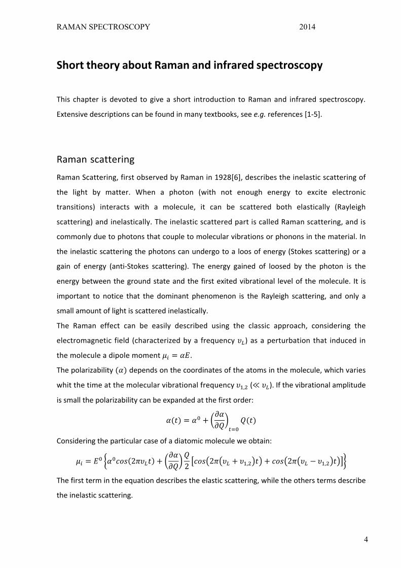
Raman scattering
Raman Scattering, first observed by Raman in 1928[6], describes the inelastic scattering of
the light by matter. When a photon (with not enough energy to excite electronic
transitions) interacts with a molecule, it can be scattered both elastically (Rayleigh
scattering) and inelastically. The inelastic scattered part is called Raman scattering, and is
commonly due to photons that couple to molecular vibrations or phonons in the material. In
the inelastic scattering the photons can undergo to a loos of energy (Stokes scattering) or a
gain of energy (anti-‐Stokes scattering). The energy gained of loosed by the photon is the
energy between the ground state and the first exited vibrational level of the molecule. It is
important to notice that the dominant phenomenon is the Rayleigh scattering, and only a
small amount of light is scattered inelastically.
The Raman effect can be easily described using the classic approach, considering the
electromagnetic field (characterized by a frequency 𝜐!) as a perturbation that induced in
the molecule a dipole moment 𝜇! = 𝛼𝐸.
The polarizability (𝛼) depends on the coordinates of the atoms in the molecule, which varies
whit the time at the molecular vibrational frequency 𝜐!,! (≪ 𝜐!). If the vibrational amplitude
is small the polarizability can be expanded at the first order:
𝛼(𝑡) = 𝛼! +𝜕𝛼𝜕𝑄 !!!
𝑄(𝑡)
Considering the particular case of a diatomic molecule we obtain:
𝜇! = 𝐸! 𝛼!𝑐𝑜𝑠 2𝜋𝜐!𝑡 +𝜕𝛼𝜕𝑄
𝑄2 𝑐𝑜𝑠 2𝜋 𝜐! + 𝜐!,! 𝑡 + 𝑐𝑜𝑠 2𝜋 𝜐! − 𝜐!,! 𝑡
The first term in the equation describes the elastic scattering, while the others terms describe
the inelastic scattering.

RAMAN SPECTROSCOPY 2014
5
For a complete description of the phenomena a quantum approach is necessary. In the
quantum description the vibrational energy of the molecule is quantized, and the interaction
with the light is described using the perturbation theory.
The incoming photon excites the molecule to a virtual state, which decay to a lower energy
state. As described in figure 1.a, the Stokes scattering involve a transition from the ground
state (1) and a successive transition to an exited level (2), while the anti-‐Stokes involve a first
transition from an excited state to the virtual level. Because at room temperature the number
of molecules in the ground state is higher than the number of molecules in the excited state,
the Stokes scattering has an higher probability than the anti-‐Stokes scattering (fig 1.b).
Figure 1. a) schematic picture of the Raman process in a two-‐level system, and b) the resulting Raman spectrum.
Using the quantum approach the intensity of scattered light can be estimated as:
𝐼!"#$%& ∝𝜐! − 𝜐!,!
!
𝜐!,!𝜕𝛼𝜕𝑄
!
𝑛! ; 𝐼!"#$!!"#$%& ∝𝜐! + 𝜐!,!
!
𝜐!,!𝜕𝛼𝜕𝑄
!
𝑛!
Two important results come directly from those expressions; first, Raman scattering only
takes place if
𝜕𝛼𝜕𝑄 ≠ 0
Moreover, the Stokes scattered light is more intense that the anti-‐Stokes, because of the
Boltzmann factor (𝑛! > 𝑛!).

RAMAN SPECTROSCOPY 2014
6
Raman spectroscopy
In a Raman spectroscopy experiment, the sample is irradiated with monochromatic light; the
light frequency has to be chosen in order to avoid the excitation of electronic levels.
The scattered light is detected, in order to collect the vibrational spectra of the sample. As
described in the previous section, the frequencies of the light scattered by a molecule with m
Raman active vibrations (with characteristic frequencies 𝜐!, 𝜐! ) will be
𝜐!"#$$,! = 𝜐! ± 𝜐! 𝑖 ∈ (1,𝑚)
Because of the stronger intensity of the Stokes lines respect to the anti-‐Stokes, usually only
the light at energy lower than the laser energy is collected.
Therefore, the Raman spectrum is composed by several bands, related to the different
vibrations that characterize the molecule.
Raman spectroscopy allows studying only those vibrations that modulates the polarizability of
the molecule under investigation (!"!"≠ 0). For this reason complementary techniques are
important in order to characterize the vibrational spectrumof a molecule.
Infrared spectroscopy
Infrared spectroscopy takes advantage from the absorption phenomena. In this case, the
photon excites a direct transition between the ground state and one of the higher
vibration states of the molecule. Photons can only be absorbed if their frequency exactly
matches the frequency of a particular molecular vibration in the material. A schematic
illustration of the principle behind IR spectroscopy is reported in Fig. 2.
Figure 2 The principle of infrared absorption. a) Photons with energies hω1, hω2, andhω3 hit a two-‐level system. Only hω2 has the same energy as the difference between the two vibrational states, and is therefore absorbed. b) The resulting infrared absorbance spectrum.

RAMAN SPECTROSCOPY 2014
7
Measuring the intensity of the non-‐absorbed light at different frequencies, it is possible to
determine the spectrum (IR-‐spectrum) of the molecule.
The first difference between Raman and infrared is in the light source used in the two
different techniques. While Raman spectroscopy requires a monochromatic light with
frequency higher than the characteristic vibrational frequencies, Infrared spectroscopy
requires polychromatic light with frequencies in the infrared region (~50-‐4000 cm-‐1).
Moreover the selection rule of the Infrared absorption is different than the one for the
Raman scattering. For IR absorption a molecular vibration can be observed only if it
modulates the dipole moment:
𝜕𝜇𝜕𝑄 ≠ 0
This means, for example, that diatomic homonuclear molecules do not give rise to any bands
in the IR spectra since those do not have a dipole moment.

RAMAN SPECTROSCOPY 2014
8
Vibrational modes
This chapter has the aim to introduce briefly some knowledge about vibrational modes. More
information can be found in the references[1,3,5,7].
A molecule containing n atoms has 3n degrees of freedom; considering that 6 of this
configuration describe pure translation and 3 pure rotations (non-‐linear molecules), there are
3n-‐6 (3n-‐5 for linear molecules) motions (vibrations) that change the relative position of the
atoms. In each vibration the atoms carry out a harmonic motion.
Vibrations are usually divided in symmetric and ant symmetric. Moreover, different kind of
vibration can occur: stretching (vibration in the same direction of a bond), bending (oscillation
that implies a change of angle between two bonds), rocking (oscillation that implies a change
of angle between a group of atoms), wagging (change in angle of the plane of a group of
atoms), twisting (change in angle of the planes of two groups of atoms).
Classic Harmonic oscillator
The simplest possible model that can be considered to describe the molecular vibration is the
harmonic oscillator. For a diatomic molecule composed of two atoms of mass 𝑚! and 𝑚! the
classical frequency is given by
𝜐 =12𝜋 𝐾(
1𝑚!
+1𝑚!)
where K represent the force constant of the bond of the two atoms.
More generically, the N atoms of a molecule can be considered to oscillate with small
amplitude around the equilibrium position. The equations of the motion are therefore
𝑚!𝜕!𝑞!𝜕𝑡 = − 𝐾!"
!!
!!!𝑞!
Where 𝑞! represent the displacement from the equilibrium position. Several numerical
methods can be used to solve this set of equation.

RAMAN SPECTROSCOPY 2014
9
In general, approximation can be done to roughly evaluate the vibration of a specific group of
atoms in the molecule. For example, because hydrogen is usually lighter than all the other
elements, the stretching frequencies of bond as C-‐H, O-‐H etc. are really higher compared to
other stretching frequencies (C-‐O, for example). Therefore vibration that involve hydrogen
atoms are almost independent from the rest of the molecule.

RAMAN SPECTROSCOPY 2014
10
Experimental Vibrational spectroscopy is a powerful tool in order to investigate a material at the
molecular level. The interaction of the incident light with matter might occur by light
scattering, as in the case of Raman experiment, or by light absorption, transmission or
reflection, as is the case in an IR experiment. The method that best applies to a certain
material highly depends on the properties of the sample, such as optical transparency,
smoothness, amount of impurities, chemical composition etc.
A troublesome case is the luminescence arising from ionic impurities present in
(unfortunately) many samples. If the luminescence is too strong, the Raman signal could be
as bad as swamped, and your spectrum is practically useless! There is however a way to
overcome this problem, and that is to increase the wavelength of the incident light.
Approaching the infrared limit (λ≈1064 nm), luminescent excitations are usually
minimized.
In this experimental project some different samples are provided to you, which you should
investigate by applying Raman spectroscopy. You should really find out as much as you can
about these materials before you start your experiments.
Be aware of the fact that some chemicals could be harmful, hence do not inhale any
amount.

RAMAN SPECTROSCOPY 2014
11
Materials and experiments
The materials at your disposal for this lab are listed in the table below.
Planar structure Liquid/solid
Methanol
CH3OH
L
Ethanol
C2H5OH
L
Isopropyl alcohol
(2-‐propanol)
C3H7OH
L
1-‐pentanol
C5H11OH
L
2-‐propanone
(acetone)
C3H6O
L
Dimethyl carbonate
(DMC)
C3H6O3
L
Ethylene carbonate
(EC)
C3H4O3
S
Toluene
C7H8
L
The first 4 elements are alcohols, while acetone is a ketone, dimethyl carbonate and
ethylene carbonate are carbonate esters, and finally toluene is an aromatic hydrocarbon.
Moreover, you have two EC/DMC mixtures (50:50 and unknown).

RAMAN SPECTROSCOPY 2014
12
Those molecules are composed only by Hydrogen, Carbon and Oxygen. Therefore, they
present really similar bonds (and spectra). Frequencies relate to those bonds and Tables
with the characteristic frequencies of those molecule can be find on reference [3],[9].
The aim of this lab is to obtain the Raman spectra of the different molecules and assign
each peak to the corresponding vibrational mode, both by using tabulated values and
comparing the different spectra. Moreover, you should discuss the differences among
the spectra of the different molecules and the different bonds (frequencies, shapes and
intensities).
As last part of the exercise, you should analyze the two mixtures of DMC/EC and
determine the concentration of the unknown mixture.
Now, in order to be able to perform the experiment in the lab you should prepare
yourself on the following issues:
a. Search in the literature for vibrational spectra/Raman spectra of the different
molecules
b. Answer to the questions you find at the end of this manual.
c. Think about the differences among the different compounds. Some of them show
similar structures, so you can expect similar spectra. The following questions
should prepare you and help you discussing your results.
• Which molecules have O-‐H bonds? Where in the spectra (at which
frequencies) do you expect to find the O-‐H vibrations? Same question about
the C-‐O bond.
• Which molecules show the C-‐H bond? Which differences do you expect in the
C-‐H band among the different alcohols? Same question about the C-‐C bond.
• You can observe both the single and the double CO bond. Using the theory of
the simple harmonic oscillator, what do you expect to see in the spectra
𝜐!!! 𝜐!!! =? ?
• Which differences do you expect in the spectra of DMC/EC?
• Which differences do you expect in the spectra of EC (solid) and the spectra
of the EC contained in the mixtures (liquid)?

RAMAN SPECTROSCOPY 2014
13
Laser safety Concerning laser safety there is a really important rule: do not look directly into the laser
beam or a reflection of the laser beam at any time for one simple reason: it will damage
your eyes. The reason why laser light is so dangerous is that it is very well focused and since
it is monochromatic all photons will be focused on the exact same spot of the eye. This might
burn some of the cells in the eye and thus cause blindness. The laboratory is equipped with
laser protection glasses, these should be worn at all times when aligning and running the
measurements. In addition, avoid wearing reflecting object (such as rings, belts, etc.).
All the samples that you will analyze are sealed in glass vials; do not open the vials. Whether
the vials seem to be broken or not sealed properly, avoid contact with the samples and
contact the lab's responsible.

RAMAN SPECTROSCOPY 2014
14
References. [1]. N.B. Colthup, L.H. Daly, and S.E. Wiberley. Introduction to infrared and Raman spectroscopy,
Academic Press Inc., 3rd edition, 1990.
[2] Practical Raman Spectroscopy. Gardiner and Graves (Eds.) Springer-Verlag Berlin Heidelberg
1989(chapter 3)
[3] P. Larkin, Infrared and Raman Spectroscopy; Principles and Spectral Interpretation,
Elsevier, 2011
[4] D. A. Long. Raman Spectroscopy, McGraw-Hill, 1977.
[5] A. Fadini, and F.-M. Schnepel. Vibrational Spectroscopy, Methods and applications, John Wiley &
Sons, 1989. (chapter 2)
[6] C. V. Raman and K. S. Krishnan, Nature 121, 501 (1928).
[7] A. D. Boardman, D. E. O’connor, and P. A. Young, Symmetry and its Applications in Science,
McGraw-Hill, 1973.
[8] Kazuo Nakamoto, Infrared and Raman spectra of Inorganic and Coordination compounds, Part A,
Wiley pubbl., 6th edition.
[9] Ernö Pretsch, Philippe Buhlmann, Martin Badertscher, Structure determination of organic
compounds – table of spectral data

RAMAN SPECTROSCOPY 2014
15
Questions to answer before the laboratory work 1) Light can be described using both wavelength and frequency (𝜆𝜐 = 𝑐). The wavelength is
expressed in meter (nm or µm) while frequency is expressed in Hz [GHz]. However, other
units are often used instead of frequency. Those units are cm-‐1 (wavenumber, 𝜐) and eV
(energy associated to the single photon, E). The conversion can be done using the
relations
𝜐 =𝑘2𝜋 =
1𝜆 𝐸 = ℎ𝜈 =
ℎ𝜆
Therefore, the conversion factors from µm to GHz, cm-‐1 and eV are
𝜐 𝐺𝐻𝑧 =3
𝜆(𝜇𝑚) 10! 𝜐 𝑐𝑚!! =
1𝜆(𝜇𝑚) 10
! 𝐸 𝑒𝑉 =1.24𝜆(𝜇𝑚)
Express the following intervals in GHz, cm-‐1 and eV
Xray UV visible Near IR mid IR Far IR THz radiation
0.01-‐10 nm 10-‐400 nm 400-‐700 nm 0.75-‐1.4 µm 1.4-‐15 µm 15-‐1000 µm 0.1-‐1 mm
2) During your Raman experiment you will use a 532 nm Laser source. If you have molecular
vibrations at 2000, 3000 and 3500 cm-‐1, which frequencies you will see in the corresponding
Raman spectra (note: the Raman spectra is composed by the scattered light!)?
3) Harmonic oscillator can be used to understand molecular vibration. Using
𝜐 =12𝜋 𝐾(
1𝑚!
+1𝑚!)
Calculate the vibrational frequencies for the following bond:
C-‐H (K=4.9 N/cm)
N-‐H (K=6.4 N/cm)
O-‐H (K=7.2 N/cm)
C-‐O (K=5.1 N/cm)
C-‐C (K=4.4 N/cm)

RAMAN SPECTROSCOPY 2014
16
4) Derive the expressions for the in-‐phase (symmetrical) and out-‐of-‐phase (anti symmetrical)
vibrational stretching mode for the two following linear molecules
M – M – M
M1 – M 2– M 1
Note: in both case you have two harmonic oscillators with the same force constant. you have to
impose same frequency and amplitude (they only differ in the phase shift)
Using those formula, try to figure out the vibrational frequency related to the following
configuration (𝐾!!!~5.1 𝑁/𝑐𝑚 ; 𝐾!!!~4.4 𝑁/𝑐𝑚)
(H3C) – (OH) (methanol)
(H3C) – (CH2-‐OH) (Ethanol)
(H3C)– (CO) – (CH3) (acetone)
(H3C-‐O) – (CO) – (O-‐CH3) and (H3C) – (O-‐CO-‐O) – (CH3) (DMC)
note: you have to consider the groups of atoms in the brackets as single unit which mass is equal to
the sum of the masses of the atoms. Be careful that in both acetone and DMC the center unit is
actually (C=O), and therefor only the carbon atom in bonded to the neighbor carbons. After the
experiment you can compare those rough values with the experimental ones.